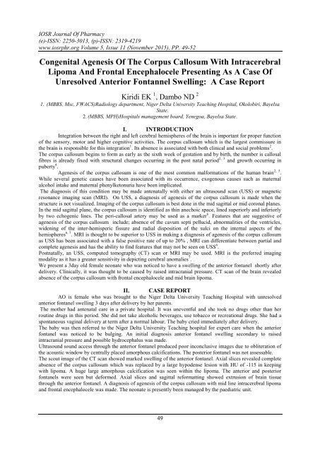
Congenital Agenesis Of The Corpus Callosum With Intracerebral Lipoma And Frontal Encephalocele Presenting As A Case Of Unresolved Anterior Fontannel Swelling: A Case Report
The IOSR Journal of Pharmacy (IOSRPHR) is an open access online & offline peer reviewed international journal, which publishes innovative research papers, reviews, mini-reviews, short communications and notes dealing with Pharmaceutical Sciences( Pharmaceutical Technology, Pharmaceutics, Biopharmaceutics, Pharmacokinetics, Pharmaceutical/Medicinal Chemistry, Computational Chemistry and Molecular Drug Design, Pharmacognosy & Phytochemistry, Pharmacology, Pharmaceutical Analysis, Pharmacy Practice, Clinical and Hospital Pharmacy, Cell Biology, Genomics and Proteomics, Pharmacogenomics, Bioinformatics and Biotechnology of Pharmaceutical Interest........more details on Aim & Scope).
The IOSR Journal of Pharmacy (IOSRPHR) is an open access online & offline peer reviewed international journal, which publishes innovative research papers, reviews, mini-reviews, short communications and notes dealing with Pharmaceutical Sciences( Pharmaceutical Technology, Pharmaceutics, Biopharmaceutics, Pharmacokinetics, Pharmaceutical/Medicinal Chemistry, Computational Chemistry and Molecular Drug Design, Pharmacognosy & Phytochemistry, Pharmacology, Pharmaceutical Analysis, Pharmacy Practice, Clinical and Hospital Pharmacy, Cell Biology, Genomics and Proteomics, Pharmacogenomics, Bioinformatics and Biotechnology of Pharmaceutical Interest........more details on Aim & Scope).

Create successful ePaper yourself
Turn your PDF publications into a flip-book with our unique Google optimized e-Paper software.
IOSR Journal <strong>Of</strong> Pharmacy<br />
(e)-ISSN: 2250-3013, (p)-ISSN: 2319-4219<br />
www.iosrphr.org Volume 5, Issue 11 (November 2015), PP. 49-52<br />
<strong>Congenital</strong> <strong>Agenesis</strong> <strong>Of</strong> <strong>The</strong> <strong>Corpus</strong> <strong>Callosum</strong> <strong>With</strong> <strong>Intracerebral</strong><br />
<strong>Lipoma</strong> <strong>And</strong> <strong>Frontal</strong> <strong>Encephalocele</strong> <strong>Presenting</strong> <strong>As</strong> A <strong>Case</strong> <strong>Of</strong><br />
<strong>Unresolved</strong> <strong>Anterior</strong> <strong>Fontannel</strong> <strong>Swelling</strong>: A <strong>Case</strong> <strong>Report</strong><br />
Kiridi EK 1 , Dambo ND 2<br />
1. (MBBS, Msc, FWACS)Radiology department, Niger Delta University Teaching Hospital, Okolobiri, Bayelsa<br />
State.<br />
2. (MBBS, MPH)Hospitals management board, Yenegoa, Bayelsa State.<br />
I. INTRODUCTION<br />
Integration between the right and left cerebral hemispheres of the brain is important for proper function<br />
of the sensory, motor and higher cognitive activities. <strong>The</strong> corpus callosum which is the largest commissure in<br />
the brain is responsible for this integration 1 . Its absence is associated with both clinical and social problems 2 .<br />
<strong>The</strong> corpus callosum begins to form as early as the sixth week of gestation and by birth, the number is callosal<br />
fibres is already fixed with structural changes occurring in the post natal period 2, 3 and growth occurring in<br />
puberty 4 .<br />
<strong>Agenesis</strong> of the corpus callosum is one of the most common malformations of the human brain 2, 5 .<br />
While several genetic causes have been associated with its occurrence, exogenous causes such as maternal<br />
alcohol intake and maternal phenylketonuria have been implicated.<br />
<strong>The</strong> diagnosis of this condition may be made antenatally with either an ultrasound scan (USS) or magnetic<br />
resonance imaging scan (MRI). On USS, a diagnosis of agenesis of the corpus callosum is made when the<br />
structure is not visualized. Imaging of the corpus callosum is best done in the mid sagittal or mid coronal planes.<br />
In the mid sagittal plane, the corpus callosum is identified as thin anechoic space, lined superiorly and inferiorly<br />
by two echogenic lines. <strong>The</strong> peri-callosal artery may be used as a marker 6 . Features that are suggestive of<br />
agenesis of the corpus callosum include; absence of the cavum septi pellucid, abnormalities of the ventricles,<br />
widening of the inter-hemisperic fissure and radial disposition of the sulci on the internal aspects of the<br />
hemispheres 6, 7 . MRI is thought to be superior to USS in making a diagnosis of agenesis of the corpus callosum<br />
as USS has been associated with a false positive rate of up to 20% , MRI can differentiate between partial and<br />
complete agenesis and has the ability to find features that may not be seen on USS 6 .<br />
Postnatally, an USS, computed tomography (CT) scan or MRI may be used. MRI is the preferred imaging<br />
modality as it has a greater sensitivity in depicting cerebral anomalies 7 .<br />
We present a 3day old female neonate who was noticed to have a swelling of the anterior fontanel shortly after<br />
delivery. Clinically, it was thought to be caused by raised intracranial pressure. CT scan of the brain revealed<br />
absence of the corpus callosum with frontal encephalocele and mid brain lipoma.<br />
II. CASE REPORT<br />
AO is female who was brought to the Niger Delta University Teaching Hospital with unresolved<br />
anterior fontanel swelling 3 days after delivery by her parents.<br />
<strong>The</strong> mother had antenatal care in a private hospital. It was uneventful and she took no drugs other than her<br />
routine drugs in this period. She did not take alcoholic beverages, use tobacco or recreational drugs. She had a<br />
spontaneous vaginal delivery at term after a normal labour. <strong>The</strong> baby cried immediately after delivery.<br />
<strong>The</strong> baby was then referred to the Niger Delta University Teaching hospital for expert care when the anterior<br />
fontanel was noticed to be bulging. An initial diagnosis anterior fontanel swelling secondary to raised<br />
intracranial pressure and possible hydrocephalus was made.<br />
Ultrasound sound access through the anterior fontanel produced poor inconclusive images due to obliteration of<br />
the acoustic window by centrally placed amorphous calcifications. <strong>The</strong> posterior fontanel was not assessable.<br />
<strong>The</strong> scout image of the CT scan showed marked swelling of the anterior fontanel. Axial slices revealed complete<br />
absence of the corpus callosum which was replaced by a large hypodense lesion with HU of -115 in keeping<br />
with lipoma. A huge large amorphous calcification was seen within the lipoma. <strong>The</strong> anterior and posterior<br />
fontanels were seen but deformed. Axial slices and sagittal reformatting showed extrusion of brain tissue<br />
through the anterior fontanel. A diagnosis of agenesis of the corpus callosum with mid line intracerebral lipoma<br />
and frontal encephalocele was made. <strong>The</strong> neonate is presently been managed by the paediatric unit.<br />
49
<strong>Congenital</strong> <strong>Agenesis</strong> <strong>Of</strong> <strong>The</strong> <strong>Corpus</strong> <strong>Callosum</strong> <strong>With</strong> <strong>Intracerebral</strong> <strong>Lipoma</strong> <strong>And</strong> <strong>Frontal</strong> <strong>Encephalocele</strong><br />
Figure 1. Scout radiograph showing anterior fontanel swelling.<br />
Figure 2 (left image): Axial CT scan image showing dilated posterior horns of the lateral ventricles. (right<br />
image): Axial CT scan image showing pointed anterior fontanelles of the lateral ventricles.<br />
♯Both images show of a large area of central hypodensity having an HU of -115 amorphous calcification within<br />
it.<br />
Figure 3. Axial slices and sagittal reformats showing protrusion of brain tissue through the anterior fontanel<br />
50
<strong>Congenital</strong> <strong>Agenesis</strong> <strong>Of</strong> <strong>The</strong> <strong>Corpus</strong> <strong>Callosum</strong> <strong>With</strong> <strong>Intracerebral</strong> <strong>Lipoma</strong> <strong>And</strong> <strong>Frontal</strong> <strong>Encephalocele</strong><br />
III. DISCUSSION<br />
While agenesis of the corpus callosum has been described as being one of the common brain<br />
malformations 2 , its incidence in Nigeria is undocumented. This may be due to the paucity of the required<br />
imaging equipment, dearth of skilled personnel and poor use of health facilities by patients.<br />
While the MRI has been described as the best imaging modality for use in cases of agenesis of the<br />
corpus callosum, a CT scan was what was available to us. CT findings in this condition include; parallel<br />
appearing lateral ventricles, colpocephaly (dilated posteriorly) and pointed frontal horns. Variable findings<br />
include a midline cyst or a lipoma 8. Colpocephaly, pointed frontal horns and a lipoma were seen in our patient<br />
Differential diagnosis include periventricular leukomalacia, lobar holoprosencephaly and hydrocephalus. Chiari<br />
2 malformation has been associated with colpocephaly 8 .<br />
While there has been an increase in the number of Nigerian women assessing antenatal imaging by way<br />
of ultrasound scans in recent times, antenatal imaging is still largely unregulated as there are no national<br />
guidelines on the antenatal imaging and a large number of people offering the service do not have the required<br />
knowledge and skill needed for obstetric imaging 9 . A mid trimester scan has the ability to point out this<br />
abnormality 6, 7 depending on when it occurred and the parents of the baby would have been properly counselled.<br />
<strong>The</strong> clinical presentation in this condition varies and may be mild in which the patient may have learning<br />
difficulties or severe in which the patient may have significant neuropsychiatric deficit 10 . Management depends<br />
on the clinical features that are present and may require multidisciplinary care.<br />
IV. CONCLUSION<br />
This presentation serves to highlight the role of radiology in the prenatal diagnosis and its importance in the<br />
management of patients. AO had initially been thought to have a hydrocephalus and this was clarified with the<br />
use of a CT scan. <strong>The</strong>re are very few facilities for advanced imaging in our setting 11 and this persistently serves<br />
as a drawback to adequate patient care . Also there is a need for urgent regulation for radiologic practice in<br />
Nigeria with emphasis on qualification of practitioners. This will help clinicians in counselling patients<br />
especially when prenatal diagnosis is required.<br />
CONSENT<br />
Consent was given by the parents of AO for images to be used in this publication.<br />
COMPETING INTERESTS: None<br />
51
<strong>Congenital</strong> <strong>Agenesis</strong> <strong>Of</strong> <strong>The</strong> <strong>Corpus</strong> <strong>Callosum</strong> <strong>With</strong> <strong>Intracerebral</strong> <strong>Lipoma</strong> <strong>And</strong> <strong>Frontal</strong> <strong>Encephalocele</strong><br />
REFERENCES<br />
[1]. Derbali F, Ben Fradj F, Rezgui A, Karmani M et al. <strong>The</strong> <strong>Corpus</strong> <strong>Callosum</strong> <strong>Agenesis</strong>. American Journal<br />
of Medical <strong>Case</strong> <strong>Report</strong>s. 2015; 3(2):27-29.<br />
[2]. Luders E, Thompson PM, Toga AW. <strong>The</strong> Development of the <strong>Corpus</strong> <strong>Callosum</strong> in the Healthy Human<br />
Brain. <strong>The</strong> Journal of neuroscience : the official journal of the Society for Neuroscience. 2010;<br />
30(33):10985-10990. doi:10.1523/JNEUROSCI.5122-09.2010.<br />
[3]. Schell-Apacik CC, Wagner K, Bihler M, et al. <strong>Agenesis</strong> and Dysgenesis of the <strong>Corpus</strong> <strong>Callosum</strong>:<br />
Clinical, Genetic and Neuroimaging Findings in a Series of 41 Patients. American journal of medical<br />
genetics Part A. 2008; 146A(19):2501-2511. doi:10.1002/ajmg.a.32476.<br />
[4]. Chavarria MC, Sánchez FJ, Chou Y-Y, Thompson PM, Luders E. Puberty in the <strong>Corpus</strong> <strong>Callosum</strong>.<br />
Neuroscience. 2014;265:1-8. doi:10.1016/j.neuroscience.2014.01.030.<br />
[5]. Chiappedi M, Bejor M. <strong>Corpus</strong> callosum agenesis and rehabilitative treatment. Italian Journal of<br />
Pediatrics. 2010;36:64. doi:10.1186/1824-7288-36-64.<br />
[6]. Santo, S., D'Antonio, F., Homfray, T., Rich, P et al. Counseling in fetal medicine: agenesis of the corpus<br />
callosum. Ultrasound Obstet Gynecol, 2012;40: 513–521. doi: 10.1002/uog.12315<br />
[7]. Singh S, Garge S. <strong>Agenesis</strong> of the corpus callosum. Journal of Pediatric Neurosciences. 2010;5(1):83-85.<br />
doi:10.4103/1817-1745.66662<br />
[8]. Jones BV. Callosal dysgenesis in Diagnostic Imaging Pediatrics. Donelly (editor) 2005. Amirsys, Utah.<br />
[9]. Enakpene CA, Morhason-Bello IO, Marinho AO, et al. Clients’ reasons for prenatal ultrasonography in<br />
Ibadan, South West of Nigeria. BMC Women’s Health. 2009;9:12. doi:10.1186/1472-6874-9-12.<br />
[10]. Chiappedi M, Fresca A, Baschenis IMC. Complete <strong>Corpus</strong> <strong>Callosum</strong> <strong>Agenesis</strong>: Can It Be Mild? <strong>Case</strong><br />
<strong>Report</strong>s in Pediatrics. 2012;2012:752751. doi:10.1155/2012/752751.<br />
[11]. Kiridi EK, Madufuro GOC, Dambo ND. Craniocerebral CT Findings in Patients with Clinically<br />
Diagnosed Stroke in Yenagoa Bayelsa State Nigeria. AASCIT Journal of Medicine. 2015; 1(3): 43-45.<br />
52