Greiner-Ultrasound-Leseprobe
To conduct an ultrasound is a clinical art – the more so, if it is to be a good ultrasound; and who would even think of it without this sense of entitlement? Possibly the beginner, for whom this book was written, containing advice for the ultrasound examination of the abdomen and other body regions with numerous case studies, black and white or with colour, with or without contrast medium, interventional or without needle and drainage, simple or complex (and purposely only loosely structured according to organ systems, similar to that which is characteristic of the everyday working environment) – and without any claim to completeness. Sonography requires taking a very close look and an adequate degree of creative association, as well as fantasy and clinical ability for deduction – not too much, but also not too little either.
To conduct an ultrasound is a clinical art – the more so, if it is to be a good
ultrasound; and who would even think of it without this sense of entitlement?
Possibly the beginner, for whom this book was written, containing advice
for the ultrasound examination of the abdomen and other body regions with numerous case studies, black and white or with colour, with or without contrast medium, interventional or without needle and drainage, simple or complex (and purposely only loosely structured according to organ systems, similar to that which is characteristic of the everyday working environment) – and without any claim to completeness.
Sonography requires taking a very close look and an adequate degree of creative association, as well as fantasy and clinical ability for deduction –
not too much, but also not too little either.

Create successful ePaper yourself
Turn your PDF publications into a flip-book with our unique Google optimized e-Paper software.
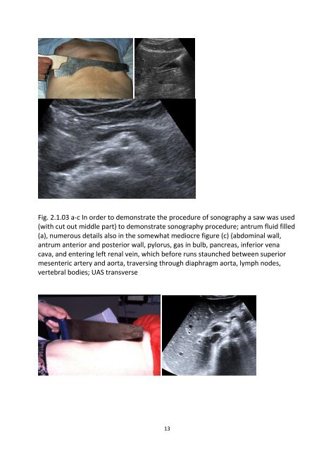
Fig. 2.1.03 a-c In order to demonstrate the procedure of sonography a saw was used<br />
(with cut out middle part) to demonstrate sonography procedure; antrum fluid filled<br />
(a), numerous details also in the somewhat mediocre figure (c) (abdominal wall,<br />
antrum anterior and posterior wall, pylorus, gas in bulb, pancreas, inferior vena<br />
cava, and entering left renal vein, which before runs staunched between superior<br />
mesenteric artery and aorta, traversing through diaphragm aorta, lymph nodes,<br />
vertebral bodies; UAS transverse<br />
13