Greiner-Ultrasound-Leseprobe
To conduct an ultrasound is a clinical art – the more so, if it is to be a good ultrasound; and who would even think of it without this sense of entitlement? Possibly the beginner, for whom this book was written, containing advice for the ultrasound examination of the abdomen and other body regions with numerous case studies, black and white or with colour, with or without contrast medium, interventional or without needle and drainage, simple or complex (and purposely only loosely structured according to organ systems, similar to that which is characteristic of the everyday working environment) – and without any claim to completeness. Sonography requires taking a very close look and an adequate degree of creative association, as well as fantasy and clinical ability for deduction – not too much, but also not too little either.
To conduct an ultrasound is a clinical art – the more so, if it is to be a good
ultrasound; and who would even think of it without this sense of entitlement?
Possibly the beginner, for whom this book was written, containing advice
for the ultrasound examination of the abdomen and other body regions with numerous case studies, black and white or with colour, with or without contrast medium, interventional or without needle and drainage, simple or complex (and purposely only loosely structured according to organ systems, similar to that which is characteristic of the everyday working environment) – and without any claim to completeness.
Sonography requires taking a very close look and an adequate degree of creative association, as well as fantasy and clinical ability for deduction –
not too much, but also not too little either.

Create successful ePaper yourself
Turn your PDF publications into a flip-book with our unique Google optimized e-Paper software.
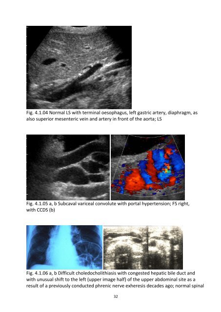
Fig. 4.1.04 Normal LS with terminal oesophagus, left gastric artery, diaphragm, as<br />
also superior mesenteric vein and artery in front of the aorta; LS<br />
Fig. 4.1.05 a, b Subcaval variceal convolute with portal hypertension; FS right,<br />
with CCDS (b)<br />
Fig. 4.1.06 a, b Difficult choledocholithiasis with congested hepatic bile duct and<br />
with unusual shift to the left (upper image half) of the upper abdominal site as a<br />
result of a previously conducted phrenic nerve exheresis decades ago; normal spinal<br />
32