

A-and-R-Classification-Criteria-Macrophage-Activation-Syndrome-2016
A-and-R-Classification-Criteria-Macrophage-Activation-Syndrome-2016
A-and-R-Classification-Criteria-Macrophage-Activation-Syndrome-2016

You also want an ePaper? Increase the reach of your titles
YUMPU automatically turns print PDFs into web optimized ePapers that Google loves.
ARTHRITIS & RHEUMATOLOGY<br />
DOI 10.1002/ART.39332<br />
VC <strong>2016</strong>, American College of Rheumatology<br />
SPECIAL ARTICLE<br />
<strong>2016</strong> <strong>Classification</strong> <strong>Criteria</strong> for<br />
<strong>Macrophage</strong> <strong>Activation</strong> <strong>Syndrome</strong> Complicating<br />
Systemic Juvenile Idiopathic Arthritis<br />
A European League Against Rheumatism/American College of Rheumatology/<br />
Paediatric Rheumatology International Trials Organisation<br />
Collaborative Initiative<br />
Angelo Ravelli, 1 Francesca Minoia, 2 Sergio Davı, 2 AnnaCarin Horne, 3 Francesca Bovis, 2<br />
Angela Pistorio, 2 Maurizio Arico, 4 Tadej Avcin, 5 Edward M. Behrens, 6 Fabrizio De Benedetti, 7<br />
Lisa Filipovic, 8 Alexei A. Grom, 8 Jan-Inge Henter, 3 Norman T. Ilowite, 9 Michael B. Jordan, 8<br />
Raju Khubch<strong>and</strong>ani, 10 Toshiyuki Kitoh, 11 Kai Lehmberg, 12 Daniel J. Lovell, 8 Paivi Miettunen, 13<br />
Kim E. Nichols, 14 Seza Ozen, 15 Jana Pachlopnik Schmid, 16 Athimalaipet V. Ramanan, 17<br />
Ricardo Russo, 18 Rayfel Schneider, 19 Gary Sterba, 20 Yosef Uziel, 21 Carol Wallace, 22<br />
Carine Wouters, 23 Nico Wulffraat, 24 Erkan Demirkaya, 25 Hermine I. Brunner, 8<br />
Alberto Martini, 1 Nicolino Ruperto, 2 <strong>and</strong> R<strong>and</strong>y Q. Cron, 26 on behalf of the<br />
Paediatric Rheumatology International Trials Organisation, the Childhood Arthritis <strong>and</strong><br />
Rheumatology Research Alliance, the Pediatric Rheumatology Collaborative Study Group,<br />
<strong>and</strong> the Histiocyte Society<br />
This criteria set has been approved by the European League Against Rheumatism (EULAR) Executive<br />
Committee <strong>and</strong> the American College of Rheumatology (ACR) Board of Directors. This signifies that the<br />
criteria set has been quantitatively validated using patient data, <strong>and</strong> it has undergone validation based on<br />
an independent data set. All EULAR/ACR-approved criteria sets are expected to undergo intermittent<br />
updates.<br />
The ACR is an independent, professional, medical <strong>and</strong> scientific society that does not guarantee, warrant,<br />
or endorse any commercial product or service.<br />
This article is published simultaneously in the March <strong>2016</strong><br />
issue of Annals of the Rheumatic Diseases.<br />
Supported by the American College of Rheumatology, the<br />
European League Against Rheumatism, <strong>and</strong> the Paediatric Rheumatology<br />
International Trials Organisation.<br />
1 Angelo Ravelli, MD, Alberto Martini, MD: Universita<br />
degli Studi di Genova <strong>and</strong> Istituto Giannina Gaslini, Genoa, Italy;<br />
2 Francesca Minoia, MD, Sergio Davı, MD, Francesca Bovis, BiolD,<br />
Angela Pistorio, MD, PhD, Nicolino Ruperto, MD, MPH: Istituto<br />
Giannina Gaslini, Genoa, Italy;<br />
3 AnnaCarin Horne, MD, PhD,<br />
Jan-Inge Henter, MD, PhD: Karolinska Institute <strong>and</strong> Karolinska<br />
University Hospital Solna, Stockholm, Sweden; 4 Maurizio Arico, MD:<br />
Azienda Sanitaria Provinciale 7, Ragusa, Italy; 5 Tadej Avcin, MD:<br />
University Children’s Hospital, Ljubljana, Slovenia;<br />
6 Edward M.<br />
1
2 RAVELLI ET AL<br />
Behrens, MD: The Children’s Hospital of Philadelphia, Philadelphia,<br />
Pennsylvania;<br />
7 Fabrizio De Benedetti, MD: Ospedale Pediatrico<br />
Bambino Gesu, Rome, Italy; 8 Lisa Filipovich, MD, Alexei A. Grom,<br />
MD, Michael B. Jordan, MD, Daniel J. Lovell, MD, MPH, Hermine<br />
I. Brunner, MD: Cincinnati Children’s Hospital Medical Center,<br />
Cincinnati, Ohio; 9 Norman T. Ilowite, MD: Albert Einstein College of<br />
Medicine <strong>and</strong> Children’s Hospital at Montefiore, Bronx, New York;<br />
10 Raju Khubch<strong>and</strong>ani, MD: Jaslok Hospital <strong>and</strong> Research Centre,<br />
Mumbai, India; 11 Toshiyuki Kitoh, MD, PhD: Aichi Medical University,<br />
Nagakute, Japan; 12 Kai Lehmberg, MD: University Medical Center,<br />
Hamburg, Germany;<br />
13 Paivi Miettunen, MD: University of<br />
Calgary, Calgary, Alberta, Canada; 14 Kim E. Nichols, MD: St. Jude<br />
Children’s Research Hospital, Memphis, Tennessee; 15 Seza Ozen, MD:<br />
Hacettepe University, Ankara, Turkey; 16 Jana Pachlopnick Schmid,<br />
MD, PhD: University Children’s Hospital, Zurich, Switzerl<strong>and</strong>; 17 Athimalaipet<br />
V. Ramanan, MD: Bristol Royal Hospital for Children, Bristol,<br />
UK;<br />
18 Ricardo Russo, MD: Hospital de Pediatria Juan P.<br />
Garrahan, Buenos Aires, Argentina;<br />
19 Rayfel Schneider, MBBCh:<br />
University of Toronto <strong>and</strong> Hospital for Sick Children, Toronto, Ontario,<br />
Canada; 20 Gary Sterba, MD: Mount Sinai Medical Center, Miami<br />
Beach, Florida; 21 Yosef Uziel, MD: Meir Medical Centre, Kfar Saba,<br />
Israel; 22 Carol Wallace, MD: Seattle Children’s Hospital <strong>and</strong> University<br />
of Washington, Seattle; 23 Carine Wouters, MD, PhD: University<br />
Hospital Leuven, Leuven, Belgium; 24 Nico Wulffraat, MD: Wilhelmina<br />
Children’s Hospital <strong>and</strong> University Medical Center Utrecht,<br />
Uthrecht, The Netherl<strong>and</strong>s; 25 Erkan Demirkaya, MD: Gulhane Military<br />
Medical Faculty, Ankara, Turkey; 26 R<strong>and</strong>y Q. Cron, MD, PhD:<br />
University of Alabama at Birmingham.<br />
Address correspondence to Angelo Ravelli, MD, Pediatria II,<br />
Istituto G. Gaslini, Largo G. Gaslini 5, 16147 Genoa, Italy. E-mail:<br />
angeloravelli@ospedale-gaslini.ge.it.<br />
Submitted for publication April 2, 2015; accepted in revised<br />
form November 30, 2015.<br />
Objective. To develop criteria for the classification<br />
of macrophage activation syndrome (MAS) in patients<br />
with systemic juvenile idiopathic arthritis (JIA).<br />
Methods. A multistep process, based on a combination<br />
of expert consensus <strong>and</strong> analysis of real patient<br />
data, was conducted. A panel of 28 experts was first<br />
asked to classify 428 patient profiles as having or not<br />
having MAS, based on clinical <strong>and</strong> laboratory features<br />
at the time of disease onset. The 428 profiles comprised<br />
161 patients with systemic JIA–associated MAS <strong>and</strong><br />
267 patients with a condition that could potentially be<br />
confused with MAS (active systemic JIA without evidence<br />
of MAS, or systemic infection). Next, the ability<br />
of c<strong>and</strong>idate criteria to classify individual patients as<br />
having MAS or not having MAS was assessed by evaluating<br />
the agreement between the classification yielded<br />
using the criteria <strong>and</strong> the consensus classification of<br />
the experts. The final criteria were selected in a consensus<br />
conference.<br />
Results. Experts achieved consensus on the classification<br />
of 391 of the 428 patient profiles (91.4%).<br />
A total of 982 c<strong>and</strong>idate criteria were tested statistically.<br />
The 37 best-performing criteria <strong>and</strong> 8 criteria obtained<br />
from the literature were evaluated at the consensus conference.<br />
During the conference, 82% consensus among<br />
experts was reached on the final MAS classification criteria.<br />
In validation analyses, these criteria had a sensitivity<br />
of 0.73 <strong>and</strong> a specificity of 0.99. Agreement between<br />
the classification (MAS or not MAS) obtained using the<br />
criteria <strong>and</strong> the original diagnosis made by the treating<br />
physician was high (k 5 0.76).<br />
Conclusion. We have developed a set of classification<br />
criteria for MAS complicating systemic JIA <strong>and</strong><br />
provided preliminary evidence of its validity. Use of<br />
these criteria will potentially improve underst<strong>and</strong>ing of<br />
MAS in systemic JIA <strong>and</strong> enhance efforts to discover<br />
effective therapies, by ensuring appropriate patient<br />
enrollment in studies.<br />
Introduction<br />
<strong>Macrophage</strong> activation syndrome (MAS) is the<br />
term used to describe a potentially life-threatening complication<br />
of systemic inflammatory disorders, which<br />
occurs most commonly in systemic juvenile idiopathic<br />
arthritis (JIA) <strong>and</strong> in its adult equivalent, adult-onset<br />
Still’s disease (1–4), although its occurrence in patients<br />
with other autoimmune or autoinflammatory conditions,<br />
i.e., adult- <strong>and</strong> childhood-onset systemic lupus<br />
erythematosus (5,6), Kawasaki disease (7,8), <strong>and</strong> periodic<br />
fever syndromes (9,10), is being reported with<br />
increased frequency. MAS is characterized by an overwhelming<br />
inflammatory reaction due to an uncontrolled<br />
<strong>and</strong> dysfunctional immune response involving the continual<br />
activation <strong>and</strong> expansion of T lymphocytes <strong>and</strong><br />
macrophages, which results in massive hypersecretion of<br />
proinflammatory cytokines (11,12).<br />
Characteristic clinical features of MAS are high,<br />
nonremitting fever, hepatosplenomegaly, generalized<br />
lymphadenopathy, central nervous system dysfunction,<br />
<strong>and</strong> hemorrhagic manifestations. Typical laboratory<br />
abnormalities include pancytopenia, increased levels of<br />
ferritin, liver enzymes, lactate dehydrogenase, triglycerides,<br />
D-dimers, <strong>and</strong> soluble interleukin-2 (IL-2) receptor<br />
a (also known as soluble CD25 [sCD25]), <strong>and</strong> decreased<br />
fibrinogen levels. A typical histopathologic feature of<br />
MAS is the accumulation of well-differentiated macrophages<br />
exhibiting hemophagocytic activity in bone<br />
marrow biopsy specimens or aspirates (13). Although the<br />
prevalence of MAS among patients with systemic JIA has<br />
been estimated to be ;10%, recent reports suggest that<br />
subclinical MAS may occur in as many as 30-40% of<br />
patients with systemic JIA (14,15).<br />
MAS can result in progressive multi-organ failure<br />
<strong>and</strong> eventually a fatal outcome if unrecognized. Recent<br />
studies indicate a mortality rate of 8% (16,17), making
EULAR/ACR CLASSIFICATION CRITERIA FOR MAS 3<br />
timely diagnosis <strong>and</strong> prompt initiation of appropriate treatment<br />
imperative. However, early recognition of MAS is<br />
often challenging, given the lack of a single pathognomonic<br />
clinical or laboratory feature. Furthermore, histopathologic<br />
features of hemophagocytosis may not be present in the<br />
initial stages (18,19) <strong>and</strong> lack specificity for hemophagocytic<br />
syndromes (20). In addition, features of MAS may be<br />
difficult to distinguish from other conditions that may present<br />
with overlapping manifestations, such as flares of systemic<br />
JIA or systemic infections. Recently, a wide disparity<br />
in the frequency <strong>and</strong> severity of the classic clinical <strong>and</strong> laboratory<br />
features across patients has been described (16,17).<br />
The difficulties in making the diagnosis of MAS<br />
<strong>and</strong> its clinical heterogeneity, together with the recent<br />
advances in its treatment <strong>and</strong> in underst<strong>and</strong>ing of its pathophysiology<br />
<strong>and</strong> underlying genetic defects (11,21–23),<br />
emphasize the need for accurate criteria to aid physicians<br />
in appropriately classifying patients as having MAS to<br />
facilitate enrollment into clinical studies. The recognition<br />
that the syndrome is clinically similar to hemophagocytic<br />
lymphohistiocytosis (HLH) has led some to recommend<br />
the use of the HLH-2004 diagnostic guidelines (24). An<br />
alternative approach is based on application of the preliminary<br />
diagnostic guidelines for MAS complicating systemic<br />
JIA (25). However, although both sets of guidelines have<br />
been utilized for detecting MAS in patients with systemic<br />
JIA, each has several limitations (26). The primary purpose<br />
of the international collaborative project described<br />
herein, conducted under the auspices of the European<br />
League Against Rheumatism, the American College of<br />
Rheumatology, <strong>and</strong> the Paediatric Rheumatology International<br />
Trials Organisation (PRINTO), was to develop a set<br />
of classification criteria for MAS complicating systemic<br />
JIA, based on a combination of expert consensus, available<br />
evidence from the medical literature, <strong>and</strong> analysis of real<br />
patient data.<br />
Methods<br />
A multistep process was used in developing the classification<br />
criteria <strong>and</strong> included the following phases: 1) a Delphi<br />
survey of international pediatric rheumatologists, aimed at<br />
identifying MAS features potentially suitable for inclusion in<br />
classification criteria (27); 2) large-scale data collection on<br />
patients with systemic JIA–associated MAS <strong>and</strong> patients with<br />
2 other conditions that potentially could be confused with<br />
MAS; 3) a web-based procedure for ascertaining consensus<br />
among experts; 4) selection of c<strong>and</strong>idate criteria through statistical<br />
analyses; 5) selection of final classification criteria in a consensus<br />
conference; <strong>and</strong> 6) cross-sectional validation of final<br />
classification criteria. Health professionals <strong>and</strong> patient/parent<br />
representatives were not included in the study task force<br />
because the project did not involve any issues of specific interest<br />
to these stakeholders. In particular, none of the study assessments<br />
required the participation of health professionals, <strong>and</strong> no<br />
patient/parent-reported outcomes were incorporated.<br />
Data collection on patients with MAS <strong>and</strong> patients<br />
with conditions that could be confused with MAS. The design,<br />
inclusion criteria, <strong>and</strong> data collection procedures of this portion<br />
of the project have been described in detail previously<br />
(16,17,26). Briefly, international pediatric rheumatologists <strong>and</strong><br />
pediatric hematologists were invited to participate in a retrospective<br />
cohort study of patients with systemic JIA–associated<br />
MAS or with 1 of 2 conditions that could potentially be<br />
confused with MAS, i.e., active systemic JIA not complicated<br />
by MAS <strong>and</strong> systemic infection.<br />
For patients with MAS, information on laboratory features<br />
at 3 time points (the last visit before onset of MAS, the<br />
time of MAS onset, <strong>and</strong> the period of full-blown MAS) was<br />
collected. Because the classification criteria were aimed at<br />
identification of MAS in its earlier stages, only laboratory data<br />
recorded at the time of onset were retained. Data at the time<br />
of presentation in patients with conditions that could be confused<br />
with MAS were also obtained. Except for blood cell<br />
counts <strong>and</strong> acute-phase reactant levels, values of laboratory<br />
parameters were tested using both the original values provided<br />
by each local laboratory <strong>and</strong> the values st<strong>and</strong>ardized according<br />
to the SI unit system based on their normal ranges, as previously<br />
reported (16).<br />
A total of 1,111 patients (362 with systemic JIA–associated<br />
MAS, 404 with active systemic JIA without MAS, <strong>and</strong> 345 with<br />
systemic infection) were reported by 95 pediatric subspecialists<br />
practicing in 33 countries in 6 continents. Pediatric subspecialists<br />
who provided these data are listed in Appendix A. The features of<br />
the patients with MAS <strong>and</strong> the comparison patients have been<br />
described elsewhere (16,17,26).<br />
Web-based procedures for ascertaining consensus<br />
among experts. At present, there is no single feature that is<br />
pathognomonic for MAS. Furthermore, no prior validated<br />
diagnostic or classification criteria are available. In order to<br />
classify patients as having or not having MAS, we therefore<br />
decided to use expert consensus as the “gold st<strong>and</strong>ard.” Based<br />
on publication records <strong>and</strong> experience in the care of children<br />
with MAS <strong>and</strong> related disorders, a panel of 28 experts (20<br />
pediatric rheumatologists <strong>and</strong> 8 pediatric hematologists) was<br />
created.<br />
The experts were asked to classify a total of 428 patient<br />
profiles as having or not having MAS, based on the clinical <strong>and</strong><br />
laboratory features recorded at disease onset. The 428 profiles<br />
were selected r<strong>and</strong>omly from among the 1,111 patients whose<br />
data were collected <strong>and</strong> comprised 161 patients with MAS, 140<br />
patients with active systemic JIA without evidence of MAS, <strong>and</strong><br />
127 patients with systemic infection. Selection bias was unlikely,<br />
as the characteristics of patients who were selected <strong>and</strong> those<br />
who were not selected were comparable (data not shown). The<br />
experts were intentionally kept unaware of the original diagnosis<br />
<strong>and</strong> overall course of each patient.<br />
Each patient profile included information about the<br />
presence or absence of key clinical manifestations, <strong>and</strong> the values<br />
of laboratory parameters <strong>and</strong> normal ranges at the respective<br />
institutions. Based on these data, all experts were asked to<br />
classify each patient as having or not having MAS. The minimum<br />
required level of agreement among experts was set at<br />
80%. If an 80% consensus was not attained, the patient profile<br />
was discussed in a further round. Profiles for which consensus<br />
had not been achieved after the final round were declared
4 RAVELLI ET AL<br />
uninterpretable <strong>and</strong> discarded from further analyses. Three<br />
rounds of voting were used, with prior vote data <strong>and</strong> recorded<br />
comments available to all of the participants before each vote<br />
took place, to augment the number of consensus decisions. All<br />
web-based consensus procedures were conducted by PRINTO.<br />
Selection of best classification criteria through statistical<br />
analyses. All statistical analyses used for selecting the<br />
best classification criteria were conducted only on the sample<br />
of patients for whom the experts achieved consensus about the<br />
diagnosis of MAS or non-MAS. Cutoff values for laboratory<br />
tests were calculated with the receiver operator characteristic<br />
(ROC) curve method, by identifying the point on the ROC<br />
curve that best discriminated between patients classified by the<br />
experts as having MAS <strong>and</strong> those classified as not having MAS.<br />
The aim of this exercise was to assess the ability of c<strong>and</strong>idate<br />
criteria to classify individual patients as having or not<br />
having MAS, <strong>and</strong> to evaluate the agreement between the classification<br />
yielded by the criteria <strong>and</strong> the consensus classification<br />
of experts. C<strong>and</strong>idate classification criteria were partly derived<br />
from the literature <strong>and</strong> partly generated from the study data.<br />
Literature criteria included the following: 1) the preliminary<br />
diagnostic guidelines for MAS complicating systemic<br />
JIA (25), 2) the same guidelines modified by addition of the<br />
item ferritin at various threshold levels (500, 1,000 or 1,500<br />
ng/ml), <strong>and</strong> 3) the HLH-2004 diagnostic guidelines (24),<br />
adapted by eliminating 3 of the 8 items because information<br />
about presence of hemophagocytosis was not available for<br />
both comparison groups, <strong>and</strong> neither natural killer (NK) cell<br />
activity nor sCD25 levels were determined in all patients.<br />
<strong>Criteria</strong> obtained from the study data were generated in 2<br />
ways: 1) through the evaluation, by the project steering committee,<br />
of all combinations of clinical <strong>and</strong> laboratory variables<br />
(see some examples in Supplementary Table 1, on the Arthritis<br />
& Rheumatology web site at http://onlinelibrary.wiley.com/doi/<br />
10.1002/art.39332/abstract) (combination of criteria approach),<br />
<strong>and</strong> 2) by assigning weights to clinical <strong>and</strong> laboratory variables,<br />
on the basis of their association with the diagnosis of MAS<br />
made by the experts, through multivariable logistic regression<br />
analysis. For each combination of variables that were significantly<br />
associated with the diagnosis of MAS in logistic regression<br />
models, the rule was to convert the odds ratio of each<br />
variable to its percentage value out of a total of 100%. Each set<br />
of criteria was then composed of a group of variables whose<br />
sum of weights made up a total score of 100 (MAS score). The<br />
cutoff value in the MAS score that was associated with the<br />
higher likelihood of the presence of MAS was obtained by calculating<br />
the point on the ROC curve that corresponded to the<br />
highest sensitivity <strong>and</strong> specificity.<br />
A total of 982 c<strong>and</strong>idate classification criteria were<br />
tested. For each set of criteria, we calculated the sensitivity<br />
(ability of the criteria to identify a patient as having MAS who<br />
had been classified as having MAS according to the expert<br />
panel), the specificity (ability of the criteria to identify a<br />
patient as not having MAS who had been classified as not having<br />
MAS by the experts), the positive <strong>and</strong> negative predictive<br />
value, the area under the ROC curve (AUC), <strong>and</strong> the kappa<br />
value for agreement between the classification yielded by the<br />
criteria <strong>and</strong> the classification made by the experts. Although<br />
Figure 1. Patient samples evaluated in the study <strong>and</strong> results of web-based expert evaluations. MAS 5 macrophage activation syndrome;<br />
sJIA 5 systemic juvenile idiopathic arthritis.
EULAR/ACR CLASSIFICATION CRITERIA FOR MAS 5<br />
there was one single model with the highest predictive value<br />
(criterion no. 929; Supplementary Table 1), we generated multiple<br />
combinations for comparison because we believed that<br />
less predictive models might have more face validity with the<br />
experts. Nevertheless, it was established that in order to qualify<br />
for inclusion in expert voting procedures at the consensus<br />
conference, a set of classification criteria should demonstrate<br />
a kappa value of $0.85, a sensitivity of $0.80, a specificity<br />
of $0.93, <strong>and</strong> an AUC of $0.90. An exception was made for<br />
the historical literature criteria, which were retained for further<br />
consideration even if they did not meet all statistical<br />
requirements.<br />
Selection of the final classification criteria at consensus<br />
conference. The International Consensus Conference on<br />
MAS <strong>Classification</strong> <strong>Criteria</strong> was held in Genoa, Italy on<br />
March 21–22, 2014. The meeting was attended by all 28<br />
experts who participated in web-based consensus evaluations<br />
<strong>and</strong> was facilitated by 2 moderators (HIB <strong>and</strong> NR) with<br />
expertise in nominal group technique. The overall goal of the<br />
meeting was to decide on a preliminary set of classification<br />
criteria using a combination of statistical <strong>and</strong> consensus formation<br />
techniques.<br />
A plenary session was first held to present the scope,<br />
methodology, <strong>and</strong> flow of the project, the results of the Delphi<br />
survey, the characteristics of patients included in the data collection,<br />
the results of web-based consensus procedures <strong>and</strong> of<br />
statistical analyses of c<strong>and</strong>idate classification criteria, <strong>and</strong> the<br />
methodology of the nominal group technique. Participants<br />
were then r<strong>and</strong>omized into 2 equal-sized nominal groups <strong>and</strong><br />
were asked to rank using nominal group technique, independently<br />
of each other <strong>and</strong> based on the evaluation of both ease<br />
of use <strong>and</strong> credibility (face/content validity) <strong>and</strong> statistical performance<br />
(particularly in terms of sensitivity, specificity, <strong>and</strong><br />
kappa value), the 5 best classification criteria from 5 (highest)<br />
to 1 (lowest). All experts were connected by their laptops to a<br />
central computer <strong>and</strong> submitted all of their rankings electronically.<br />
A series of repeated independent voting sessions was<br />
held until the top 3 classification criteria were selected by each<br />
voting group. Then an 80% consensus was attained on the best<br />
(final) set of classification criteria, in a session with members<br />
of the 2 nominal groups combined.<br />
Analysis of the association between the variables included<br />
in the final classification criteria <strong>and</strong> the web-based experts’<br />
consensus evaluations. The association between the final classification<br />
criteria <strong>and</strong> the web-based evaluations made by the<br />
experts was assessed by multiple logistic regression analysis,<br />
which used as explanatory variables the individual items<br />
included in the final classification criteria <strong>and</strong> as the dependent<br />
variable the web-based expert consensus on classification<br />
of patients as having or not having MAS. The effect was<br />
expressed in terms of odds ratios, <strong>and</strong> 95% confidence intervals<br />
were calculated; statistical significance was tested by likeli-<br />
Table 1. Comparison of clinical <strong>and</strong> laboratory features at disease onset between patients classified by the 28-member expert panel as having<br />
macrophage activation syndrome (MAS) (n 5 95) <strong>and</strong> those classified as not having MAS (n 5 296)<br />
No. with<br />
available<br />
data<br />
Patients classified as<br />
having MAS<br />
No. (%) or<br />
median (IQR)*<br />
No. with<br />
available<br />
data<br />
Patients classified as<br />
not having MAS<br />
No. (%) or<br />
median (IQR)*<br />
Clinical manifestations<br />
Fever 94 93 (98.9) 294 278 (94.6) 0.08<br />
Hepatomegaly 94 68 (72.3) 295 75 (25.4) ,0.0001<br />
Splenomegaly 92 53 (57.6) 294 67 (22.8) ,0.0001<br />
Lymphadenopathy 91 48 (52.8) 292 73 (25.0) ,0.0001<br />
Central nervous system involvement 93 40 (43.0) 292 25 (8.6) ,0.0001<br />
Hemorrhagic manifestations 92 25 (27.2) 294 16 (5.4) ,0.0001<br />
Heart involvement 94 27 (28.7) 294 34 (11.6) ,0.0001<br />
Lung involvement 95 27 (28.4) 294 40 (13.6) 0.0009<br />
Kidney involvement 95 16 (16.8) 295 16 (5.4) 0.0004<br />
Laboratory results<br />
Hemoglobin, gm/dl 95 9.9 (8.0–11.2) 289 10.9 (9.4–12.2) ,0.0001<br />
White blood cell count, 310 9 /liter 95 8.1 (3.2–12.8) 289 15.3 (9.9–20.1) ,0.0001<br />
Neutrophil count, 310 9 /liter 82 3.7 (1.5–8.0) 236 9.4 (5.1–14.2) ,0.0001<br />
Platelet count, 310 9 /liter 95 98 (57–141) 290 385 (286–551) ,0.0001<br />
Erythrocyte sedimentation rate, mm/hour 90 28 (17–65) 245 70 (39–93) ,0.0001<br />
C-reactive protein, mg/dl 85 8.7 (2.4–16.1) 282 8.2 (2.4–15.6) 0.63<br />
Aspartate aminotransferase, units/liter 93 171 (98–436) 284 30 (22–45) ,0.0001<br />
Alanine aminotransferase, units/liter 91 115 (43–283) 284 18 (12–34) ,0.0001<br />
Lactate dehydrogenase, units/liter 81 1,560 (801–2,400) 248 482 (362–688) ,0.0001<br />
Triglycerides, mg/dl 86 267 (192–358) 186 123 (96–160) ,0.0001<br />
Albumin, gm/dl 79 3.0 (2.6–3.5) 252 3.7 (3.2–4.1) ,0.0001<br />
Serum sodium, mEq/liter 80 136 (133–140) 259 138 (136–141) 0.003<br />
Fibrinogen, mg/dl 88 220 (148–345) 226 500 (356–650) ,0.0001<br />
Ferritin, ng/ml 90 9,094 (2,000–19,767) 244 268 (62–938) ,0.0001<br />
D-dimer, ng/ml 48 3,579 (1,834–7,373) 94 1,638 (528–3,325) ,0.0001<br />
* Clinical manifestations are reported as the number (%) <strong>and</strong> laboratory results as the median (interquartile range [IQR]).<br />
P
6 RAVELLI ET AL<br />
Table 2. Univariate analysis of the ability of specific variables to distinguish patients with macrophage activation<br />
syndrome from comparison patients, as assessed using the 391 patients for whom expert consensus was<br />
achieved<br />
No. of patients<br />
with available<br />
data OR (95% CI)* P<br />
Clinical features<br />
Central nervous system involvement 385 8.1 (4.5–14.4) ,0.0001<br />
Hepatomegaly 389 7.7 (4.5–12.9) ,0.0001<br />
Hemorrhagic manifestations 386 6.5 (3.3–12.8) ,0.0001<br />
Fever 388 5.4 (0.7–40.9) 0.106<br />
Splenomegaly 386 4.6 (2.8–7.6) ,0.0001<br />
Lymphoadenopathy 383 3.4 (2.1–5.5) ,0.0001<br />
Kidney involvement 390 3.5 (1.7–7.4) 0.0008<br />
Heart involvement 388 3.1 (1.7–5.5) 0.0001<br />
Lung involvement 389 2.5 (1.4–4.4) 0.0011<br />
Active arthritis 390 1.6 (1.0–2.6) 0.0587<br />
Laboratory features<br />
Non-normalized values<br />
Ferritin .684 ng/ml 334 111.6 (26.7–465.8) ,0.0001<br />
Platelet count #181 3 10 9 /liter 385 84.3 (40–177.5) ,0.0001<br />
Aspartate aminotransferase .48 units/liter 377 51.9 (21.7–124.4) ,0.0001<br />
Lactate dehydrogenase .853 units/liter 329 20.0 (10.7–37.3) ,0.0001<br />
Triglycerides .156 mg/dl 272 19.6 (9.4–40.8) ,0.0001<br />
Alanine aminotransferase .36 units/liter 375 18.0 (9.4–34.3) ,0.0001<br />
Fibrinogen #360 mg/dl 314 11.5 (6.3–21.0) ,0.0001<br />
Neutrophil count #3.7 3 10 9 /liter 318 5.9 (3.4–10.5) ,0.0001<br />
D-dimer .1,350 ng/ml 142 5.7 (2.4–13.4) ,0.0001<br />
Erythrocyte sedimentation rate #30 mm/hour 335 5.6 (3.3–9.5) ,0.0001<br />
Albumin #3.6 gm/dl 331 5.5 (3.0–10.1) ,0.0001<br />
Hemoglobin #8.5 gm/dl 384 4.9 (2.7–8.8) ,0.0001<br />
White blood cell count #10.2 3 10 9 /liter 384 4.6 (2.8–7.5) ,0.0001<br />
Serum sodium #133, mEq/liter 339 2.9 (1.7–5.0) 0.0002<br />
C-reactive protein .0.86 mg/dl 367 2.4 (1.0–5.9) 0.0501<br />
Normalized values<br />
Ferritin .2,773.6 ng/ml 334 23.9 (12.7–44.8) ,0.0001<br />
Aspartate aminotransferase .44.4 units/liter 377 47.4 (21.6–103.9) ,0.0001<br />
Alanine aminotransferase .23 units/liter 375 39.4 (14.1–110.5) ,0.0001<br />
Lactate dehydrogenase .238.8 units/liter 329 39.2 (18.2–84.6) ,0.0001<br />
Triglycerides .193.9 mg/dl 272 16.3 (8.7–30.8) ,0.0001<br />
Fibrinogen #318.9 mg/dl 314 11.0 (6.1–20.0) ,0.0001<br />
Albumin #3.9 gm/dl 331 6.4 (3.6–11.3) ,0.0001<br />
D-dimer .1,320.3 ng/ml 142 5.2 (2.1–12.7) ,0.0001<br />
Serum sodium #135 mEq/liter 339 3.3 (1.9–5.6) ,0.0001<br />
*OR5 odds ratio; 95% CI 5 95% confidence interval.<br />
hood ratio test. The AUC of the model was used as an indicator<br />
of its predictive ability. The purpose of this post-consensus<br />
analysis was to evaluate which were the variables that most<br />
influenced the experts’ decision to classify the patients as having<br />
or not having MAS.<br />
Validation of final classification criteria. Validation analysis<br />
was performed by assessing the performance of the criteria,<br />
in terms of sensitivity, specificity, negative predictive value, positive<br />
predictive value, AUC, <strong>and</strong> kappa value, in discriminating<br />
patients with MAS from patients with the 2 conditions with<br />
which MAS could be confused (combined in a single group),<br />
using the original diagnosis made by the caring physician (i.e.,<br />
the investigator who entered the patient’s data in the study web<br />
site) as the gold st<strong>and</strong>ard. This analysis was performed on the<br />
sample of patients not used for expert evaluations (n 5 683).<br />
Only patients with available data on all items included in the<br />
final classification criteria were used for the analyses.<br />
Results<br />
Results of web-based process for ascertainment<br />
of expert consensus. After 3 rounds of web-based evaluations,<br />
the experts achieved consensus on the classification<br />
of 391 (91.4%) of the 428 patient profiles examined (Figure<br />
1). A total of 95 patients were classified as having MAS<br />
by the experts, 88 of whom had also been diagnosed as having<br />
MAS by the treating physician; the original diagnosis<br />
had been systemic JIA without MAS in 3 patients <strong>and</strong> systemic<br />
infection in 4. A total of 296 patients were classified<br />
by the experts as not having MAS, 47 of whom had been<br />
diagnosed as having MAS by the treating physician. Thirtyseven<br />
patient profiles for which 80% consensus among<br />
experts was not reached were discarded. A comparison of
EULAR/ACR CLASSIFICATION CRITERIA FOR MAS 7<br />
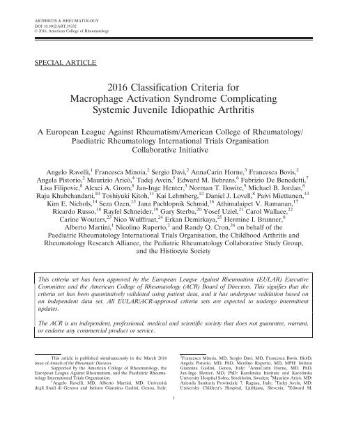
Figure 2. <strong>Criteria</strong> for the classification of macrophage activation syndrome in patients with systemic juvenile idiopathic arthritis. Laboratory abnormalities<br />
should not be otherwise explained by the patient’s condition, such as concomitant immune-mediated thrombocytopenia, infectious hepatitis,<br />
visceral leishmaniasis, or familial hyperlipidemia.<br />
clinical <strong>and</strong> laboratory features between patients diagnosed<br />
by the experts as having MAS <strong>and</strong> those diagnosed as not<br />
having MAS is shown in Table 1. Overall, patients whose<br />
condition was classified as MAS by the experts had more<br />
severe clinical <strong>and</strong> laboratory features than those classified<br />
as not having MAS.<br />
C<strong>and</strong>idate clinical <strong>and</strong> laboratory variables<br />
determined by univariate analysis. In univariate analyses,<br />
the 10 variables found to have the greatest ability to distinguish<br />
patients with MAS from comparison patients were<br />
as follows: ferritin level, platelet count, levels of aspartate<br />
transaminase (AST), lactate dehydrogenase, triglycerides,<br />
alanine transaminase (ALT), <strong>and</strong> fibrinogen, central nervous<br />
system involvement, hepatomegaly, <strong>and</strong> hemorrhagic<br />
manifestations (Table 2). These variables qualified for inclusion<br />
in logistic regression analyses aimed at generating c<strong>and</strong>idate<br />
classification criteria through the MAS score method<br />
described above. However, ALT levels were excluded owing<br />
Table 3. Logistic regression model to assess association between<br />
the variables included in the final classification criteria <strong>and</strong> the<br />
experts’ consensus of patients’ classification as having or not having<br />
macrophage activation syndrome*<br />
Explanatory variable OR (95% CI) P†<br />
Ferritin .684 ng/ml .999.999 (40.62.999.999) ,0.0001<br />
Platelet count<br />
237.0 (18.22.999.999) ,0.0001<br />
#181 3 10 9 /liter<br />
Triglycerides .156 mg/dl 18.3 (2.0–163.9) 0.009<br />
Fibrinogen #360 mg/dl 16.0 (2.0–126.4) 0.009<br />
Aspartate aminotransferase<br />
.48 units/liter<br />
10.0 (1.3–78.4) 0.029<br />
* The experts’ consensus on patients’ classification as having or not<br />
having macrophage activation syndrome (from the web-based process)<br />
was the dependent variable. Complete data were available on<br />
227 patients. The area under the receiver operating characteristic<br />
curve of the model was 0.99. OR 5 odds ratio; 95% CI 5 95% confidence<br />
interval.<br />
† By likelihood ratio test.<br />
to their close correlation with AST levels <strong>and</strong> a slightly lower<br />
statistical performance. They were replaced by neutrophil<br />
count or albumin level, depending on the model.<br />
C<strong>and</strong>idate classification criteria sets. Of the<br />
982 sets of criteria tested, 45 were retained for further<br />
evaluation at the consensus conference. Of these, 37 (20<br />
generated through the combination of criteria approach<br />
<strong>and</strong> 17 obtained with the MAS score method) were represented<br />
by criteria that met the statistical requirements<br />
described in Methods, <strong>and</strong> 8 were criteria derived from<br />
the literature. The statistical performance of the 20 best<br />
c<strong>and</strong>idate criteria is presented in Supplementary Table<br />
1 (on the Arthritis & Rheumatology web site at http://<br />
onlinelibrary.wiley.com/doi/10.1002/art.39332/abstract).<br />
The definition of <strong>and</strong> statistics on all 982 criteria tested are<br />
available from the corresponding author upon request.<br />
Final classification criteria selected at face-toface<br />
conference. During the consensus conference, 7<br />
voting sessions were held among the 28 experts until 3<br />
top classification criteria sets remained (criteria nos. 466,<br />
472, <strong>and</strong> 929; Supplementary Table 1). Notably, the same<br />
criteria were selected independently by the 2 nominal<br />
groups, confirming convergent validity of the selection<br />
process. After the last voting session, 82% consensus was<br />
reached on the final definition (criteria no. 472; Supplementary<br />
Table 1). A subsequent open face-to-face discussion<br />
among all experts led to the decision to include in<br />
the final definition the presence of fever as a m<strong>and</strong>atory<br />
criterion <strong>and</strong> the requirement that the patient should<br />
have known or suspected systemic JIA. The final classification<br />
criteria selected at the consensus conference are<br />
presented in Figure 2.<br />
Association between final classification criteria<br />
<strong>and</strong> experts’ web-based consensus evaluations. For<br />
this multivariable analysis, complete data were available<br />
on 227 patients. The logistic regression model to evaluate
8 RAVELLI ET AL<br />
which were the variables included in the final classification<br />
criteria that most influenced the experts’ decision<br />
to classify a patient as having or not having MAS is<br />
presented in Table 3. All variables were independently<br />
correlated with the experts’ diagnoses. However, the<br />
association was much stronger for ferritin <strong>and</strong> platelet<br />
count. The AUC of the model was 0.99.<br />
Preliminary validation of the final criteria set. The<br />
evaluation of the ability of the new classification criteria to<br />
discriminate MAS from comparator conditions in the<br />
patient sample not included in the expert evaluations<br />
(n 5 415 of 683) showed a sensitivity of 0.73, a specificity of<br />
0.99, a positive predictive value of 97.4%, a negative predictive<br />
value of 85.9%, an AUC of 0.86, <strong>and</strong> a kappa value for<br />
agreement between the diagnosis yielded by the criteria <strong>and</strong><br />
the diagnosis made by the treating physician of 0.76.<br />
Discussion<br />
Using a consensus process as well as a statistical<br />
approach, we have developed a new set of classification<br />
criteria for MAS complicating systemic JIA (Figure 2).<br />
Because the criteria were established against comparison<br />
samples composed of patients with either a rheumatologic<br />
condition (active systemic JIA without evidence<br />
of MAS) or a nonrheumatologic condition (systemic<br />
infection), they may be of interest to a specialists in a<br />
range of disciplines.<br />
The classification criteria include only laboratory<br />
variables <strong>and</strong> no clinical manifestations, with the exception<br />
of fever. This choice is in accordance with the common<br />
view that suspicion of MAS is most commonly raised by<br />
detection of subtle laboratory alterations, whereas clinical<br />
symptoms are often delayed <strong>and</strong>/or similar to those<br />
observed in other conditions (28). In a previous analysis of<br />
the MAS sample included in the present study, we found<br />
that 4 of the 5 laboratory tests that are part of the criteria<br />
(ferritin, platelet count, AST, <strong>and</strong> triglycerides) were<br />
among the parameters that showed a change of .50%<br />
between the last visit before the onset of MAS <strong>and</strong> the<br />
onsetofMAS(16).Inthesamestudy, ferritin levels exhibited<br />
the largest change over time, which underscores the<br />
major importance of this feature in MAS detection <strong>and</strong><br />
supports its use as a m<strong>and</strong>atory criterion. The key value of<br />
ferritin in the classification of MAS was corroborated by<br />
the observation that it was the parameter that had the<br />
greatest influence on the experts’ classification of patients<br />
as having or not having MAS (Table 3).<br />
Although the presence of fever did not enable<br />
discrimination between MAS <strong>and</strong> comparator illnesses<br />
as it was found in all or nearly all patients in each sample,<br />
the expert panel considered fever a prerequisite for<br />
the classification of MAS. The cardinal diagnostic role<br />
of fever is substantiated by the observation that it was<br />
the mostly highly ranked clinical feature identified in<br />
the Delphi survey (27). Unfortunately, we lacked reliable<br />
information on the pattern of fever in the 3 patient<br />
groups. However, it is generally accepted that the onset<br />
of MAS is heralded by a shift from the high-spiking<br />
intermittent pattern typical of active systemic JIA to a<br />
continuous nonremitting pattern (3,4,29).<br />
The detection of macrophage hemophagocytosis<br />
in bone marrow biopsy specimens or aspirates or reticuloendothelial<br />
organ biopsy specimens is another frequent<br />
<strong>and</strong> characteristic feature of MAS. However, because<br />
hemophagocytosis is often absent during the early stages<br />
of MAS (18,19) <strong>and</strong> its demonstration requires an invasive<br />
procedure, the expert panel deemed it not necessary<br />
for the classification of systemic JIA–associated MAS.<br />
Notably, the demonstration of hemophagocytosis is not<br />
m<strong>and</strong>atory in either the HLH-2004 or the preliminary<br />
MAS diagnostic guidelines (24,25).<br />
Although possibly useful for diagnostic purposes,<br />
the classification criteria are primarily intended for use in<br />
clinical trials <strong>and</strong> research studies. The criteria exhibited<br />
high accuracy <strong>and</strong> face/content validity in consensus <strong>and</strong><br />
statistical evaluations, but it should be taken into account<br />
that they were developed using expert consensus as the<br />
gold st<strong>and</strong>ard. It also should be noted that the experts<br />
were asked to differentiate MAS from non-MAS conditions<br />
by reviewing the clinical features <strong>and</strong> laboratory values<br />
recorded at a single point in time (i.e., at disease<br />
onset), <strong>and</strong> were unaware of the patient’s clinical course,<br />
laboratory values over time, response to treatment, or<br />
outcome. This information was, however, available to the<br />
treating physician, who made the original diagnosis in the<br />
clinical setting. This disparity in the available information<br />
may partially explain the high proportion of patients who<br />
were diagnosed by the treating physician as having MAS<br />
but classified by the experts as not having MAS (47 of<br />
161; 29.2%). It is conceivable that because the experts<br />
were provided with only the information relevant to the<br />
development of the criteria, they tended to confirm the<br />
diagnosis of MAS only in straightforward <strong>and</strong> unambiguous<br />
cases.<br />
It is therefore important to emphasize that the classification<br />
criteria may not capture all instances of MAS<br />
seen in the routine clinical setting, particularly those with<br />
subtle onset or incomplete clinical expression. Notably, of<br />
the 47 patients for whom the treating physician’s diagnosis<br />
of MAS was not confirmed by the experts, 30 (63.8%) also<br />
did not meet the final classification criteria, 11 (23.4%)<br />
could not be assessed due to lack of data on the laboratory<br />
variables needed to apply the criteria, <strong>and</strong> only 6 (12.8%)
EULAR/ACR CLASSIFICATION CRITERIA FOR MAS 9<br />
were classified as having MAS according to the final classification<br />
criteria. In addition, only 18 (46.6%) of the 37<br />
patients for whom the experts could not agree on the diagnosis<br />
were classified as having MAS according to the final<br />
classification criteria. These findings underscore the consistency<br />
of the experts’ evaluations <strong>and</strong> support the validity<br />
of the final classification criteria.<br />
The fact that the cutoff values for platelet count<br />
<strong>and</strong> fibrinogen level included in the criteria are within the<br />
normal range of routine laboratory assessments may be<br />
regarded as clinically implausible. The same may apply to<br />
the cutoffs for AST <strong>and</strong> triglycerides, which are only<br />
slightly above the upper limits of normal. However, it is<br />
widely recognized that children with active systemic JIA<br />
often have increased platelet counts (e.g., .600 2 800 3<br />
10 9 /liter) as well as elevated fibrinogen levels (e.g., .500–<br />
600 mg/dl) as part of the underlying inflammatory process<br />
(30,31). Thus, a paradoxically normal platelet count or<br />
fibrinogen level in the setting of otherwise prominent systemic<br />
inflammation may raise the suspicion of MAS<br />
(16,28). Because the levels of serum transaminases <strong>and</strong><br />
triglycerides are generally normal in children with systemic<br />
JIA who do not have other coexistent pathologic conditions<br />
(e.g., infectious hepatitis or familial hyperlipidemia),<br />
their simple increase above the upper normal limits, combined<br />
with the other clinical <strong>and</strong> laboratory parameters<br />
included in the criteria, may be sufficient to herald the<br />
occurrence of MAS. This fits with the real-world patient<br />
data used in these studies to establish cutoff values that<br />
distinguish between children with JIA who have MAS<br />
<strong>and</strong> those who do not have MAS.<br />
A secondary objective of the present project was<br />
analysis of the role of change in laboratory findings<br />
over time in the detection of MAS. However, this exercise<br />
was performed only for descriptive purposes, i.e.,<br />
to identify <strong>and</strong> rank the laboratory parameters for<br />
which change over time was deemed by the experts as<br />
most important or useful for early detection of MAS.<br />
Because serial laboratory results were available for<br />
patients with MAS but not for the comparator groups,<br />
we could not establish the threshold level of change in<br />
each parameter that had the greatest sensitivity <strong>and</strong><br />
specificity for the diagnosis of MAS. This precluded<br />
the ability to incorporate the change in laboratory<br />
values over time in the classification criteria. Due to<br />
space constraints, this analysis is reported in a separate<br />
manuscript (32).<br />
Recently there have been several reports, from<br />
r<strong>and</strong>omized controlled clinical trials <strong>and</strong> from postmarketing<br />
experience, of MAS occurring in patients with<br />
systemic JIA being treated with the cytokine blockers<br />
canakinumab <strong>and</strong> tocilizumab (33–35). Because these<br />
agents inhibit the biologic effects of IL-1 <strong>and</strong> IL-6,<br />
respectively, which are among the proinflammatory<br />
cytokines involved in the physiopathology of MAS<br />
(11,36), it is conceivable that MAS episodes developing<br />
during treatment with these biologic agents may occur<br />
in the absence of fever or some of the typical laboratory<br />
abnormalities of the syndrome. Clinical symptoms in<br />
patients with systemic JIA–associated MAS receiving<br />
tocilizumab were found to be milder than those in<br />
patients not receiving this treatment (37). Preliminary<br />
analyses in patients who developed MAS while receiving<br />
tocilizumab or canakinumab have shown that a few<br />
cases did not meet the new criteria, due to the absence<br />
of fever or a peak ferritin level of ,684 ng/ml (38,39).<br />
More data from real-world clinical practice are needed<br />
to establish whether the criteria should be refined to<br />
increase their power to identify MAS occurring during<br />
treatment with IL-1 <strong>and</strong> IL-6 inhibitors.<br />
Our study should be interpreted in light of<br />
some potential caveats. Patient data were collected<br />
through retrospective review of clinical charts, <strong>and</strong> retrospective<br />
analysis is subject to missing <strong>and</strong> possibly<br />
erroneous data. However, because all patient profiles<br />
were reviewed by the experts <strong>and</strong> the diagnosis of<br />
MAS or non-MAS was confirmed only when a high<br />
level of consensus was reached, the impact of this<br />
potential limitation was likely minimized. Some<br />
important diagnostic parameters of MAS, such as<br />
sCD25 <strong>and</strong> sCD163 levels <strong>and</strong> NK cell activity, could<br />
not be assessed due to their unavailability in some of<br />
the patient samples. However, these biomarkers are<br />
not routinely assessed, nor are they timely, in most<br />
pediatric rheumatology centers.<br />
In summary, we have developed a set of classification<br />
criteria for MAS complicating systemic JIA<br />
<strong>and</strong> provided preliminary evidence of their validity.<br />
These criteria will help st<strong>and</strong>ardize the design <strong>and</strong> conduct<br />
of future clinical trials <strong>and</strong> research studies <strong>and</strong><br />
contribute to enhancing knowledge <strong>and</strong> awareness of<br />
the syndrome.<br />
ACKNOWLEDGMENTS<br />
The authors thank the PRINTO employees, Simona<br />
Angioloni, Chiara Pallotti, Michele Pesce, Mariangela Rinaldi,<br />
<strong>and</strong> Luca Villa, for technical assistance during the study <strong>and</strong> for<br />
technical <strong>and</strong> secretarial assistance in the organization of the<br />
consensus conference.<br />
AUTHOR CONTRIBUTIONS<br />
All authors were involved in drafting the article or revising it<br />
critically for important intellectual content, <strong>and</strong> all authors approved<br />
the final version to be published. Dr. Ravelli had full access to all of
10 RAVELLI ET AL<br />
the data in the study <strong>and</strong> takes responsibility for the integrity of the<br />
data <strong>and</strong> the accuracy of the data analysis.<br />
Study conception <strong>and</strong> design. Ravelli, Minoia, Davı, Horne, Martini,<br />
Ruperto, Cron.<br />
Acquisition of data. Ravelli, Minoia, Davı, Horne, Arico, Avcin,<br />
Behrens, De Benedetti, Filipovic, Grom, Henter, Ilowite, Jordan,<br />
Khubch<strong>and</strong>ani, Kitoh, Lehmberg, Lovell, Miettunen, Nichols, Ozen,<br />
Pachlopnik Schmid, Ramanan, Russo, Schneider, Sterba, Uziel, Wallace,<br />
Wouters, Wulffraat, Demirkaya, Brunner, Martini, Ruperto, Cron.<br />
Analysis <strong>and</strong> interpretation of data. Ravelli, Minoia, Horne, Bovis,<br />
Pistorio, Brunner, Ruperto, Cron.<br />
REFERENCES<br />
1. Prieur AM, Stephan JL. <strong>Macrophage</strong> activation syndrome in<br />
rheumatic diseases in children. Rev Rhum Ed Fr 1994;61:447–51.<br />
In French.<br />
2. Grom AA, Passo M. <strong>Macrophage</strong> activation syndrome in systemic<br />
juvenile rheumatoid arthritis. J Pediatr 1996;129:630–2.<br />
3. Sawhney S, Woo P, Murray KJ. <strong>Macrophage</strong> activation syndrome:<br />
a potentially fatal complication of rheumatic disorders.<br />
Arch Dis Child 2001;85:421–6.<br />
4. Ravelli A, Martini A. <strong>Macrophage</strong> activation syndrome. In:<br />
Lehman TH, Cimaz R, editors. Pediatric rheumatology. Amsterdam:<br />
Elsevier; 2008. p. 55–63.<br />
5. Parodi A, Davi S, Pringe AB, Pistorio A, Ruperto N, Magni-<br />
Manzoni S, et al, for the Lupus Working Group of the Paediatric<br />
Rheumatology European Society. <strong>Macrophage</strong> activation syndrome<br />
in juvenile systemic lupus erythematosus: a multinational<br />
multicenter study of thirty-eight patients. Arthritis Rheum 2009;<br />
60:3388–99.<br />
6. Avcin T, Tse SM, Schneider R, Ngan B, Silverman ED. <strong>Macrophage</strong><br />
activation syndrome as the presenting manifestation of<br />
rheumatic diseases in childhood. J Pediatr 2006;148:683–6.<br />
7. Kumar S, Vaidyanathan B, Gayathri S, Rajam L. Systemic onset<br />
juvenile idiopathic arthritis with macrophage activation syndrome<br />
misdiagnosed as Kawasaki disease: case report <strong>and</strong> literature<br />
review. Rheumatol Int 2013;33:1065–9.<br />
8. Simonini G, Pagnini I, Innocenti L, Calabri GB, de Martino M,<br />
Cimaz R. <strong>Macrophage</strong> activation syndrome/hemophagocytic lymphohistiocytosis<br />
<strong>and</strong> Kawasaki disease [letter]. Pediatr Blood<br />
Cancer 2010;55:592.<br />
9. Rigante D, Capoluongo E, Bertoni B, Ansuini V, Chiaretti A,<br />
Piastra M, et al. First report of macrophage activation syndrome<br />
in hyperimmunoglobulinemia D with periodic fever syndrome.<br />
Arthritis Rheum 2007;56:658–61.<br />
10. Rossi-Semerano L, Hermeziu B, Fabre M, Kone-Paut I. <strong>Macrophage</strong><br />
activation syndrome revealing familial Mediterranean<br />
fever. Arthritis Care Res (Hoboken) 2011;63:780–3.<br />
11. Ravelli A, Grom AA, Behrens EM, Cron RQ. <strong>Macrophage</strong> activation<br />
syndrome as part of systemic juvenile idiopathic arthritis: diagnosis,<br />
genetics, pathophysiology <strong>and</strong> treatment. Genes Immun 2012;<br />
13:289–98.<br />
12. Grom AA, Mellins ED. <strong>Macrophage</strong> activation syndrome: advances<br />
towards underst<strong>and</strong>ing pathogenesis. Curr Opin Rheumatol<br />
2010;22:561–6.<br />
13. Ravelli A. <strong>Macrophage</strong> activation syndrome. Curr Opin Rheumatol<br />
2002;14:548–52.<br />
14. Behrens EM, Beukelman T, Paessler M, Cron RQ. Occult macrophage<br />
activation syndrome in patients with systemic juvenile<br />
idiopathic arthritis. J Rheumatol 2007;34:1133–8.<br />
15. Bleesing J, Prada A, Siegel DM, Villanueva J, Olson J, Ilowite<br />
NT, et al. The diagnostic significance of soluble CD163 <strong>and</strong> soluble<br />
interleukin-2 receptor a-chain in macrophage activation<br />
syndrome <strong>and</strong> untreated new-onset systemic juvenile idiopathic<br />
arthritis. Arthritis Rheum 2007;56:965–71.<br />
16. Minoia F, Davi S, Horne A, Demirkaya E, Bovis F, Caifeng L,<br />
et al, on behalf of the Pediatric Rheumatology International Trials<br />
Organization, the Childhood Arthritis <strong>and</strong> Rheumatology Research<br />
Alliance, the Pediatric Rheumatology Collaborative Study Group,<br />
<strong>and</strong> the Histiocyte Society. Clinical features, treatment, <strong>and</strong> outcome<br />
of macrophage activation syndrome complicating systemic<br />
juvenile idiopathic arthritis: a multinational, multicenter study of<br />
362 patients. Arthritis Rheumatol 2014;66:3160–9.<br />
17. Minoia F, Davi S, Horne A, Bovis F, Demirkaya E, Akikusa J,<br />
et al, Pediatric Rheumatology International Trials Organization,<br />
Childhood Arthritis <strong>and</strong> Rheumatology Research Alliance, Pediatric<br />
Rheumatology Collaborative Study Group, Histiocyte Society.<br />
Dissecting the heterogeneity of macrophage activation<br />
syndrome complicating systemic juvenile idiopathic arthritis.<br />
J Rheumatol 2015;42:994–1001.<br />
18. Bode SF, Lehmberg K, Maul-Pavicic A, Vraetz T, Janka G,<br />
Stadt UZ, et al, Recent advances in the diagnosis <strong>and</strong> treatment<br />
of hemophagocytic lymphohistiocytosis. Arthritis Res Ther 2012;<br />
14:213.<br />
19. Arico M, Janka G, Fischer A, Henter JI, Blanche S, Elinder G,<br />
et al, FHL Study Group of the Histiocyte Society. Hemophagocytic<br />
lymphohistiocytosis: report of 122 children from the International<br />
Registry. Leukemia 1996;10:197–203.<br />
20. Ho C, Yao X, Tian L, Li FY, Podoltsev N, Xu ML. Marrow<br />
assessment for hemophagocytic lymphohistiocytosis demonstrates<br />
poor correlation with disease probability. Am J Clin Pathol<br />
2014;141:62–71.<br />
21. Weaver LK, Behrens EM. Hyperinflammation, rather than<br />
hemophagocytosis, is the common link between macrophage<br />
activation syndrome <strong>and</strong> hemophagocytic lymphohistiocytosis.<br />
Curr Opin Rheumatol 2014;26:562–9.<br />
22. Zhang M, Behrens EM, Atkinson TP, Shakoory B, Grom AA,<br />
Cron RQ. Genetic defects in cytolysis in macrophage activation<br />
syndrome. Curr Rheumatol Rep 2014;16:439.<br />
23. Kaufman KM, Linghu B, Szustakowski JD, Husami A, Yang F,<br />
Zhang K, et al. Whole-exome sequencing reveals overlap<br />
between macrophage activation syndrome in systemic juvenile<br />
idiopathic arthritis <strong>and</strong> familial hemophagocytic lymphohistiocytosis.<br />
Arthritis Rheumatol 2014;66:3486–95.<br />
24. Henter JI, Horne A, Arico M, Egeler RM, Filipovich AH,<br />
Imashuku S, et al. HLH-2004: diagnostic <strong>and</strong> therapeutic guidelines<br />
for hemophagocytic lymphohistiocytosis. Pediatr Blood<br />
Cancer 2007;48:124–31.<br />
25. Ravelli A, Magni-Manzoni S, Pistorio A, Besana C, Foti T,<br />
Ruperto N, et al. Preliminary diagnostic guidelines for macrophage<br />
activation syndrome complicating systemic juvenile idiopathic<br />
arthritis. J Pediatr 2005;146:598–604.<br />
26. Davi S, Minoia F, Pistorio A, Horne A, Consolaro A, Rosina S,<br />
et al, on behalf of the Paediatric Rheumatology International<br />
Trials Organisation, the Childhood Arthritis <strong>and</strong> Rheumatology<br />
Research Alliance, the Pediatric Rheumatology Collaborative<br />
Study Group, <strong>and</strong> the Histiocyte Society. Performance of current<br />
guidelines for diagnosis of macrophage activation syndrome<br />
complicating systemic juvenile idiopathic arthritis. Arthritis<br />
Rheumatol 2014;66:2871–80.<br />
27. Davi S, Consolaro A, Guseinova D, Pistorio A, Ruperto N,<br />
Martini A, et al. An international consensus survey of diagnostic<br />
criteria for macrophage activation syndrome in systemic juvenile<br />
idiopathic arthritis. J Rheumatol 2011;38:764–8.<br />
28. Kelly A, Ramanan AV. Recognition <strong>and</strong> management of macrophage<br />
activation syndrome in juvenile arthritis. Curr Opin Rheumatol<br />
2007;19:477–81.<br />
29. Stephan J, Kone-Paut I, Galambrun C, Mouy R, Bader-Meunier<br />
B, Prieur A. Reactive haemophagocytic syndrome in children<br />
with inflammatory disorders: a retrospective study of 24 patients.<br />
Rheumatology (Oxford) 2001;40:1285–92.<br />
30. Pelkonen P, Swanljung K, Siimes MA. Ferritinemia as an indicator<br />
of systemic disease activity in children with systemic<br />
juvenile rheumatoid arthritis. Acta Paediatr Sc<strong>and</strong> 1986;75:<br />
64–8.
EULAR/ACR CLASSIFICATION CRITERIA FOR MAS 11<br />
31. De Benedetti F, Massa M, Robbioni P, Ravelli A, Burgio GR,<br />
Martini A. Correlation of serum interleukin-6 levels with joint<br />
involvement <strong>and</strong> thrombocytosis in systemic juvenile rheumatoid<br />
arthritis. Arthritis Rheum 1991;34:1158–63.<br />
32. Ravelli A, Minoia F, Davi S, Horne AC, Bovis F, Pistorio<br />
A, et al. Expert consensus on dynamics of laboratory tests<br />
for diagnosis of macrophage activation syndrome complicating<br />
systemic juvenile idiopathic arthritis. RMD Open 2015;<br />
1:e000161.<br />
33. Nigrovic PA, Mannion M, Prince FH, Zeft A, Rabinovich CE,<br />
van Rossum MA, et al. Anakinra as first-line disease-modifying<br />
therapy in systemic juvenile idiopathic arthritis: report of fortysix<br />
patients from an international multicenter series. Arthritis<br />
Rheum 2011;63:545–55.<br />
34. Ruperto N, Brunner HI, Quartier P, Constantin T, Wulffraat N,<br />
Horneff G, et al. Two r<strong>and</strong>omized trials of canakinumab in systemic<br />
juvenile idiopathic arthritis. N Engl J Med 2012;367:2396–406.<br />
35. De Benedetti F, Brunner HI, Ruperto N, Kenwright A, Wright<br />
S, Calvo I, et al. R<strong>and</strong>omized trial of tocilizumab in systemic<br />
juvenile idiopathic arthritis. N Engl J Med 2012;367:2385–95.<br />
36. Strippoli R, Caiello I, de Benedetti F. Reaching the threshold: a<br />
multilayer pathogenesis of macrophage activation syndrome.<br />
J Rheumatol 2013;40:761–7.<br />
37. Shimizu M, Nakagishi Y, Kasai K, Yamasaki Y, Miyoshi M,<br />
Takei S, et al. Tocilizumab masks the clinical symptoms of systemic<br />
juvenile idiopathic arthritis-associated macrophage activation<br />
syndrome: the diagnostic significance of interleukin-18 <strong>and</strong><br />
interleukin-6. Cytokine 2012;58:287–94.<br />
38. De Benedetti F, Scheider R, Weitzman S, Devlin C, Daimaru K,<br />
Yokota S, et al. <strong>Macrophage</strong> activation syndrome in patients<br />
with systemic juvenile idiopathic arthritis treated with tocilizumab<br />
[abstract]. Pediatr Blood Cancer 2015;62 Suppl 1:S5.<br />
39. Grom AA, Ilowite NT, Pascual V, Brunner HI, Martini A,<br />
Lovell D, et al. Rate <strong>and</strong> clinical presentation of macrophage<br />
activation syndrome in patients with systemic juvenile idiopathic<br />
arthritis treated with canakinumab. Arthritis Rheumatol <strong>2016</strong>;68:<br />
218–28.<br />
APPENDIX A: PARTICIPATING PHYSICIANS<br />
The following physicians contributed patient data for use in<br />
the study: Mario Abinun, MD (Newcastle, UK); Amita Aggarwal,<br />
MD (Lucknow, India); Jonathan Akikusa, MD (Melbourne, Victoria,<br />
Australia); Sulaiman M. Al-Mayouf, MD (Riyadh, Saudi Arabia);<br />
Maria Alessio, MD (Naples, Italy); Jordi Anton, MD (Barcelona,<br />
Spain); Maria Teresa Apaz, MD (Cordoba, Argentina); Itziar<br />
Astigarraga, MD (Bilbao, Spain); Nuray A. Ayaz, MD (Istanbul,<br />
Turkey); Patrizia Barone, MD (Catania, Italy); Blanca Bica, MD<br />
(Rio de Janeiro, Brazil); Isabel Bolt, MD (Berne, Switzerl<strong>and</strong>);<br />
Luciana Breda, MD (Chieti, Italy); Vyacheslav Chasnyk, MD (Saint<br />
Petersburg, Russian Federation); Rol<strong>and</strong>o Cimaz, MD (Florence,<br />
Italy); Fabrizia Corona, MD (Milan, Italy); Ruben Cuttica, MD<br />
(Buenos Aires, Argentina); Gianfranco D’Angelo, MD (Ancona,<br />
Italy); Zane Davidsone, MD (Riga, Latvia); Carmen De Cunto, MD<br />
(Buenos Aires, Argentina); Jaime De Inocencio, MD (Madrid,<br />
Spain); Eli Eisenstein, MD (Jerusalem, Israel); S<strong>and</strong>ra Enciso, MD<br />
(Mexico City, Mexico); Graciela Espada, MD (Buenos Aires, Argentina);<br />
Michel Fischbach, MD (Munster, Germany); Michael Frosch,<br />
MD (Munster, Germany); Romina Gallizzi, MD (Messina, Italy);<br />
Maria Luz Gamir, MD (Madrid, Spain); Yi-Jin Gao, MD (Shangai,<br />
China); Thomas Griffin, MD (Charlotte, NC); Soad Hashad, MD<br />
(Tripoli, Libya); Teresa Hennon, MD (Buffalo, NY); Gerd Horneff,<br />
MD (Sankt Augustin, Germany); Zeng Huasong, MD (Guangzhou,<br />
China); Adam Huber, MD (Halifax, Nova Scotia, Canada); Norman<br />
Ilowite, MD (New York, NY); Antonella Insalaco, MD (Rome,<br />
Italy); Maka Ioseliani, MD (Tbilisi, Georgia); Marijia Jelusic-Drazic,<br />
MD (Zagreb, Croatia); Michael Jeng, MD (Stanford, CA); Agneza<br />
Kapovic, MD (Zagreb, Croatia); Ozgur Kasapcopur, MD (Istanbul,<br />
Turkey); Toshiyuki Kitoh, MD (Nagakute, Japan); Isabelle<br />
Kone-Paut, MD (Paris, France); Sheila Knupp Feitosa de Oliveira,<br />
MD (Rio de Janeiro, Brazil); Bianca Lattanzi, MD (Ancona, Italy);<br />
Loredana Lepore, MD (Trieste, Italy); Caifeng Li, MD (Beijing,<br />
China); Jeffrey M. Lipton, MD (New York, NY); Silvia Magni-Manzoni,<br />
MD (Rome, Italy); Despoina Maritsi, MD (Athens, Greece); Deborah<br />
McCurdy, MD (Orange, CA); Rosa Merino, MD (Madrid, Spain);<br />
Velma Mulaosmanovic, MD (Sarajevo, Bosnia <strong>and</strong> Herzegovina); Susan<br />
Nielsen, MD (Copenhagen, Denmark); Priyankar Pal, MD (Kolkata,<br />
India); Sampath Prahalad, MD (Atlanta, GA); Donato Rigante, MD<br />
(Rome, Italy); Ingrida Rumba-Rozenfelde, MD (Riga, Latvia); Claudia<br />
Saad Magalhaes, MD (Botucatu, Brazil); Helga Sanner, MD (Oslo,<br />
Norway); Sujata Sawhney, MD (New Delhi, India); Wafaa M. Sewairi,<br />
MD (Riyadh, Saudi Arabia); Bita Shakoory, MD (Philadelphia, PA);<br />
Susan Shenoi, MD (Seattle, WA); Artur Silva Clovis, MD (Sao Paulo,<br />
Brazil); Valda Stanevicha, MD (Riga, Latvia); Kimo C. Stine, MD (Little<br />
Rock, AR); Gordana Susic, MD (Belgrade, Serbia); Flavio Sztajnbok,<br />
MD (Rio de Janeiro, Brazil); Syuji Takei, MD (Kagoshima City, Japan);<br />
Hasan Tezer, MD (Ankara, Turkey); Ralf Trauzeddel, MD (Berlin,<br />
Germany); Elena Tsitsami, MD (Athens, Greece); Erbil Unsal, MD<br />
(Izmir, Turkey); Olga Vougiouka, MD (Athens, Greece); Lehn K.<br />
Weaver, MD (Philadelphia, PA); Jennifer Weiss, MD (Hackensack, NJ);<br />
Sheila Weitzman, MD (Toronto, Ontario, Canada); Mabruka Zletni,<br />
MD (Tripoli, Libya).