

AAHS ASPN ASRM - 2013 Annual Meeting - American Association ...
AAHS ASPN ASRM - 2013 Annual Meeting - American Association ...
AAHS ASPN ASRM - 2013 Annual Meeting - American Association ...

Create successful ePaper yourself
Turn your PDF publications into a flip-book with our unique Google optimized e-Paper software.
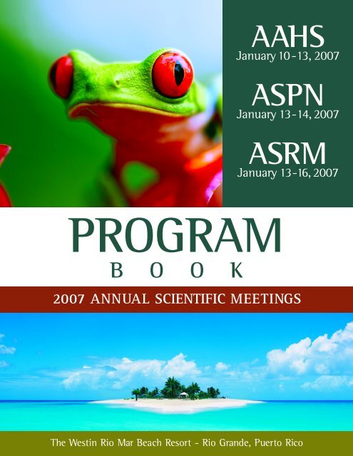
PROGRAM<br />
B O O K<br />
<strong>AAHS</strong><br />
January 10-13, 2007<br />
<strong>ASPN</strong><br />
January 13-14, 2007<br />
<strong>ASRM</strong><br />
January 13-16, 2007<br />
2007 ANNUAL SCIENTIFIC MEETINGS<br />
?<br />
The Westin Rio Mar Beach Resort - Rio Grande, Puerto Rico
The Westin Rio Mar<br />
Beach Resort<br />
?
TABLE OF CONTENTS<br />
<strong>AAHS</strong> Board of Directors . . . . . . . . . . . . . . . . . . . . . . . . . . . . . . . . . . . . . . . . . . . . . . . . . . . . . . . . . . . . . . . . . . . . . . . . . . . . . . . . . . . . . . . . . . . . . . . . . . 1<br />
<strong>AAHS</strong> Committees . . . . . . . . . . . . . . . . . . . . . . . . . . . . . . . . . . . . . . . . . . . . . . . . . . . . . . . . . . . . . . . . . . . . . . . . . . . . . . . . . . . . . . . . . . . . . . . . . . . . . . . . 2<br />
<strong>AAHS</strong> Historical Information . . . . . . . . . . . . . . . . . . . . . . . . . . . . . . . . . . . . . . . . . . . . . . . . . . . . . . . . . . . . . . . . . . . . . . . . . . . . . . . . . . . . . . . . . . . . . . 3<br />
Hand Surgery Endowment Contributor List . . . . . . . . . . . . . . . . . . . . . . . . . . . . . . . . . . . . . . . . . . . . . . . . . . . . . . . . . . . . . . . . . . . . . . . . . . . . . . 4-6<br />
<strong>ASPN</strong> Council Members . . . . . . . . . . . . . . . . . . . . . . . . . . . . . . . . . . . . . . . . . . . . . . . . . . . . . . . . . . . . . . . . . . . . . . . . . . . . . . . . . . . . . . . . . . . . . . . . . . . 7<br />
<strong>ASPN</strong> Committees . . . . . . . . . . . . . . . . . . . . . . . . . . . . . . . . . . . . . . . . . . . . . . . . . . . . . . . . . . . . . . . . . . . . . . . . . . . . . . . . . . . . . . . . . . . . . . . . . . . . . . . . 8<br />
<strong>ASPN</strong> Historical Information . . . . . . . . . . . . . . . . . . . . . . . . . . . . . . . . . . . . . . . . . . . . . . . . . . . . . . . . . . . . . . . . . . . . . . . . . . . . . . . . . . . . . . . . . . . . . . 9<br />
<strong>ASRM</strong> Council Members . . . . . . . . . . . . . . . . . . . . . . . . . . . . . . . . . . . . . . . . . . . . . . . . . . . . . . . . . . . . . . . . . . . . . . . . . . . . . . . . . . . . . . . . . . . . . . . . . 10<br />
<strong>ASRM</strong> Committees . . . . . . . . . . . . . . . . . . . . . . . . . . . . . . . . . . . . . . . . . . . . . . . . . . . . . . . . . . . . . . . . . . . . . . . . . . . . . . . . . . . . . . . . . . . . . . . . . . . 11-12<br />
<strong>ASRM</strong> Historical Information . . . . . . . . . . . . . . . . . . . . . . . . . . . . . . . . . . . . . . . . . . . . . . . . . . . . . . . . . . . . . . . . . . . . . . . . . . . . . . . . . . . . . . . . . . . . . 13<br />
(insert tab labeled GENERAL)<br />
Messages from the Program Chairs . . . . . . . . . . . . . . . . . . . . . . . . . . . . . . . . . . . . . . . . . . . . . . . . . . . . . . . . . . . . . . . . . . . . . . . . . . . . . . . . . . . . . . . 14<br />
General Announcements . . . . . . . . . . . . . . . . . . . . . . . . . . . . . . . . . . . . . . . . . . . . . . . . . . . . . . . . . . . . . . . . . . . . . . . . . . . . . . . . . . . . . . . . . . . . . . . . . 15<br />
Social Events & Tours . . . . . . . . . . . . . . . . . . . . . . . . . . . . . . . . . . . . . . . . . . . . . . . . . . . . . . . . . . . . . . . . . . . . . . . . . . . . . . . . . . . . . . . . . . . . . . . . . . . 16<br />
2007 Exhibitor Listing . . . . . . . . . . . . . . . . . . . . . . . . . . . . . . . . . . . . . . . . . . . . . . . . . . . . . . . . . . . . . . . . . . . . . . . . . . . . . . . . . . . . . . . . . . . . . . . 17-19<br />
CME Information . . . . . . . . . . . . . . . . . . . . . . . . . . . . . . . . . . . . . . . . . . . . . . . . . . . . . . . . . . . . . . . . . . . . . . . . . . . . . . . . . . . . . . . . . . . . . . . . . . . . 20-22<br />
Presenters’ Disclosure Listing . . . . . . . . . . . . . . . . . . . . . . . . . . . . . . . . . . . . . . . . . . . . . . . . . . . . . . . . . . . . . . . . . . . . . . . . . . . . . . . . . . . . . . . . . . . . . 23<br />
Future <strong>Annual</strong> <strong>Meeting</strong> Locations . . . . . . . . . . . . . . . . . . . . . . . . . . . . . . . . . . . . . . . . . . . . . . . . . . . . . . . . . . . . . . . . . . . . . . . . . . . . . . . . . . . . . . . . 24<br />
(insert tab labeled <strong>AAHS</strong>)<br />
<strong>AAHS</strong> Wednesday Day-At-A-Glance . . . . . . . . . . . . . . . . . . . . . . . . . . . . . . . . . . . . . . . . . . . . . . . . . . . . . . . . . . . . . . . . . . . . . . . . . . . . . . . . . . . . . . 25<br />
La Federacion del Mano Inaugural <strong>Meeting</strong> . . . . . . . . . . . . . . . . . . . . . . . . . . . . . . . . . . . . . . . . . . . . . . . . . . . . . . . . . . . . . . . . . . . . . . . . . . . . . . 26<br />
Bioskills Workshops . . . . . . . . . . . . . . . . . . . . . . . . . . . . . . . . . . . . . . . . . . . . . . . . . . . . . . . . . . . . . . . . . . . . . . . . . . . . . . . . . . . . . . . . . . . . . . . . . . . . . . 26<br />
Specialty Day Program: Rapid Recovery - The Fast Track . . . . . . . . . . . . . . . . . . . . . . . . . . . . . . . . . . . . . . . . . . . . . . . . . . . . . . . . . . . . . . . . . . 27<br />
Specialty Day Program Handouts . . . . . . . . . . . . . . . . . . . . . . . . . . . . . . . . . . . . . . . . . . . . . . . . . . . . . . . . . . . . . . . . . . . . . . . . . . . . . . . . . . . . . 27-50<br />
<strong>AAHS</strong> Thursday Day-At-A-Glance . . . . . . . . . . . . . . . . . . . . . . . . . . . . . . . . . . . . . . . . . . . . . . . . . . . . . . . . . . . . . . . . . . . . . . . . . . . . . . . . . . . . . . . . 51<br />
Keynote Speaker: Bob Jamieson . . . . . . . . . . . . . . . . . . . . . . . . . . . . . . . . . . . . . . . . . . . . . . . . . . . . . . . . . . . . . . . . . . . . . . . . . . . . . . . . . . . . . . . . . 54<br />
<strong>AAHS</strong> Friday Day-At-A-Glance . . . . . . . . . . . . . . . . . . . . . . . . . . . . . . . . . . . . . . . . . . . . . . . . . . . . . . . . . . . . . . . . . . . . . . . . . . . . . . . . . . . . . . . . . . . 55<br />
J. Joseph Danyo Presidential Invited Lecturer: Robert D. Beckenbaugh, MD . . . . . . . . . . . . . . . . . . . . . . . . . . . . . . . . . . . . . . . . . . . . . . . 56<br />
Comprehensive Hand Surgery Review Course . . . . . . . . . . . . . . . . . . . . . . . . . . . . . . . . . . . . . . . . . . . . . . . . . . . . . . . . . . . . . . . . . . . . . . . . . . . . . 58<br />
<strong>AAHS</strong> Invited Speaker: Richard Kogan, MD . . . . . . . . . . . . . . . . . . . . . . . . . . . . . . . . . . . . . . . . . . . . . . . . . . . . . . . . . . . . . . . . . . . . . . . . . . . . . . . 59<br />
(insert tab labeled <strong>AAHS</strong>/<strong>ASRM</strong>/<strong>ASPN</strong>)<br />
<strong>AAHS</strong>/<strong>ASRM</strong>/<strong>ASPN</strong> Saturday Day-at-a-Glance . . . . . . . . . . . . . . . . . . . . . . . . . . . . . . . . . . . . . . . . . . . . . . . . . . . . . . . . . . . . . . . . . . . . . . . . . . . . 60<br />
<strong>AAHS</strong>/<strong>ASRM</strong>/<strong>ASPN</strong> Presidents’ Invited Lecturer: Richard H. Gelberman, MD . . . . . . . . . . . . . . . . . . . . . . . . . . . . . . . . . . . . . . . . . . . . . . . . 61<br />
<strong>ASRM</strong> Masters Series in Microsurgery . . . . . . . . . . . . . . . . . . . . . . . . . . . . . . . . . . . . . . . . . . . . . . . . . . . . . . . . . . . . . . . . . . . . . . . . . . . . . . . . . . . . 62<br />
(insert tab labeled <strong>ASPN</strong>)<br />
<strong>ASPN</strong> Saturday Day-At-A-Glance . . . . . . . . . . . . . . . . . . . . . . . . . . . . . . . . . . . . . . . . . . . . . . . . . . . . . . . . . . . . . . . . . . . . . . . . . . . . . . . . . . . . . . . . 63<br />
Invited Speaker: Prof. Xavier Navarro Acebes, MD, PhD . . . . . . . . . . . . . . . . . . . . . . . . . . . . . . . . . . . . . . . . . . . . . . . . . . . . . . . . . . . . . . . . . . . 64<br />
Invited Speaker: Jianguang Xu, MD, PhD . . . . . . . . . . . . . . . . . . . . . . . . . . . . . . . . . . . . . . . . . . . . . . . . . . . . . . . . . . . . . . . . . . . . . . . . . . . . . . . . 65<br />
Invited Speaker: Prof. Rolfe Birch . . . . . . . . . . . . . . . . . . . . . . . . . . . . . . . . . . . . . . . . . . . . . . . . . . . . . . . . . . . . . . . . . . . . . . . . . . . . . . . . . . . . . . . . 65<br />
<strong>ASPN</strong> Sunday Day-At-A-Glance . . . . . . . . . . . . . . . . . . . . . . . . . . . . . . . . . . . . . . . . . . . . . . . . . . . . . . . . . . . . . . . . . . . . . . . . . . . . . . . . . . . . . . . . . . 66<br />
Invited Speaker: Tessa Gordon, PhD . . . . . . . . . . . . . . . . . . . . . . . . . . . . . . . . . . . . . . . . . . . . . . . . . . . . . . . . . . . . . . . . . . . . . . . . . . . . . . . . . . . . . . 68<br />
(insert tab labeled <strong>ASRM</strong>)<br />
<strong>ASRM</strong> Sunday Day-At-A-Glance . . . . . . . . . . . . . . . . . . . . . . . . . . . . . . . . . . . . . . . . . . . . . . . . . . . . . . . . . . . . . . . . . . . . . . . . . . . . . . . . . . . . . . . . . 71<br />
Godina Lecturer: Ming Huei Cheng, MD, MHA . . . . . . . . . . . . . . . . . . . . . . . . . . . . . . . . . . . . . . . . . . . . . . . . . . . . . . . . . . . . . . . . . . . . . . . . . . . 75<br />
<strong>ASRM</strong> Monday Day-At-A-Glance . . . . . . . . . . . . . . . . . . . . . . . . . . . . . . . . . . . . . . . . . . . . . . . . . . . . . . . . . . . . . . . . . . . . . . . . . . . . . . . . . . . . . . . . . 76<br />
<strong>ASRM</strong> Presidential Invited Lecturer: Ronald M. Zuker, MD, FRCSC, FACS, FAAP . . . . . . . . . . . . . . . . . . . . . . . . . . . . . . . . . . . . . . . . . . . . 79<br />
Composite Tissue Allotransplantation Update Session . . . . . . . . . . . . . . . . . . . . . . . . . . . . . . . . . . . . . . . . . . . . . . . . . . . . . . . . . . . . . . . . . . . . . 80<br />
<strong>ASRM</strong> Tuesday Day-At-A-Glance . . . . . . . . . . . . . . . . . . . . . . . . . . . . . . . . . . . . . . . . . . . . . . . . . . . . . . . . . . . . . . . . . . . . . . . . . . . . . . . . . . . . . . . . . 81<br />
Buncke Lecturer: James Urbaniak, MD . . . . . . . . . . . . . . . . . . . . . . . . . . . . . . . . . . . . . . . . . . . . . . . . . . . . . . . . . . . . . . . . . . . . . . . . . . . . . . . . . . . 83<br />
(insert tab labeled ABSTRACTS)<br />
Abstract Table of Contents . . . . . . . . . . . . . . . . . . . . . . . . . . . . . . . . . . . . . . . . . . . . . . . . . . . . . . . . . . . . . . . . . . . . . . . . . . . . . . . . . . . . . . . . . . . . . . 84
2006-2007 <strong>AAHS</strong> BOARD OF DIRECTORS<br />
President Ronald Palmer, MD<br />
President-Elect N. Bradly Meland, MD<br />
Vice-President Scott Kozin, MD<br />
Secretary A. Lee Osterman, MD FACS<br />
Treasurer Richard E. Brown, MD<br />
Historian Keith Brandt, MD<br />
Parliamentarian Brian Adams, MD<br />
Past Presidents Susan Mackinnon, MD<br />
Richard A. Berger, MD, PhD<br />
Directors At Large George Landis, MD<br />
Peter Murray, MD<br />
Nash Naam, MD<br />
Nicholas Vedder, MD<br />
Affiliate Directors Julianne Howell, PT MS CHT<br />
Christine Novak, PT MS<br />
Aviva Wolff, MA, OTR, CHT<br />
1
<strong>AAHS</strong> COMMITTEES<br />
Please join us in thanking the <strong>AAHS</strong> committees for their work in 2006.<br />
EDUCATION COMMITTEE<br />
Jaiyoung Ryu, MD FACS, Chair<br />
Timothy J. Best, MD, MSc, FRCSC<br />
Kevin Chung, MD<br />
Paula Galaviz, OT<br />
Lisa J. Gould, MD, PhD<br />
Kevin Plancher, MD<br />
Renata Vanja Weber, MD<br />
FINANCE COMMITTEE<br />
Richard E. Brown, MD, Chair<br />
Susan Mackinnon, MD<br />
N. Bradly Meland, MD<br />
Ronald Palmer, MD<br />
MEMBERSHIP: ACTIVE COMMITTEE<br />
Steven McCabe, MD, Chair<br />
John D. Bauer, MD<br />
Amy L. Ladd<br />
Steven L. Moran, MD<br />
Raj Sood, MD<br />
Robert Spinner, MD<br />
MEMBERSHIP: AFFILIATE COMMITTEE<br />
Carin Jean Wulf, OT, CHT, Chair<br />
Gail Groth, OTR/L CHT MHS<br />
Rebecca von der Heyde, MS, OTR/L, CHT<br />
NOMINATING COMMITTEE<br />
Susan Mackinnon, MD, Chair<br />
Maureen Hardy, PT MS CHT<br />
M. Ather Mirza, MD<br />
Michael Neumeister, MD<br />
Warren Schubert, MD<br />
William Swartz, MD<br />
PROGRAM COMMITTEE<br />
A. Lee Osterman, MD FACS, Chairperson<br />
Jorge L. Orbay, MD, Co-Chairperson<br />
Randipsingh Bindra, MD<br />
Diana D. Carr, MD<br />
Kevin Chung, MD<br />
M. Ather Mirza, MD<br />
Christine Novak, PT MS<br />
Kevin Plancher, MD<br />
Kirsten Westberg, MD<br />
RESEARCH GRANTS COMMITTEE<br />
Michael Neumeister, MD, Chair<br />
Matthew A. Bernstein, MD<br />
Christine J. Cheng, MD<br />
Loree K. Kalliainen, MD, FACS<br />
TECHNOLOGY COMMITTEE<br />
George Landis, MD<br />
Coleen T. Gately, PT, DPT, MS<br />
David Netscher, MD<br />
Eric Rothenberg, MD, FACS<br />
Stephen Schnall, MD<br />
Hugh L. Vu, MD, MPH<br />
2
<strong>AAHS</strong> HISTORICAL INFORMATION<br />
<strong>AAHS</strong> PAST PRESIDENTS<br />
J. Joseph Danyo, MD 1970-1972<br />
Henry Burns, MD 1972-1973<br />
Ray A. Elliott, Jr., MD 1973-1974<br />
James Borden, MD 1974-1975<br />
Kim K. Lie, MD 1975-1976<br />
Frank L. Thorne, MD 1976-1977<br />
Lawrence R. Werschky, MD 1977-1978<br />
Robert T. Love, MD 1978-1979<br />
Arnis Freiberg, MD 1979-1980<br />
Thomas J. Krizek, MD 1980-1981<br />
George L. Lucas, MD 1981-1982<br />
Garry S. Brody, MD 1982-1983<br />
James G. Hoehn, MD 1983-1984<br />
Peter C. Linton, MD 1984-1985<br />
Wallace H.J. Chang, MD 1985-1986<br />
Austin D. Potenza, MD 1986-1987<br />
Lee E. Edstrom, MD 1987-1988<br />
C. Lin Puckett, MD 1988-1989<br />
Robert J. Demuth, MD 1989-1990<br />
Wyndell H. Merritt, MD 1990-1991<br />
Frederick R. Heckler, MD 1991-1992<br />
Robert D. Beckenbaugh, MD 1992-1993<br />
David J. Smith, Jr., MD 1993-1995<br />
James W. May, Jr., MD 1995-1996<br />
Robert H. Brumfield, Jr., MD 1996-1997<br />
Robert C. Russell, MD 1997-1998<br />
Peter C.amadio, MD 1998-1999<br />
William M. Swartz, MD 1999-2000<br />
William Blair, MD 2000-2001<br />
Robert Buchanan, MD 2001-2002<br />
Alan Freeland, MD 2002-2003<br />
Allen Van Beek, MD 2003-2004<br />
Richard Berger, MD 2004-2005<br />
Susan Mackinnon, MD 2005-2006<br />
PRESIDENTIAL INVITED LECTURERS<br />
Harold E. Kleinert, MD 1989<br />
Arthur C. Rettig, MD 1990<br />
Paul W. Brand, MD 1991<br />
Ronald L. Linschied, MD 1993<br />
Guy Foucher, MD 1995<br />
Michael R. Harrison, MD 1996<br />
Dallas D. Raines 1997<br />
John Texter, MD 1998<br />
Vincent R. Hentz, MD 1999<br />
Nancy Dickey, MD 2000<br />
Michael Wood, MD 2001<br />
Francisco Rosas 2002<br />
Arnold-Peter Weiss, MD 2003<br />
Susan Mackinnon, MD 2004<br />
Elvin Zook, MD 2004<br />
Gavin Menzies 2005<br />
3<br />
KEYNOTE SPEAKERS<br />
William L. White, MD 1978<br />
John W. Madden, MD 1979<br />
Harold E. Kleinert, MD 1980<br />
J. William Littler, MD 1981<br />
Clifford C. Snyder, MD 1982<br />
Robert A. Chase, MD 1983<br />
Richard J. Smith, MD 1984<br />
James M. Hunter, MD 1985<br />
Bernard McC. O’Brien, MD 1986<br />
Erle E. Peacock, Jr., MD 1988<br />
Michael Jabelay, MD 1989<br />
Robert M. McFarlane, MD 1990<br />
James H. Dobyns, MD 1991<br />
Adrian E. Flatt, MD 1992<br />
John B. Carlson, PhD 1993<br />
Pat Clyne 1995<br />
David M. Evans, FRCS 1996<br />
Eugene Nelson, MD 1997<br />
Fritz Klein 1998<br />
Janet L.Babb 1999<br />
Frank E. Jones, MD 2000<br />
Joseph Buckwalter, MD 2001<br />
Linda Cendales, MD 2002<br />
Arnold-Peter Weiss, MD 2003<br />
Terry L. Whipple, MD, FACS 2005<br />
Jeff Lichtman, MD, PhD 2006<br />
CLINICIAN/TEACHER OF THE YEAR<br />
Forst Brown, MD 1995<br />
Robert Beckenbaugh, MD 1996<br />
James Hoehn, MD 1997<br />
Alan Freeland, MD 1998<br />
Wyndell Merritt, MD 1999<br />
Peteramadio, MD 2000<br />
Anthony DeSantolo, MD 2002<br />
Michael Jabaley, MD 2002<br />
Maureen Hardy 2002<br />
Sterling Mutz, MD 2002
HAND SURGERY ENDOWMENT<br />
The following companies are recognized as Corporate Sponsors for their generous donations in 2006.<br />
Hand Innovations $20,000<br />
Synthes $20,000<br />
<strong>American</strong> Surgical Centers $2,000<br />
Slack Inc. $1,000<br />
The following individuals are recognized for their ongoing financial commitment to the Hand Surgery Endowment<br />
DIAMOND<br />
(Cumulative giving totaling $20,000 or more)<br />
Joseph Danyo, MD<br />
Alan Freeland, MD<br />
Robert Schenck, MD<br />
EMERALD<br />
(Cumulative giving up to $10,000)<br />
N. Bradly Meland, MD<br />
Ronald Palmer, MD<br />
Miguel Saldana, MD<br />
James Schlenker, MD<br />
RUBY<br />
(Cumulative giving up to $5,000)<br />
Anthony Brown, MD<br />
Ali Seif, MD<br />
Allen Van Beek, MD<br />
SAPPHIRE<br />
(Cumulative giving up to $3,000)<br />
Stephan Ariyan, MD<br />
Mark Baratz, MD<br />
Rocco Barbieri, MD<br />
John Bax, MD<br />
William Benson, MD<br />
Beth Ann Bergman, MD<br />
Phillip Blevins, MD<br />
Forst Brown, MD<br />
Richard Brown, MD<br />
William Brown, MD<br />
Robert Buchanan, MD<br />
A. Lawrence Cervino, MD<br />
David T.W. Chiu, MD<br />
Jerry Chow, MD<br />
James Clayton, MD<br />
Mark Cohen, MD<br />
David Conner, MD<br />
James Cullington, MD<br />
Donald Ditmars, MD<br />
Richard Fox, MD<br />
Felix Freshwater, MD<br />
Robert Gardere, MD<br />
William Geissler, MD<br />
Neil Green, MD<br />
Robert Harris, MD<br />
Fred Heckler, MD<br />
David Hildreth, MD<br />
James Hoehn, MD<br />
Peter Hui, MD<br />
Richard Idler, MD<br />
Ted Jackson, MD<br />
Ronald Joseph, MD<br />
Scott Kozin, MD<br />
George Landis, MD<br />
John Lang, MD<br />
W.P. Andrew Lee, MD<br />
William Leighton, MD<br />
JoAnn Levitan, MD<br />
Kim Lie, MD<br />
Ho Min Lim, MD<br />
Shelia Lindley, MD<br />
Robert Love, MD<br />
George Lucas, MD<br />
Susan Mackinnon, MD<br />
James May, Jr., MD<br />
W. R. McArthur, MD<br />
Vaughn Meyer, MD<br />
Susan Michlovitz, PT, PhD<br />
Ather Mirza, MD<br />
Hiram Morgan, MD<br />
Nash Naam, MD<br />
Daniel Nagle, MD<br />
A Lee Osterman, MD<br />
Edward Palmer, MD<br />
Swainathan Rajan, MD<br />
Norman Rappaport, MD<br />
Jaiyoung Ryu, MD<br />
Amorn Salyapongse, MD<br />
Somprasong Songcharoen, MD<br />
William Swartz, MD<br />
Nicholas Vedder, MD<br />
Eric Wegener, MD<br />
Michael White, MD<br />
Eric Wyble, MD<br />
TOPAZ<br />
(Cumulative giving of $1,000)<br />
Alejandro Badia, MD<br />
Kyle Bickel, MD<br />
Randy Bindra, MD<br />
Robert Costarella, MD<br />
Richard Demuth, MD<br />
Michael Kalisman, MD<br />
Albert Weiss, MD<br />
HONOR ROLL<br />
(Cumulative giving less than $1,000)<br />
Dorit Aaron, MA, OTR,CHT<br />
Govind Acharya, MD<br />
Brian Adams, MD<br />
Lisa Adams, CVE, CHT<br />
Medhi Adham, MD<br />
Galaa Agban, MD<br />
Joseph Agris, MD<br />
John Alipit, MD<br />
Bernard Alpert, MD<br />
Peteramadio, MD<br />
Chittur Ananthakrishnan, MD<br />
Dimitri Anastakis, MD<br />
William Anderson, MD<br />
Cary Andras, MD<br />
Michael Angel, MD<br />
4<br />
Alexander Angelides, MD<br />
Mallory Anthony, RPT<br />
John Anton, MD<br />
Dori Ann Appleman, OTR,CHT<br />
Thomas Arganese, MD<br />
Enrique Armenta, MD<br />
William Armiger, MF<br />
Laurence Arnold, MD<br />
Michael Aron, MD<br />
Kenneth Arthur, MD<br />
Tyrone Artz, MD<br />
Stanley Askin, MD<br />
Edward Athanasian, MD<br />
John Attwood, MD<br />
John Aversa, MD<br />
Samir Azer, MD<br />
Medhi Balakhani, MD<br />
Joyce Baldwin, OTR/L, CHT<br />
George Balfour, MD<br />
Brent Bamberger. MD<br />
Adel Barakat, MD<br />
John Barham, MD<br />
David Barker, MD<br />
Bruce Barton, MD<br />
M. L. Barton, MD<br />
Lynn Bassini, OTR, CHT<br />
Basilio Bautista, MD<br />
Carlos Bazan, MD<br />
Michael Beatty, MD<br />
Robert Beckenbaugh, MD<br />
Lawrence Bell, MD<br />
Keith Bengtson, MD<br />
Louis Benoist, MD<br />
Leon Benson, MD<br />
David Bierwagen, PT, CHT<br />
David Bikoff, MD<br />
David Billmore, MD<br />
David Birkbeck, MD<br />
Roderick Birnie, MD<br />
Allen Bishop, MD<br />
Donald Bittner, MD<br />
William Blair, MD<br />
Elizabeth Blake, MD<br />
Donna Blood, OTR/L, CHT<br />
Richard Bloomenstein, MD<br />
Leonard Bodell, MF<br />
Maria Bonazinga<br />
Michael Born, MD<br />
William Boss, MD<br />
Lawrence Bowen, MD<br />
David Bozentka, MD<br />
Timothy Bradley, MD<br />
Keith Brandt, MD<br />
Nancy Branz, PT, MS<br />
Laurence Brenner, MD<br />
Anthony Brentlinger, MD<br />
Bruce Brewer, MD<br />
James Brinkman, MD<br />
Garry Brody, MD<br />
Linda Brown, ND<br />
Mary Lynn Brown, MD<br />
Roger Brown, MD<br />
Robert Brumfield, MD<br />
Mark Buchman, MD<br />
Robert Buckley, MD<br />
Geoffrey Buncke, MD<br />
Gregory Buncke, MD<br />
Kim Buchstaber, OTR<br />
Rudolf Buntic, MD<br />
Mary Burns, OTR/L,CHT<br />
Christine Burridge, PT, CHT<br />
Vincent Butera, MD<br />
Marcia Buzzelli, PT, MS<br />
Carmine Calabrese, MD<br />
Elethea Caldwell, MD<br />
Catherine Calvey<br />
Michael Campbell<br />
Nancy Cannon, OTR, CHT<br />
David Caplin, MD<br />
James Carey, MD<br />
Ronaldo Carneiro, MD<br />
Ann Carrillo, OTR<br />
Glenn Carwell, MD<br />
Phyllis Chang, MD<br />
Wallace Chang, MD<br />
James Chao, MD<br />
John Chapple, MD<br />
Lawrence Chase, MD<br />
Andre Chaves, MD<br />
Eugene Cherny, MD<br />
Vradej Chinookoswong, MD<br />
Raj Chowdary, MD<br />
Robert Chuinard, MD<br />
Duke Chung, MD<br />
Michael Clark, MD<br />
Michael Clendenin, MD<br />
June Clopton, MD<br />
Tyson Cobb, MD<br />
Roberta Cohen, OTR<br />
Richard Coin, MD<br />
Larry Colen, MD<br />
Lee Colony, MD<br />
Charles Combs, MD<br />
Michelen Craft-Maynor<br />
Lester Cramer, MD<br />
Evan Crandall, MD<br />
Gregory Croll, MD<br />
Terry Cromwell, MD<br />
Robert Crow, MD<br />
Bohdan Czepak, MD<br />
Suman Das, MD<br />
Brian Davies, MD<br />
Bert Davis, MD<br />
Jayne Dederichs, OTR/L<br />
Fanny De le Cruz, MD<br />
A. Lee Dellon, MD
HONOR ROLL CON’T<br />
Heather Delp, LPT<br />
Jack Demos, MD<br />
Gloria DeOlarte, MD<br />
Michael DePriest, MD<br />
Robin De rose, OTR<br />
Robert Derkash, MD<br />
Sanjay Desai, MD<br />
Antonio DeSantolo, MD<br />
Susan DeStefano, OTR/L<br />
Gloria De Vore, OTR<br />
Gene Deune, MD<br />
Thomas DeWire, MD<br />
Prabhu Dhalla, MD<br />
Michal Diaz, MD<br />
Thomas DiBenedetto, MD<br />
Wayne Dickason, MD<br />
John Dietrich, MD<br />
Joseph Disa, MD<br />
Susan Doehr, OTR<br />
Sam Dovelle, OTR<br />
Gregory Dowbak, MD<br />
David Drake, MD<br />
John Drewniany, MD<br />
Marc Drimmer, MD<br />
Gale DuPont, OTR/L<br />
William Dzwierzynski, MD<br />
Janice Eaton, OTR/L, CHT<br />
Charles Eaton, MD<br />
Herbert Ecker, MD<br />
Lee Edstrom , MD<br />
Robert Ellis, MD<br />
James Esch, MD<br />
Gregory Evans, MD<br />
John Faillace, MD<br />
John Fatti, MD<br />
Bohdan Fedczuk, MD<br />
Harodl Fenner, MD<br />
Mciahel Ferdinands, MD<br />
John Finley, MD<br />
David Fischer, MD<br />
Gregory Fisher, MD<br />
Stephen Fisher, MD<br />
David Fitz, MD<br />
Richard Flaherty, MD<br />
Sandra Fletchall, OTR CHT, MPA<br />
Michael Flood, MD<br />
Waldo Floyd, MD<br />
Martin Fox, MD<br />
Paul Fragner, MD<br />
Arnis Freiberg, MD<br />
Scott Fried, MD<br />
Jeff Friedman, MD<br />
Mia Fuller, MS, OTR, CHT<br />
Stephen Fuller, MD<br />
Gerard Gabel, MD<br />
Unmeshchandra Gadaria, MD<br />
Sylvia Gagnon, MD<br />
Paula Galaviz, OT<br />
Randi Galli, MD<br />
Peter Galpin, MD<br />
Greg Ganske, MD<br />
Carlos Garcia Moral, MD<br />
Walter Garst, MD<br />
William Garvin, MD<br />
Shelai Gassler, OTR, CHT<br />
Trenton Gause, MD<br />
Eusebio Gaw, MD<br />
Michael Genoff, MD<br />
Margaret Geringer, OTR<br />
Gunter Germann, MD<br />
Royal Gerow, MD<br />
Marilyn Giln, OTR, CHT<br />
Terry Fillian, MD<br />
Kenn Given, MS<br />
Lawrence Glassman, MD<br />
Shellye Godfrey, OTR/L, CDE, CHT<br />
Alan Gold, MD<br />
Nelson Godlberg, MD<br />
Stephen Goldstein, MD<br />
Gregg Godnstrom, MD<br />
Federico Gonzalez, MD<br />
Marc Gottlieb, MD<br />
Joel Grad, MD<br />
Wendell Gray, MD<br />
Lawrence Gray, MD<br />
Daniel Greenwald, MD<br />
Jack Greider, MD<br />
John Griggs, MD<br />
John Grossman, MD<br />
Gail Groth, OTR/L, CHT, MHS<br />
Amit Gupta, MD<br />
Roxanne Guy, MD<br />
Mutaz Habal, MD<br />
Jane Haher, MD<br />
Paul Haiduk, MD<br />
Geoffrey Hallock, MD<br />
Michael Halls, MD<br />
Yousif Hamati, MD<br />
Robert Hansen, MD<br />
Paul Harkins, MD<br />
Richard Harkness, MD<br />
William Hart, MD<br />
Thomas Harter, MD<br />
David Haskell, MD<br />
Christopher Hauge, MD<br />
Chester Haverback, MD<br />
Robert Havlik, MD<br />
Tom Hayakawa, MD<br />
John Heieck, MD<br />
Darrell Henderson, MD<br />
Douglas Hendricks, MD<br />
Karen Henehan, MD<br />
Charles Hergrueter, MD<br />
Ralph Herms, MD<br />
Gregory Hill, MD<br />
Blayne Hirche, MD<br />
Christine Hoban, OTR, CHT<br />
Leslie Holcombe, OTR, CHT<br />
Ellen Horvitz, MS, PT, CHT<br />
Barney Horvath, MD<br />
Arden Hothem, MD<br />
Homer House, MD<br />
Patrick Houvet, MD<br />
Lon Howard, MD<br />
Pamela Howard, PT<br />
Richard Howard, MD<br />
David Huang, MD<br />
Mary Hubbell, CHT, CWCE<br />
William Huffaker, MD<br />
Edward Hughes, Jr., MD<br />
Thomas Hunt, MD<br />
Kenneth Hunter, MD<br />
Mary Isaacson, OTR<br />
Cindy Ivy, CHT<br />
Deborah Jacob-Maas<br />
Marshall Jemison, MD<br />
Chet Janecki, MD<br />
Raymond Janevicius, MD<br />
David Janssen, MD<br />
M.R. Jayasanker, MD<br />
David Jensen, MD<br />
Theron Jernigan, PT<br />
Stiles Jewett, MD<br />
Craig Johnson, MD<br />
Susan Johnson-Melat, OTR, CHT/CVE<br />
5<br />
William Jones, MD<br />
Jesse Jupiter, MD<br />
Ramasamy Kalimuthu, MD<br />
Loree Kalliainen<br />
Ann Kammien, PT<br />
Shin Kang, MD<br />
Reza Karimipour, MD<br />
Scott Kasden, MD<br />
Martin Kassan, MD<br />
Joanne Kassimir, CHT, OTR<br />
Richard Katz, MD<br />
Mark Kehn, MD<br />
Patrick Kelly, MD<br />
Mark Kendall, MD<br />
Carolyn Kerrigan, MD<br />
Martin Kessler, MD<br />
Lawrence Ketch, MD<br />
Roger Khouri, MD<br />
Suheil Khuri, MD<br />
Kyo Kim, MD<br />
Myung Kim, MD<br />
Woo-Kyung Kim, MD<br />
Yong Kim, MD<br />
Charles Kincaid, MD<br />
Gabriel Kind, MD<br />
Eugene King<br />
Lynn King, OTR, CHT<br />
John Kitzmiller, MD<br />
Howard Klein, MD<br />
Richard Knauft, MD<br />
Todd Koch, MD<br />
Richard Korentager, MD<br />
Bruce Kraemer, MD<br />
David Kupfer, MD<br />
Mine Kurtay, MD<br />
Stuart Kuschner, MD<br />
Joe Kutz, MD<br />
Jean Labelle, MD<br />
Daniel Labs, MD<br />
Juanita Laflin, CST<br />
John Lamana, MD<br />
Lauren Lancaster<br />
Richard Landry, MD<br />
Ann Lang, MA, OTR, CHT<br />
Edward Lanigan, MD<br />
Mary Beth Laplant, LPT<br />
Donald Leatherwood, MD<br />
Peter Ledoux, MD<br />
Charles Lee, MD<br />
Hans Lee, MD<br />
Howard Lee, MD<br />
James Lehman, MD<br />
Charles Leinberry, MD<br />
Lori Lenef, OTR/L<br />
Carl Lentz, MD<br />
Malcolm Lesavoy, MD<br />
Mark Leslie, MD<br />
John Lettieri, MD<br />
Scott Levin, MD<br />
Richard Levin, MD<br />
George Levine, MD<br />
Carolyn Levis, MD<br />
Jonathon Lewis, MD<br />
Terry Light, MD<br />
Peter Linden, MD<br />
John Lindsey, MD<br />
Charles Loguda, MD<br />
Linda Loya, MA, OTR,CHT<br />
Karen Luckett, OTR, CHT<br />
Steven Macht, MD<br />
Joy MacDermid, MD<br />
Douglas Mackenzie, MD<br />
Larry Maddy, RN, OTR/L, CHT<br />
Alexander Majidian, MD<br />
Matthew Malerich, MD<br />
Parvaiz Malik, MD<br />
Stephen Maloff, MD<br />
Christopher Maloney, MD<br />
Harold Mancusi-Ungaro, MD<br />
Mark Mandel, MD<br />
John Mara, MD<br />
Hallene Maragh, MD<br />
Norberto Marfori, MD<br />
Andrew Markiewitz, MD<br />
James Marshall, MD<br />
Kenneth Marshall, MD<br />
David Martin, MD<br />
Rosendo Martinez, MD<br />
Nalin Master, MD<br />
Howard Matsuba, MD<br />
William Mayhall, MD<br />
Alexander McArthur, MD<br />
John McAvoy, MD<br />
Steven McCabe, MD<br />
Thomas McChesney, MD<br />
Pamela McFarlane, MA, OTR, CHT<br />
John McGill, MD<br />
Nancy McHugh, OTR/L<br />
Craig McKee, MD<br />
Mehul Mehta, MD<br />
Keith Melancon, MD<br />
Mark Melhorn, MD<br />
Charles Melone, MD<br />
Jayasanker Menon, MD<br />
Craig Merrell, MD<br />
Wyndell Merritt, MD<br />
Louis Mes, MD<br />
Philip Metz, MD<br />
Claudia Meuli-Simmen, MD<br />
John Miller, MD<br />
Fred Miller, MD<br />
Norbert Ming, MD<br />
Sinesio Misol, MD<br />
Jose Monsivais, MD<br />
Carlos Montero, MD<br />
Kenneth Moore, MD<br />
William Moore, MD<br />
Seid Moosavi, MD<br />
Thomas Mordick, MD<br />
Richard Morgan, MD<br />
Donald Morris, MD<br />
Stephen Morris, MD<br />
Robert Morrison, MD<br />
Keith Morrison, MD<br />
Robert Morrow, MD<br />
Kenneth Murray, MD<br />
Peter Murray, MD<br />
Eid Mustafa, MD<br />
Stephen Naso, MD<br />
Chet Nastala, MD<br />
Peter Nathan, MD<br />
Michael Neumeister, MD<br />
Andrew Newman, MD<br />
Frank Newman, MD<br />
Mary Lynn Newport, MD<br />
William Nickell, MD<br />
Thomas Nipper, MD<br />
Christine Novak, MD<br />
Renee O’Sullivan, MD<br />
William Ogden, MD<br />
Walter Okunski, MD<br />
Elizabeth Ouellette, MD<br />
Winston Parkhill, MD<br />
Douglas Parks, MD<br />
Samuel Parry, MD<br />
Jay Patel, MD
HONOR ROLL CON’T<br />
Norman Payea, MD<br />
Shirley Pearson, OTR, MS<br />
Edward Pechter, MD<br />
William Pederson, MD<br />
Joseph Perlman, MD<br />
James Pertsch, MD<br />
Mary Son Pesco, MD<br />
Donald Pfeiffer, MD<br />
Howard Philips, MD<br />
Guy Pierret, MD<br />
Alan Pillersdorf, MD<br />
Jean Pilllet, MD<br />
James Pinkham, MD<br />
Miguel Pirella-Cruz, MD<br />
Subbarao Polineni, MD<br />
Jay Pomerance, MD<br />
Barry Poole, OTR, CHT<br />
Rasa Poorman, OTR, CHT<br />
Edward Powers, MD<br />
Julian Pribaz, MD<br />
George Primiano, MD<br />
Sergio Proserpi, MD<br />
Talmage Raine, MD<br />
Oscar Ramirez, MD<br />
Lorna Ramos, MA, OT<br />
James Raphael, MD<br />
Georg Rappold, MD<br />
Gregory Rauscher, MD<br />
Vincent Reale, MD<br />
Larry Reaves, MD<br />
Michael Reed, MD<br />
Loka Reddy, MD<br />
David Rehak, MD<br />
Michael Rench, MD<br />
Kevin Renfree, MD<br />
Charles Renner, OTR<br />
Charles Resnick, MD<br />
William Reus, MD<br />
Mary Reuterfors, MA, OTR, CHT<br />
Scott Riley, MD<br />
William Riley, Jr., MD<br />
Jeff Robb, MD<br />
Bradford Roberg, MD<br />
Dwight Roberson, MD<br />
Craig Roberts, MD<br />
Celia Robinson, MA, PT, CHT<br />
John Robinson, MD<br />
Alfredo Rodriguez, MD<br />
Frank Rogers, MD<br />
Allen Rosen, MD<br />
Arthur Rosenstock, MD<br />
Douglas Ross, MD<br />
Malcolm Roth, MD<br />
Eric Rothenberg, MD<br />
Paul Rottler, MD<br />
Leo Rozmaryn, MD<br />
Ronald Rusko, MD<br />
Robert Russell, MD<br />
Scott Sagerman, MD<br />
Harilaos Sakellarides, MD<br />
Chester Sakura, MD<br />
Jeffrey Salomon, MD<br />
Mona Samaan<br />
Alejandro Sanchez, MD<br />
Richard Sanders, MD<br />
Gordon Sasaki, MD<br />
Sandra Saunders, RPT, PT, CHT<br />
Robert Savage, MD<br />
Linda Schaffstall, OTR/L, CHT<br />
John Schantz, MD<br />
James Scheu, MD<br />
Kenneth Schiffman, MD<br />
Will Schlaff, MD<br />
Linda Schoenhals, MS, RPT<br />
Anne Schofield, OTR/L,CHT<br />
Warren Schubert, MD<br />
Frank Schuler, MD<br />
Karen Schultz, MS, OTR, CHT<br />
Timothy Schurman, MD<br />
John Seaberg, MD<br />
Kristin Seabol, MD<br />
Houshang Seradge, MD<br />
Donald Serafin, MD<br />
Jacob Sharp, MD<br />
Jay Shenaq, MD<br />
Saleh Shenaq, MD<br />
Randolph Sherman, MD<br />
Kenneth Shestak, MD<br />
Sung Shin, MD<br />
Raymond Shively, MD<br />
J.F. Showalter, MD<br />
Aamir Siddiqui, MD<br />
Jessica Siegers, OTR/L<br />
Maria Siemionow, MD<br />
Roger Simpson, MD<br />
Martin Skie, MD<br />
Joesph Slade, MD<br />
Paul Slater, MD<br />
Anthony Smith, MD<br />
Dell Parker Smith, MD<br />
Raymond Smith, MD<br />
Bendy So, MD<br />
Roger Sobel, MD<br />
Norman Sogioka, MD<br />
Raj Sood, MD<br />
Dean Sotereanos, MD<br />
John Sparrow, MD<br />
Jerome Spivack, MD<br />
T. Sreecharana, MD<br />
James Stafford, MD<br />
William Starr, MD<br />
J. Stayman, MD<br />
Scott Steinmann, MD<br />
Charlene Stennett, MD<br />
Peter Stern, MD<br />
Michael Sternschein, MD<br />
Thomas Stevenson, MD<br />
Curtis Steyers, MD<br />
Harold Stokes, MD<br />
Mary Stoliker, MS, OTR, CHT<br />
Tristan Stronger, MD<br />
Kevin Strathy, MD<br />
Berish Strauch, MD<br />
William Strinden, MD<br />
Chi-Tsung Su, MD<br />
John Swinburne, MD<br />
Raymond Takahashi, MD<br />
John Taras, MD<br />
Julia Terzis, MD<br />
Howard Tepper, MD<br />
Ben Thebaut, MD<br />
Lawrence Thompson, MD<br />
Grant Thomson, MD<br />
James Thornton, MD<br />
Jennifer Thurn, OTR, CHT<br />
David Toivonen, MD<br />
Bruce Topol, MD<br />
Steven Topper, MD<br />
Allen Tracy, MD<br />
Richard Troiano, MD<br />
Deborah Trojanowski, MD<br />
Thomas Trumble, MD<br />
Robert Tucker, MD<br />
Tsu Tsai, MD<br />
Christopher Ubinger, MD<br />
6<br />
Joseph Upton, MD<br />
James Urbaniak, MD<br />
Frederick Valauri, MD<br />
Napoleon Valdez, MD<br />
David Van Brunt, MD<br />
Robert Van Demark, MD<br />
Peter Van Giesen, MD<br />
Scott Vann, MD<br />
Benjamin Van Raalte, MD<br />
George Veasy, MD<br />
Tina Veraldi, OTR, CHT<br />
Nicholas Vienowicz, MD<br />
Charles Verheyden, MD<br />
Jeffrey Visotsky, MD<br />
Thomas Von Gillern, MD<br />
Frank Walchak, MD<br />
Lorenzo Walker, MD<br />
William Wallace, MD<br />
Madlyn Walton, OTR, CHT<br />
Robert Walton, MD<br />
Huan Wang, MD<br />
Robert Wanless, MD<br />
Robert Waterhouse, MD<br />
Paul Wavak, MD<br />
Burton Weber, MD<br />
Donald Wehmeyer, MD<br />
Larry Weinstein, MD<br />
Beth Weiss, OTR<br />
Mark Wells, MD<br />
Terry Westfield, MD<br />
Linda Westphal, OTR/L<br />
Michael Wheatley, MD<br />
John Wheeler, MD<br />
Nick Wheeler, MD<br />
Richard Whipple, MD<br />
Carolyn White<br />
David White, MD<br />
Richard White, MD<br />
David Whiteman, MD<br />
Robert Wilcox, MD<br />
Brandon Wilhelmi, MD<br />
Carl Williams, MD<br />
Barbara Winthrop-Rose, OTR, CHT<br />
Edward Withers, MD<br />
Eugene Wittenstrom, MD<br />
David Witzke, MD<br />
Levent Yalcin, MD<br />
Scott Young, MD<br />
John Yousif, MD<br />
William Zamboni, MD<br />
Jerrold Zeitels, MD<br />
Michael Zenn, MD<br />
D. Ziarkowski-Herb, PT, CHT<br />
Paul Zidel, MD<br />
Richard Zienowicz, MD<br />
Elvin Zook, MD
2006-2007 <strong>ASPN</strong> COUNCIL<br />
President Rajiv Midha, MD<br />
President-Elect Gregory R. D. Evans, MD, FACS<br />
Vice President Robert C. Russell, MD<br />
Secretary Howard M. Clarke, MD, PhD<br />
Treasurer Warren Schubert, MD<br />
Immediate Past President Maria Siemionow, MD, PhD<br />
Past President Steven McCabe, MD<br />
Historian Paul S. Cederna, MD<br />
Council Member at Large Ivica Ducic, MD, PhD<br />
Council Member at Large Allan J. Belzberg, MD<br />
Council Member at Large Thomas H.H. Tung, MD<br />
7
<strong>ASPN</strong> COMMITTEES<br />
Please join us in thanking the following <strong>ASPN</strong> committees who have helped make the 2006-year successful.<br />
PROGRAM COMMITTEE<br />
Robert Spinner, MD, Chairperson<br />
Joseph Rosen, MD<br />
Nash Naam, MD<br />
Ivica Ducic, MD, PhD<br />
David Netscher, MD<br />
Melanie Urbanchek, PhD<br />
Peter Evans, MD<br />
David Weinstein, MD, PhD<br />
Nancy McKee, MD, FRCSC<br />
Jose Monsivais, MD<br />
Martijn Malessy, MD, PhD<br />
Robert Tiel, MD<br />
Rajiv Midha, MD, Ex-Officio<br />
MEMBERSHIP COMMITTEE<br />
Gregory R. D. Evans, MD, FACS, Chairperson<br />
Howard M. Clarke, MD, PhD<br />
Jose Monsivais, MD<br />
Warren Hammert, DDS, MD<br />
Rajiv Midha, MD, Ex-Officio<br />
NOMINATING COMMITTEE<br />
Maria Siemionow, MD, PhD, Chairperson<br />
Allan Belzberg, MD<br />
Martijn Malessy, MD, PhD<br />
Warren Schubert, MD<br />
Jonathan Winograd, MD<br />
Steven McCabe, MD, Ex-Officio<br />
BYLAWS COMMITTEE<br />
Melanie Urbanchek, PhD, Chairperson<br />
Paul Cederna, MD<br />
Loree Kalliainen, MD<br />
William Kuzon, MD, PhD<br />
Warren Schubert, MD<br />
Rajiv Midha, MD, Ex- Officio<br />
FINANCE COMMITTEE<br />
Robert Russell, MD, Chairperson<br />
Martijn Malessy, MD, PhD<br />
Thomas Tung, MD<br />
Rajiv Midha, MD, Ex-Officio<br />
TECHNICAL EXHIBITS COMMITTEE<br />
Gregory R. D. Evans, MD, FACS, Chairperson<br />
William Kuzon, MD, PhD<br />
Robert Spinner, MD<br />
Rajiv Midha, MD, Ex-Officio<br />
EDUCATION COMMITTEE<br />
Dimitri Anastakis, MD, Chairperson<br />
A. Lee Dellon, MD<br />
Robert Spinner, MD<br />
Rajiv Midha, MD, Ex-Officio<br />
TIME AND PLACE COMMITTEE<br />
Robert Russell, MD, Chairperson<br />
Maria Siemionow, MD, PhD<br />
Gregory R. D. Evans, MD, FACS<br />
Thomas Tung, MD<br />
Steven McCabe, MD<br />
Rajiv Midha, MD<br />
Allan Belzberg, MD<br />
Howard M. Clarke, MD, PhD<br />
Warren Schubert, MD<br />
Paul Cederna, MD<br />
Ivica Ducic, MD, PhD<br />
NEWSLETTER COMMITTEE<br />
Nash Naam, MD, Editor<br />
Robert Spinner, MD, Assistant Editor<br />
Christine Novak, PT, MS, Assistant Editor<br />
WEB SITE COMMITTEE<br />
Paul Cederna, MD, Chairperson<br />
David Brown, MD, FACS<br />
Rajiv Midha, MD<br />
CODING AND REIMBURSEMENT COMMITTEE<br />
Keith Brandt, MD<br />
8
<strong>ASPN</strong> HISTORICAL INFORMATION<br />
FOUNDING COUNCIL (Established April 19, 1990)<br />
Warren Breidenbach, MD<br />
Thomas Brushart, MD<br />
David Chiu, MD<br />
A. Lee Dellon, MD<br />
Richard Ehrlichman, MD<br />
Nelson Goldberg, MD<br />
Roger Khouri, MD<br />
Howard Klein, MD<br />
Susan Mackinnon, MD<br />
Hallene Maragh, MD<br />
Wyndell Merritt, MD<br />
Michael Orgel, MD<br />
Elliot Rose, MD<br />
Joseph Rosen, MD<br />
Brooke Seckel, MD<br />
Saleh Shenaq, MD<br />
Thomas Stevenson, MD<br />
Berish Strauch, MD<br />
Julia K.Terzis, MD, PhD<br />
Allen Van Beek, MD<br />
H. Bruce Williams, MD<br />
<strong>ASPN</strong> PAST PRESIDENTS<br />
Julia K. Terzis, MD, PhD 1990-1992<br />
A. Lee Dellon, MD 1992-1993<br />
Berish Strauch, MD 1993-1994<br />
H. Bruce Williams, MD 1994-1995<br />
Susan E. Mackinnon, MD 1995-1996<br />
Wyndell Merritt, MD 1996-1997<br />
Allen Van Beek, MD 1997-1998<br />
Saleh Shenaq, MD 1998-1999<br />
David T. W. Chiu, MD 1999-2001<br />
Nancy H. McKee, MD 2001-2002<br />
William M. Kuzon, Jr., MD, PhD 2002-2003<br />
Keith E. Brandt, MD 2003-2004<br />
Steven McCabe, MD 2004-2005<br />
Maria Siemionow, MD, PhD 2005-2006<br />
9
2006-2007 <strong>ASRM</strong> EXECUTIVE COUNCIL MEMBERS<br />
President L. Scott Levin, MD, FACS<br />
President-Elect Lawrence B. Colen, MD<br />
Vice-President A. Lee Dellon, MD<br />
Secretary Peter C. Neligan, MD<br />
Treasurer Keith E. Brandt, MD<br />
Treasurer-Elect Joseph M. Serletti, MD, FACS<br />
Immediate Past President William C. Pederson, MD<br />
Senior Members-At-Large Gregory R. D. Evans, MD, FACS<br />
Michael W. Neumeister, MD<br />
Junior Members-At-Large Charles E. Butler, MD<br />
Alexander Y. Shin, MD<br />
Historian Neil F. Jones, MD<br />
10
<strong>ASRM</strong> COMMITTEES<br />
Please join us in thanking the following <strong>ASRM</strong> committees who have helped make the 2006-year successful.<br />
ABPS REPRESENTATIVE<br />
L. Scott Levin, MD, FACS<br />
AD HOC BREAST COMMITTEE<br />
Joseph M. Serletti, MD, FACS, Chairperson<br />
Elisabeth K. Beahm, MD<br />
Neil A. Fine, MD, FACS<br />
Maurice Nahabedian, MD<br />
Geoffrey L. Robb, MD<br />
Aldona J. Spiegel, MD<br />
Michael R. Zenn, MD<br />
AD HOC COMMITTEE ON COMPLEX<br />
RECONSTRUCTION<br />
Fu-Chan Wei, MD, FACS, Chairperson<br />
Lawrence B. Colen, MD<br />
Gunter Germann, MD<br />
William C. Pederson, MD<br />
Julian J. Pribaz, MD<br />
Milan Stevanovic, MD<br />
AD HOC COMMITTEE ON COMPOSITE<br />
TISSUE ALLOTRANSPLANTATION<br />
Robert L. Walton, MD, FACS, Chairperson<br />
A. Lee Dellon, MD<br />
Neil F Jones, MD<br />
L. Scott Levin, MD, FACS<br />
Susan E. Mackinnon, MD<br />
Maria Siemionow, MD, Ph.D.<br />
Thomas Tung, MD<br />
Fu-Chan Wei, MD, FACS<br />
AD HOC COMMITTEE ON GENITOURINARY<br />
RECONSTRUCTION<br />
Lawrence B. Colen, MD, Chairperson<br />
Charles E Butler, MD<br />
David W. Chang, MD<br />
AD HOC HEAD & NECK<br />
SUBSPECIALITY COMMITTEE<br />
Peter G. Cordeiro, MD, Chairperson<br />
Lawrence J. Gottlieb, MD<br />
Michael Miller, MD<br />
Julian J. Pribaz, MD<br />
AD HOC LOWER EXTREMITY<br />
SUBSPECIALITY COMMITTEE<br />
L. Scott Levin, MD, FACS, Chairperson<br />
Christopher Attinger, MD<br />
Marko Bumbasirevic, MD<br />
Raymond M. Dunn, MD<br />
AD HOC MEDIA RELATIONS COMMITTEE<br />
L. Scott Levin, MD, FACS, Chairperson<br />
Maria Siemionow, MD, Ph.D.<br />
Allen L. Van Beek, MD<br />
AD HOC MICROSURGICAL<br />
RESEARCH COMMITTEE<br />
Maria Siemionow, MD, PhD, Chairperson<br />
A. Lee Dellon, MD<br />
Neil F Jones, MD<br />
Peter C. Neligan, MD<br />
Michael W. Neumeister, MD<br />
AD HOC OUTREACH COMMITTEE<br />
Randy Sherman, MD, Chairperson<br />
Gunter Germann, MD<br />
L. Scott Levin, MD, FACS<br />
Robert C. Russell, MD<br />
Fu-Chan Wei, MD, FACS<br />
AD HOC UPPER EXTREMITY<br />
SUBSPECIALTY COMMITTEE<br />
Nicholas B. Vedder, MD, Chairperson<br />
Warren Breidenbach, MD<br />
Gunter Germann, MD<br />
Jonathan Isaacs, MD<br />
Neil F Jones, MD<br />
ASPS BOARD REPRESENTATIVE<br />
Robert L. Walton, MD, FACS<br />
AUDIT COMMITTEE<br />
Jeffrey D. Friedman, MD, Chairperson<br />
Elisabeth K. Beahm, MD<br />
Michael R. Zenn, MD<br />
BUNCKE LECTURESHIP COMMITTEE<br />
William C. Pederson, MD, Chairperson<br />
David W. Chang, MD<br />
L. Scott Levin, MD, FACS<br />
Geoffrey L. Robb, MD<br />
G. Ian Taylor, AO, FRACS, FACS FRCS<br />
Michael R. Zenn, MD<br />
BYLAWS COMMITTEE<br />
Julian J. Pribaz, MD, Chairperson<br />
Gregory M. Buncke, MD<br />
Anthony A. Smith, MD<br />
CLINICAL GUIDELINES<br />
& OUTCOMES COMMITTEE<br />
Nicholas B. Vedder, MD, Chairperson<br />
Howard N. Langstein, MD<br />
Peter C. Neligan, MD<br />
Michael W. Neumeister, MD<br />
11
<strong>ASRM</strong> COMMITTEES CON’T<br />
CPT/RUC COMMITTEE<br />
Keith E. Brandt, MD, Chairperson<br />
Gregory M. Buncke, MD<br />
Raymond M. Dunn, MD<br />
Daniel J. Nagle, MD<br />
Scott D. Oates, MD<br />
William C. Pederson, MD<br />
Michael R. Zenn, MD<br />
EDUCATION COMMITTEE<br />
Howard N. Langstein, MD, Chairperson<br />
Paul S. Cederna, MD, FACS<br />
Neil A. Fine, MD, FACS<br />
Maria Siemionow, MD, Ph.D.<br />
ELECTRONIC COMMUNICATIONS<br />
COMMITTEE<br />
Charles E Butler, MD, Chairperson<br />
Keith E. Brandt, MD, Ex-Officio<br />
William Dzwierzynski, MD<br />
Howard N. Langstein, MD<br />
Peter Murray, MD<br />
ENDOWMENT COMMITTEE<br />
Robert L. Walton, MD, FACS, Chairperson<br />
Keith E. Brandt, MD<br />
Joseph James Disa, MD<br />
William A. Zamboni, MD<br />
FINANCE COMMITTEE<br />
Lawrence B. Colen, MD, Chairperson<br />
Keith E. Brandt, MD, Ex-Officio<br />
Frederick J. Duffy, Jr, MD<br />
Joseph M. Serletti, MD, FACS<br />
GODINA FELLOWSHIP<br />
SELECTION COMMITTEE<br />
Lawrence B. Colen, MD, Chairperson<br />
Zoran M. Arnez, MD, PhD<br />
David W. Chang, MD<br />
L. Scott Levin, MD, FACS<br />
Peter C. Neligan, MD<br />
MEMBERSHIP COMMITTEE<br />
Lawrence B. Colen, MD, Chairperson<br />
A. Lee Dellon, MD, Ex-Officio<br />
Elisabeth K. Beahm, MD<br />
Gabriel M. Kind, MD<br />
Peter Murray, MD<br />
Milan Stevanovic, MD<br />
NOMINATING COMMITTEE<br />
William C. Pederson, MD, Chairperson<br />
Allen T. Bishop, MD<br />
David W. Chang, MD<br />
Gunter Germann, MD<br />
Robert C. Russell, MD<br />
PROGRAM COMMITTEE<br />
Michael R. Zenn, MD, Chairperson<br />
Joseph M. Serletti, MD, FACS, Ex-Officio<br />
Elisabeth K. Beahm, MD<br />
Allen T. Bishop, MD<br />
Gunter Germann, MD<br />
Gabriel M. Kind, MD<br />
Alexander Y. Shin, MD<br />
Milan Stevanovic, MD<br />
Joseph Upton, MD<br />
PSEF REPRESENTATIVE<br />
Gregory R.D. Evans, MD, FACS<br />
TECHNICAL EXHIBITS COMMITTEE<br />
Peter C. Neligan, MD, Chairperson<br />
L. Scott Levin, MD, FACS<br />
William Lineaweaver, MD<br />
TIME & PLACE COMMITTEE<br />
William C. Pederson, MD, Chairperson<br />
Robert L. Walton, MD, FACS<br />
Ronald M. Zuker, MD, FRCSC<br />
12
<strong>ASRM</strong> HISTORICAL INFORMATION<br />
1983 FOUNDING COUNCIL<br />
James B. Steichen, MD<br />
Berish Strauch, MD<br />
Julia K. Terzis, MD, PhD<br />
James R. Urbaniak, MD<br />
Allen L. Van Beek, MD<br />
YEAR PRESIDENT ANNUAL MEETING SITE FOUNDERS/GODINA LECTURERS<br />
1985 Berish Strauch, MD Las Vegas, NV Henry J. Buncke, MD<br />
Founders’ Lecturer<br />
1986 James R. Urbaniak, MD New Orleans, LA Harold E. Kleinert, MD<br />
Founders’ Lecturer<br />
1987 Joseph E. Kutz, MD San Antonio, TX Robert D. Acland, MD<br />
Founders’ Lecturer<br />
1988 H. Bruce Williams, MD Baltimore, MD Berish Strauch, MD<br />
Founders’ Lecturer<br />
1989 James B. Steichen, MD Seattle, WA G. Ian Taylor, FRCS, FRACS<br />
Founders’ Lecturer<br />
1990 Allen L. Van Beek, MD Toronto, Ontario, Canada Andrew Lightbody<br />
Founders’ Lecturer<br />
1991 Michael B. Wood, MD Orlando, FL Alain Gilbert, MD<br />
Founders’ Lecturer<br />
1992 Andrew J. Weiland, MD Scottsdale, AZ Edgar Biemer, MD<br />
Founders’ Lecturer<br />
1993 Graham Lister, MD Kansas City, MO Algimantas Narakas<br />
Founders’ Lecturer<br />
Lawrence B. Colen, MD<br />
Godina Lecturer<br />
1994-95 Robert C. Russell, MD Marco Island, FL Nguyen Huy Phan, MD<br />
Founders’ Lecturer<br />
Mark A. Schusterman, MD<br />
Godina Lecturer<br />
1995-96 Ralph T. Manktelow, MD Tucson, AZ Fu Chan Wei, MD<br />
Founders’ Lecturer<br />
Randy Sherman, MD<br />
Godina Lecturer<br />
1996-97 James A. Nunley, MD Boca Raton, FL James R. Urbaniak, MD<br />
Founders’ Lecturer<br />
Zoran M. Arnez, MD<br />
Godina Lecturer<br />
1997-98 William M. Swartz, MD Scottsdale, AZ H. Bruce Williams, MD<br />
Founders’ Lecturer<br />
L. Scott Levin, MD<br />
Godina Lecturer<br />
1998-99 David T. W. Chiu, MD Waikoloa, HI Julia K. Terzis, MD<br />
Founders’ Lecturer<br />
Phillip Blondeel, MD<br />
Godina Lecturer<br />
1999-2000 Daniel Nagle, MD Miami, FL Allen Van Beek , MD<br />
Founders’ Lecturer<br />
Gregory R. D. Evans, MD<br />
Godina Lecturer<br />
2000-2001 Saleh M. Shenaq, MD San Diego, CA Wayne Morrision, MD, FRACS<br />
Founders’ Lecturer<br />
Roger Khouri, MD<br />
Godina Lecturer<br />
2001-2002 Randy Sherman, MD Cancun, Mexico Robert Russell, MD<br />
Founders’ Lecturer<br />
William Zamboni, MD<br />
Godina Lecturer<br />
2002-2003 Julia K. Terzis, MD, PhD Kauai, HI Panayotis Soucacos, MD<br />
Founders’ Lecturer<br />
Raymond Dunn, MD<br />
Godina Lecturer<br />
2003-2004 Ronald M. Zuker, MD Palm Springs, CA Ralph Manktelow, MD<br />
Founders’ Lecturer<br />
Milomir Ninkovic, MD, PhD<br />
Godina Lecturer<br />
2004-2005 Robert L. Walton, MD, FACS Fajardo, Puerto Rico Isao Koshima, MD<br />
Founders’ Lecturer<br />
Michael Neumeister, MD, FRCSC, FACS<br />
Godina Lecturer<br />
2005-2006 William C. Pederson, MD Tucson, AZ David Chang, MD, FACS<br />
Godina Lecturer<br />
13
MESSAGES FROM THE PROGRAM CHAIRS<br />
The 2007 annual meeting of the <strong>American</strong> <strong>Association</strong> for Hand Surgery will be held at the Westin Rio Mar Beach Golf Resort and Spa in Puerto Rico from January 10 to 13, 2007. The theme is<br />
Hand Treasures of the Caribbean and promises the attendee an adventurer’s chest of clinical pearls, educational rubies, diamonds of friendship, and exciting nuggets of gold.<br />
The program begins on Wednesday, January 10 with a speciality day program emphasizing the techniques that insure rapid recovery from hand and wrist injuries. The afternoon lets you rate your<br />
golf swing against a pro, take a bioskills course, and enjoy the amenities of the resort. Save your energy for the welcome reception that night.<br />
Thursday, January 11, begins bright and early with six instructional courses, a panel on the Evolution of the Hand, chaired by Amy Ladd, MD and featuring noted anthropologist Mary Marzke, PhD<br />
and neurologist Frank Wilson, MD author of The Hand: How its use shapes the Brain. Richard A. Berger, MD, PhD updates the state of wrist and hand prostheses. After free papers, the morning<br />
session concludes with the keynote speaker, Bob Jamison, ABC national and former White House correspondent. Following lunch, the learning opportunities continue with six more ICLs and an<br />
update on coding strategies by Daniel Nagle, MD.<br />
Friday, January 12, continues the adventure with six more ICLs, dual free paper sessions, a panel challenging the experts on distal radius fracture cases, and two keynote lectures by Robert D.<br />
Beckenbaugh, MD and Eduardo Zancolli III, MD. The afternoon is dedicated to Peter Murray, MD’s popular Comprehensive Hand Surgery Review that updates every aspect of hand surgery. Friday<br />
evening is not to be missed. It begins with a performance by Richard Kogan, MD, a psychiatrist and concert pianist who analyzes the tortured creativity of Gershwin through his letters, medical<br />
problems, and music. Step directly from there into the <strong>AAHS</strong> dinner dance and party that would make the Pirates of the Caribbean jealous.<br />
Saturday, January 13, is our collaborative program with the <strong>American</strong> Society for Reconstructive Microsurgery and the <strong>American</strong> Society for Peripheral Nerve. Panels include Upper Extremity Injury<br />
in Modern Warfare detailing the recent advances in caring for severe extremity wounds and a Brachial Plexus panel full of practical management tips and nerve transfer specifics. Richard Gelberman,<br />
MD will be the Presidents’ keynote speaker.<br />
The afternoon golf tournament underlines the fact that outside your front door there are championship golf, tennis, multiple pools, and an ocean beach. When feeling less athletic, enjoy the world<br />
class spa, five gourmet restaurants, and casino. Close by, explore the natural beauty of El Yunque rain forest, visit the forts of Old San Juan, and salsa dance to the Latin nightlife.<br />
Thank you for being a part of this exciting, educational, and enjoyable experience.<br />
A. Lee Osterman, MD Jorge L. Orbay, MD<br />
<strong>AAHS</strong> 2007 Program Chair <strong>AAHS</strong> 2007 Program Co-Chair<br />
Thank you for attending the 16th annual <strong>American</strong> Society for Peripheral Nerve scientific meeting at the luxurious Westin Rio Mar Beach Resort in Rio Grande, Puerto Rico.<br />
On Saturday morning, joint sessions with the <strong>AAHS</strong> and <strong>ASRM</strong> will convene. A panel on Upper Extremity Injuries in Modern Warfare will begin the conference. Dr. Richard Gelberman will deliver<br />
the Presidents Invited Lecture on: Identifying Targets for Clinical and Research Excellence in 2007. Outstanding nerve papers from the 3 societies will be presented. A panel of international experts<br />
will review new trends and perspectives in Brachial Plexus Surgery 2007.<br />
Saturday afternoon will include 3 invited speakers: Xavier Navarro Acebes on Tube Repair: Advances Towards an Artificial Nerve Graft; Jianguang Xu on C7 Nerve Transfer: Past, Present, Future; and<br />
Rolfe Birch on Iatropathic Injuries of Peripheral Nerves. There will be one session with clinical and research papers.<br />
On Sunday, a host of instructional courses will start off the day: on brachial plexus birth palsy, cortical reorganization, reinnervating muscle, peripheral nerve tumors, and intraoperative monitoring.<br />
An <strong>ASRM</strong>/<strong>ASPN</strong> panel on Free Functioning Muscle Transfer is planned. Invited Speaker Tessa Gordon will discuss Emerging Strategies to Improve Outcome of Nerve Injury. Free papers will be<br />
admixed in the morning and afternoon sessions.<br />
In addition to the outstanding scientific program, a lively social agenda and beautiful weather is being forecasted. The <strong>ASPN</strong> Welcome Reception on Saturday evening will foster informal interactions<br />
with family and friends. Additional recreational opportunities can be anticipated at various venues – including the golf course, pool, beach, spa, or restaurants, not to mention the other attractions<br />
on the island.<br />
All in all, this meeting promises to be a memorable experience.<br />
Robert J. Spinner, M.D.<br />
<strong>ASPN</strong> 2007 Program Chair<br />
The 23rd annual meeting of the <strong>ASRM</strong> this January 2007 promises to be one of our best yet. Our meeting venue returns us to beautiful Puerto Rico, this time to the incomparable Westin Rio Mar<br />
Beach Golf Resort and Spa. The meeting has a longstanding tradition of presenting cutting edge topics in microsurgery, panel discussions, and instructional courses applicable to the clinician as<br />
well as the researcher and this will continue. Structural changes to the instructional part of the meeting as well as marquis entertainment for the social portion of the meeting will mark the 2007<br />
meeting as one not to be missed.<br />
This meeting will reflect suggestions the membership has made over the years to improve the <strong>ASRM</strong> meeting experience. One often noted complaint was not enough family and free time during<br />
the day to enjoy the venue and camaraderie outside the meeting. This complaint has been addressed by significantly restructuring the meeting. This year’s meeting will have significant free time<br />
each afternoon and plenty of activities to fill them with. Over 20 hours of free afternoon time have been programmed in. No reason to “skip” presentations or panels now. By one o’clock each<br />
afternoon, instructional activities will be curtailed and the free time begins! While we have chosen to make this prime time available for your enjoyment, our goal has been to maintain the high<br />
level of educational content and to avoid the reduction of CME available for the meeting. We have done this in a number of creative ways. While the number of presentations will remain constant,<br />
the time for presentation has been curtailed to 3 minutes. This has been partially offset by a necessary and requested increase in discussion time for each paper. Evening panels have been added.<br />
Self-study computer modules will also be available throughout the meeting for additional and valuable patient safety CME.<br />
Our speakers will continue to be top notch. Our president’s invited speaker will be Past President Ronald Zuker, MD. The Buncke lecturer will be one of the founders of our society, James Urbaniak,<br />
MD, who still practices microsurgery. Elisabeth Beahm, MD will preside over the 2007 Master’s Series which will present interactive video presentations from leaders in their fields. Panel discussions<br />
will feature facial transplantation, optimization of results in head and neck reconstruction, and new ideas in limb salvage.<br />
Other members have asked for more presentation and discussion of complications and their management. Others still would like a forum to present interesting and unique individual cases which<br />
would be of interest to us as microsurgeons. These single cases do not often warrant formal presentation on the podium. Both of these interests will be served in evening sessions. First, we will<br />
be presenting an award for the “Best Microsurgical Save” of the year. Submitted cases will be judged by an expert panel that will score cases based on degree of complication, originality of the<br />
microsurgical salvage, and the overall value of the case. The ultimate winner will be decided by the membership present. No formal attire is required and yes, the bar will be open. This should allow<br />
exposure to and discussion of complications in a fun and relaxing format. The second award will be for the “Best Microsurgical Case” of the year. We all do great cases throughout the year. Here’s<br />
your chance to share one with your colleagues and have bragging rights for a year by winning. Again panelists will critique the cases and the membership present will pick the winner. Solicitations<br />
for these awards will be sent throughout the year and can be submitted to me via the <strong>ASRM</strong> website.<br />
Perhaps most exciting is our announcement of Branford Marsalis as our featured entertainer. Monday night will feature a night of jazz with Branford Marsalis and his band. Branford has been very<br />
active with Harry Connick Jr. providing much needed relief for musicians affected by Hurricane Katrina through Habitat for Humanity. Our <strong>ASRM</strong> concert will benefit these efforts. This will be a<br />
night remembered for all time.<br />
Our Puerto Rican venue offers plenty of activities for our indulgence. Beautiful beaches, lush rain forests, daytrips to San Juan, golf, and poolside lounging are only a few of the attractions that<br />
await us.<br />
Michael Zenn, MD, FACS<br />
<strong>ASRM</strong> 2007 Program Chair<br />
14<br />
<strong>AAHS</strong><br />
<strong>ASPN</strong><br />
<strong>ASRM</strong>
GENERAL ANNOUNCEMENTS<br />
MEETING SERVICE HOURS (subject to change)<br />
Stop by our meeting services desk in the Rio Mar Atrium for meeting information or assistance.<br />
Wednesday, January 10 7:00am - 6:00pm<br />
Thursday, January 11 7:00am - 4:00pm<br />
Friday, January 12 9:00am - 6:00pm<br />
Saturday, January 13 6:30am - 5:00pm<br />
Sunday, January 14 6:30am - 1:30pm; 6:30pm - 9:30pm<br />
Monday, January 15 6:30am - 1:00pm; 8:00pm - 10:00pm<br />
Tuesday, January 16 6:30am - 12:00pm<br />
<strong>AAHS</strong> POSTER PRESENTATION VIEWING HOURS<br />
The <strong>AAHS</strong> Poster Presentations will be placed near the Rio Mar Foyer. Posters will be available for viewing Wednesday - Saturday. If you are a presenter, please<br />
have your posters set up prior to 12:00pm on Wednesday and taken down prior to 12:00pm on Saturday. The <strong>American</strong> <strong>Association</strong> for Hand Surgery will not<br />
be responsible for any poster that is not removed within the time allotted.<br />
COMMERCIAL EXHIBITS<br />
The commercial exhibits will be located in Rio Mar Ballroom 5 and Foyer. A variety of commercial exhibits are featured at the meeting, enabling the attendees<br />
to learn about the technological advances pertaining to upper extremity surgery, neurosurgery and reconstructive microsurgery, and to meet key suppliers. Please<br />
refer to the Exhibit Listing in this book.<br />
EXHIBIT HOURS<br />
Wednesday, January 10 6:30am to 11:00am <strong>AAHS</strong> ONLY exhibits open<br />
Thursday, January 11 7:00am to 1:30pm <strong>AAHS</strong> ONLY exhibits open<br />
Friday, January 12 6:30 am – 11:00am <strong>AAHS</strong> ONLY/ <strong>AAHS</strong>-<strong>ASRM</strong>-<strong>ASPN</strong> JOINT exhibits open<br />
Saturday, January 13 6:30am to 9:30am <strong>AAHS</strong>-<strong>ASRM</strong>-<strong>ASPN</strong> JOINT exhibits open<br />
2:00pm to 4:00pm <strong>AAHS</strong>-<strong>ASRM</strong>-<strong>ASPN</strong> JOINT exhibits open<br />
Sunday, January 14 6:00am to 1:00pm <strong>AAHS</strong>-<strong>ASRM</strong>-<strong>ASPN</strong> JOINT and <strong>ASRM</strong> ONLY exhibits open<br />
Monday, January 15 6:00am to 12:30pm <strong>ASRM</strong> ONLY exhibits open<br />
Tuesday, January 16 6:00am to 10:30am <strong>ASRM</strong> ONLY exhibits open<br />
SPEAKER READY ROOM HOURS<br />
The Speaker Ready Room will be located in San Cristobal on the Rio Mar Ballroom level.<br />
Wednesday, January 10 6:30am - 2:00pm<br />
Thursday, January 11 6:30am - 4:00pm<br />
Friday, January 12 6:30am - 5:00pm<br />
Saturday, January 13 6:00am - 5:00pm<br />
Sunday, January 14 6:00am - 5:00pm<br />
Monday, January 15 6:00am - 1:00pm<br />
Tuesday, January 16 6:30am - 11:00am<br />
DRESS CODE<br />
Casual attire. Ties are discouraged for any session or function.<br />
MESSAGE BOARD/CYBER CAFE<br />
A message board will be set up near <strong>Meeting</strong> Services in Rio Mar Atrium. Please refer to the message board for meeting notices and general announcements.<br />
A Cyber Cafe will be located in the meeting area.<br />
The <strong>AAHS</strong>/<strong>ASRM</strong>/<strong>ASPN</strong> would like to thank Kinetikos Medical Inc./KMI<br />
for their generous sponsorship of the Cyber Cafe<br />
15
SOCIAL EVENTS<br />
Social events are offered to encourage networking and to enhance your meeting experience. Many of the events are included in your registration fee, and we encourage you to purchase tickets for<br />
your guests for all social events. We recommend that you purchase guest tickets in advance, as they will be available on a very limited basis at the meeting. Tickets will be collected at all events.<br />
<strong>AAHS</strong> Welcome Reception<br />
Wednesday, January 10: 6:00pm – 7:00pm<br />
Cost: One ticket included in <strong>AAHS</strong> registration. Additional tickets at $40 each.<br />
Tropical breezes and ocean views set the tone for a grand kick-off to a successful week ahead. Join us at Club Coqui as we enjoy the dramatic beauty of the Island’s outdoor scenery.<br />
<strong>AAHS</strong> Invited Speaker: Richard Kogan, MD<br />
Friday, January 12: 6:00pm - 7:30pm<br />
Cost: Complimentary to <strong>AAHS</strong> registrants and guests.<br />
“Music and Medicine: George Gershwin”<br />
George Gershwin (1898-1937) was one of the greatest composers in <strong>American</strong> history, writing memorable songs and concert pieces until his untimely death at age 38 of a brain tumor. Concert pianist<br />
and physician Dr. Richard Kogan will discuss Gershwin's life from a medical and psychiatric perspective and will perform Rhapsody in Blue and other examples of Gershwin's glorious music.<br />
Richard Kogan has a distinguished career both as a concert pianist and as a psychiatrist. He has been praised for his "eloquent, compelling and exquisite playing" by the New York Times and the Boston<br />
Globe wrote that "Kogan has somehow managed to excel at the world's two most demanding professions." He has gained international renown for his groundbreaking work on the connections between<br />
music and healing and on the influence of medical and psychiatric illnesses on the creative output of composers such as Mozart Beethoven, Schumann, Tchaikovsky, and Gershwin. His work forms the<br />
basis for the Yamaha DVD series entitled "Richard Kogan: Music and the Mind".<br />
Dr. Kogan is a graduate of the Juilliard School of Music and of Harvard College and Harvard Medical School. He completed his psychiatry residency training at NYU. He currently has a private practice<br />
of psychiatry in New York City and is affiliated with the Weill - Cornell Medical School as Director of its Human Sexuality Program.<br />
<strong>AAHS</strong> Awards Dinner Dance<br />
Friday, January 12: 7:30pm – 8:00pm Reception; 8:00pm – 11:00pm Dinner and Dancing<br />
Cost: One ticket included in <strong>AAHS</strong> registration. Additional adult tickets are $175 each; tickets for children and young adults ages 5 – 18 @ $50 each.<br />
A picture perfect evening will unfold during this entertaining evening of dining and dancing to Puerto Rico’s hottest dance band, Kamaleon. They entertained us in 2005, and now they’re back by popular<br />
demand to get the party started and keep it going all night long. Dress casual and prepare yourself for a night to remember. Festive beverages, hors d’ oeuvres, dinner wines and a multi-course<br />
gourmet dinner are all included in the adult ticket. An alternate menu and beverage selection will be served to children and young adults ages 5 – 18.<br />
11th <strong>Annual</strong> Day at the Links<br />
Saturday, January 13: 12:30pm Shotgun Start<br />
Cost: $215 per player. Tickets are non-refundable.<br />
A shamble format tournament will take place on the Westin’s River Course, Greg Norman’s first Caribbean design. The 6,945 yard course rolls along the Mameyes River, framed by vistas of mountains and<br />
sea. Ideal for players of all skill levels, and typical of Norman’s courses, all 18 holes can rate the title of “signature.” Prizes will be awarded during our post event reception to 1st, 2nd and 3rd place teams,<br />
and individuals who win the longest drive, longest putt and closest to the pin competitions. Tournament registration is very limited this year, and may not be available for on-site registration. To be paired<br />
with specific players, requests must be submitted at the Registration Desk by noon on Friday, January 12. Tournament costs include shuttle service, box lunch, greens fees, cart, tournament coordination,<br />
prizes, range balls and an after golf reception. Pro-Line clubs are available to rent with advance reservation at $35 per set.<br />
<strong>ASRM</strong> International Reception<br />
Saturday, January 13: 6:00pm – 7:30pm<br />
Cost: One ticket included in <strong>ASRM</strong> registration. Additional tickets at $50 each.<br />
Join us as we kick off the <strong>ASRM</strong> <strong>Annual</strong> <strong>Meeting</strong> with our International Reception, highlighting the countries represented in the organization. Hear our colleagues reflect on what <strong>ASRM</strong> has meant<br />
to them and their organizations. Tropical hors d’oeuvres and cocktails will be served on the Ocean Terrace.<br />
The <strong>ASRM</strong> would like to thank ASSI for their generous sponsorship of this reception.<br />
<strong>ASPN</strong> Welcome Reception<br />
Saturday, January 13: 6:30pm – 8:00pm<br />
Cost: One ticket included in <strong>ASPN</strong> registration. Additional tickets at $50 each.<br />
Tropical breezes and ocean views will be the setting for this lively gathering at Club Coqui. Join us as we network and enjoy the dramatic beauty of the Island’s outdoor scenery.<br />
<strong>ASRM</strong> Branford Marsalis Jazz Charity Concert Event<br />
Monday, January 15: 8:30pm – 10:30pm<br />
Cost: One ticket included in <strong>ASRM</strong> registration. Additional tickets at $100 each.<br />
Experience a once-in-a-lifetime music event featuring one of the most popular jazz artists of our time. In a very limited, intimate setting at the Westin, Grammy Award winning saxophonist Branford<br />
Marsalis will perform for <strong>ASRM</strong> to benefit Habitat for Humanity’s Musicians’ Village. The devastation caused by Hurricane Katrina forced many of New Orleans’ musicians to flee the city. The Musicians'<br />
Village, an effort co-chaired by Marsalis, has endeavored to build 81 Habitat-constructed homes in the Upper Ninth Ward for displaced New Orleans musicians. Learn more about this accomplished artist<br />
and the project at www.branfordmarsalis.com. This ticket is for a charitable event sponsored by the <strong>ASRM</strong> a 501(c) 3 not for profit organization. Net proceeds from this event will be contributed to The<br />
New Orleans Habitat Musicians’ Village.<br />
The <strong>ASRM</strong> would like to thank Smith and Nephew for their generous co-sponsorship of this event.<br />
OPTIONAL TOURS AND ACTIVITIES<br />
The Westin offers its guests a wide variety of organized Island tours and daily youth and adult activities that take place at The Westin. When you arrive at The Westin, you’ll have an opportunity to visit<br />
with the concierge to plan the activities of your choice. Some of the fee-based and complimentary activities available daily: Deep Sea Fishing, Power Walks, Wave Runners, Catamaran Sailing, Hair Braiding,<br />
Family Pool and Lawn Games, Bingo, Yoga Classes, Iguana Feeding, Volleyball, Horseback Riding, Sunset Sail, Basketball, Dive Trip, Aqua Aerobics, Latin Dance, Iguana Kids Club, Casino, Tennis, Golf,<br />
Shopping, and much more. If you prefer to join in with fellow association members, we’ve made special arrangements for a few private tours that can be reserved in advance. On-site ticket purchases<br />
will be limited, and some tours may be unavailable. If tour ticket minimum sales are not met, some tours may be cancelled. In these cases, all advance reservation payments will be fully refunded.<br />
El Yunque Rain Forest Tour<br />
Offered: Saturday, January 13, 1:00pm – 5:00pm; Sunday and Monday, 14th and 15th, 1:30 pm – 5:30 pm.<br />
Cost: $44 per adult; $40 per child ages 6 – 12 years of age<br />
The most visited natural sight on the island, El Yunque is the only tropical rain forest in the U.S. National Forest System. Spanning 28,000 acres and reaching an elevation of 3,624 feet, the area receives<br />
over 100 million gallons of rainfall each year. Explore leaf-canopied paths on this guided walking tour and learn about the endangered Puerto Rican parrot or the famous coquí frogs as you walk amongst<br />
over 240 different species of trees, ferns and flowers. You’ll also have the opportunity to visit the Yohaku observation tower and see a spectacular view of St. Thomas. Wear your swimsuit under your<br />
clothing and enjoy a refreshing swim under a stunning waterfall. Transportation, guide, park admission, bottled water and gratuity included. Some incline and decline walking is required.<br />
Old San Juan Historical Walking Tour and Shopping<br />
Offered: Saturday, January 13, 12:00 pm – 5:00pm; Sunday, 14th, 1:00 p.m. – 6:00 p.m.; Monday, 15th 1:30 pm – 6:30 pm.<br />
Cost: $55.50 adult; $50 per child ages 3 – 6 years of age<br />
Forty-Five minutes from the Westin, find cobblestone streets, colorful buildings, centuries-old fortresses, street merchants and charming shops, all bathed by a tropical breeze. Meet the legendary Old San<br />
Juan. This brief guided walking tour is followed by time to explore the quaint shops and eateries of Old San Juan on your own. Transportation, guide, bottled water and gratuity included.<br />
Bioluminescent Bay Evening Kayak Tour<br />
Offered: Saturday, January 13, 7:45 pm – 11 pm<br />
Cost: $87 adult; $78 per child ages 6 – 12 years of age<br />
For something truly magical, the world-renowned bioluminescence of the Laguna Grande in Fajardo cannot be missed. Spend twilight kayaking above what is considered one of the brightest displays of<br />
mysterious blue-green light emitted by dinoflagellates in the world. This relaxing kayak tour can be enjoyed by adults and children over age 6 who are willing and able to paddle. Transportation, guide,<br />
kayak gear, bottled water and gratuity included. This tour has limited availability.<br />
<strong>ASRM</strong> “Pamper Package” Guest Program<br />
Sunday, January 14, 7:00 pm – 9:00 pm<br />
Cost: One ticket included in <strong>ASRM</strong>’s Spouse/Guest Fee. A la carte tickets are $25 for adults, and $15 for children age 12 – 18.<br />
We’ve designed this special evening event exclusively for our <strong>ASRM</strong> guest attendees to coincide with the Scientific <strong>Meeting</strong>’s evening programming schedule. Join us for an inside look at health and<br />
beauty through the eyes of a professional esthetician. The staff of the resort’s famed Mandara Spa is our host for this up close and personal gathering where we’ll learn about the tricks of the trade for<br />
restoring our youthful, glowing skin. The facial demonstration will be followed by an opportunity to sample luxury products from the lines of La THERAPIE and Elemis. Festive beverages and sweet treats<br />
are all part of our Pamper Package. Participation is limited; sign up early to be guaranteed a seat. Please, no children under age 12.<br />
16
AMERICAN SOCIETY OF<br />
PLASTIC SURGEONS<br />
Booth: 23<br />
Emily Matzelle<br />
<strong>American</strong> Society of Plastic Surgeons<br />
444 East Algonquian Rd.<br />
Arlington Heights, IL 60005<br />
Phone- 847-228-9900 fax- 847-981-5482<br />
email- ejm@plasticsurgery.org<br />
website- www.plasticsurgery.org<br />
The <strong>American</strong> Society of Plastic Surgeons is the largest organization of board-certified<br />
plastic surgeons in the world. With 6,000 members, the society is recognized as a leading<br />
authority and information source on cosmetic and reconstructive plastic surgery. The society<br />
represents physicians certified by The <strong>American</strong> Board of Plastic Surgery or The Royal<br />
College of Physicians and Surgeons of Canada.<br />
ANATOMY GIFTS REGISTRY<br />
Booth: 33<br />
Brenda Bardsley<br />
7522 Connelley Drive, Suite P<br />
Hanover, MD 21076<br />
phone- 410-553-0525 fax-410-553-0502<br />
email- info@anatomicgift.com<br />
website- www.anatomicgift.com<br />
Anatomy Gifts Registry is a non-profit Donor Registry specializing in procurement, preservation<br />
and distribution of human tissue on an individualized investigator basis. AGR is<br />
licensed by the State of New York as a Non-Transplant Anatomic Bank. For information<br />
on specimen availability or application please call (410) 553-0525 or e-mail<br />
info@anatomicgift.com.<br />
ANGIOTECH (FORMERLY SURGICAL SPECIALTIES CORPORATION)<br />
Booth: 34<br />
Erika Fry<br />
100 Dennis Drive, Reading, PA 19606<br />
Phone- 610-404-1000 fax- 610-404-2061<br />
e-mail- efry@angio.com<br />
website- www.angiotech.com / www.surgicalspecialties.com<br />
Angiotech, formerly Surgical Specialties Corporation, features a wide rage of Sharpoint®<br />
micro-sutures and microsurgical knives – designed to meet the specific demands of the<br />
micro-surgeon. They also offer micro-training materials including: manuals, non-sterile<br />
sutures and PracticeRat. Stop by our booth and take a macro view of your full line micro<br />
company.<br />
APTIS<br />
Booth: 14<br />
Barbara Chesher<br />
5 River Hill Rd., Louisville, KY 40207<br />
phone- 502-561-4240 fax-502-585-0009<br />
email- barabrac@aptismedical.com<br />
website- www.anatomicgift.com<br />
APTIS MEDICAL specializes in taking current concepts to the next level. With the success<br />
of the Scheker DRUJ prosthesis, Aptis has provided a product that totally replaces the<br />
DRUJ, prevents subluxation and allows the bearing of weight. Other new and innovated<br />
designs for joint replacement are soon to follow<br />
ASCENSION ORTHOPEDICS<br />
Booth: 9<br />
Patsy Peterson<br />
8700 Cameron Rd., #100<br />
Austin, TX 78723<br />
phone- 512-836-5001 fax-512-836-5145<br />
email- ppeterson@ascensionortho.com<br />
website- www.ascensionortho.com<br />
Ascension Orthopedics is dedicated to the research, development, manufacture and distribution<br />
of revolutionary orthopedic devices for the hand, upper extremity, and foot. The<br />
company founders are pioneers in the material science to combat the debilitating effects<br />
of arthritis.<br />
17<br />
ASSI<br />
Booth: 20<br />
Proud sponsor of the <strong>ASRM</strong> International Reception<br />
Marie Bonazinga<br />
300 Shames Drive, Westbury, NY 11590<br />
phone- 516-333-2570 fax-516-997-4948<br />
email- cbowen@accuratesurgical.com<br />
website- www.accuratesurgical.com<br />
ASSI will feature the Engler Breast Retractor, the Stanger C Brest Retractors and the new<br />
Lalonde Breast Sizers, the Lalonde Percutaneous Bone Clamp with K-wire guide, Face Lift<br />
Retractors, Campbell Lip Awl, Matarasso Lipo Roller and SuperCut Face Lift Scissors, ASSI’s<br />
Bipolar Scissors, Micro Monopolar Forceps, the Surex Sural Nerve Extractor and Nerve<br />
Holding/Cutting Forceps. ASSI’s Hand Crafted Microsurgical Instruments and Clinical<br />
Microvascular Clamps.<br />
BIOMET TRAUMA<br />
Booth: 1<br />
Charlie Eaton<br />
100 Interpace Pkwy., Parsippany, NJ 7054<br />
phone- 973-299-9300 fax-973-299-0391<br />
email- charlie.eaton@ebimed.com<br />
website- ebimed.com<br />
Biomet-New Jersey (formerly EBI, L.P.) develops and markets a full range of internal and<br />
external orthopedic devices used in fracture fixation of the upper extremity. This includes<br />
the OptiLock Distal Radius Plating System, Uniflex Humeral Nail and a variety of distal<br />
radius fixators.<br />
BME<br />
Booth: 11<br />
Lisa May<br />
Marketing Specialist<br />
14785 Omicron Dr., #205, San Antonio, TX 78245<br />
Phone- 800-880-6528 fax- 210-677-0355<br />
email- lisamay@BME-tx.com<br />
website- www.bme-tx.com<br />
COOK MEDICAL<br />
Booth: 19<br />
Rachel Barnhill<br />
750 Daniels Way, Bloomington, IN 47404<br />
phone- 812-339-2235 fax-812-339-3704<br />
email- rachel.barnhill@cook-inc.com<br />
website- www.cookmedical.com<br />
Cook® presents two unique products: The Cook-Swartz implantable Doppler Blood Flow<br />
probe with new DP-M250 Monitor offering the latest technology for continuous confirmation<br />
of vascular patency. Surgisis®, an absorbable porcine small intestinal submucosa<br />
biomaterial that provides a scaffold for host tissue remodeling, creating a natural, costeffective<br />
alternative to surgical repair.<br />
DVO<br />
Booth: 3<br />
Rachel Bushong<br />
720 East Winona Ave., Warsaw, IN 46580<br />
phone- 877-777-9382 fax-574-258-1542<br />
email- rachel.bushong@dvosolutions.com<br />
website- www.dvosolutions.com<br />
DVO Solutions creates unique implant and instrumentation solutions for the upper<br />
extremities market. DVO will be showcasing our new Volar Plate System incorporating lag<br />
screw and CoverLoc Technology. We will also be displaying our MIfx System that contains<br />
our Dorsal IM Plate and Intrafocal Pin Plate.<br />
HOLOGIC, INC.<br />
Booth: 10<br />
Nicole Athanas<br />
35 Cosby Drive, Bedford, MA 1730<br />
phone- 781-999-7342 fax-781-280-0668<br />
email- nathanas@hologic.com<br />
website- www.hologic.com<br />
Fluoroscan® mini C-arm X-ray systems from Hologic are designed for orthopedic surgeons<br />
performing minimally invasive surgical procedures of the extremities, as well as for lowdose,<br />
in-office imaging.
HAND SURGERY ENDOWMENT<br />
Booth: TABLE TOP<br />
Laura Downes Leeper, CAE<br />
20. N. Michigan Ave., Suite 700<br />
Chicago, IL 60602<br />
Phone- 312-236-3307<br />
email- downes@isms.org<br />
The Hand Surgery Endowment supports education and research to improve patient care<br />
and safety of the hand and upper extremities. The Endowment funds scholarships and<br />
awards; public awareness, research projects and <strong>AAHS</strong> strategic initiatives that promote<br />
optimal hand and upper extremity health and safety.<br />
INTEGRA<br />
Booth: 29<br />
Proud sponsor of the <strong>ASPN</strong> Invited Speakers<br />
Jon Trout<br />
311 Enterprise Dr., Plainsboro, NJ 8536<br />
Phone- 609-275-0500 fax- 609-799-3297<br />
e-mail- jtrout@integra-ls.com<br />
website- www.integra-ls.com<br />
Integra develops, manufactures, and markets medical devices for neuro-trauma and neurosurgery,<br />
plastic and reconstructive surgery and general surgery. Integra’s peripheral<br />
nerve surgery products include NeuraGen for completely severed nerves and<br />
NeuraWrap for compressed, scarred or partially injured nerves.<br />
KINETIKOS MEDICAL INC./KMI<br />
Booth: 28<br />
Danielle Macrorie<br />
6005 Hidden Valley Rd., Suite 180<br />
Carlsbad, CA 92011<br />
Phone- 760-448-1700 ext.1422 fax- 760-448-1739<br />
e-mail- dmarcrorie@visitkmi.com<br />
MED LINK USA<br />
Booth: 30<br />
Tod Kellen<br />
PO BOX 42483, Des Moines, IA 50323<br />
Phone- 800-762-7921 fax- 800-329-5990<br />
e-mail- tkellen@medlinkusa.com<br />
website- www.medlinkusa.com<br />
Proud sponsor of the Cyber Cafe<br />
MEDARTIS<br />
Booth: 6<br />
Erin Graybill<br />
127 W. Street Road, Suite 203, Kennett Square, PA 19348<br />
Phone- 610-961-6101 fax- 610-961-6108<br />
e-mail- erin.graybill@medartis.com<br />
website- www.medartis.com<br />
Medartis is committed to providing surgeons and operating theatre personnel in orthopedic<br />
and cranio-maxillofacial surgery with innovative implants, instruments and services<br />
that represent advances in osteosynthesis and thus patients’ quality of life. To help it<br />
do justice to this ambitious aim, Medartis relies heavily on close collaboration with surgeons,<br />
scientists, universities and hospitals<br />
MICRINS SURGICAL<br />
Booth: 31<br />
Bern Teitz<br />
28438 Ballard Dr., Lake Forest, IL 60048<br />
Phone- 847-549-1410 fax- 847-549-1510<br />
e-mail- bern@micrins.com<br />
Micrins Features well over 3500 different instruments and accessories for Aesthetic, Hand,<br />
Micro and Reconstructive Surgery. Our product offering includes: The Micrins® brand of<br />
Hand Held Surgical Instruments, Razor-Edge Scissors, and Genuine Stille Surgical<br />
Instruments. The Micrins booth is conveniently located in the center of the exhibit hall.<br />
18<br />
MICROSURGERY INSTRUMENTS, INC<br />
Booth: 16<br />
Nancy Kang<br />
7211 Regency Sq. Blvd #223, Houston, TX 77036<br />
Phone- 713-664-4707 fax- 713-664-8873<br />
e-mail- mirriusa@microsurgeryusa.com<br />
Microsurgery Instruments, Inc. is one of the leading suppliers for surgical loupes and<br />
instruments in the United States and many other countries. We are well known in a large<br />
number of surgical fields including: Orthopedics, Plastic Surgery, Hand Surgery, ENT,<br />
Urology, General Surgery, Neurosurgery, and in many other more fields.<br />
NDI<br />
Booth: 18<br />
Jim Gabriel<br />
One Chagrin Highlands, 2000 Auburn Dr., Ste 320, Cleveland, OH 44122<br />
Phone- 216-378-9106 fax- 216-378-9116<br />
e-mail- jgabriel@ndimedical.com<br />
website- www.ndimedical.com<br />
NDI Medical is a neurostimulation medical device company. The Checkpoint stimulator<br />
is a surgical tool that provides surgeons with a reliable method of applying electrical stimulation<br />
to exposed motor nerves and muscle tissue to locate and identify nerves. The<br />
variable parameters provide reliable feedback while testing nerve and muscle excitability.<br />
NEXA ORTHOPEDICS<br />
Booth: 13<br />
Patricia Dorgan<br />
Marketing Services Manager<br />
11035 Roselle Street, San Diego, CA 92121<br />
Phone- 800-835-8480 fax- 858-866-0661<br />
e-mail- pdorgan@nexaortho.com<br />
website- www.nexaortho.com<br />
Nexa Orthopedics, the technology leader in products for reconstructive surgery of the<br />
extremities, is introducing its first products in its new pyrocarbon implant line. Please stop<br />
by the booth to see our new pyrocarbon CMI (basal thumb) and PI 2 (trapezium), and<br />
Radial Head systems.<br />
SMALL BONE INNOVATIONS<br />
Booth: 5<br />
Carolyn Desautels<br />
Global <strong>Meeting</strong>s Manager<br />
1711 S. Pennsylvania Ave., Morrisville, PA 19067<br />
Phone- 215-428-1791 fax- 215-428-1805<br />
e-mail- cdesautels@totalsmallbone.com<br />
website- www.totalsmallbone.com<br />
Small Bone Innovations, Inc. (“SBI”) is focused on the needs of the small bone & joint<br />
surgeon resulting in surgeon designed and clinically proven products. SBI is continually<br />
expanding its portfolio to become the worldwide leader in the design, development,<br />
manufacture, and marketing of upper and lower extremity medical devices.<br />
SMITH AND NEPHEW<br />
Booth: 15<br />
Jennifer Waddell<br />
Educational Conference Coordinator Proud co-sponsor of the <strong>ASRM</strong> concert event.<br />
11775 Starkey Rd., Largo, FL 33773<br />
With an emphasis on customer-driven technology and innovation, our goal at Smith &<br />
Nephew is to continue anticipating the future of excisional debridement. VersaJet is a<br />
specialized fluid jet instrument that enables surgeons to easily grasp, cut and remove<br />
damaged tissue and contaminants from surgical, traumatic and chronic wounds and<br />
burns in a precise and safe manner.
SPRINGER<br />
Booth: TABLE TOP<br />
Acasia Dalmau<br />
233 Spring St., New York, NY 10013<br />
Phone- 212-460-1600 fax- 201-272-1832<br />
e-mail- exhibits-ny@springer.com<br />
website- www.springer.com<br />
Stop by and browse Springer’s selection of Hand Surgery publications, and pick up your free<br />
sample copy of HAND, the new official journal of the <strong>American</strong> <strong>Association</strong> for Hand Surgery.<br />
STRYKER ORTHOPAEDICS<br />
Booth: 7 & 8 FOR <strong>AAHS</strong><br />
AND 21 FOR JOINT<br />
Peter Valente<br />
325 Corporate Dr., Mahwah, NJ 7430<br />
Phone- 201-831-5276 fax- 201-831-6453<br />
e-mail- perter.valente@stryker.caom<br />
Stryker range of Upper Extremities and Hand Products offers a full range of options from<br />
Intramedullary nails, external fixation devices, screws, pins, plates and new orthobiologic<br />
solutions for fracture treatment. Industry recognized product lines such as T2®,<br />
Hoffmann, Asnis III, Variax and Profyle Hand Products.<br />
SYNOVIS<br />
Booth: 24 & 25<br />
Britt Maganzini<br />
739 Industral Lane, Birmingham, AL 35211<br />
Phone- 205-941-0111<br />
e-mail- britt.maganzini@synovismicro.com<br />
Synovis MCA, “the microsurgeon’s most trusted resource,” provides innovative advancements<br />
and technologies for microsurgeons. The COUPLER and GEM MicroClips are precise<br />
instruments that help save valuable OR time and enable microsurgeons to perform<br />
highly effective anastomotic surgical procedures faster, easier and more dependably than<br />
traditional suture anastomosis. The GEM Neurotube® is an absorbable woven polyglycolic<br />
acid mesh tube designed for peripheral nerve repair and reconstruction.<br />
Introducing Veritas® Collagen Matrix, an advanced technology in soft tissue repair, saves<br />
time, is biocompatible and remodelable, has exceptional handling and comes in a variety<br />
of sizes. Biover Microvascular Clamps are disposable and produce a consistent measured<br />
pressure. S&T® microsurgical instruments are crafted from premium materials and<br />
built from a solid foundation of research, expert cooperation and engineering. The<br />
Varioscope® is the world’s smallest head-mounted surgical microscope.<br />
SYNTHES<br />
Booth: 2<br />
Lisa Kauffman<br />
1301 Goshen Parkway, West Chester, PA 19380<br />
Phone- 610-719-6873 fax- 610-719-6533<br />
e-mail- Kauffman.lisa@synthes.com<br />
website- www.synthes.com<br />
Proud sponsor of the Bottled Water<br />
and <strong>AAHS</strong> President’s Dinner<br />
Proud sponsor of the <strong>ASPN</strong> website and<br />
<strong>ASRM</strong> “Best Microsurgical Awards”<br />
Synthes CMF is the sole authorized North <strong>American</strong> manufacturer and distributor of AO<br />
ASIF instruments and implants for internal fixation of craniomaxillofacial and mandibular<br />
trauma and reconstruction. Our product offering includes distraction osteogenesis<br />
devices, resorbable plating, and bone graft substitutes. Synthes CMF also supports North<br />
<strong>American</strong> AO ASIF Continuing Education courses.<br />
19<br />
THE GUATEMALA HEALING<br />
HANDS FOUNDATION<br />
Booth: 12<br />
Mona Lipson<br />
290 6th Ave., Brooklyn, NY 11215<br />
Phone- 718-768-5927<br />
e-mail- monalipson@hotmail.com<br />
website- www.guatemalahands.org<br />
GHHF is a nonprofit organization dedicated to improving the quality and availability of<br />
hand care in Guatemala through education, surgery, and therapy. Specializing in the<br />
treatment of congenital and hand injuries, we aim to reach the Guatemalan population<br />
through medical missions led by a volunteer team of specialized and skilled surgeons,<br />
therapists, and dedicated volunteers.<br />
THIEME PUBLISHERS<br />
Booth: 17<br />
Ross Lumpkin or Daniel Schiff<br />
333 Seventh Ave., New York, NY 10001<br />
Phone- 212-584-4706 fax- 212-947-1112<br />
e-mail- grace.eassese@thieme.com<br />
website- www.thieme.com<br />
Established in 1886, Thieme is a major international publisher with offices in New York,<br />
Stuttgart, and Singapore. The company publishes the Journal of Reconstructive<br />
Microsurgery (Editor-in-Chief, Berish Strauch, MD), and recently released the new edition<br />
of the best selling book, Atlas of Microvasular Surgery by Berish Strauch and Han-Liang Yu.<br />
TRI MED, INC.<br />
Booth: 4<br />
Jim Fassett<br />
869 Main Street, Walpale, MA 2081<br />
Phone- 508-668-0988 fax- 508-668-0212<br />
e-mail- trimedortho@verizon.net<br />
website- www.trimedortho.com<br />
VIOPITIX, INC<br />
Booth: 26<br />
Denise Yarmlak<br />
44061-B Old Warm Springs Blvd., Fremont, CA 94538<br />
Phone- 510-226-5806 x 217<br />
e-mail- yarmlakd@vioptix.com<br />
The ViOptix Tissue Oximeter is a proprietary breakthrough product and technology that<br />
enables non-invasive, direct, real-time measurement of local tissue oxygen saturation.<br />
The ViOptix Tissue Oximeter is used in reconstructive flap surgeries and digit replantation<br />
to assess and monitor tissue viability post-operatively, thereby improving medical<br />
outcomes and decreasing cost.
<strong>AAHS</strong> CONTINUING MEDICAL EDUCATION<br />
<strong>AAHS</strong> MEETING OBJECTIVES:<br />
1. To present clinical and basic science studies on a variety of hand and upper extremity problems.<br />
2. To integrate principles of hand therapy with surgical management of hand and upper extremity problems.<br />
3. To review principles of managing common hand and upper extremity problems.<br />
4. To enhance the intellectual discourses of the annual meeting through an integrated program with the related surgical societies of the <strong>AAHS</strong>, <strong>ASRM</strong> and the <strong>ASPN</strong>.<br />
5. To implement bioskills courses and enhance management of fractures and arthritis.<br />
6. To present hand therapy issues related to the evidence-based intervention for a variety of hand problems and fractures.<br />
7. To encourage the exchange of knowledge and expertise of the various specialties involved with hand surgery.<br />
METHODS TO ACHIEVE THESE OBJECTIVES WILL BE:<br />
1. Original research will be presented as papers in an open session that will encourage audience participation.<br />
2. Recognized experts will present instructional courses and panels on hand and upper extremity problems.<br />
3. Hand therapy principles will be reviewed on a special focused day and throughout the meeting.<br />
4. A combined meeting with outstanding papers contributed by the <strong>AAHS</strong>, <strong>ASRM</strong> and <strong>ASPN</strong> with a focus on nerve , Brachial Plexus injury, and war injuries.<br />
5. Bioskills courses will be presented on operative procedures related to fractures and arthritis using cadaver dissections to illustrate surgical techniques.<br />
ACCREDITATION/CME<br />
The <strong>American</strong> Society of Plastic Surgeons® (ASPS®) is accredited by the Accreditation Council for Continuing Medical Education (ACCME) to provide continuing<br />
medical education for physicians. The Plastic Surgery Educational Foundation(r) (PSEF®) is the clinical education arm of the ASPS. The PSEF designates this<br />
educational activity for Category I credit towards the <strong>American</strong> Medical <strong>Association</strong> Physician’s Recognition Award as follows (Credit hours subject to change):<br />
<strong>AAHS</strong> ANNUAL MEETING<br />
January 10-13, 2007 13.75 hours<br />
COMPREHENSIVE HAND SURGERY REVIEW COURSE<br />
January 12, 2007 5.25 hours<br />
<strong>AAHS</strong>/<strong>ASRM</strong>/<strong>ASPN</strong> COMBINED DAY<br />
January 13, 2007 4.0 hours<br />
Additional CME hours are available for Instructional/Bioskills Courses on an hour-for-hour basis, awarded solely based on registration lists, as follows:<br />
Course # CME<br />
Bioskills Workshops BW1 - BW6 2.0 hours each<br />
Instructional Courses 102 - 107 1.0 hour each<br />
Instructional Courses 108 - 113 1.0 hour each<br />
Instructional Courses 115 - 120 1.0 hour each<br />
Credit hours are subject to program changes.<br />
COPYRIGHT<br />
All of the proceedings of the annual meeting, including the presentations of scientific papers, are intended solely for the benefit of the membership of the <strong>American</strong><br />
<strong>Association</strong> for Hand Surgery. No statement or presentation made at the meeting is to be regarded as dedicated to the public domain. Any statement or presentation<br />
is to be regarded as limited publication only and all property rights in the material presented, including common law copyright, are expressly reserved to the<br />
speaker or to theamerican <strong>Association</strong> for Hand Surgery. Any sound reproduction, transcript, or other use of material presented at the meeting without the permission<br />
of the speaker or the <strong>American</strong> <strong>Association</strong> for Hand Surgery is prohibited to the full extent of common law copyright in such material.<br />
THE USE OF CAMERAS OR PHOTOGRAPHIC EQUIPMENT IS NOT PERMITTED DURING THE PRESENTATION OF SCIENTIFIC PAPERS.<br />
20
<strong>ASPN</strong> CONTINUING MEDICAL EDUCATION<br />
<strong>ASPN</strong> MEETING OBJECTIVES:<br />
1. To present and discuss experimental techniques in nerve research.<br />
2. To provide exposure to emerging technologies in the management of nerve and extremity injuries.<br />
3. To understand electrodiagnostic evaluation of acute and chronic nerve injuries.<br />
4. To present and discuss the management of nerve injury and nerve tumors.<br />
5. To present and discuss evolving management of nerve injury causing pain.<br />
6. To present and discuss the physiology of the neuromuscular junction and muscle reinnervation.<br />
METHODS TO ACHIEVE THESE OBJECTIVES WILL BE:<br />
1. Scientific presentations on current and recent advances in research on nerve injury and repair.<br />
2. Seminars from invited experts on specialized topics related to reinnervation, reconstruction and nerve tumors.<br />
3. Invited lectures from clinical scientists and researchers in nerve biology, pathophysiology and injury.<br />
ACCREDITATION/CME<br />
The <strong>American</strong> Society of Plastic Surgeons® (ASPS®) is accredited by the Accreditation Council for Continuing Medical Education (ACCME) to provide continuing<br />
medical education for physicians. The Plastic Surgery Educational Foundation® (PSEF®) is the clinical education arm of the ASPS. The PSEF designates this<br />
educational activity for Category I credit towards the <strong>American</strong> Medical <strong>Association</strong> Physician’s Recognition Award as follows (Credit hours subject to change):<br />
<strong>AAHS</strong>/<strong>ASRM</strong>/<strong>ASPN</strong> COMBINED DAY<br />
January 13, 2007 4.0 hours<br />
<strong>ASPN</strong> ANNUAL MEETING<br />
January 13 - 14, 2007 13.0 hours<br />
Additional CME hours are available for Instructional Courses on an hour-for-hour basis, awarded solely based on registration lists, as follows:<br />
COURSE # CME<br />
Instructional Courses 201 - 205 1.00 hour each<br />
Credit hours are subject to program changes.<br />
COPYRIGHT<br />
All of the proceedings of the annual meeting, including the presentations of scientific papers, are intended solely for the benefit of the membership of the<br />
<strong>American</strong> Society for Peripheral Nerve. No statement or presentation made at the meeting is to be regarded as dedicated to the public domain. Any statement<br />
or presentation made at the meeting is to be regarded as limited publication only and all property rights in the material presented, including common<br />
law copyright, are expressly reserved to the speaker or to the <strong>American</strong> Society for Peripheral Nerve. Any sound reproduction, transcript, or other use of material<br />
presented at the meeting without the permission of the speaker or the <strong>American</strong> Society for Peripheral Nerve is prohibited to the full extent of common<br />
law copyright in such material.<br />
DISCLAIMER<br />
The content of this program is presented solely for educational purposes and is intended for use by medical practitioners in the peripheral nerve specialty. This<br />
material is intended to express the opinions, techniques or approaches of the authors and presenters, which may be beneficial and/or of interest to other practitioners.<br />
Sponsorship of this program is not to be construed, in any fashion, as an endorsement of the materials presented.<br />
The view expressed and the subject material presented in the course of any activities sponsored by the <strong>American</strong> Society for Peripheral Nerve including lectures,<br />
seminars, instructional courses, or otherwise, represent the personal views of the individual participants and do not represent the opinion of the <strong>American</strong> Society<br />
for Peripheral Nerve. The Society assumes no responsibilities for such views or materials, and hereby expressly disclaims any and all warranties, expressed or<br />
implied, for the content of any Society sponsored presentations. Further, the Society hereby acknowledges that while its broad purpose is to promote the development<br />
and exchange of knowledge pertaining to peripheral nerve regeneration; it does so only in the contest of a private forum without making any representations<br />
to the public whatsoever. Accordingly, the Society declares that its primary purpose is to benefit only its physician members, and responsibility of the<br />
Society for acts or omissions of Society members dealing with the public is hereby expressly disclaimed.<br />
THE USE OF CAMERAS OR PHOTOGRAPHIC EQUIPMENT IS NOT PERMITTED DURING THE PRESENTATION OF SCIENTIFIC PAPERS.<br />
21
<strong>ASRM</strong> CONTINUING MEDICAL EDUCATION<br />
<strong>ASRM</strong> MEETING OBJECTIVES:<br />
1. To provide the membership with an update on the state of the art techniques for microsurgical and complex reconstruction via<br />
peer reviewed scientific presentations on clinical and basic science research.<br />
2. To provide scientific and academic interaction and foster collaboration amongst the <strong>American</strong> <strong>Association</strong> for Hand Surgery,<br />
the <strong>American</strong> Society for Peripheral Nerve and the <strong>American</strong> Society for Reconstructive Microsurgery.<br />
3. To provide education opportunities for practicing surgeons, residents and fellows.<br />
4. To provide the attendees a focused update on specific topics in the form of expert panel presentations.<br />
5. To encourage participation by colleagues from around the world.<br />
METHODS TO ACHIEVE THESE OBJECTIVES WILL BE:<br />
1. Free papers to be presented in the open forum that allows audience discussion.<br />
2. National and international experts will provide different instructional courses on each day of the meeting.<br />
3. Panels by national and international experts will discuss conventional, new and controversial aspects of microsurgery and complex reconstruction.<br />
4. Patient Safety CME will be available by meeting participation and specialized computer modules available throughout the program.<br />
5. Specific mailing and invitations will increase participation of colleagues from around the world.<br />
6. Daily social events will provide interaction amongst colleagues, societies, and trainees.<br />
7. A conjoint scientific session including panels, instructional courses will be maintained on Saturday with the <strong>AAHS</strong> and on Sunday with the <strong>ASPN</strong>.<br />
ACCREDITATION/CME<br />
The <strong>American</strong> Society of Plastic Surgeons® (ASPS®) is accredited by the Accreditation Council for Continuing Medical Education (ACCME) to provide continuing<br />
medical education for physicians. The Plastic Surgery Educational Foundation® (PSEF®) is the clinical education arm of the ASPS. The PSEF designates this<br />
educational activity for Category I credit towards the <strong>American</strong> Medical <strong>Association</strong> Physician’s Recognition Award as follows (Credit hours subject to change):<br />
<strong>AAHS</strong>/<strong>ASRM</strong>/<strong>ASPN</strong> Combined Day<br />
January 13, 2007 4.0 hours<br />
<strong>ASRM</strong> Master Series in Microsurgery<br />
January 13, 2007 3.5 hours<br />
<strong>ASRM</strong> <strong>Annual</strong> <strong>Meeting</strong><br />
January 14 - 16, 2007 16.5 hours<br />
Additional CME hours are available for Instructional Courses and Patients Safety Computerized Presentations on an hour-for-hour basis, awarded solely based<br />
on registration lists, as follows:<br />
Course # CME<br />
Patients Safety Computerized Presentations 1.00 hour each<br />
Instructional Courses 301 - 307 1.00 hour each<br />
Instructional Coruses 308 - 314 1.00 hour each<br />
Instructional Courses 315 - 321<br />
Credit hours are subject to program changes.<br />
1.00 hour each<br />
COPYRIGHT<br />
All of the proceedings of the annual meeting, including the presentations of scientific papers, are intended solely for the benefit of the membership of the <strong>American</strong><br />
Society for Reconstructive Microsurgery. No statement or presentation made at the meeting is to be regarded as dedicated to the public domain. Any statement or<br />
presentation is to be regarded as limited publication only and all property rights in the material presented, including common law copyright, are expressly reserved to<br />
the speaker or to the <strong>American</strong> Society for Reconstructive Microsurgery. Any sound reproduction, transcript, or other use of material presented at the meeting without<br />
the permission of the speaker or the <strong>American</strong> Society for Reconstructive Microsurgery is prohibited to the full extent of common law copyright in such material.<br />
DISCLAIMER<br />
The views expressed and the subject material presented in the course of any activities sponsored by the <strong>American</strong> Society for Reconstructive Microsurgery including<br />
lectures, seminars, instructional courses, or otherwise, represent the personal views of the individual participants and do not represent the opinion of the<br />
<strong>American</strong> Society for Reconstructive Microsurgery. The Society assumes no responsibility for such views or materials, or implied, for the content of any Society<br />
sponsored presentations. Further, the Society hereby acknowledges that while its broad purpose is to promote the development and exchange of knowledge pertaining<br />
to the practice of microsurgery; it does so only in the context of a private forum without making any representation to the public whatsoever. Accordingly,<br />
the Society declares that its primary purpose is to benefit only its members, and responsibility of the Society for acts or omissions of Society members dealing<br />
with the public is hereby expressly disclaimed.<br />
THE USE OF CAMERAS OR PHOTOGRAPHIC EQUIPMENT IS NOT PERMITTED DURING THE PRESENTATION OF SCIENTIFIC PAPERS.<br />
22
<strong>American</strong> <strong>Association</strong> for Hand Surgery<br />
<strong>American</strong> Society for Reconstructive Microsurgery<br />
<strong>American</strong> Society for Peripheral Nerve<br />
2007 <strong>Annual</strong> <strong>Meeting</strong><br />
PRESENTERS’ DISCLOSURES<br />
THE FOLLOWING ANNUAL MEETING PRESENTERS HAVE INDICATED THE FOLLOWING DISCLOSURES:<br />
Chris Attinger, MD, serves on the speakers’ bureau for Integra, Johnson & Johnson and Smith & Nephew.<br />
Robert D. Beckenbaugh, MD, has financial ties to Ascension Orthopedics.<br />
Allan Belzberg, MD, receives research funding from DOD and UCB.<br />
Charles Butler, MD<br />
William P. Cooney, MD has a know-how license with Small Bone Innovation.<br />
Scott G. Edwards, MD serves as a consultant for Medartis.<br />
Alan Freeland, MD, receives departmental and institutional support from AONA and royalties from Elsevier Publishing Company.<br />
William Geissler, MD, serves as a consultant to Acumed<br />
Gerald H. Jordan, MD, receives royalties from C & S Surgical. Along with that he is a board member for Engineers and Doctors and<br />
a lecturer for CIEF. He also serves as a consultant for <strong>American</strong> Medical Systems and is involved in clinical trials with the following<br />
companies: Engineers & Doctors, Mentor, <strong>American</strong> Medical Systems, Auxilium.<br />
Don Lalonde, MD, serves as a consultant for ASSI.<br />
W. P. Andrew Lee, MD, serves as a consultant for Biomimetic Pharmaceutical Inc.<br />
Luis R. Scheker, MD, owns a patent for a total distal radioulnar joint prosthesis.<br />
Robert J. Spinner, MD, receives royalties for work licensed through Mayo Clinic to a privately held company for contributions<br />
related to the use of nerve signal modulation to treat central, autonomic, and peripheral nervous system disorders, including pain.<br />
Mayo Clinic receives royalties and owns equity in this company. The company does not currently license or manufacture any drug or<br />
device in the medical field.<br />
John Taras, MD has received an honorarium from Integra.<br />
The following presenters will include a discussion of an “off-label” or other non-FDA-approved, investigational use of medical devices<br />
or pharmaceutical products during their presentation. They will disclose that the product is not labeled for use under discussion or that<br />
the product is still investigational.<br />
Nicholas Vedder, MD<br />
THE FOLLOWING PRESENTERS DID NOT SUBMIT DISCLOSURE DOCUMENTS:<br />
Prof. Xavier Navarro Acebes, MD, PhD<br />
Richard Berger, MD, PhD<br />
Richard E. Brown, MD<br />
Bernard Chang, MD<br />
David T. W. Chiu, MD<br />
Peter G. Cordeiro, MD<br />
Zoe Dailliana, MD<br />
Coleen Gately, MS, PT, DPT<br />
Ralph Gilbert, MD<br />
Tessa Gordon, PhD<br />
Michael R. Hausman, MD<br />
Joy MacDermid, PhD, PT<br />
Wyndell H. Merritt, MD<br />
Peter C. Neligan, MD<br />
Jorge L. Orbay, MD<br />
Ronald Palmer, MD<br />
William C. Pederson, MD<br />
Matthew Putnam, MD<br />
Mark Rekant, MD<br />
David Ring, MD<br />
Teri Skirven, OTR/L, CHT<br />
Joseph Slade, III, MD<br />
Hans-Ulrich Steinau, MD<br />
Rebecca von der Heyde, MS, OTR, CHT<br />
Fu-Chan Wei, MD, FACS<br />
Andrew Weiland, MD<br />
Aviva Wolff, OTR/L, CHT<br />
Jianguang Xu, MD, PhD<br />
Ron Yu, MD<br />
The remainder of the annual meeting presenters have indicated in writing that they have nothing to disclose and/or will not discuss any “off-label”<br />
or other non-FDA-approved, investigation use of a medical device or pharmaceutical product.<br />
23
FUTURE ANNUAL MEETING LOCATIONS<br />
<strong>AAHS</strong><br />
2008 ANNUAL MEETING<br />
January 9 - 12, 2008<br />
The Hyatt Century Plaza Hotel and Spa<br />
Beverly Hills, California<br />
The 2008 <strong>Annual</strong> <strong>Meeting</strong>s with have an added touch of contemporary glamour and style at the Hyatt Regency Century Plaza.<br />
Perfectly situated in the fashionable Beverly Hills area, this elegant hotel offers easy access to all the sights and attractions that<br />
make Los Angeles great. Newly renovated guest rooms, world-class spa and fitness center and scintillating dining makes this<br />
luxury hotel feel more like a resort. Bring the whole family and enjoy a winter retreat that offers something for everyone.<br />
2009 ANNUAL MEETING<br />
January 7 - 10, 2009<br />
Grand Wailea Resort Hotel and Spa<br />
Maui, Hawaii<br />
2010 ANNUAL MEETING<br />
January 6 - 9, 2010<br />
Boca Raton Resort and Club<br />
Boca Raton, Florida<br />
<strong>ASPN</strong><br />
2008 ANNUAL MEETING<br />
January 12 - 13, 2008<br />
The Hyatt Century Plaza Hotel and Spa<br />
Beverly Hills, California<br />
2009 ANNUAL MEETING<br />
January 10 - 11, 2009<br />
Grand Wailea Resort Hotel and Spa<br />
Maui, Hawaii<br />
2010 ANNUAL MEETING<br />
January 9 - 10, 2010<br />
Boca Raton Resort and Club<br />
Boca Raton, Florida<br />
24<br />
<strong>ASRM</strong><br />
2008 ANNUAL MEETING<br />
January 12 - 15, 2008<br />
The Hyatt Century Plaza Hotel and Spa<br />
Beverly Hills, California<br />
2009 ANNUAL MEETING<br />
January 10 - 13, 2009<br />
Grand Wailea Resort Hotel and Spa<br />
Maui, Hawaii<br />
2010 ANNUAL MEETING<br />
January 9 - 12, 2010<br />
Boca Raton Resort and Club<br />
Boca Raton, Florida
<strong>AAHS</strong><br />
DAY-AT-A-GLANCE<br />
Wednesday, January10, 2007<br />
6:30am - 2:00pm Speaker Ready Room San Cristobal<br />
6:30am - 7:30am Continental Breakfast with Exhibitors Rio Mar Foyer & Ocean Terrace<br />
7:00am - 6:00pm <strong>Meeting</strong> Services and Cyber Cafe Rio Mar Atrium<br />
7:30am - 1:30pm Specialty Day Program: Rapid Recovery - The Fast Track Rio Mar 6<br />
10:45am - 11:00am Break with Exhibitors Rio Mary Foyer<br />
1:00pm - 5:00pm La Federacion del Mano Inaugural <strong>Meeting</strong> Rio Mar 7<br />
3:30pm - 5:30pm Bioskills Workshops<br />
BW-1 Current Techniques for PyroCarbon MCP/PIP/CMC Rio Mar 1<br />
Arthroplasty<br />
BW-2 Current Techniques for DRUJ Disorder Rio Mar 2<br />
BW-3 Treatment of Distal Radius Fractures Rio Mar 8<br />
BW-4 Open & Arthroscopic Treatment of Thumb CMC Arthritis Rio Mar 9<br />
BW-5 Minimal Resection Total Wrist Arthroplasty; Rio Mar 10<br />
Surgical Technique & Case Review<br />
BW-6 Hand & Upper Extremity Solutions Sea Gull<br />
6:00pm - 7:00pm <strong>AAHS</strong> Welcome Reception Club Coqui & Poolside Terrace<br />
25
<strong>AAHS</strong><br />
Wednesday, January 10, 2007<br />
6:30am – 7:30pm Continental Breakfast with Exhibitors<br />
7:30am – 1:30pm Specialty Day Program: Rapid Recovery<br />
The Fast Track<br />
Experts will present the latest techniques to treat and<br />
rehabilitate your patient for fastest recovery following<br />
hand and wrist injuries. Hear how to get your<br />
athlete, work injured patient, or family friend back to<br />
work and play as quick as possible using new<br />
surgical and rehabilitation methods. Relative risks<br />
compared to more “traditional” methods will also be<br />
discussed. And, don’t forget to join us for a little<br />
side show of golf – test your skills and win a prize.<br />
Brian Adams, MD, Program Co-Chair<br />
Aviva Wolff, OTR/L, CHT, Program Co-Chair<br />
7:30am – 7:35am President’s Welcome<br />
Ronald Palmer, MD<br />
7:35am – 7:45am Overview – “When Can Recovery be Rapid?”<br />
Brian Adams, MD<br />
Aviva Wolff, OTR/L, CHT<br />
7:45am – 8:00am Outcomes - How Do We Measure<br />
Rapid Recovery?<br />
Joy Macdermid, PT, PhD, CHT<br />
Wrist Injuries: “The Express Line”<br />
Christine Novak, PT, Moderator<br />
8:00am – 8:10am Scaphoid Fractures<br />
Randall Culp, MD<br />
8:10am – 8:20am Distal Radial Fractures<br />
Jorge Orbay, MD<br />
8:20am – 8:35am Recovery After Wrist Fractures<br />
Dorit H. Aaron, MA, OTR/L, CHT, FAOTA<br />
8:35am – 8:55am Ligament Injuries<br />
Richard Berger, MD, PhD<br />
Julianne Howell, PT, MS, CHT<br />
8:55am – 9:10am Panel/Discussion<br />
9:10am – 10:45am Digital Injuries – “Hold Fast”<br />
Brian Adams, MD, Moderator<br />
9:10am – 9:30am Metacarpal and Phalangeal Fractures<br />
Michael Bednar, MD<br />
Terri Skirven, MS, OTR/L, CHT<br />
9:30am – 9:50am PIP Fracture Dislocations<br />
Joseph Slade, MD<br />
Paul Brach, MS, PT, CHT<br />
9:50am – 10:10am Extensor Tendon Injuries<br />
Brian Adams, MD<br />
Julianne W. Howell, MS, PT, CHT<br />
10:10am – 10:30am Flexor Tendon Injuries<br />
Peter Amadio, MD<br />
Rebecca von der Heyde, MS, OTR, CHT<br />
10:30am – 10:45 Panel/Discussion<br />
10:45am – 11:00am Break with Exhibitors<br />
Pediatric Injuries: “What Can We Learn From Kids?”<br />
11:00am – 11:20am Upper Extremity Fractures: Hand, Wrist<br />
and Forearm<br />
Scott Kozin, MD<br />
Dorit H. Aaron, MA, OTR/L, CHT, FAOTA<br />
Sports Injuries: “How the Athletes Do It – In a NY Minute”<br />
Susan Michlovitz, PT, PhD, CHT, Moderator<br />
26<br />
11:20am – 11:30am Surgical Treatment – When to fix, When to wait<br />
Andrew Weiland, MD<br />
11:30am – 11:40am Protective Gear – Rules, Regulations,<br />
Requirements<br />
Michelle Carlson, MD<br />
11:40am – 11:50am Methods of Protection – Splints, Padding<br />
Coleen Gately, MS, PT, DPT<br />
11:50am – 12:00pm Casting Techniques for Sports Activities<br />
Ronald Palmer, MD<br />
12:00pm – 12:10pm The Injured Golfer<br />
Aviva Wolff, OTR/L, CHT<br />
Case Presentations: When Rapid is Not So Rapid<br />
12:10pm – 12:20pm My Experience with Firefighters<br />
Joy Macdermid, PT, PhD, CHT<br />
12:20pm – 12:30pm My Experience with the Not So Healthy Worker<br />
Susan Michlovitz, PT, PhD, CHT<br />
12:30pm – 12:40pm Rapid Recovery and Nerve Injury, an Oxymoron?<br />
Christine Novak, MS,PT, PhD(c)<br />
12:40pm – 1:00pm Panel/Discussion<br />
1:00pm – 1:30pm Rate Your Golf Swing against the Pro<br />
1:00pm – 5:00pm La Federacion del Mano Inaugural <strong>Meeting</strong><br />
Eduardo Zancolli, III, MD<br />
Course is complimentary but pre-registration is required.<br />
3:30pm – 5:30pm Bioskills Workshops<br />
BW-I Current Techniques for PyroCarbon<br />
MCP/PIP/CMC Arthroplasty<br />
Course Description: Use of the Ascension MCP/PIP/PHS implants will be demonstrated<br />
in this workshop. The course is designed to provide a comprehensive overview of the<br />
PyroCarbon Implants, design rationale, patient selection, surgical technique and postoperative<br />
therapy. Cadaveric models will be used to demonstrate MCP/PIP/PHS implant<br />
insertion and soft tissue repair.<br />
Course Objectives: Participants should have an understanding of indication, proper<br />
technique and post operative therapy.<br />
Faculty: Robert Beckenbaugh, MD<br />
BW-2 Current Techniques for DRUJ Disorder<br />
Course Description: Use of the Ascension First Choice DRUJ System will be demonstrated<br />
in this workshop. The course will introduce a new Partial Resurfacing implant<br />
designed to replace the articular region of the distal ulna. The course will provide a<br />
comprehensive overview of DRUJ disorders, discuss both implant options offered in the<br />
First Choice DRUJ System, the Partial Resurfacing implant and a Total Ulnar Head<br />
Implant, design rationale, patient selection, surgical technique and postoperative therapy.<br />
Cadaveric models will be used to demonstrate the surgical technique of both the<br />
Partial Resurfacing Implant and Total Ulnar Head Implant.<br />
Course Objectives: Participants should have an understanding of Indication, proper<br />
technique and post operative therapy.<br />
Faculty: Brian Adams, MD<br />
BW-3 Treatment of Distal Radius Fractures<br />
Course Description: There will be a surgical demonstration on the treatment of distal<br />
radius fractures using the OptiLock® Distal Radius Plating System with patented<br />
SphereLock TM technology. Emphasis will be on indications, design rationale, surgical technique,<br />
post-operative rehabilitation and follow-up.<br />
Faculty: Melvin P. Rosenwasser, MD<br />
BW-4 Open and Arthroscopic Treatment of the<br />
Thumb CMC Arthritis<br />
Course Description: A number of different methods can be successfully used for treatment<br />
of osteoarthritis of the CMC joint. This workshop will focus on strategies for the<br />
use of a minimally invasive approach to treat earlier stage OA utilizing a biodegradable<br />
resurfacing implant.<br />
Course Faculty: Randall Culp, MD<br />
BW-5 Minimal Resection Total Wrist Arthroplasty;<br />
Surgical Technique and Case Review<br />
Course Description: Reproducible positive results with total wrist arthroplasty rely heavily<br />
on the instrumentation and surgical technique. A new, anatomic wrist instrumentation<br />
system (and implant) will be presented, focusing on minimal bone resection and<br />
consistent results.<br />
Course Faculty: William Cooney, MD<br />
BW-6 Hand & Upper Extremity Solutions<br />
6:00pm – 7:00pm <strong>AAHS</strong> Welcome Reception<br />
See page 16 for details<br />
Sponsored by:<br />
Sponsored by:<br />
Sponsored by:<br />
Sponsored by:<br />
Sponsored by:<br />
Sponsored by:
SCAPHOID FRACTURES: RAPID RECOVERY – THE FAST TRACK<br />
RANDALL W. CULP, M.D.<br />
I. Epidemiology<br />
A. Among all wrist injuries, the incidence is second only to fractures of the distal radius fracture 6<br />
B. Approximately 345,000 scaphoid fractures per year in the U.S. 17<br />
C. Nearly 70% of carpal fractures involve the scaphoid 19<br />
D. <strong>Annual</strong> incidence in Denmark: 38 per 100,000 men, 8 per 100,000 women 18<br />
E. Average age group: 15-29 years 18<br />
F. Nonunion rate of 92% in fractures displaced < 1 mm 9<br />
G. Estimated that there are 17,250 to 34,500 nonunions per year despite proper treatment 20<br />
II. Historical Perspective<br />
A. Nonoperative Treatment<br />
1. Closed cast immobilization for stable non-displaced fracture<br />
a. The undisputed recommendation<br />
b. Union rate up to 95%<br />
c. Location of fracture determines healing time, duration immobilization10 1) Distal pole – 8 weeks<br />
2) Waist – 12 weeks<br />
3) Proximal pole – 5-6 months<br />
d. 30 year follow up, 56 patients, 10% nonunion rate 8<br />
2. Type of Immobilization: Support can be found for nearly every type of<br />
cast and position!!!!<br />
a. Wrist Position: volar flexion vs. dorsiflexion, ulnar deviation vs.<br />
2, 6, 11, 16<br />
radial deviation<br />
1) No clinical data to support one wrist position over another<br />
2) Typically fractures amenable to cast immboliziation are “stable”<br />
and require protection rather than reduction – probably explains<br />
why wrist position has little effect on union rates<br />
3) Practially speaking – wrist position should be such that radiographs<br />
after cast application show anatomic coaptation of fracments, with<br />
normal carpal alignment<br />
b. Thumb spica cast (long or short) most commonly used<br />
1) Long arm vs. short arm (Gellman 198910 )<br />
a) Fractures of the middle or proximal third of the<br />
scaphoid-immobilization of the elbow resulted in<br />
significantly shorter time to union<br />
b) Fractures of the distal third – did well regardless of<br />
elbow inclusion<br />
2) Forearm rotation in short arm cast induces fracture site<br />
motion (cadaver study) 15<br />
c. Inclusion of additional digits<br />
1) Dehne 7<br />
– inclusion of the index and middle finger<br />
2) Taleisnik 26<br />
– include index/middle if “very unstable”<br />
3) Clay5 – no difference in incidence of nonunion rates<br />
with or without thumb spica component<br />
27
B. Operative Treatment<br />
12, 13<br />
1. Herbert & Fisher<br />
a. “New scaphoid screw”<br />
b. Volar approach<br />
c. 100% union rate in acute fractures<br />
2. Rettig & Kollias 12-23<br />
a. 12 athletes, acute waist scaphoid fx, ORIF with Herbert screw<br />
b. Avg union rate 90% by 9.8 weeks<br />
c. Return to sports in less than 6 weeks<br />
All volar approaches require division of volar carpal ligaments!!!<br />
III. Percutaneous Scaphoid Fixation<br />
A. Minimally invasive – avoids division of important volar carpal<br />
ligaments and excessive soft tissue dissection<br />
B. Indications<br />
1. High Performance Athlete<br />
2. “Critical need to return to work or activity”<br />
C. The Data –<br />
1. Nondisplaced scaphoid waist fractures<br />
a. Bond, Shin, McBride Dao3 1) Prospective randomized study – cast vs. percutaneous<br />
screw fixation (25 patients)<br />
2) Acute stable waist fractures<br />
3) Time to healing with surgery 7.09 weeks vs. 11.62<br />
weeks for cast (p=0.0003)<br />
4) Return to military duty paralleled healing time (p=0.0001)<br />
5) Minimal complications<br />
6) No nonunions or delayed unions<br />
b. Adolfsson, Lindau, Arner1 1) Multicenter, prospective, randomized<br />
2) 53 patients with undisplaced scaphoid waist fractures<br />
3) Percutaneous Acutrak screw vs. cast immobilization (short arm cast)<br />
4) No statistical difference between groups for rate of union and<br />
time to union (but radiographs not obtained at interval periods)<br />
5) Improved motion at 16 weeks for screw group<br />
6) No difference in grip strength<br />
7) Acute fixation with percutaneous screw allows early mobilization<br />
without adverse affect on fracture healing<br />
c. Yip, Wu, Chang, So 27<br />
1) 49 patients with scaphoid waist stable fracture<br />
2) volar percutaneous technique: AO screw or Alphatec screw<br />
3) Average time to fracture union 12 weeks<br />
4) 16 patients with trapezium erosion<br />
2. Displaced Scaphoid Waist Fractures – volar Percutaneous Technique<br />
a. Jeon, Oh, Park, Ihn, Kim14 1) 13 displaced waist fracture<br />
2) Volar, non-cannulated screw placement<br />
3) Union at 9.2 weeks, 100% union<br />
4) Return to work ranged from 1 day to 3 weeks<br />
5) One case RSD<br />
28
3. Arthroscopic Assisted Dorsal Percutaneous Techniques<br />
a. Slade, Gutow, Geissler 25<br />
1) 27 acute scaphoid fractures: 17 waist, 109 proximal pole<br />
2) Arthroscopic assisted, dorsal percutaneous fixation<br />
3) 100% union at 12 weeks<br />
IV. Mini-Open Techniques<br />
A. Definition – what constitutes “mini-open” technique?<br />
1. Is it an incision less than 2 cm, or is it a technique that does not<br />
violate ligamentous structures and avoids excessive tissue dissection?<br />
2. Definition not standardized and as such, many techniques may be<br />
included in this category.<br />
B. Dorsal Mini-Open<br />
1. Excellent for proximal pole scaphoid fracture<br />
a. Rettig, Raskin 24<br />
1) 17 patients<br />
2) Acute unstable proximal pole<br />
3) Retrograde fixation with Herbert Screw via dorsal approach<br />
4) Average time to union 10 weeks<br />
5) ORIF is better means than casting to reduce complications<br />
of delayed union, nonunion, osteonecrosis<br />
C. Volar Mini-Open<br />
1. Techniques that utilize small incision to expose volar lip of<br />
scaphoid or used to ronguer portion of trapezium to gain access to scaphoid<br />
V. Does Volar vs. Dorsal Matter?<br />
A. Chan, McAdams 4<br />
1. Cadaveric study to compare central positioning of screw via dorsal<br />
or volar percutaneous placement<br />
2. Proximal dorsal placement allowed for more central placement of<br />
screw compared to volar, however authors conceded that it remained<br />
unclear if a more central screw placement translated to improved<br />
clinical outcome<br />
29
REFERENCES<br />
1. Adolfsson, L; Lindau T; Arner, M: Acutrak screw fixation versus cast immobilisation for undisplaced<br />
scaphoid waist fractures. J Hand Surg [Br], 26(3): 192-5, 2001.<br />
2. Bao, JL: Wrist position in closed reduction of fractured carpal scaphoid. An experimental observation.<br />
Chin Med J [Engl], 105(1): 55-9, 1992.<br />
3. Bond, CD; Shin, AY; McBride MT; and Dao, KD: Percutaneous screw fixation or cast immobilization<br />
for nondisplaced scaphoid fractures. J Bone Joint Surg, 83A: 263-277, 2001.<br />
4. Chan KW and McAdams TR: Central screw placement in percutaneous screw scaphoid fixation: a<br />
cadaveric comparison of proximal and distal techniques. J Hand Surg [Am], 29(1): 74-9, 2004.<br />
5. Clay, NR; Dias, JJ; Costigan, PS; Gregg, PJ; Barton, NJ: Need the thumb be immobilized in scaphoid<br />
fractures? A randomized prospective trial. J Bone Joint Surg, 73B: 828-832, 1991.<br />
6. Cooney, WP; Dobyns, JH; Linscheid, RL: Fractures of the scaphoid: a rational approach to treatment.<br />
Clin Orthop, 149: 90-94, 1980<br />
7. Dehne, E; Deffer, PA; Feighney, RE: Patho mechanics of the fracture of the carpal navicular.<br />
J Trauma, 4: 96-114, 1954.<br />
8. Duppe, H; Johnell, O; Lundborg, G; Karlsson, M; Redlund-Johnell, I: Long-term results of fracture<br />
of the scaphoid. A follow-up study of more than thirty years. J Bone Joint Surg [Am], 76(2):<br />
249-252, 1994.<br />
9. Eddeland, A; Eiken, O; Hellgren, E; Ohlsson, NM: Fractures of the Scaphoid. Scan J Plast Reconst Surg,<br />
9: 234-239, 1975.<br />
10. Gellman, H; Caputo, RJ; Carter, V; Aboulafia, A; McKay, M: Comparison of short and long thumb-spica<br />
casts for non-displaced fractures of the carpal scaphoid [see comments]. J bone Joint Surg [Am], 71 (3):<br />
354-7, 1989.<br />
11. Hambidge, JE; Desai, VV; Schranz, PJ; Compson, JP; Davis, TR; Barton, NJ: Acute fractures of the<br />
scaphoid. Treatment by cast immobilization with the wrist in flexion or extension? J Bone Joint Surg<br />
[Br], 81 (1): 91-2, 1999.<br />
12. Herbert, TJ; Fisher, WE: Management of the fractured scaphoid using a new bone screw. J Bone Joint Surg,<br />
66B: 114-123, 1984.<br />
13. Herbert, TJ; Fisher, WE; Leicester, AW: The Herbert bone screw: a ten year perspective. J Hand Surg [Br],<br />
17 (4); 415-9. 1992.<br />
14 Jeon, IH; Oh, CW; Park, BC; Ihn, JC; Kim, PT: Minimal invasive percutaneous Herbert screw fixation<br />
in acute unstable scaphoid fracture. Hand Surg, 8(2): 213-8, 2003.<br />
15. Kaneshiro, SA; Failla, JM; Tashman, S: Scaphoid fracture displacement with forearm rotation in a<br />
short-arm thumb-spica cast. J Hand surg [Am], 24(5); 984-91, 1999.<br />
16. King, RJ; Mackenney, RP; Elnur, S: Suggested method for closed treatment of fractures of the carpal<br />
scaphoid: hypothesis supported by dissection and clinical practice. J R Soc Med, 75(11): 860-7, 1982.<br />
17. Kuschner, SH; Lane, CS; Brien, WW; Gellman, H: Scaphoid fractures and scaphoid nonunion. Diagnosis<br />
and treatment. Orthop Rev, 23(11): 861-71, 1994.<br />
30
18. Larson, CF; Brondum, V; Skov, O: Epidemiology of scaphoid fractures in Osense, Denmark. Acta<br />
Orthop Scand, 63: 216-218, 1992.<br />
19. Lourie, GM: Carpal Fractures. In <strong>American</strong> Society for Surgery of the Hand: Hand Surgery Update 2.<br />
Edited by Light, TR, Rosemont, <strong>American</strong> Academy of Orthopaedic Surgeons, 1999.<br />
20. Osterman, AL; Mikulics, M: Scaphoid nonunion. Hand Clin, 14: 437-455, 1988.<br />
21. Rettig, AC: Fractures in the hand in athletes. Instr Course Lect, 47: 187-90, 1998.<br />
22. Rettig, AC; Kollias, SC: Internal fixation of acute stable scaphoid fractures in the athlete.<br />
Am J Sports Med, 24(2): 182-6, 1996.<br />
23. Rettig, AC; Weidenbener, EJ; Gloyeske, R: Alternative management of mid-third scaphoid fractures<br />
in the athlete. Am J Sports Med, 22(5): 711-4, 1994.<br />
24. Rettig, ME, Raskin, KB: Retrograde compression screw fixation of acute proximal pole scaphoid<br />
fractures. J Hand Surg [Am], 24(6): 1206-10, 1999.<br />
25. Slade, JF, 3 rd ; Gutow, AP; Geissler, WB: Percutaneous internal fixation of scaphoid fractures via an<br />
arthroscopically assisted dorsal approach. Bone Joint Surg [Am], 84-A, Suppl 2: 21-36, 2002.<br />
26. Taleisnik, J: The Wrist. pp. 118, Edited 118, New York, Churchill Livingstone, 1985.<br />
27. Yip, HS; Wu, WC; Chang, RY; So, TY: Percutaneous cannulated screw fixation of acute scaphoid<br />
waist fractures. J Hand Surg [Br], 27(1): 42-6, 2002.<br />
31
The Treatment of Unstable Distal Radius Fractures with the<br />
DVR Plate and the Extended FCR Approach<br />
Orbay J.L, Badia A., Indriago I., Khouri R.K., Gonzalez E., and Fernandez D.L.<br />
Introduction: We present our clinical experience with a new internal fixation method for the general treatment of the<br />
unstable distal radius fracture. The DVR plate applied through the extended FCR approach allows the volar management<br />
of complex distal radius fractures regardless of their direction of instability. This technique provides stable internal fixation<br />
and allows early function while avoiding the extensor tendon problems that have plagued dorsal plate fixation.<br />
Methods: We treated unstable distal radius fractures, mostly dorsally displaced, through the extended FCR approach. This<br />
is an extension of the classic FCR approach in which dorsal exposure is obtained by releasing the radial septum and mobilizing<br />
the proximal radius. It provides sufficient exposure to manage articular displacement, apply bone graft and treat nascent<br />
malunions. Internal fixation was provided by the DVR plate, which is a fixed angle device, designed for volar fixation<br />
of dorsally unstable distal radius fractures.<br />
Results: We followed 127 patients presenting with 136 unstable distal radius fractures for an average of 27 weeks. All<br />
cases were treated with the DVR plate through the extended FCR approach. The fractures were classified according to the<br />
“Comprehensive Classification of Long Bone Fractures” and to the direction of instability. The clinical results were evaluated<br />
radiographically and functionally. The average final range of motion was 60 deg. of dorsiflexion, 58 deg. of volar flexion,<br />
82 deg. of pronation and 79 deg. of supination. The grip strength was 77% of the contralateral side. No external fixation<br />
was needed. Functional use of the hand was allowed on the first post-op week. Splinting was utilized for four weeks.<br />
There were no cases of plate failure or loss of reduction. Complications consisted of one case of dorsal tendon irritation<br />
from an excessive long peg, treated by hardware removal and two cases of transient regional pain syndrome.<br />
Conclusion: The general treatment of unstable distal radius fractures with internal fixation and early function is possible<br />
with the use of the DVR plate and the extended FCR approach. This technique presents a low complication rate and minimizes<br />
tendon problems.<br />
32
References:<br />
Recovery after Wrist Fractures:<br />
Post Op Management of DRFx and Scaphoid Fx<br />
Dorit H. Aaron, MA, OTR, CHT, FAOTA<br />
<strong>AAHS</strong> PR 2006<br />
DHA2@aol.com<br />
Aaron DH, Stegink-Jansen CW (2000). Rehabilitation: Matching Patient Priorities & Performance with Pathology &<br />
Tissue Healing. OT Practice 5(8), 11-15.<br />
Dell PC, Dell RB (2002). Management of Carpal Fractures and Dislocations. Rehabilitation of the Hand and Upper<br />
Extremity: 5th Ed. Editors: Mackin, Callahan, Skirven, Schneider, Osterman. Mosby, St. Louis. 1171-1184.<br />
Harris JE, MacDermid JC, Roth J (2005). The International Classification of Functioning as an explanatory model of<br />
health after distal radius fracture: a cohort study. Helath Qual Life Outcomes. Nov;3-73<br />
Lichtman DM, Alexander AH (1997). Part IX: Wrist Therapy & Rehabiliation. The Wrist & Its Disorders.WB Saunders,<br />
Philadelphia. 693-714.<br />
Laseter, GF (2002). Therapist’s Management of Distal Radius Fractures. Rehabilitation of the Hand and Upper<br />
Extremity: 5th Ed. Editors: Mackin, Callahan, Skirven, Schneider, Osterman. Mosby, St. Louis. 1136-1155.<br />
LaStayo PC, Chidgey LK(Editors) (1996). The Wrist: Special Issue. Journal of Hand Therapy. 9(2), 81-183.<br />
MacDermid, JC, Richards, R.S., Roth, JH (2001). Distal Radius Fracture: A Prospective Outcome Study of 275 Patients.<br />
Journal of Hand Therapy, Vol 14(2), p 154-169.<br />
Michlovitz, SL, LaStayo, PC, Alzner, S, Watson, E. (2001). Distal Radius Fractures: Therapy Practice Patterns. Journal of<br />
Hand Therapy. Vol 14(4), p249-257.<br />
Orbay JL, Touhami A (2006). Current concepts in volar fixed-angle fixation of t unstable distal radius fractures. Clin<br />
Orthop Rleat Res. Apr;445:58-67<br />
Rosental TD, Blazar PE (2006). Functional outcome and complications after volar plating for dorsally displaced, unstable<br />
fractures of the distal radius. J Hand Surg (Am). Mar; 31(3):359-65<br />
Skirven TM, Osterman AL, (2002). Clinical Examination of the Wrist. Rehabilitation of the<br />
Extremity: 5<br />
Hand and Upper<br />
th Ed. Editors: Mackin, Callahan, Skirven,<br />
•Schneider, Osterman. Mosby, St. Louis. 1099-1116.<br />
Smith, DW, Brou, KE, Henry, MH (2004). Early Active Rehabilitation for Operatively Stabilized Distal Radius Fractures.<br />
Journal of Hand Therapy. Vol 17(1), p 43-49.<br />
Ustunb T.B., World Health Organization, Geneva, Switzerland<br />
Kimberly Goldie Staines, OTR, CHT for selected photos.<br />
Dorit H. Aaron, MA OTR CHT FAOTA<br />
Houston, Texas USA<br />
33
Metacarpal and Phalangeal Fractures<br />
Michael S. Bednar, MD<br />
Chief, Section of Hand Surgery<br />
Associate Professor<br />
Dept. of Orthopaedic Surgery and Rehabilitation<br />
Loyola University – Chicago<br />
Introduction<br />
Incidence – metacarpal and phalangeal fractures – 10% of all fractures<br />
Location<br />
Distal phalanx (45-50%)<br />
Metacarpal (30-35%)<br />
Proximal phalanx (15-20%)<br />
Middle phalanx (8-12%)<br />
“Phalangeal fracture of the hand. An analysis of gender and age-related incidence and aetiology.” De Jonge. JHS 19B,<br />
1994.<br />
10-29 y.o. – sports<br />
40-60 y.o. – industrial injures, highest incidence<br />
>70 y.o. – accidental falls<br />
Classification of phalangeal fractures – modified from Belsky, Jupiter, Axelrod<br />
Location Pattern Skeleton Deformity Soft tissue Assoc Injury Reaction to Motion<br />
base transverse simple angulation closed skin stable<br />
shaft oblique impacted dorsal/palmar open tendon unstable<br />
neck spiral comminuted lateral avulsion ligament<br />
head avulsion bone loss malrotation burnnerve<br />
physis shortening crush blood vessel<br />
X-rays<br />
AP and true lateral of individual digit<br />
Oblique helpful when fracture close to joint<br />
Pre-and post-reduction views<br />
Management<br />
Considerations<br />
Associated soft tissue injury<br />
Age of:<br />
Patient<br />
Injury<br />
Associated diseases<br />
Patient motivation<br />
Socioeconomic factors<br />
Principles of treatment<br />
Accurate fracture reduction<br />
Movement of uninvolved fingers to prevent stiffness<br />
Elevation of extremity to limit edema<br />
Immobilization in intrinsic positive position<br />
Early remobilization of injured finger<br />
Fracture consolidation<br />
Closed non-displaced fracture – protected motion can start within the first 21 days depending on stability<br />
Fractures of diaphyseal phalanges have prolonged healing times<br />
Middle phalanx – 10-14 weeks<br />
Proximal phalanx – 5-7 weeks<br />
Comminuted fractures and those requiring ORIF take longer to consolidate<br />
34
Metacarpal Fractures<br />
Metacarpal Shaft Fractures<br />
Stable<br />
splint, fracture brace, cast<br />
include wrist and MP joint<br />
Unstable<br />
Longitudinal percutaneous Kirschner wire<br />
Oblique percutaneous Kirschner wire<br />
Transverse Kirschner wires inserted into neighboring metacarpal<br />
Intramedullary rods<br />
Inteosseous wire<br />
Lag screws<br />
Plate<br />
Goals of treatment<br />
Least invasive procedure<br />
Simplest<br />
Produces least amount of soft tissue injury<br />
Attain sufficient stability<br />
Promote union<br />
Allow motion<br />
Metacarpal Neck Fractures<br />
Ring and Small Finger<br />
40 – percutaneous pinning, ORIF<br />
Index and Middle Finger<br />
>15 - ORIF<br />
Phalangeal Fractures<br />
Phalangeal shaft fractures<br />
Nondisplaced and stable<br />
Majority of fractures<br />
Cast, or splint with buddy tape<br />
Follow closely over 3-4 weeks for evidence of displacement<br />
Angulation .>10∞<br />
Shortening < 2 mm<br />
Bone appostion > 50%<br />
Any malrotation<br />
Displaced transverse and short oblique fractures<br />
Proximal phalanx<br />
Longitudinal pinning thru metacarpal head Belsky, Eaton, Lane. JHS 9A, 1984<br />
0.035 “ (1.0 mm) or 0.045” (1.1 mm) pin is passed on one side of the extensor tendon<br />
joint must be reduced and compressed before advancement of wire<br />
Pin ends in subchondral bone of proximal phalanx at PIP joint<br />
Use adjacent fingers for control of rotation thru the fracture<br />
Cast or splint for 3 weeks<br />
Crossed K-wire placement<br />
Configuration provides most resistance to torsion and distraction in transverse fractures, equal to longitudinal<br />
wires for bending<br />
Viegas, et al. J Hand Surg 13A, 1988<br />
In proximal phalanx, easier to place for phalangeal base fractures, more difficult for midshaft fractures<br />
35
Method of choice for middle phalanx fractures<br />
Long consolidation time for fracture<br />
Allows early ROM of PIP and DIP joints<br />
Multiple planes<br />
For oblique fractures<br />
Pins perpendicular to fracture resist bending, torsion, and distraction<br />
Pins placed perpendicular to fracture resists compressive load<br />
Intramedullary nailing Gonzalez, et al. J Hand Surg 20A, 1995<br />
Multiple pre-bent 0.8 mm are passed proximally to distally thru drill holes made near the MP joint<br />
2-3 rods are placed into the proximal phalanx and end at the subchondral bone distally<br />
rod is cut flush with the bone proximally<br />
hand is placed in a dorsal MP blocking splint for 4 weeks and begins immediate PIP motion<br />
results<br />
average angulation 2∞,<br />
worst 12 lateral angulation,<br />
4∞hyperextension<br />
average AROM of PIP 89∞<br />
4 pts. with flexion contracture of PIP joint, worst 20<br />
TAM 238∞, (191∞ –269∞)<br />
Long oblique fracture<br />
Definition - fracture whose length is twice the diameter of the bone at the fracture site<br />
When closed reduction possible:<br />
Hold reduction with percutaneous reduction clamp<br />
Insert multiple K-wires perpendicular to the bone and fracture<br />
Insert > 2 lags screws, each separated by 2 head diameters<br />
Phalangeal neck fracture<br />
May occur at middle or proximal phalanx<br />
Phalangeal head is hyperextended<br />
Failure to treat leads to permanent loss of flexion<br />
Technique<br />
Place K-wire longitudinally thru distal phalanx<br />
Hyperextend distal bone thru IP joint to capture head of fractured phalanx<br />
Reduce fracture by inserting pin down the shaft of the broken phalanx<br />
Complications<br />
Pin tract infection<br />
Reported as high as 18% Botte, et al, Clin Orthop, 276, 1992<br />
No tension should be present at pin exit site<br />
Infection more likely when patient splinted and motion at pin site allowed<br />
Flexion contracture<br />
More likely when fracture is near PIP joint<br />
More likely with soft tissue disruption<br />
36
References:<br />
J. Agee. Treatment principles for proximal and middle phalangeal fractures. Orthopedic Clinics of North<br />
America.1992;1:35-40.<br />
E. Beatty, T. R. Light, R. J. Belsole and J. A. Ogden. Wrist and hand skeletal injuries in children .Hand<br />
Clinics.1990;4:723-38.<br />
M. R. Belsky, R. G. Eaton and L. B. Lane. Closed reduction and internal fixation of proximal phalangeal fractures. Journal<br />
of Hand Surgery - <strong>American</strong> Volume.1984;5:725-9.<br />
M. J. Botte, J. L. Davis, B. A. Rose, H. P. von Schroeder, H. Gellman, E. M. Zinberg and R. A. Abrams. Complications of<br />
smooth pin fixation of fractures and dislocations in the hand and wrist. Clinical Orthopaedics & Related<br />
Research.1992;276:194-201.<br />
B. K. Bryan and E. N. Kohnke. Therapy after skeletal fixation in the hand and wrist. Hand Clinics.1997;4:761-76.<br />
J. T. Capo and H. Hastings, 2nd. Metacarpal and phalangeal fractures in athletes. Clinics in Sports Medicine.1998;3:491-<br />
511.<br />
J. J. De Jonge, J. Kingma, B. van der Lei and H. J. Klasen. Phalangeal fractures of the hand. An analysis of gender and<br />
age-related incidence and aetiology. Journal of Hand Surgery - British Volume.1994;2:168-70.<br />
M. W. Elmaraghy, A. W. Elmaraghy, R. S. Richards, S. J. Chinchalkar, R. Turner and J. H. Roth. Transmetacarpal<br />
intramedullary K-wire fixation of proximal phalangeal fractures. Annals of Plastic Surgery.1998;2:125-30.<br />
A. E. Freeland, W. B. Geissler and A. P. Weiss. Surgical treatment of common displaced and unstable fractures of the<br />
hand. Instructional Course Lectures.2002;185-201.<br />
M. H. Gonzalez and R. F. Hall, Jr. Intramedullary fixation of metacarpal and proximal phalangeal fractures of the hand.<br />
Clinical Orthopaedics & Related Research.1996;327:47-54.<br />
M. H. Gonzalez, C. M. Igram and R. F. Hall. Intramedullary nailing of proximal phalangeal fractures. Journal of Hand<br />
Surgery - <strong>American</strong> Volume.1995;5:808-12.<br />
S. H. Kozin, J. J. Thoder and G. Lieberman. Operative treatment of metacarpal and phalangeal shaft fractures. Journal of<br />
the <strong>American</strong> Academy of Orthopaedic Surgeons.2000;2:111-21.<br />
S. G. Lee and J. B. Jupiter. Phalangeal and metacarpal fractures of the hand. Hand Clinics.2000;3:323-32.<br />
H. S. Matloub, P. L. Jensen, J. R. Sanger, B. K. Grunert and N. J. Yousif. Spiral fracture fixation techniques. A biomechanical<br />
study. Journal of Hand Surgery - British Volume.1993;4:515-9.<br />
S. F. Viegas, E. L. Ferren, J. Self and A. F. Tencer. Comparative mechanical properties of various Kirschner wire configurations<br />
in transverse and oblique phalangeal fractures. Journal of Hand Surgery - <strong>American</strong> Volume.1988;2:246-53.<br />
37
Introduction<br />
<strong>AAHS</strong> SPECIALTY DAY PROGRAM 2007<br />
“Pilon & PIP Fracture- Dislocation”<br />
Joseph F. Slade, III, MD<br />
Associate Professor & Director<br />
Hand and Upper Extremity Service<br />
Department of Orthopaedics & Rehabilitation<br />
Yale University School of Medicine<br />
Joseph.slade@yale.edu<br />
“Rapid Recovery- The Fast Track”<br />
9:30-9:50AM<br />
<strong>AAHS</strong> 2007 ANNUAL MEETING<br />
PUERTO RICO<br />
Wednesday, January 10th, 2007<br />
Pilon fractures are comminuted intra-articular fractures of the base of the middle phalanx. These fractures are<br />
a result of axial loading which cause a disruption of articular rim of the base of the middle phalanx both the dorsal and<br />
volar articular surface. This injury results in central articular depression and widening of the base of the proximal phalanx.<br />
Stern reported that pilon fractures treated with external fixation resulted in similar results from those treated with<br />
ORIF, but without the associated complications of open repair. Salter determined that early motion of articular injuries<br />
resulted in healing and remodeling of an injured joint surface. Schenck applied Salter’s principles and design an orthotic<br />
traction splint which permitted passive motion while applying continuous traction. This traction splint used ligamentotaxis<br />
to mold the injured base of the middle phalanx articular surface to the condyles of the proximal phalanx during<br />
healing. There were two concerns about continuous traction in the treatment of pilon fractures. The first, was the ability<br />
of traction alone to prevent levering at the fracture site as it attempted to glide around the Condyles. The second,<br />
was the force required to maintain reduction. These problems were solved by the placement of a fulcrum just distal to<br />
the fracture site. The application of a lever reduces the forces required to maintain fracture reduction. The fulcrum also<br />
acts as a check to joint subluxation as the joint glides through a full arc of motion. The dynamic traction external fixator<br />
maintains congruent reduction of a pilon fracture while restoring hand function by permitting early initiation of<br />
both active and passive motion protocols.<br />
Anatomy of PIP Joint<br />
The PIP joint is a constrained hinge joint whose stability is conferred by both the matched bone contouring at the joint interface<br />
and the capsular complex composed of stout lateral cords and mobile volar plate. The head of the proximal phalanx is cam shaped<br />
and composed of a bicondylar head with a central groove. The doubly concave surface of the base of the middle phalanx is divided by<br />
a midline tongue to guide the joint through its eccentric arc of motion. The main lateral stabilizer of this joint is the proper collateral<br />
ligament. This ligament originates from the head of the proximal phalanx and inserts into the base of the middle phalanx. The proper<br />
collateral ligament is joined to the volar plate by shroud-like fibers of the accessory collateral ligament. These two structures function<br />
as a composite unit to resist both the lateral and hyperextension stresses on the joint. In extension the volar plate is tight and the collateral<br />
ligament is moderately lax. As the joint flexes the collateral ligament tightens over the larger volar condlyes to seat the base of<br />
the middle phalanx firmly against the proximal phalangeal head. In flexion, the volar plate is lax. The average ROM at the PIP joint<br />
is approximately 110 degrees.<br />
38
Mechanics of Injury<br />
The mechanism of injury most commonly associated with pilon fractures of the PIP joint is a sudden axial loading to the extended<br />
digit. These injuries include dorsal , volar and combined rimmed fractures which permits a widening of the shattered base.<br />
Cadaveric studies in our lab identified dorsal fracture –dislocations and the pilon fracture s as the most common injuries sustained<br />
when an sudden axial load was applied to a digit. Dorsal fracture –dislocations were most common when a fully extended PIP was<br />
axially impacted . This is due in part to the laxity of the collateral ligaments when the joint is in full extension, permitting increased dorsal<br />
translation of the middle phalanx and an increased load on its volar surface and the volar plate. With a hyperextension moment on the PIP<br />
joint, the base of the middle phalanx is forced dorsally resulting in a shearing of its volar base on the proximal phalangeal head and a dorsal<br />
dislocation of the joint. With flexion of the PIP joint the base of the proximal phalanx is more firmly seated and axial loading resulted in a<br />
significant increase in the number of pilon fractures generated following injury, both the intrinsic and extrinsic tendon systems act as deforming<br />
forces on the joint. The strong sublimus insertion causes a flexion moment on the distal portion of the middle phalanx, while the lateral<br />
bands tend to collapse the joint in flexion and rotate the proximal end of the middle phalanx dorsally. These forces explain the difficulty in<br />
maintaining a concentric reduction. To maintain a reduction, treatment must re-balance these forces . To allow motion of the injured joint,<br />
the stabilizing forces must be realized throughout the full arc of motion.<br />
Treatment Options for Pilon Fractures<br />
Extensive comminution makes anatomic restoration of the articular surface impossible.<br />
The goal is restoration of a congruent gliding surface.<br />
1. Splinting- results in stiffness<br />
2. Traction<br />
dynamic traction external fixator<br />
span the pip joint and permit early active motion<br />
orthotic traction splint<br />
passive motion only<br />
ORIF<br />
Standard ORIF- increase complications<br />
Limited ORIF to restore central depression<br />
bone graft- as needed<br />
Dynamic external fixator<br />
Volar Arthroplasty<br />
Boney reconstruction of Dorsal rim<br />
Dynamic external fixator<br />
Hemi-Hamate Chondral Arthroplasty<br />
Dynamic external fixator<br />
Technique of Application of PIP Dynamic Distraction External Fixation:<br />
Manual closed reduction is performed on the injured digit. If closed reduction of the proximal interphalangeal joint is unattainable,<br />
a percutaneous reduction can be accomplished using minimal incisions with fluroscopic guidance. Concentric reduction of the joint must be<br />
obtainable prior to frame application. The dynamic distraction splint is assembled using three 0.045-inch K-wires placed parallel to each other<br />
and perpendicular to the lateral axis of the digit (figure). The lateral axis of the finger is a plane where the dorsal forces acting on the skin are<br />
perfectly balanced by the palmar forces, resulting in minimal soft tissue gliding and important feature in pin placement to reduce pin tract<br />
infection. Using a mini C-arm fluoroscopy for pin placement, the first K-wire is placed through the rotational center of the head of the proximal<br />
phalanx as seen on the lateral fluoroscopic view. The free ends of the wire on both sides are bent at right angles and distally along the<br />
long axis of the digit. The ends of the wire are formed into hooks for application of the dental loop rubber bands.<br />
39
A second parallel 0.045-inch k-wire is passed through the distal metaphysis or condlye of the middle phalanx. The free ends of this wire are<br />
also bent at a right angle and directed distally. This wire is parallel to the first wire and its ends are also fashioned into hooks for attachment<br />
of the dental loops. The proximal and distal pins are bent into hook form with a distance between the hooks of 2.5 cm. These two wires bridge<br />
the joint and with the dental loop rubber bands become the engine for continuous distraction to maintain concentric joint reduction.<br />
Ligamentous traction maintains reduction by transferring the forces of distraction to the surrounding soft tissue of the joint, which hold the<br />
fracture alignment. The final K-wire is parallel to other two wires and passes through the mid-diaphysis of the middle phalanx in the mid-axial<br />
line. Its free ends are cut short and bent around the limbs of the first wire to maintain the alignment of the first wire in the same plane with<br />
the digit as it courses through its flexion and extension arc of motion. This wire is provides a palmar translatory moment to aid in balancing<br />
the dorsally-directed forces of displacement. Dental loop rubber bands are then applied between the two hooks in sufficient quantity (usually<br />
three for each pair of hooks) to distract the joint and maintain its reduction throughout full range of motion. Joint reduction through a complete<br />
arc of motion is confirmed with radiographic imaging.<br />
Postoperative Management<br />
Post-operatively, successful restoration of hand function is greatly aided by supervised hand therapy for both active and passive<br />
motion at both interphalangeal joints. Patients are started on an immediate gentle motion protocol within a few days following surgery as<br />
swelling decreases and joint motion increases . Weekly radiographs are obtained to confirm joint reduction . If dorsal subluxation is identified,<br />
applying more dental loop rubber bands until reduction is obtained increases traction. This is a powerful distraction device, and if over distraction<br />
is detected, the number of rubber bands used for traction is reduced. Local wound care to pin tract sites is provided once a day with dilute<br />
hydrogen peroxide and sterile cotton swabs. The use of medicated ointments for pin tract care is not recommended, as these compounds prevent<br />
drainage, and may cause contact dermatitis with prolonged use. It has been shown with this dynamic splint that full recovery of flexion<br />
can be attained . Strict compliance with a supervised hand rehabilitation program is critical if patients are to regain full active extension of the<br />
PIP joint. The device is removed when radiographs demonstrate bony union, usually at five to six weeks. Prior to device removal, lateral radiographs<br />
with the rubber bands removed are obtained in full flexion and extension to confirm that concentric joint reduction is maintained out<br />
of traction . Therapy is continued after splint removal for one to two months, to recover motion and strengthen the hand.<br />
Results<br />
We reported on a series of 9 fracture-dislocations of the PIP joint , treated with this dynamic distraction external fixation device. All<br />
fractures healed without complications recurrent dorsal dislocation or infection. Average flexion measured 94 degrees (range 85 to 115) and<br />
average extension deficit measured 14.5 degrees (range 0 to 35). DIP motion averaged 60 degrees of flexion and 9.5 degrees extension lag.<br />
One patient required a return to the operating room at 5 months for extensor tenolysis, and had restoration of active extension measuring 7<br />
degrees. While all patients recovered functional digital flexion, there was a marked improvement in recovery of extension among the 5 patients<br />
treated with supervised hand therapy when compared with a group of 4 patients treated exclusively with a home exercise program.<br />
Figures<br />
Below are schematic representation and photographs of the dynamic traction external fixator created intraoperatively from Kirschner<br />
wires and dental bands that spans the proximal interphalangeal joint and provides continuous traction throughout a full range of motion of<br />
the joint. The dynamic distraction splint is assembled using three 0.045-inch Kirschner wires placed parallel to each other and perpendicular to<br />
the lateral axis of the digit. Arrow A points to the placement of the first 0.045 inch K-wire is placed through the rotational center of the head<br />
of the proximal phalanx and is parallel to the joint surface. Arrow B points to the placement of the 2nd wire, also parallel to the joint surface<br />
of DIP joint and the first wire. The free ends of both wires are fashioned into hooks for attachment of the dental loops rubber bands. The distance<br />
between the hooks is 2.5cm. The final wire is parallel to the other two wires and passes through the middle phalanx in the mid-axial<br />
line. Its free ends are cut short and bent around the limbs of the first K-wire. This pin acts as a fulcrum to maintain congruent reduction<br />
through a full arc of motion. Dental loop rubber bands are then placed between the two hooks in sufficient quantity (usually three per pair of<br />
hooks) to distract the joint and maintain its reduction throughout full range of motion. Finally, joint reduction through a complete arc of<br />
motion is confirmed with radiographic imaging.<br />
40
Reference:<br />
1. Hastings H II, Ernst JMJ: Dynamic External Fixation for Fractures of the Proximal Interphalangeal Joint.<br />
Hand Clinics. Nov. 1993; Vol. 9(4): 659-674.<br />
2. Krakauer JD, Stern PJ: Hinged Device for Fractures Involving the Proximal Interphalangeal Joint. Clin. Orthop. Rel. Res.<br />
1996; No.327: 29-37.<br />
3. Schenck RR: Dynamic traction and early passive movement for fractures of the proximal interphalangeal joint.<br />
J Hand Surg 1986; 11A: 850-858.<br />
4. Schenck RR: The Dynamic Traction Method. Hand Clinics. May 1994;10(2): 187-198.<br />
5. Schuind F, Cooney WP III, Burny F, An KN: Small External Fixation Devices for the Hand and Wrist. Clin Orthop<br />
Rel Res. 1993; No.293: 77-82.<br />
6. Slade JF 3d, Chrostowski JH, Pomerance J, Mc Auliffe J, Wolfe SW: Treatment of Unstable Fractures of the Proximal<br />
Interphalangeal Joint with Dynamic Traction and Immediate Active Motion. Orthopaedic Transactions 19(1):<br />
127; 1995.<br />
7. Slade, JF 3d; Gutow, A; Cohen, M; Wolfe, SW: Can a Distractor/Fixator Prevent Dorsal Subluxation of PIP<br />
Fracture-Dislocations? Orthop Trans.19(3):828, 1996.<br />
8. Slade, JF 3d; Choi, JY; Wolfe, SW: A Cadaveric Model of the Unstable Fracture-Dislocation of the<br />
Proximal Interphalangeal Joint . Orthop Trans.21(1):120, 1997.<br />
9. Slade, JF 3d; Choi, JY; Panjabi, MM; Wolfe, SW: The Influence of Joint Position on Fracture Type and Soft<br />
Injuries of Proximal Interphalangeal Joint Injuries. Orthop Trans.21(1):349, 1997.<br />
10. Slade, JF 3d; Wolfe, SW; Gutow, A: Dynamic Distraction Fixation of Unstable Fractures of the PIP Joint.<br />
(Movie)-Copyright ASSH, 17 minutes, 1996.<br />
11. Stern PJ, Roman RJ, Kiefhaber TR, McDonaough JJ: Pilon Fractures of the Proximal Interphalangeal Joint.<br />
J Hand Surg. 1991;16A: 844-850.<br />
12. Wolfe SW, Katz LD: Intra-articular impaction fractures of the phalanges. J Hand Surg. 20A:327-333.<br />
41
New Techniques for Flexor Tendon Rehabilitation<br />
Peter C. Amadio, MD<br />
What Happens with No Rehabilitation<br />
The tendon Heals<br />
It doesn’t move well<br />
The purpose of Rehabilitation<br />
Increase Tendon Excursion<br />
Either promote or at least not interfere with healing<br />
Interaction of<br />
Patient<br />
Injury<br />
Repair/surgical technique<br />
Therapy/therapist<br />
Modalities<br />
Passive Motion<br />
Active Motion<br />
Synergistic/Hybrid methods<br />
Loading/Mechanical stimulation<br />
Physical<br />
Ultrasound/Other<br />
Modified Synergistic Therapy (Tanaka/Zhao)<br />
42
References<br />
Boyer, M. I., R. H. Gelberman, et al. (2001). “Intrasynovial flexor tendon repair. An experimental study comparing low<br />
and high levels of in vivo force during rehabilitation in canines.” Journal of Bone & Joint Surgery - <strong>American</strong><br />
Volume. 83-A(6): 891-9.<br />
Boyer, M. I., J. W. Strickland, et al. (2003). “Flexor tendon repair and rehabilitation: state of the art in 2002.”<br />
Instructional Course Lectures. 52: 137-61.<br />
Dobbe, J. G., N. E. van Trommel, et al. (1999). “A portable device for finger tendon rehabilitation that provides an isotonic<br />
training force and records exercise behaviour after finger tendon surgery.” Medical & Biological<br />
Engineering & Computing. 37(3): 396-9.<br />
Dobbe, J. G., N. E. van Trommel, et al. (2002). “Patient compliance with a rehabilitation program after flexor tendon<br />
repair in zone II of the hand.” Journal of Hand Therapy. 15(1): 16-21.<br />
Elliot, D. (2002). “Primary flexor tendon repair—operative repair, pulley management and rehabilitation.” Journal of Hand<br />
Surgery - British Volume. 27(6): 507-13.<br />
Gelberman, R. H., M. I. Boyer, et al. (1999). “The effect of gap formation at the repair site on the strength and excursion<br />
of intrasynovial flexor tendons. An experimental study on the early stages of tendon-healing in dogs.” Journal<br />
of Bone & Joint Surgery - <strong>American</strong> Volume. 81(7): 975-82.<br />
Goldfarb, C. A., F. Harwood, et al. (2001). “The effect of variations in applied rehabilitation force on collagen concentration<br />
and maturation at the intrasynovial flexor tendon repair site.” Journal of Hand Surgery - <strong>American</strong> Volume.<br />
26(5): 841-6.<br />
Grewal, R., S. S. Saw, et al. (1999). “Passive and active rehabilitation for partial lacerations of the canine flexor digitorum<br />
profundus tendon in zone II.” Journal of Hand Surgery - <strong>American</strong> Volume. 24(4): 743-50.<br />
Rosberg, H. E., K. S. Carlsson, et al. (2003). “What determines the costs of repair and rehabilitation of flexor tendon<br />
injuries in zone II? A multiple regression analysis of data from southern Sweden.” Journal of Hand Surgery -<br />
British Volume. 28(2): 106-12.<br />
Zhao, C., P. C. Amadio, et al. (2002). “Remodeling of the gliding surface after flexor tendon repair in a canine model in<br />
vivo.” Journal of Orthopaedic Research. 20(4): 857-62.<br />
Zhao, C., P. C. Amadio, et al. (2002). “Effect of synergistic motion on flexor digitorum profundus tendon excursion.”<br />
Clinical Orthopaedics & Related Research.(396): 223-30.<br />
43
Core Sutures<br />
New Techniques for Flexor Tendon Repair in Zone 2<br />
Peter C. Amadio, MD<br />
Multiple Strands: 4 vs 2 or 6<br />
Suture Caliber: 3-0<br />
Locking Loops: YES<br />
Novel Materials<br />
Suture Placement<br />
Repair at a Distance (Brunelli)<br />
Knot Placement: NOT palmar<br />
Peripheral Sutures<br />
Locking Loops: YES<br />
FDS Repair in Zone 2: MGH Repair of ONE SLIP; remove the other?<br />
Partial Lacerations: Trimming<br />
Surface Modification/Lubrication<br />
44
References<br />
Angeles, J. G., H. Heminger, et al. (2002). “Comparative biomechanical performances of 4-strand core suture repairs for<br />
zone II flexor tendon lacerations.” Journal of Hand Surgery - <strong>American</strong> Volume. 27(3): 508-17.<br />
Barrie, K. A., S. L. Tomak, et al. (2000). “The role of multiple strands and locking sutures on gap formation of flexor<br />
tendon repairs during cyclical loading.” Journal of Hand Surgery - <strong>American</strong> Volume. 25(4): 714-20.<br />
Churei, Y., T. Yoshizu, et al. (1999). “Flexor tendon repair in a rabbit model using a “core” of extensor retinaculum with<br />
synovial membrane. An experimental study.” Journal of Hand Surgery - British Volume. 24(3): 267-71.<br />
Hatanaka, H. and P. R. Manske (1999). “Effect of the cross-sectional area of locking loops in flexor tendon repair.”<br />
Journal of Hand Surgery - <strong>American</strong> Volume. 24(4): 751-60.<br />
McLarney, E., H. Hoffman, et al. (1999). “Biomechanical analysis of the cruciate four-strand flexor tendon repair.”<br />
Journal of Hand Surgery - <strong>American</strong> Volume. 24(2): 295-301.<br />
Momose, T., P. C. Amadio, et al. (2000). “The effect of knot location, suture material, and suture size on the gliding<br />
resistance of flexor tendons.” Journal of Biomedical Materials Research. 53(6): 806-11.<br />
Momose, T., P. C. Amadio, et al. (2001). “Suture techniques with high breaking strength and low gliding resistance:<br />
experiments in the dog flexor digitorum profundus tendon.” Acta Orthopaedica Scandinavica. 72(6): 635-41.<br />
Moneim, M. S., K. Firoozbakhsh, et al. (2002). “Flexor tendon repair using shape memory alloy suture: a biomechanical<br />
evaluation.” Clinical Orthopaedics & Related Research.(402): 251-9.<br />
Paillard, P. J., P. C. Amadio, et al. (2002). “Gliding resistance after FDP and FDS tendon repair in zone II: an in vitro<br />
study.” Acta Orthopaedica Scandinavica. 73(4): 465-70.<br />
Papaloizos, M. Y., N. Scharer, et al. (2000). “Cross stitch peripheral tendon repair: a mechanical comparison with core<br />
stitch techniques.” Chirurgie de la Main. 19(2): 128-33.<br />
Singer, G., E. Ebramzadeh, et al. (1998). “Use of the Taguchi method for biomechanical comparison of flexor-tendonrepair<br />
techniques to allow immediate active flexion. A new method of analysis and optimization of technique to<br />
improve the quality of the repair.” Journal of Bone & Joint Surgery - <strong>American</strong> Volume. 80(10): 1498-506.<br />
Smith, A. M. and D. M. Evans (2001). “Biomechanical assessment of a new type of flexor tendon repair.” Journal of<br />
Hand Surgery - British Volume. 26(3): 217-9.<br />
Tang, J. B., C. Z. Pan, et al. (1999). “A biomechanical study of Tang’s multiple locking techniques for flexor tendon<br />
repair.” Chirurgie de la Main. 18(4): 254-60.<br />
Taras, J. S., J. S. Raphael, et al. (2001). “Evaluation of suture caliber in flexor tendon repair.” Journal of Hand Surgery -<br />
<strong>American</strong> Volume. 26(6): 1100-4.<br />
Veitch, A., K. Firoozbakhsh, et al. (2000). “In vitro biomechanical evaluation of the double loop suture for flexor tendon<br />
repair.” Clinical Orthopaedics & Related Research.(377): 228-34.<br />
Winters, S. C., R. H. Gelberman, et al. (1998). “The effects of multiple-strand suture methods on the strength and excursion<br />
of repaired intrasynovial flexor tendons: a biomechanical study in dogs.” Journal of Hand Surgery -<br />
<strong>American</strong> Volume. 23(1): 97-104.<br />
Xie, R. G., S. Zhang, et al. (2002). “Biomechanical studies of 3 different 6-strand flexor tendon repair techniques.”<br />
Journal of Hand Surgery - <strong>American</strong> Volume. 27(4): 621-7.<br />
45
RAPID RECOVERY: Pediatric Injuries<br />
Upper Extremity Fractures: Hand Wrist and Forearm Hand Therapy<br />
Dorit Haenosh Aaron, MA OTR CHT FAOTA<br />
AASH, PR 2006<br />
Houston, Texas dha2@aol.com<br />
SUMMARY<br />
•Children heal faster and more efficiently then adults<br />
•Children with UE fx rarely need formal therapy, usually they are referred for therapy if there are complications<br />
such as persistent pain, decreased ROM, or refusal to use the hand.<br />
•Care taker of a child has a primary role in the rehabilitative process of a child<br />
•Children do not always understand why certain things are done “to them” and therefore<br />
may not be cooperative<br />
•In post op cases, children must first understand to “do no harm” to a healing part<br />
•Therapy must take the form of “play” whenever possible<br />
•Most fractures heal uneventfully and therapy, if needed, consists of splinting and a home program<br />
•Splinting fabrication needs to take in to consideration the “Houdini Effect”<br />
REFERENCES<br />
•**Aaron D (2006). Pediatric Hand Therapy. In Henderson and Pehoski’s editors: Hand Function in the<br />
Child, Foundations for Remediation ((367-400). St Louis, Mosby.<br />
•Davis JL, Crick JC (1988). Pediatric hand injuries. Type and general treatment considerations. AORN Jr,<br />
48 (2):237-239, 242-235, 248-249<br />
•Mahbir RC et al (2001). Pediatric hand fractures: a review. Pediatr Emerg Care, 17(3):153-6<br />
•Thompson T (2004). Strategies and techniques to enhance wearing compliance of splints in pediatrics.<br />
Advance for OT Practitioners, 17:15-15<br />
•Valencia J, Leyva F, Gomez-Bajo GJ (2005). Pediatric Hand Trauma. Clin Orthop Relat Res. (432):77-86<br />
•Roberts A (2000). Special Considerations in Children’s Fractures. In Gupta A at al editors: The Growing<br />
Hand (519-530). St Louis, Mosby<br />
•Most of the art in this presentation was done by the children of Schneider Children’s Hospital In Israel<br />
www.schneider.org.il<br />
THANK YOU!<br />
46
Pediatric Upper Extremity Fractures<br />
Scott Kozin, MD<br />
Dorit H. Aaron, MA, OTR/L, CHT<br />
I. Properties of Children<br />
a. Different than adults<br />
b. Short attention span<br />
c. Honest, open, and love life<br />
d. Literal interpretation<br />
e. No secondary gain<br />
f. Literal interpretation<br />
g. Loose- tend NOT to stiffen!!!!!!!<br />
II. Evaluation of Stiffness in Children<br />
a. Why??<br />
b. Evaluation<br />
i. History of injury<br />
ii. Treatment<br />
iii. Immobilization- length and position<br />
c. X-rays/ Advanced imaging studies<br />
III. Treatment- non-operative<br />
a. Time and patience<br />
b. Time for remodeling<br />
c. Therapy<br />
i. Low load prolonged stretch<br />
ii. Total end range time<br />
iii. Modalities<br />
d. Exercise- play!!<br />
IV. Treatment- operative<br />
a. Correct underlying deformity<br />
b. Restore anatomy<br />
c. Joint release<br />
d. Arthroplasty- resurfacing<br />
V. A Glimpse in to the Future<br />
a. What can we learn form children?<br />
b. Fetal healing<br />
c. Beredjiklian et al<br />
<strong>American</strong> <strong>Association</strong> for Hand Surgery<br />
Specialty Day 2007<br />
Rapid Recovery- The Fast Track<br />
References<br />
Brink HE, Miller GJ, Beredjiklian PK, Nicoll SB. Serum-dependent effects on adult and fetal tendon fibroblast migration<br />
and collagen expression. Wound Repair Regen (2006 Mar-Apr) 14(2):179-86<br />
Favata M, Beredjiklian PK, Zgonis MH, Beason DP, Crombleholme TM, Jawad AF Soslowsky LJ . Regenerative properties<br />
of fetal sheep tendon are not adversely affected by transplantation into an adult environment. J Orthop Res (2006 Nov)<br />
24(11):2124-32<br />
Gausepohl T, Mader K, Pennig D. Mechanical distraction for the treatment of posttraumatic stiffness of the elbow in<br />
children and adolescents. J Bone Joint Surg Am (2006 May) 88(5):1011-21<br />
Lambertz D, Mora I, Grosset JF, Perot C. Evaluation of musculotendinous stiffness in prepubertal children and adults,<br />
taking into account muscle activity.<br />
J Appl Physiol (2003 Jul) 95(1):64-72<br />
47
References<br />
RAPID RECOVERY & NERVE INJURY: AN OXYMORON?<br />
Rapid Recovery – The Fast Track<br />
<strong>American</strong> <strong>Association</strong> for Hand Surgery<br />
January 10, 2007<br />
Christine B. Novak, PT, MSc, PhD(c)<br />
University Health Network, Toronto, Ontario<br />
1. Anastakis DJ, Chen R, Davis KD, Mikulis D. Cortical plasticity following upper extremity injury and<br />
reconstruction. Clin Plast Surg 2005; 32:617-634.<br />
2. Bach-y-Rita P. Central nervous system lesions: sprouting and unmasking in rehabilitation. Arch Phys<br />
Med Rehab 1981; 62:413-417.<br />
3. Bach-y-Rita P. Brain plasticity as a basis for recovery of function in humans. Neuropsychologia 1990;<br />
28:547-554.<br />
4. Chalidapong P, Sananpanick K, Klaphajone J. Electromyographic comparison of various exercises to<br />
improve elbow flexion following intercostal nerve transfer. J Bone Joint Surg 2006; 88B:620-622.<br />
5. Chen R, Anastakis DJ, Haywood CT, Mikulis DJ, Manktelow RT. Plasticity of the human motor system<br />
following muscle reconstruction: a magnetic stimulation and functional magnetic resonance imaging<br />
study. Clinical Neurophysiology 2003; 114:2434-2446.<br />
6. Cusick CG, Wall JT, Whiting Jr. JH, Wiley RG. Temporal progression of cortical reorganization following<br />
nerve injury. Brain Res 1990; 537:355-358.<br />
7. Dellon AL, Curtis RM, Edgerton MT. Reeducation of sensation in the hand after nerve injury and repair.<br />
Plast Reconstr Surg 1974; 53:297-305.<br />
8. Florence SL, Boydston LA, Hackett TA, Taub Lachoff H, Strata F, Niblock MM. Sensory enrichment after<br />
peripheral nerve injury restores cortical, not thalamic, receptive field organization. Eur J Neurosci 2001;<br />
13:1775-1766.<br />
9. Lundborg G. Brain plasticity and hand surgery: an overview. J Hand Surg 2000; 25B:242-252.<br />
10. Lundborg G. Nerve Injury and Repair. Churchill Livingstone; 2005.<br />
11. Mackinnon SE, Novak CB. Nerve Transfers. Hand Clin 1999; 15:643-666.<br />
12. Malessy MJ, Bakker D, Dekker AJ et al. Functional magnetic resonance imaging and control over the<br />
biceps muscle after intercostal - musculocutaneous nerve transfer. J Neurosurg 2003; 98:261-268.<br />
13. Malessy MJ, Thomeer RT, van Dijk JG. Changing central nervous system control following intercostal<br />
nerve transfer. J Neurosurg 1998; 89:568-574.<br />
14. Mandruch M, Bezuhly M, Anastakis DJ et al. Serial fMRI of adaptive changes in primary sensorimotor<br />
cortex following thumb reconstruction. Neurology 2002; 59:1278-1281.<br />
15. Mano Y, Chuma T, Watanabe I. Cortical reorganization in training. J Electromyogr Kines 2003; 13:57-62.<br />
16. Novak CB, Mackinnon SE. Treatment of a proximal accessory nerve injury with a nerve transfer.<br />
Laryngoscope 2004; 114:1482-1484.<br />
17. Novak CB, Tung TH, Mackinnon SE. Patient outcome following a thoracodorsal to musculocutaneous<br />
nerve transfer for reconstruction of elbow flexion. Br J Plast Surg 2003; 55:416-419.<br />
18. Pascual-Leone A, Cammarota A et al. Modulation of motor cortical outputs to the reading hand of<br />
braille readers. Ann Neurol 1993; 34:33-77.<br />
19. Pascual-Leone A, Torres F. Plasticity of the sensorimotor cortex representation of the reading finger in<br />
Braille readers. Brain 1993; 116 (Pt 1):39-52.<br />
20. Tung TH, Novak CB, Mackinnon SE. Nerve transfers to the biceps and brachialis branches to improve<br />
elbow flexion strength after brachial plexus injuries. J Neurosurg 2003; 98:313-318.<br />
50
<strong>AAHS</strong><br />
DAY-AT-A-GLANCE<br />
Thursday, January 11, 2007<br />
6:30am - 4:00pm Speaker Ready Room San Cristobal<br />
6:30am - 7:30am Financial Instructional Course<br />
101 Cost-Effective and Tax-Efficient Managed Money for Physicians Rio Mar 1<br />
7:00am - 4:00pm <strong>Meeting</strong> Services Rio Mar Atrium<br />
7:00am - 8:00am Continental Breakfast with Exhibitors Rio Mar Foyer & Ocean Terrace<br />
7:30am - 8:30am Instructional Courses<br />
102 Treatment of Basal Joint Arthritis: More than Just Trapeziectomy Rio Mar 1<br />
103 Treating Scapholunate Instability: A Gap Can Get You into Trouble Rio Mar 2<br />
104 Emerging Concepts in the Treatment of Common Tendonopathies Rio Mar 3<br />
105 Solving the Failed Carpal/Cupital Tunnel Decompression Rio Mar 4<br />
106 Improving the Outcome of Flexor Tendon Repair Rio Mar 7<br />
107 I Read it in The Journal. Should I Change My Practice? Rio Mar 8<br />
8:30am - 8:45am President/Program Chair Welcome<br />
HAND Editor<br />
ASSH Presidential Welcome Rio Mar 6<br />
8:45am - 9:45am Throwing Darts on the Back Nine: What Every Hand Surgeon<br />
Should Know about Evolution & the Skilled Human Hand Rio Mar 6<br />
9:45am - 10:15am Wrist & Hand Joint Replacement: A Prosthetic Update Rio Mar 6<br />
10:15am - 10:25am Presentation of 2006 Vargas Trip to Romania Rio Mar 6<br />
10:25am - 10:55am Break with Exhibitors Rio Mar Foyer<br />
10:55am - 12:15pm Concurrent Scientific Paper Session 1A Rio Mar 6<br />
10:55am - 12:15pm Concurrent Scientific Paper Session 1B Caribbean 2 & 3<br />
12:15pm - 1:00pm Keynote Speaker: Bob Jamieson Rio Mar 6<br />
1:00pm - 1:30pm Lunch with Exhibitors Rio Mar Foyer & Ocean Terrace<br />
1:00pm - 2:00pm Hand Journal Editorial Board Luncheon Egret<br />
1:30pm - 2:30pm Instructional Courses<br />
108 Advances In Extensor Tendon Repair and Rehabilitation: From Mallets to Motion Rio Mar 1<br />
109 Distal Radius Malunion: Prevention And Correction Rio Mar 2<br />
110 Wide Awake Approach To Hand Surgery Rio Mar 3<br />
111 Reconstruction Of The Burned Hand In Adults and Children Rio Mar 4<br />
112 Surviving And Salvaging PIPJ Injuries Rio Mar 7<br />
113 Ulnar Wrist Pain: Understanding the Snaps, Clicks & Clunks Rio Mar 8<br />
2:40pm - 3:40pm Coding Alerts to Maximize the Work Unit Value Rio Mar 6<br />
51
<strong>AAHS</strong><br />
Thursday, January 11, 2007<br />
6:30am – 7:30am Financial Instructional Course<br />
101 Cost-Effective and Tax-Efficient Managed<br />
Money for Physicians<br />
Physicians are busy individuals that require specialized<br />
services for their wealth management needs. This<br />
discussion will focus on managed money for<br />
physicians—the most cost-effective and tax-efficient<br />
way to manage money for high income earning<br />
individuals. We will discuss current investment<br />
opportunities and markets, maximizing the use of<br />
tax-free investments, as well as how to select the right<br />
investment consultants that specialize in managing<br />
money for healthcare professionals like you, who<br />
understand you and your family’s needs best.<br />
Patrick Donnelly, Smith Barney Consulting Group<br />
Jeff Palmer, Smith Barney Consulting Group<br />
7:00am – 8:00am Continental Breakfast with Exhibitors<br />
7:30am – 8:30am Instructional Courses<br />
102 The Treatment of Basal Joint Arthritis: More<br />
than Just Trapeziectomy<br />
Optimal functional outcome following surgical<br />
treatment of arthritis at the thumb basal joint must<br />
consider normal bio-mechanics and ligamental<br />
anatomy. This course will address the various<br />
options, beyond simple trapeziectomy, and emphasize<br />
pearls and pitfalls accompanying each.<br />
Matthew M. Tomaino, MD, Moderator<br />
Alejandro Badia, MD<br />
Randall W. Culp, MD<br />
Eduardo Zancolli. III, MD<br />
103 Treating Scapholunate Instability: A Gap<br />
Can Get You into Trouble<br />
This course will update the approach to treating this<br />
common and difficult carpal instability including<br />
surgical and therapy alternatives.<br />
Mel Rossenwasser, MD, Moderator<br />
William Geissler, MD<br />
104 Emerging Concepts in the Treatment of<br />
Common Tendonopathies<br />
This course offers practical pearls to patient management<br />
of the common tendonopathies from the<br />
elbow to the hand.<br />
Wyndell Merritt, MD, Moderator<br />
Julianne Howell, PT MS CHT<br />
Nash Naam, MD<br />
105 Solving the Failed Carpal/Cubital<br />
Tunnel Decompression<br />
This instructional course will address the challenging<br />
problem of evaluation and management of the<br />
patient with problems following failed carpal and<br />
cubital tunnel surgery. Key points in the physical<br />
examination and surgical technique of “redo<br />
“surgery will be emphasized.<br />
Susan Mackinnon, MD, Moderator<br />
Christine Novak, PT MS<br />
Dean Sotereanos, MD<br />
John Taras, MD<br />
106 Improving the Outcome of Flexor Tendon Repair<br />
This session will examine both traditional methods as<br />
well as focusing on new developments in flexor<br />
tendon repair, reconstruction, and rehabilitation with<br />
the goal of optimizing functional outcomes.<br />
Nicholas Vedder, MD, Moderator<br />
Peter Amadio, MD<br />
Randipsingh Bindra, MD<br />
Michael Neumeister, MD<br />
52<br />
107 I Read It In The Journal. Should I<br />
Change My Practice?<br />
How do we use principles of critical reasoning and evdence-based<br />
practice to make clinical decisions? Can<br />
you believe what you read in the journal? Those of us<br />
in clinical practice can be overwhelmed by the deluge of<br />
information on treatment options for a variety of hand<br />
and upper extremity conditions and overwhelmed by the<br />
literature. This panel will present methods to assist in<br />
treatment choices based upon contemporary peer<br />
reviewed literature. Join a hand surgeon, hand<br />
therapist, physical therapist/epidemiologist and statistician<br />
in a lively morning discussion.<br />
Susan Michlovitz, PT, PhD, CHT, Moderator<br />
Scott Kozin, MD<br />
Joy MacDermid, PhD, PT<br />
Paul Velleman, PhD<br />
8:30am – 8:40am President/Program Chair Welcome<br />
Ronald Palmer, MD, <strong>AAHS</strong> President<br />
A. Lee Osterman, MD, <strong>AAHS</strong> Program Chair<br />
Jorge L. Orbay, MD, <strong>AAHS</strong> Program Co-Chair<br />
Elvim Zook, MD, Editor of HAND<br />
8:40am – 8:45am ASSH Presidential Welcome<br />
David M. Lichtman, MD<br />
8:45am – 9:45am Throwing Darts on the Back Nine:<br />
What Every Hand Surgeon Should Know about<br />
Evolution and the Skilled Human Hand<br />
Hand surgeon Amy Ladd, anthropologist Mary Marzke,<br />
and neurologist Frank Wilson offer a Darwinian perspective<br />
on the management of complex hand disorders.<br />
Amy Ladd, MD<br />
Mary Marzke, Phd<br />
Frank Wilson, MD<br />
Mary Marzke, Phd, Professor of Anthropology at<br />
Arizona State University, received her AB (1959) and<br />
Ph.D. (1964) from the University of California Berkeley,<br />
and her M.A. from Columbia University (1961). Her<br />
research focuses on the evolution of the human hand<br />
and bipedality.<br />
Frank R. Wilson is retired clinical professor of neurology<br />
at Stanford University Medical Center, and a cofounder<br />
and former medical director of the Peter<br />
F. Ostwald Health Program for Performing Artists at the<br />
University of California School of Medicine, San<br />
Francisco. He is a graduate of Columbia College in New<br />
York City and the University of California School of<br />
Medicine in San Francisco and is the author of The Hand:<br />
How Its Use Shapes the Brain, Language, and Human<br />
Culture, nominated for a Pulitzer Prize for nonfiction<br />
in 1998.<br />
9:45am – 10:15am Wrist and Hand Joint Replacement:<br />
A Prosthetic Update<br />
Building on the lessons learned from the creative and<br />
forward thinking of the first endoprosthetic developers,<br />
today’s implants for the wrist and hand have seen<br />
significant improvements in design, longevity, durability<br />
and ease of implantation. This lecture will provide a<br />
overview of the “state of the art” of endoprosthetic<br />
treatment of wrist and hand arthritis with a historical<br />
perspective.<br />
Richard A. Berger, MD, PhD
10:15am – 10:25am Presentation of 2006 Vargas Trip to Romania<br />
Donna Pendleton, MS, PT, CHT<br />
Lorna Ramos, MA, OTR<br />
10:25am – 10:55pm Break with Exhibitors<br />
10:55am - 12:15pm CONCURRENT SCIENTIFIC<br />
PAPER SESSION 1A<br />
*Designates resident/fellow paper presentations<br />
Moderators: Brian Adams, MD<br />
Christine Novak, PT, MS<br />
10:55am - 11:00am<br />
In-vivo 3-D Distal Radioulnar Joint Arthrokinematic Analysis During Resisted<br />
Active Pronation and Supination<br />
Institution where the work was prepared: Mayo Clinic, Rochester, MN, USA<br />
kazunari Tomita, MD; Shian Chao Tay, MBBS, FRCS, FAMS; Richard A. Berger, MD,<br />
PhD; Kimberlyamrami; Kai-Nan An, PhD<br />
11:00am - 11:05am<br />
The Effect of Wide Excision of the Distal Ulna on Radioulnar Load-Sharing<br />
Institution where the work was prepared: Wayne State University, Department of<br />
Orthopaedic Surgery, Detroit, MI, USA<br />
Gregory L. DeSilva, MD; Joseph Horton, MD; Christina Huber, MS<br />
11:05am - 11:10am<br />
The Distal Radio Ulna Joint Prosthesis as an Effective Last Resort after Failed<br />
Salvage Procedure; a Study of Functional Outcomes in 18 Cases<br />
Institution where the work was prepared: cmki, louisville, KY, USA<br />
Adam Goodwin; luis laurentin<br />
11:10am - 11:15am<br />
*The “Fovea” Sign for Defining Ulnar Wrist Pain: An Analysis of Sensitivity<br />
and Specificity<br />
Institution where the work was prepared: Mayo Clinic College of Medicine,<br />
Rochester, MN, USA<br />
Shian Chao Tay, MD; Kazunari Tomita, MD; Richard A. Berger, MD, PhD<br />
11:15am - 11:20am<br />
Failed Darrach Procedure: an Allograft Solution<br />
Institution where the work was prepared: Allegheny General Hospital, Pittsburgh,<br />
PA, USA<br />
Filippos S. Giannoulis; Jeffrey A. Greenberg, MD; Rob W. Weiser, PA-C; Dean G.<br />
Sotereanos<br />
11:20am - 11:25am<br />
Discussion<br />
Moderators: Scott Kozin, MD<br />
Susan Michlovitz, PT, PhD, CHT<br />
11:25am - 11:30am<br />
*Biomechanical Evaluation of Volar Locking Plates for Distal Radius Fractures<br />
Institution where the work was prepared: Stony Brook University, Stony Brook, NY, USA<br />
Scott Michael Levin, MD; Glenn Alan Teplitz, MD; Cory Oliver Nelson; Jonathon<br />
Devlin Botts; Yong Kwon; Frederick James Serra-Hsu<br />
11:30am - 11:35am<br />
*Biomechanical Comparison of Different Volar Fracture Fixation Plates for<br />
Distal Radius Fractures<br />
Institution where the work was prepared: University of Colorado Health Sciences<br />
Center, Denver, CO, USA<br />
Kareem Sobky, MD; Kenneth Thomas, MD; Todd Baldini; Joel Bach; Jennifer<br />
Moriatis Wolf, MD<br />
11:35am - 11:40am<br />
Why Plate? Fractures of the Distal Radius: A Unique Approach<br />
Institution where the work was prepared: M Ather Mirza MD PC, Smithtown, NY, USA<br />
M. Ather Mirza, MD; Mary Kate Reinhart, CNP<br />
11:40am - 11:45am<br />
Discussion<br />
53<br />
11:45am - 11:50am<br />
Dupuytren’s Diathesis Revisited- Modification of an Important Prognostic<br />
Indicator<br />
Institution where the work was prepared: University of Manchester, Manchester,<br />
United Kingdom<br />
Sandip Hindocha, MBChB; John K. Stanley, MCh, Orth, FRCS; Stewart J. Watson,<br />
MRCP, FRCS; Ardeshir Bayat, MD, PhD<br />
11:50am - 11:55am<br />
Safety and Efficacy of Injectable Mixed Collagenase Subtypes in the Treatment<br />
of Dupuytren’s Disease, Early Phase III Results<br />
Institution where the work was prepared: Auxilium Pharmaceuticals, Inc, Malvern,<br />
PA, USA<br />
Marie Badalamente, PhD; Lawrence Hurst, MD<br />
11:55am - 12:00pm<br />
Discussion<br />
10:55am - 12:15pm CONCURRENT SCIENTIFIC<br />
PAPER SESSION 1B<br />
*Designates resident/fellow paper presentations<br />
Moderators: Peter Amadio, MD<br />
Julianne Howell, PT, MS, CHT<br />
10:55am - 11:00am<br />
*The Effect of IL-10 Overexpression on the Biomechanical and Histological<br />
Properties of Healing Tendon<br />
Institution where the work was prepared: University of Pennsylvania, Philadelphia,<br />
PA, USA<br />
Sudheer Reddy, MD; Eric T. Ricchetti; Miltiadis H. Zgonis; Heather L. Ansorge;<br />
Kenneth W. Liechty, MD; Louis J. Soslowsky, PhD; Pedro K. Beredjiklian, MD<br />
11:00am - 11:05am<br />
Flexor Tendon Repair Using a Novel Polylactide/Polyglycolide Platform:<br />
Biomechanical & Immunohistochemical Analyses<br />
Institution where the work was prepared: Stanford University, Palo Alto, CA, USA<br />
Timothy R. McAdams, MD; Vincent R. Hentz, MD; James Chang, MD<br />
11:05am - 11:10am<br />
*Evaluation of Looped Suture and New Suture Material for Tendon Repair<br />
Institution where the work was prepared: Loma Linda University, Loma Linda, CA,<br />
USA<br />
Lawrence G. Sullivan, MD; Chad Brockardt; Montri D. Wongworawat, MD; Qiang<br />
Dai, PhD; Barry Watkins, MD<br />
11:10am - 11:15am<br />
*Biomechanical Analysis of a New Ultrasound Welded Knotless Tendon<br />
Repair<br />
Institution where the work was prepared: University College Hospital,, Galway, Ireland<br />
Colin L. Riordan, MB, BCh, MRCS; Jeff Chan; Jack L Kelly; Padraic J Regan<br />
11:15am - 11:20am<br />
Discussion<br />
11:20am - 11:25am<br />
Management of the Central Extensor Tendon on the Surgical Approach for<br />
Exposure of the Proximal Interphalangeal Joint: A Biomechanical Study<br />
Institution where the work was prepared: University of New Mexico, Albuquerque,<br />
NM, USA<br />
Keikhosrow Firoozbakhsh; Deana Mercer; Alex Carvalho; Moheb S. Moneim<br />
11:25am - 11:30am<br />
Did We Find a New Method in Solving the Mallet Finger Deformity?<br />
Institution where the work was prepared: University of Medicine “Iuliu Hatieganu”,<br />
Cluj-Napoca, Romania<br />
Alexandru Georgescu, Prof, MD, PhD; Irina Capota; Ileana Matei; Filip Ardelean<br />
11:30am - 11:35am<br />
Results of Tenodermodesis for Severe Chronic Mallet Finger Deformity in<br />
Children<br />
Institution where the work was prepared: Children’s Hospital, Boston, MA, USA<br />
Tarik Kardestuncer, MD; Donald S. Bae, MD; Peter M. Waters, MD
11:35am - 11:40am<br />
*Thumb Extension Is Immediate following Extensor Indicis Proprius to<br />
Extensor Pollicis Longus Tendon Transfer Using the “Wide Awake” Approach<br />
Institution where the work was prepared: Saint John Regional Hospital, Saint<br />
John, NB, Canada<br />
Michael Bezuhly, BSc, MD; Gerald Sparkes; Amanda Higgins; Michael Neumeister;<br />
Donald H. Lalonde<br />
11:40am - 11:45am<br />
Discussion<br />
Moderators: Pedro Beredjiklian<br />
Aviva Wolff, BSC, OTR/L, CHT<br />
11:45am - 11:50am<br />
Single Incision Repair with Suture Anchors for Treatment of Distal Biceps<br />
Tendon Rupture: a 59 Cases Follow up<br />
Institution where the work was prepared: Allegheny General Hospital, Pittsburgh,<br />
PA, USA<br />
Filippos S. Giannoulis, MD; Rob W. Weiser; Dean G. Sotereanos<br />
11:50am - 11:55am<br />
Immediate Motion After Distal Biceps Repair Using a Dynamic Elbow Flexion-<br />
Assist Splint: Therapy Progression and Outcomes<br />
Institution where the work was prepared: Samaritan Hand Therapy Specialists,<br />
Corvallis, OR, USA<br />
Julianne Howell, MS, PT, CHT; James Gyovai, PT, CHT; Luis Vela, DO<br />
11:55am - 12:00pm<br />
*Giant Cell Tumor of the Tendon Sheath: Risk Factors for Recurrence<br />
Institution where the work was prepared: Mount Carmel Medical Center,<br />
Columbus, OH, USA<br />
John G. Mowbray, MD; Raymond K. Wurapa, MD; John M. Bednar, MD; Brent A.<br />
Bickel; Damon C. Adamany<br />
12:00pm - 12:05pm<br />
*Recurrent Giant Cell Tumors of the Hand; a Prospective Study<br />
Institution where the work was prepared: Henry Ford Health System, Detroit, MI,<br />
USA<br />
Craig Oser, DO; Aamir Siddiqui, MD; Joseph Musial, PhD; Peter Janevski, MD<br />
12:05pm - 12:10pm<br />
*Syndactyly Correction in Patients with Associated Syndromes<br />
Institution where the work was prepared: The Johns Hopkins School of Medicine,<br />
Baltimore, MD, USA<br />
William Dahl, BA; E. Gene Deune, MD<br />
12:10pm - 12:15pm<br />
Discussion<br />
12:15pm – 1:00pm Keynote Speaker: Bob Jamieson<br />
Covering Medicine, Wars and Politics<br />
A Native of Illinois, Jamieson served in the U.S. Navy<br />
and attended Knox College from which he received<br />
an honorary doctor of letters degree in 1996.<br />
The address will focus on ABC's unique way of covering<br />
medicine, how we have been covering the<br />
middle east and the issues that will impact every day<br />
life in the United States as well as the political<br />
landscape that will affect us all. Questions will<br />
also be taken at the end of the address.<br />
Bob Jamieson joined ABC News in 1990 as a correspondent<br />
based in New York. During more than 30<br />
years as a network television news correspondent,<br />
Jamieson has reported from all seven continents, won<br />
five National News Emmys and was cited for his<br />
coverage of 9/11 as part of the ABC News team<br />
honored with both Alfred I. DuPont and George Foster<br />
Peabody Awards. His assignments with ABC News<br />
have varied widely, from the Kurdish refugee crisis in<br />
Northern Iraq after the first Gulf War to 9/11.<br />
54<br />
Jamieson has also reported extensively from the<br />
Middle East beginning with the 1973 Yom Kippur war<br />
and subsequently the 1982 conflict in Lebanon, from<br />
Iran, Syria, Jordan and Saudi Arabia and Baghdad.<br />
Beginning in 1971, he was assigned first as national<br />
correspondent in the Chicago bureau, then White<br />
House correspondent from 1975 to 1978, then to<br />
assignments in London and as a senior national<br />
correspondent based in New York.<br />
1:00pm – 1:30pm Lunch with Exhibitors<br />
1:00pm - 2:00pm Hand Journal Editorial Board Luncheon<br />
1:30pm – 2:30pm Instructional Courses<br />
108 Advances In Extensor Tendon Repair And<br />
Rehabilitation: From Mallets To Motion<br />
This course will offer an up to date review of the<br />
current state of the art of extensor tendon repair and<br />
rehabilitation from the distal terminal tendon insertion<br />
to the proximal forearm. The participants will<br />
hear which injuries are best treated surgically, and<br />
which are best treated with splinting, casting or<br />
observation.<br />
John D. Lubahn, MD, Moderator<br />
Stephanie Sweet, MD<br />
109 Distal Radius Malunion: Prevention<br />
And Correction<br />
Malunion of the distal radius fracture is still one of the<br />
most common complications and one of the most<br />
common sources of legal action. These experts will<br />
address how to avoid the complication and, as<br />
importantly, how to restore radial anatomy.<br />
Jorge L. Orbay, MD, Moderator<br />
David Bozentka, MD<br />
110 Wide Awake Approach To Hand Surgery<br />
This course will provide the necessary information to<br />
allow the hand surgeon to perform most hand operations<br />
under pure local anesthesia without sedation and<br />
without a tourniquet. The wide awake pain free<br />
patient is able to actively move the reconstructed<br />
structures which the surgeon can observe and adjust<br />
before the skin is closed. The 4th dimension of active<br />
movement by the patient is added to the surgery.<br />
Donald H. Lalonde, MD, Moderator<br />
N. Bradly Meland, MD<br />
111 Reconstruction of the Burned Hand in Adults<br />
and Children<br />
Reconstruction of the burned hand in adults and<br />
children is presented through discussion and examples<br />
of acute and reconstructive principles. Planning<br />
reconstructive procedures requires thorough knowledge<br />
of scar contraction and burn maturation as well<br />
as surgical technique. This instructional course will<br />
provide insight into timing of surgical reconstruction<br />
and prognosis of return of maximum hand function.<br />
Roger Simpson, MD, Moderator<br />
Bruce Brewer, MD<br />
Anthony Smith, MD<br />
112 Surviving and Salvaging PIPJ Injuries<br />
This course will review the latest information on constrained<br />
and nonconstrained arthroplasty and on<br />
arthrodesis for irreparable PIPJ injuries and post<br />
traumatic arthritis.<br />
Alan Freeland, MD, Moderator<br />
Robert Beckenbaugh, MD<br />
Mark R. Belsky, MD<br />
113 Ulnar Wrist Pain: Understanding the Snaps,<br />
Clicks & Clunks<br />
This course will discuss the common causes of ulnar<br />
sided wrist pain, correlating the physical findings<br />
including snaps, clunks, and pops with the pathologic<br />
anatomy. The treatment for these common ulnar<br />
sided conditions will be discussed including splinting,<br />
non operative modalities and surgical solutions.<br />
John M. Bednar, MD, Moderator<br />
Scott G. Edwards, MD<br />
Mark Rekant, MD<br />
Teri Skirven, OTR/L, CHT<br />
2:40pm – 3:40pm Coding Alerts to Maximize the Work Unit Value<br />
The course will provide the insight into the RBRVS<br />
and coding updates, tips and pearls.<br />
Daniel Nagle, MD
<strong>AAHS</strong><br />
DAY-AT-A-GLANCE<br />
Friday, January 12, 2007<br />
6:30am - 5:00pm Speaker Ready Room San Cristobol<br />
6:30am - 7:30am Instructional Course for Non-Members<br />
114 Financial Planning for the Newly Established Surgeon Rio Mar 1<br />
7:00am - 7:30am <strong>Annual</strong> Business <strong>Meeting</strong> Breakfast (<strong>AAHS</strong> members only) Rio Mar 8<br />
7:30am - 8:30am Instructional Courses<br />
115 Resurrection of Dead Bone: Solving Kienboch’s & Avascular Non-Unions Rio Mar 1<br />
116 Pediatric Brachial Plexus Injury Rio Mar 2<br />
117 Adult Elbow Fractures Rio Mar 3<br />
118 New Concepts in Total Wrist Replacement Rio Mar 4<br />
119 Innovations in Scaphoid Care Rio Mar 9<br />
120 Post Traumatic Hand Reconstruction Rio Mar 10<br />
8:35am - 9:20am Panel: Problem Solving in Distal Radius Fracture Rio Mar 6<br />
9:00am - 6:00pm <strong>Meeting</strong> Services Rio Mar Atrium<br />
9:00am - 11:00am <strong>ASRM</strong> Strategic Planning Session Rio Mar 7<br />
9:20am - 9:50am Presidential Address Rio Mar 6<br />
9:50am - 10:25am J. Joseph Danyo Presidential Invited Lecturer: Robert D. Beckenbaugh, MD Rio Mar 6<br />
10:25am - 10:55am Break with Exhibitors Rio Mar Foyer<br />
10:55am - 12:30pm Concurrent Scientific Paper Session 2A Rio Mar 6<br />
10:55am - 12:30pm Concurrent Scientific Paper Session 2B Caribbean 2 & 3<br />
11:00am - 1:00pm <strong>ASRM</strong> Council <strong>Meeting</strong> Rio Mar 7<br />
12:30pm - 1:00pm Hand Federacion Presentation: Contributions & Influences of Argentina to Hand Surgery Rio Mar 6<br />
1:00pm - 6:15pm Comprehensive Hand Surgery Review Course Rio Mar 6<br />
2:00pm - 3:00pm <strong>AAHS</strong> Board of Directors <strong>Meeting</strong> Boardroom<br />
3:00pm - 5:30pm <strong>ASPN</strong> Council <strong>Meeting</strong> Rio Mar 7<br />
6:00pm - 7:30pm <strong>AAHS</strong> Invited Speaker: Richard Kogan, MD Rio Mar 1-3<br />
7:30pm - 11:00pm <strong>AAHS</strong> Reception & Awards Dinner Dance Caribbean Terrace & Ballroom<br />
55
<strong>AAHS</strong><br />
Friday, January 12, 2007<br />
6:30am – 7:30am Financial Instructional Course<br />
for Non-Members<br />
114 Financial Planning for the Newly<br />
Established Surgeon<br />
If you are like most busy physicians, you may lack<br />
the time needed to select the right investment<br />
consultants that best meet your specific financial<br />
needs. On the other hand, selecting the right team<br />
of investment consultants is one of the most<br />
important decisions you can make—one that can<br />
have an enormous impact on your long-term<br />
financial goals. This discussion will focus on<br />
the basic things residents or new physicians need to<br />
do in order to begin investing for themselves and<br />
their practices, and how to select the right<br />
investment consultants.<br />
Patrick Donnelly, Smith Barney Consulting Group<br />
Jeff Palmer, Smith Barney Consulting Group<br />
7:00am – 7:30am <strong>Annual</strong> Business <strong>Meeting</strong> Breakfast<br />
(attendence is limited to <strong>AAHS</strong> members only)<br />
7:30am – 8:30am Instructional Courses<br />
115 Resurrection of Dead Bone: Solving Kienboch’s<br />
and Avascular Non-Unions<br />
This instructional course will discuss the daignosis,<br />
classification and treatment options for scaphoid<br />
delayed union/nonunion with AVN, Preiser’s disease,<br />
Kienbock’s, as well as capitate AVN. Heavy emphasis<br />
will be placed on the newer armamentarium of VBG’s<br />
(vascularized bone graft) and their selective use for each<br />
of these difficult diagnostic categories.<br />
T. Greg Sommerkamp, MD, Moderator<br />
Kyle Bickel, MD, FACS<br />
Steven L. Moran, MD<br />
116 Pediatric Brachial Plexus Injury<br />
Discuss the management of pediatric brachial plexus<br />
injuries from diagnosis through treatment. Review<br />
operative indications for primary microsurgery and<br />
secondary reconstruction. Review surgical techniques,<br />
outcomes, and complications.<br />
Scott Kozin, MD, Moderator<br />
Allan J. Belzberg, MD<br />
Howard M. Clarke, MD<br />
117 Adult Elbow Fractures<br />
Elbow fractures are unforgiving. These experts will<br />
address the assessment, operative tactics, and complications<br />
associated with elbow fractures<br />
Mark Baratz, MD<br />
Michael R. Hausman, MD<br />
David Ring, MD<br />
118 New Concepts in Total Wrist Replacement<br />
This Instructional Course will provide updated<br />
information on new, precision guided replacement of<br />
the wrist. Presentations will include the current state<br />
of the art in Total Wrist Replacement with alternative<br />
procedures described along with initial results of<br />
treatment for both rheumatoid and posttraumatic<br />
arthritis of the wrist.<br />
William Cooney, III, MD, Moderator<br />
Brian Adams, MD<br />
Luis Scheker, MD<br />
119 Innovations in Scaphoid Care<br />
The faculty will review their reconstructive approach<br />
to mutilating hand injuries, including general<br />
princibles, timing and latest techniques in skeletal and<br />
soft tissue reconstructions.<br />
Alexander Shin, MD<br />
Joseph Slade, III, MD<br />
120 Post Traumatic Hand Reconstruction<br />
The faculty will review their reconstructive approach<br />
to mutilating hand injuries, including general<br />
principles, timing and latest techniques in skeletal<br />
and soft tissue reconstructions.<br />
W.P. Andrew Lee, MD, Moderator<br />
Richard E. Brown, MD<br />
Alexandru Georgescu, MD<br />
L. Scott Levin, MD, FACS<br />
56<br />
8:35am – 9:20am Panel: Problem Solving in Distal Radius<br />
Fracture<br />
Jaiyoung Ryu, MD Moderator<br />
William Geissler, MD<br />
Amy Ladd, MD<br />
Jorge L. Orbay, MD<br />
9:00am – 11:00am <strong>ASRM</strong> Strategic Planning Session<br />
9:20am – 9:50am Presidential Address<br />
Ronald Palmer, MD<br />
9:50am – 10:25am J. Joseph Danyo Presidential Invited Lecturer:<br />
Robert D. Beckenbaugh, MD<br />
10:25am – 10:55am Break with Exhibitors<br />
“Is It Fun Anymore?”<br />
Medical practice has changed drastically over the last<br />
thirty years. We have seen conversion of patient<br />
oriented caring medical treatment to business<br />
oriented money making, cost saving approaches to<br />
patient care. Questions arise with regard to the ethics<br />
and morality of such changes. Is there room to<br />
provide expert care and have fun as a physician<br />
anymore in caring for the, injured, ill or especially the<br />
profitless elderly? The answer may lie in our original<br />
reasons for going into medicine and the oath which<br />
we have all taken.<br />
Robert Beckenbaugh, MD is Professor of Orthopedic<br />
Surgery at the Mayo Clinic Rochester. His research<br />
interests include continuing work on the development<br />
of artificial joint replacements for the wrist and<br />
hand. He served as president of the <strong>AAHS</strong> from 1992-3.<br />
10:55am - 12:30pm CONCURRENT SCIENTIFIC<br />
PAPER SESSION 2A<br />
*Designates resident/fellow paper presentations<br />
Moderators: Kevin Chung, MD<br />
Susan Mackinnon, MD<br />
10:55am - 11:00am<br />
The Cause of Carpal Tunnel Syndrome?<br />
Institution where the work was prepared: University of Louisville, School of Public<br />
Health and Informatio, Louisville, KY, USA<br />
Steven J. McCabe, MD, MSc; Vasyl Pihur; Roberto S. Rosales, MD, PhD; Isam<br />
Atroshi, MD, PhD<br />
11:00am - 11:05am<br />
Comparison of Psychosocial Profile of Patients with Neuropathic Conditions<br />
Treated with and without Surgery<br />
Institution where the work was prepared: Hand and Microsurgery Center of El<br />
Paso, El Paso, TX, USA<br />
Jose Monsivais, MD; Kris Robinson, PhD, FNP<br />
11:05am - 11:10am<br />
*A Detailed Cost and Efficiency Analysis of Performing Carpal Tunnel Surgery<br />
in the Main Operating Room Versus the ambulatory Setting<br />
Institution where the work was prepared: Dalhousie University / Saint John<br />
Regional Hospital, Saint John, NB, Canada<br />
Martin R. LeBlanc, BSc, MD; Janice Lalonde, RN; Donald H. Lalonde, BSc, MSc, MD<br />
11:10am - 11:15am<br />
Pronator Syndrome: A Cadaveric Study of the True Sites of Compression<br />
Institution where the work was prepared: Southern Illinois University School of<br />
Medicine, Springfield, IL, USA<br />
Damon Cooney, MD, PhD; Reuben Bueno; Michael W Neumeister<br />
11:15am - 11:20am<br />
Outcome Study of Vascularized Ulnar Nerve Transposition in 100 Consecutive<br />
Patients with Cubital Tunnel Syndrome<br />
Institution where the work was prepared: Temple University Hospital, Philadelphia,<br />
PA, USA<br />
Julie Spears;amit Mitra; Beth Mccampbell; Ravi Kiran; John Roussalis; Eva Chavez;<br />
Avir Mitra
11:20am - 11:25am<br />
Discussion<br />
Moderators: Keith Brandt, MD<br />
John Taras, MD<br />
11:30am - 11:35am<br />
Diagnosis for Hand-Arm Vibration Syndrome<br />
Institution where the work was prepared: Medical College of Wisconsin,<br />
Milwaukee, WI, USA<br />
Dennis Kao; ji-Geng Yan, MD; Hani S. Matloub; Lin-LIng Zhang; James R. Sanger;<br />
Yuhui Yan; Danny A. Riley; Michael Agrestic; David Rowe; Paula Galaviz; Judith<br />
Marechant-Hanson; Scott Lifchez<br />
11:35am - 11:40am<br />
Neurochemical Response in Forelimb Tendons in a Rat Model of Upper<br />
Extremity WMSD<br />
Institution where the work was prepared: Temple University, Philadelphia, PA, USA<br />
Jane M. Fedorczyk, MS, PT, CHT; Ann E. Barr, DPT, PhD; Mamtaamin; Marcus J.<br />
Handy; Mary F. Barbe, PhD<br />
11:40am - 11:45am<br />
Comparison of Return to Work: Endoscopic Cubital Tunnel Release versus<br />
Anterior Subcutaneous Transposition of the Ulnar Nerve<br />
Institution where the work was prepared: Orthopaedic Specialists, Davenport, IA, USA<br />
TYSON Cobb, MD; Patrick T Sterbank, PA-C<br />
11:45am - 11:50am<br />
Peripheral Nerve Injuries and Nerve grafting<br />
Institution where the work was prepared: Boston University School of Medicine,<br />
Boston, MA, USA<br />
Harilaos Theodore Sakellarides, MD<br />
11:50am - 11:55am<br />
Humeral Shaft Fractures and Radial Nerve Palsy: To Explore or Not to Explore.<br />
. . That is the Question?<br />
Institution where the work was prepared: Grandview Medical Center, Dayton, OH,<br />
USA<br />
Matthew Heckler, DO; HB Bamberger<br />
11:55am - 12:00pm<br />
Iatrogenic Injury to the Deep Motor Branch of the Ulnar Nerve in Percutaneous<br />
Pinning of 5th Carpometacarpal Fracture Dislocations : A Cadaveric Study<br />
Institution where the work was prepared: Albert Einstein Medical Center,<br />
Philadelphia, PA, USA<br />
Minn Saing, MD; James Raphael<br />
12:00pm - 12:05pm<br />
Discussion<br />
Moderators: Allen van Beek, MD<br />
Nick Vedder, MD<br />
12:05pm - 12:10pm<br />
*Outcomes in Upper Extremity Replantation: a National Study of 16,128<br />
Replants<br />
Institution where the work was prepared: Yale University, New Haven, CT, USA<br />
Michael Chen, MD<br />
12:10pm - 12:15pm<br />
Traumatic Thumb Reconstruction by Index Pollicization<br />
Institution where the work was prepared: Mayo Clinic, Rochester, MN, USA<br />
Cesar J. Bravo, MD; Alexander Shin, MD; Allen T. Bishop, MD; Steven Moran<br />
12:15pm - 12:20pm<br />
The Free Style Concept in Harvesting Transpozition Island Perforator Flaps in<br />
the Forearm<br />
Institution where the work was prepared: University of Medicine “Iuliu Hatieganu”,<br />
Cluj-Napoca, Romania<br />
Alexandru Georgescu, Prof, MD, PhD; Irina Capota; Ileana Matei; Filip Ardelean<br />
12:20pm - 12:25pm<br />
Cryopreservation of Composite Tissue Transplants<br />
Institution where the work was prepared: University of Kentucky, Lexington, KY, USA<br />
Brian Rinker, MD; XD Cui; DY Gao; BF Fink; HC Vasconez<br />
57<br />
12:25pm - 12:30pm<br />
Discussion<br />
10:55am - 12:30pm CONCURRENT SCIENTIFIC<br />
PAPER SESSION 2B<br />
*Designates resident/fellow paper presentations<br />
10:55am - 11:30pm<br />
Moderators: Peter Murray, MD<br />
Nash Naam, MD<br />
10:55am - 11:00am<br />
*Effects of the Deep Anterior Oblique and Dorsoradial Ligaments on<br />
Trapeziometacarpal Joint Stability<br />
Institution where the work was prepared: The University of Chicago, Chicago, IL, USA<br />
Matthew Colman, BA; Daniel Paul Mass; Louis Draganich<br />
11:00am - 11:05am<br />
LRTI Carpometacarpal Joint Arthroplasty With Flexor Carpi Radialis Sparing<br />
Allograft: A Review of 30 Cases<br />
Institution where the work was prepared: Allegheny General Hospital, Pittsburgh,<br />
PA, USA<br />
Dean G. Sotereanos; Filippos S. Giannoulis; Rob W. Weiser<br />
11:05am - 11:10am<br />
Arthroscopic Cuetis Interpositional Arthroplastyof The BasilarJointof The Thumb<br />
Institution where the work was prepared: Kaiser Permanente, Bakersfield, CA, USA<br />
Albert R. Swafford, MD<br />
11:10am - 11:15am<br />
Long Term Outcome of Thumb Trapeziometacarpal Arthrodesis: A Review of<br />
178 Cases<br />
Institution where the work was prepared: Mayo Clinic, Rochester, MN, USA<br />
Marco Rizzo, MD; Steven L. Moran, MD; Alexander Y. Shin<br />
11:15am - 11:20am<br />
A New Frontier: Total Joint Arthroplasty for the Treatment of PIP Joint<br />
Arthrosis<br />
Institution where the work was prepared: The Permanente Medical Group,<br />
Sacramento, CA, USA<br />
Robert R. Slater, MD, FACS<br />
11:20am - 11:25am<br />
Discussion<br />
Moderators: Kevin Plancher, MD<br />
Dean Soteareanos, MD<br />
11:25am - 11:30am<br />
The Dorsal/Volar Method Improves Reliability in Measuring Wrist Range of<br />
Motion: An In Vitro Study of Reliability and Accuracy of Manual Goniometry<br />
Institution where the work was prepared: Hospital for Special Surgery, New York,<br />
NY, USA<br />
Aviva L. Wolff, BS, OTR, CHT; Timothy I. Carter, BA; Brian Pansy, BS; Howard J.<br />
Hillstrom, PhD; Sherri I. Backus-Saccoliti, DPT; Mark W. Lenhoff, BS; Scott W.<br />
Wolfe, MD<br />
11:30am - 11:35am<br />
Nerve Ending Distribution in Human Radiocarpal Ligaments: a Fluorescent<br />
Immunohistochemical Study<br />
Institution where the work was prepared: Mayo Clinic, Rochester, MN, USA<br />
Kazunari Tomita, MD; Richard A. Berger, MD, PhD; Evelyn Berger; Kai-Nan An,<br />
PhD; Jirachart Kraisarin, MD<br />
11:35am - 11:40am<br />
Preliminary Results after Reconstruction of the Destroyed DRUJ with an Ulnar<br />
Head Endoprosthesis<br />
Institution where the work was prepared: Dep. of Hand, Plastic & Reconstructive<br />
Surgery -BG Trauma Center, Ludwigshafen, Germany<br />
Michael Sauerbier, MD, PhD; Miriam Müller, MD; Günter Germann, MD, PhD<br />
11:40am - 11:45am<br />
Reconstruction of the TFCC using ECU Half-slip - a New Technique<br />
Institution where the work was prepared: Department of Orthopaedic Surgery,<br />
School of Medicine, Keio Univ, Tokyo, Japan<br />
Toshiyasu Nakamura, MD, PhD; Hiroyasu Ikegami; Kazuki Sato; Noriaki<br />
Nakamichi; Noriko Okuyama; Shinichiro Takayama, MD, PhD
11:45am - 11:50am<br />
*Mechanical Testing of Distal Radioulnar Instability Repair: Ligament<br />
Reconstruction vs Capsulorraphy<br />
Institution where the work was prepared: Leonard M. Miller School of Medicine,<br />
University of Miami, Miami, FL, USA<br />
Christopher J. Dy, BS, MD-Candidate; E. Anne Ouellette; Ali Malik; Veronica Diaz;<br />
Anna-Lena Makowski; Edward Milne; Andre Barreto; Loren Latta<br />
11:50am - 11:55am<br />
Discussion<br />
Moderators: Richard E Brown, MD<br />
Dan Nagle, MD<br />
12:00pm - 12:05pm<br />
*Does Thumb Immobilization Contribute to Scaphoid Fracture Stability?<br />
Institution where the work was prepared: Loma Linda University, Loma Linda, CA,<br />
USA<br />
J. Mark Schramm, MD; Minhthy Nguyen, BA; Montri D. Wongworawat, MD; Ingrid<br />
Kjellin, MD<br />
12:05pm - 12:10pm<br />
*Complications in Percutaneous Screw Fixation of Scaphoid Fractures<br />
Institution where the work was prepared: University of North Carolina Hospitals,<br />
Chapel Hill, NC, USA<br />
Brandon DuBose Bushnell, MD; Andrew McWilliams, MPH; Terry M. Messer, MD<br />
12:10pm - 12:15pm<br />
*The Use of Routine Radiography in the Evaluation of Ganglion Cysts of the<br />
Wrist<br />
Institution where the work was prepared: University of Michigan Hospital, Ann<br />
Arbor, MI, USA<br />
Andrew S. Wong, MD; Peter J.L. Jebson; Peter M. Murray, MD; Stephen D. Trigg,<br />
MD<br />
12:15pm - 12:20pm<br />
*Arthroscopic Management of Dorsal Wrist Ganglions<br />
Institution where the work was prepared: UCSF, Division of Plastic Surgery, San<br />
Francisco, CA, USA<br />
Amarjit S. Dosanjh; Scott L. Hansen, MD; Kyle Bickel, MD<br />
12:20pm - 12:25pm<br />
Discussion<br />
11:00am – 1:00pm <strong>ASRM</strong> Council <strong>Meeting</strong><br />
12:30pm – 1:00pm Hand Federacion Presentation:<br />
Contributions and Influences of Argentina<br />
to Hand Surgery<br />
History of Hand Surgery in Argentina beginning in<br />
the early 50´s and presenting all the main contributions<br />
of Argentinian surgeons helping the<br />
knowledge of our speciality.<br />
Eduardo Zancolli III, MD<br />
1:00pm – 6:15pm Comprehensive Hand Surgery Review Course<br />
Peter M. Murray, MD, Chairman<br />
Randy Bindra, MD, Co-Chairman<br />
The excellent faculty of this Comprehensive Hand<br />
Surgery Review Course will address the important<br />
topics covered on board examinations, the hand<br />
surgery certification examination and resident<br />
in-training examinations. From arthrogryposis to<br />
Z-plasty, this course will truly have it all and you<br />
will consider it time well spent.<br />
Course is complimentary, but pre-registration is required.<br />
Box lunch will be provided.<br />
1:00pm – 1:15pm Tendonopathies and Dupuytrens Contracture<br />
Tendonopathies of the hand and wrist and<br />
Dupuytrens Contracture are among the most<br />
common problems seen in hand surgery. An<br />
overview of the pathophysiology of these<br />
conditions will be provided as well as specific<br />
treatment recommendations.<br />
Peter M. Murray, MD<br />
58<br />
1:15pm – 1:35pm Compressive Neuropathies & CRPS<br />
In this lecture carpal tunnel syndrome and cubital<br />
tunnel syndrome will be reviewed. Physical examination<br />
and diagnostic modalities will be emphasized.<br />
The last portion of the presentation will review the<br />
diagnosis, treatment and long-term sequelae of<br />
complex regional pain syndrome.<br />
Daniel Nagle, MD<br />
1:35pm – 2:00pm Thumb Basal Joint Arthritis, Wrist Arthritis,<br />
Kienbock’s Disease<br />
This lecture will address the fundamentals of diagnosis<br />
and treatment for thumb basal joint arthritis,<br />
SLAC/SNAC degeneration of the wrist, and avascular<br />
necrosis of the lunate. Critical success factors<br />
necessary to obtain favorable outcomes will be<br />
emphasized.<br />
Matthew Tomaino, MD<br />
2:00pm – 2:25pm Inflammatory Arthritis of the Hand and Wrist<br />
Reconstructive options for deformities secondary to<br />
rheumatoid arthritis and other inflammatory conditions<br />
will be outlined. Indications and techniques for<br />
MCP arthroplastied, tendon reconstruction, synovectomy,<br />
wrist arthrodesis and total wrist arthroplasty<br />
with be reviewed.<br />
Brian Adams, MD<br />
2:25pm – 2:45pm Distal Radius Fractures<br />
A comprehensive review of adult distal radius fractures<br />
including the clinical evaluation, diagnostic<br />
imaging options and interpretation, indications for<br />
operative versus non-operative treatment and the<br />
current strategies and indications for the various<br />
operative treatment techniques.<br />
Peter J. L. Jebson, MD<br />
2:45pm – 2:55pm Distal Radioulnar Joint<br />
Anatomy, biomechanics and patterns of injury will be<br />
discussed. Bone and soft tissue salvage reconstructive<br />
options will be illustrated.<br />
Brian Adams, MD<br />
2:55pm – 3:10pm Scaphoid Fractures and Non-Unions<br />
A review of the clinical features, diagnostic challenges,<br />
operative and non-operative treatment<br />
options, and a contemporary approach to the patient<br />
with an acute scaphoid fracture or established<br />
non-union.<br />
Peter J. L. Jebson, MD<br />
3:10pm – 3:30pm Carpal Instability<br />
A review of the anatomy and mechanics of the wrist<br />
as it relates to carpal instability, including a review of<br />
the diagnostics and treatment of common patterns<br />
of instability.<br />
Richard Berger, MD, PhD<br />
3:30pm – 3:45pm Fractures of the Metacarpals and Phalanges<br />
Metacarpal and phalangeal fractures are among the<br />
most common injuries seen in the hand. A thorough<br />
review of the anatomy and biomechanics of these<br />
injuries will be provided. The treatment choices of<br />
closed management, percutaneous pinning, plate<br />
fixation and intramedullary rodding will be reviewed<br />
along with their technical nuances.<br />
Stephen D. Trigg, MD<br />
3:45pm – 4:00pm Flexor & Extensor Tendon Injuries<br />
This presentation will review aspects of physical exam<br />
and radiographic analysis of extension tendon injuries,<br />
technical aspects and biomechanical data and post op<br />
rehab protocols. Basic science of tendon healing and<br />
repair will also be discussed.<br />
Kevin J. Renfree, MD<br />
4:15pm – 4:30pm Infections of the Hand<br />
Comprehensive review of infections of the hand with up to<br />
date information to allow the participant to feel comfortable<br />
treating patients with their maladies and being able to<br />
successfully complete their questions on the Certificate of<br />
Added Qualifications exam.<br />
Kevin D. Plancher, MD, MS, FACS, FAAOS
4:30pm – 4:55pm Congenital Hand Differences<br />
Discuss congenital anomalies of the upper extremity<br />
including embryology, diagnosis, and treatment.<br />
Focus on associated syndromes that require accurate<br />
diagnosis and management. Review surgical<br />
techniques, outcomes, and complications.<br />
Scott H. Kozin, MD<br />
4:55pm – 5:15pm Tumors of the Hand and Wrist<br />
Discussion of the pathology, radiology and treatment of<br />
benign and malignant bone and soft tissue tumors<br />
affecting the hand and wrist.<br />
Edward A. Athanasian, MD<br />
5:15pm – 5:35pm Soft Tissue Coverage in the Hands<br />
A variety of pedicled flaps and free flaps of are available<br />
for coverage of the soft tissue defects of the hand. These<br />
flaps will be reviewed and technical tips provided. A<br />
spectrum of cases will be reviewed to illustrate the utility<br />
of each soft tissue coverage procedure.<br />
William C. Pederson, MD<br />
5:35pm – 5:50pm Tendon Transfers for the Hand<br />
Palsy of the median, ulnar or radial nerves can be<br />
devastating to hand and wrist function. Tendon<br />
transfers can provide predictable restoration of<br />
digital function. The more commonly chosen tendon<br />
transfers will be discussed along with the technical<br />
challenges unique to each set of transfers.<br />
Randipsingh Bindra, MD<br />
5:50pm – 6:05pm Vascular Disorders of the Hand/Reimplantation<br />
Vascular disorders of the hand are uncommon and<br />
the indications for reimplantation narrow. This<br />
presentation will discuss the various diagnostic<br />
challenges encountered in vascular disorders of the<br />
hand and techniques and indication for reimplantation<br />
will be discussed.<br />
Peter M. Murray, MD<br />
6:05pm – 6:15pm Questions/Adjourn<br />
3:00pm – 5:30pm <strong>ASPN</strong> Council <strong>Meeting</strong><br />
6:00pm – 7:30pm <strong>AAHS</strong> Invited Speaker: Richard Kogan, MD<br />
“Music and Medicine: George Gershwin”<br />
George Gershwin (1898-1937) was one of the greatest<br />
composers in <strong>American</strong> history, writing memorable<br />
songs and concert pieces until his untimely death at<br />
age 38 of a brain tumor. Concert pianist and physician<br />
Dr. Richard Kogan will discuss Gershwin’s life from a<br />
medical and psychiatric perspective and will perform<br />
Rhapsody in Blue and other examples of Gershwin’s<br />
glorious music.<br />
Richard Kogan has a distinguished career both as a<br />
concert pianist and as a psychiatrist. He has been<br />
praised for his “eloquent, compelling and exquisite<br />
playing” by the New York Times and the Boston Globe<br />
wrote that “Kogan has somehow managed to excel<br />
at the world’s two most demanding professions.” He<br />
has gained international renown for his groundbreaking<br />
work on the connections between music and<br />
healing and on the influence of medical<br />
and psychiatric illnesses on thecreative output of<br />
composers such as Mozart Beethoven, Schumann,<br />
Tchaikovsky, and Gershwin. His work forms the basis<br />
for the Yamaha DVD series entitled “Richard Kogan:<br />
Music and the Mind”.<br />
Dr. Kogan is a graduate of the Juilliard School of Music<br />
and of Harvard College and Harvard Medical School. He<br />
completed his psychiatry residency training at NYU. He<br />
currently has a private practice of psychiatry in New<br />
York City and is affiliated with the Weill - Cornell<br />
Medical School as Director of its Human Sexuality<br />
Program.<br />
7:30pm - 11:00pm <strong>AAHS</strong> Reception & Awards Dinner Dance<br />
See page 16 for details<br />
59
<strong>AAHS</strong>/<strong>ASRM</strong>/<strong>ASPN</strong><br />
DAY-AT-A-GLANCE<br />
Saturday, January 13, 2007<br />
6:00am - 5:00pm Speaker Ready Room San Cristobal<br />
6:30am - 7:30am Continental Breakfast with Exhibitors Rio Mar 5 & Ocean Terrace<br />
6:30am - 5:00pm <strong>Meeting</strong> Services Rio Mar Atrium<br />
7:00am - 8:00am Panel: Upper Extremity Injuries in Modern Warfare Rio Mar 6<br />
8:00am - 8:10am <strong>AAHS</strong>/<strong>ASRM</strong>/<strong>ASPN</strong> Presidents’ Welcome Rio Mar 6<br />
8:10am - 9:10am <strong>AAHS</strong>/<strong>ASRM</strong>/<strong>ASPN</strong> Presidents’ Invited Lecture: Rio Mar 6<br />
Richard H. Gelberman, MD<br />
9:10am - 9:30am Break with Exhibitors Rio Mar 5<br />
9:30am - 10:30am <strong>AAHS</strong>/<strong>ASRM</strong>/<strong>ASPN</strong> Outstanding Nerve Paper Presentations Rio Mar 6<br />
10:30am - 11:30am Panel: Brachial Plexus Surgery 2007 Rio Mar 6<br />
12:30pm - 4:00pm <strong>ASRM</strong> Master Series in Microsurgery Rio Mar 6<br />
12:30pm Shot Gun 11th <strong>Annual</strong> Day at the Links River Course<br />
6:00pm - 7:30pm <strong>ASRM</strong> International Reception Ocean Terrace<br />
6:30pm - 8:00pm <strong>ASPN</strong> Welcome Reception Club Coqui<br />
60
<strong>AAHS</strong>/<strong>ASRM</strong>/<strong>ASPN</strong><br />
Saturday, January 13, 2007<br />
6:30am – 7:30am Continental Breakfast<br />
7:00am – 8:00am Panel: Upper Extremity Injuries in<br />
Modern Warfare<br />
This symposium given by members of the active<br />
military will detail the advances in the treatment of<br />
upper extremity war injury from the battlefield<br />
through reconstruction and prosthetics.<br />
CDR Erik Hofmeister, MD<br />
CDR Michael A. Thompson, MD<br />
CDR Michael T. Mazurek, MD<br />
8:00am – 8:10am <strong>AAHS</strong>/<strong>ASRM</strong>/<strong>ASPN</strong> Presidents Welcome<br />
Ronald Palmer, MD, <strong>AAHS</strong> President<br />
L. Scott Levin, MD, FACS, <strong>ASRM</strong> President<br />
Rajiv Midha, MD, <strong>ASPN</strong> President<br />
ASPS Presidential Remarks<br />
8:10am – 9:10am <strong>AAHS</strong>/<strong>ASRM</strong>/<strong>ASPN</strong> Presidents Invited Lecture:<br />
Richard H. Gelberman, MD<br />
Identifying Targets for Clinical and Research<br />
Excellence in 2007<br />
The purpose of this presentation is to present a<br />
strategy for overcoming the fiscal and academic<br />
resource challenges that threaten research and<br />
educational productivity in 2007.<br />
Richard H. Gelberman, MD is the Fred C. Reynolds<br />
Professor and Chairman of the Department of<br />
Orthopaedic Surgery at the Washington University<br />
School of Medicine and Chairman of Orthopaedic<br />
Surgery at Barnes-Jewish Hospital in St. Louis,<br />
Missouri.<br />
9:10am – 9:30am Break with Exhibitors<br />
9:30am - 10:30am OUTSTANDING NERVE PAPER PRESENTATIONS<br />
*Designates resident/fellow paper presentations<br />
<strong>AAHS</strong><br />
Moderator: A. Lee Osterman, MD<br />
Jorge L. Orbay, MD<br />
9:30am - 9:38am<br />
Comparison of Return to Work: Endoscopic Cubital Tunnel Release versus<br />
Anterior Subcutaneous Transposition of the Ulnar Nerve<br />
Institution where the work was prepared: Orthopaedic Specialists, Davenport, IA,<br />
USA<br />
Tyson Cobb, MD; Patrick T Sterbank, PA-C<br />
9:38am - 9:40am<br />
Discussion<br />
9:40am - 9:48am<br />
A Detailed Cost and Efficiency Analysis of Performing Carpal Tunnel Surgery<br />
in the Main Operating Room Versus the ambulatory Setting<br />
Institution where the work was prepared: Dalhousie University / Saint John<br />
Regional Hospital, Saint John, NB, Canada<br />
Martin R. LeBlanc, BSc, MD; Janice Lalonde, RN; Donald H. Lalonde, BSc, MSc, MD<br />
61<br />
9:48am - 9:50am<br />
Discussion<br />
<strong>ASPN</strong><br />
Moderator: Robert J. Spinner, MD<br />
9:50am - 9:58am<br />
*The Genetic Modification of the Human Sural Nerve Using Lentiviral Vectors<br />
Institution where the work was prepared: Netherlands Institute for Neuroscience,<br />
Amsterdam, Netherlands<br />
Martijn R. Tannemaat, MD; Gerard J Boer; Joost Verhaagen; Martijn J.A. Malessy<br />
9:58am - 10:00am<br />
Discussion<br />
10:00am - 10:08am<br />
A comparison study between single, double or triple nerve transfer for shoulder<br />
abduction in avulsed brachial plexus injury: Revisiting after 1000 case<br />
experiences<br />
Institution where the work was prepared: Chang Gung University Hospital,<br />
Taoyuan, Taiwan<br />
Alexander Cardenas-Mejia, MD; Kuang-Te Chen, MD; David CC Chuang; Yu-Te<br />
Lin, MD; Paul Tulley, MD<br />
10:08am - 10:10am<br />
Discussion<br />
<strong>ASRM</strong><br />
Moderator: Michael Zenn, MD<br />
10:10am – 10:15am<br />
Cross Facial Nerve Grafting for Facial Paralysis with Incomplete Recovery<br />
Institution where the work was prepared: National Taiwan University Hospital,<br />
Taipei, Taiwan<br />
Yueh-Bih Chen Tang, MD, Ph, D; Hui-Hsiu Chang, resident; Hung-Chi Chen, MD,<br />
FACS<br />
10:15am – 10:18am<br />
Discussion<br />
10:18am – 10:26am<br />
Research on Traumatic Paraplegia: Microsurgical Connection of the Above the<br />
Lesion Cord with Peripheral Nerves (C.N.S.-P.N.S. Connection)<br />
Institution where the work was prepared: Fondazione ricerca lesioni mdollo<br />
spinale, Brescia, Italy<br />
Giorgio Brunelli, Professor<br />
10:26am – 10:30am<br />
Discussion<br />
10:30am – 11:30am Panel: Brachial Plexus Surgery 2007<br />
This panel will discuss the current status of phrenic nerve<br />
transfer and of the CNS/PNS interface as well as other new<br />
nerve transfers. In addition, the panel will talk about the<br />
management of a patient with an upper pattern injury<br />
present in six months after injury and the management of<br />
a patient with a complete brachial plexus lesion<br />
presenting 3 months after injury.<br />
Allan Belzberg, MD, Moderator<br />
Robert J. Spinner, MD, Moderator<br />
Prof. Rolfe Birch<br />
Susan Mackinnon, MD<br />
Jianguang Xu, MD, PhD, MBA<br />
12:30pm Shot Gun 11th <strong>Annual</strong> Day at the Links<br />
See page 16 for details
12:30pm – 4:00pm <strong>ASRM</strong> Master Series in Microsurgery<br />
The <strong>American</strong> Society for Reconstructive Microsurgery<br />
is pleased to present Part III of the “Masters Series in<br />
Microsurgery”. This course was instituted in 2005 to<br />
provide an in-depth description of state of the art<br />
techniques pertinent to the key flaps which<br />
incorporate the breadth of reconstructive surgery. An<br />
interactive video prepared by a master surgeon will<br />
highlight technical pearls and pitfalls and address<br />
novel solutions to critical reconstructive challenges.<br />
Each video will be followed by audience discussion<br />
and a commentary led by experts in the field.<br />
Additional registration is required. Attendance is<br />
complimentary to Resident and Fellows. A DVD of<br />
the session will be sent to all registrants of the course.<br />
Lunch will be provided.<br />
12:30pm – 12:40pm Introductory Remarks<br />
Elisabeth K. Beahm, MD, FACS, Chair<br />
12:40pm – 1:05pm Facial Recontouring<br />
Joseph Upton, MD, FACS<br />
1:05pm – 1:15pm Discussion and Comments<br />
Robert Walton, MD, FACS<br />
1:15pm – 1:40pm Thoracodorsal Artery Perforator (TDAP) Flap<br />
Moustapha Hamdi, MD<br />
1:40pm – 1:50pm Discussion and Comments<br />
Maurice Nahabedian, MD, FACS<br />
1:50pm – 2:15pm Superior Gluteal Artery Perforator (SGAP) Flap<br />
Bernard Chang, MD, FACS<br />
2:15pm – 2:25pm Discussion and Comments<br />
Robert J. Allen, MD<br />
2:25pm – 2:40pm Break<br />
2:40pm – 3:05pm Vascularized Epiphyseal Transfer<br />
Marco Innocenti, MD<br />
3:05pm – 3:15pm Discussion and Comments<br />
L. Scott Levin, MD, FACS<br />
3:15pm – 3:40pm Esophageal Reconstruction<br />
Hung-Chi Chen, MD<br />
3:40pm – 3:50pm Discussion and Comments<br />
Ron Yu, MD<br />
6:00pm – 7:30pm <strong>ASRM</strong> International Reception<br />
Join us as we kick off the <strong>ASRM</strong> <strong>Annual</strong> <strong>Meeting</strong> with our<br />
International Reception, highlighting the countries<br />
represented in the organization. Hear our colleagues<br />
reflect on what <strong>ASRM</strong> has meant to them and their<br />
organizations. Tropical hors d’oeuvres and cocktails will be<br />
served on the Ocean Terrace.<br />
The <strong>ASRM</strong> would like to thank ASSI<br />
for their generous sponsorship<br />
of this reception.<br />
6:30pm - 8:00pm <strong>ASPN</strong> Welcome Reception<br />
Tropical breezes and ocean views will be the setting<br />
for this lively gathering at Club Coqui. Join us as<br />
we network and enjoy the dramatic beauty of the<br />
Island's outdoor scenery.<br />
62
<strong>ASPN</strong><br />
DAY-AT-A-GLANCE<br />
Saturday, January 13, 2007<br />
6:00am - 5:00pm Speaker Ready Room San Cristobal<br />
1:00pm - 2:00pm Invited Speaker: Prof. Xavier Navarro Acebes, MD, PhD Caribbean 2 & 3<br />
2:00pm - 3:30pm Scientific Paper Presentations A Caribbean 2 & 3<br />
3:30pm - 4:00pm Break with Exhibitors Rio Mar 5<br />
4:00pm - 4:30pm Invited Speaker: Jianguang Xu, MD, PhD, MBA Caribbean 2 & 3<br />
4:30pm - 5:00pm Invited Speaker: Prof. Rolfe Birch Caribbean 2 & 3<br />
6:30pm - 8:00pm <strong>ASPN</strong> Welcome Reception Club Coqui &<br />
Ocean terrace<br />
63
<strong>ASPN</strong><br />
Saturday, January 13, 2007<br />
1:00pm – 2:00pm Invited Speaker: Prof. Xavier Navarro<br />
Acebes, MD, PhD<br />
Prof. Xavier Navarro Acebes, MD, PhD, Dept. Cell<br />
Biology, Physiology and Immunology, Institute of<br />
Neurosciences, Universitat Autonoma de Barcelona,<br />
Bellaterra, Spain<br />
Tube Repair: Advances towards an Artificial<br />
Nerve Graft<br />
Tube repair has emerged as an effective alternative to<br />
direct suture or short grafts for repairing severed<br />
peripheral nerves. However, simple tubes have a limit<br />
to regeneration depending upon the length of the<br />
gap. The characteristics of the guide wall, in terms of<br />
permeability, durability and adhesiveness, also influence<br />
regeneration. Taking into account the importance<br />
of the cellular component in regeneration, the<br />
development of an artificial graft, composed of a<br />
biocompatible nerve guide filled with a neurotropic<br />
matrix and seeded with Schwann cells, is a reasonable<br />
option to enhance nerve regeneration and to<br />
become an alternative to long autologous nerve<br />
grafting. Over the past years several of the techniques<br />
required to develop a transplantation program using<br />
Schwann cells prepared in culture have been achieved.<br />
However, the origin and the density of transplanted<br />
cells determine the outcome. Coadjuvant treatment<br />
with FK506 allows for enhanced regeneration and<br />
also for preventing rejection of heterologous<br />
Schwann cell transplants.<br />
2:00pm - 3:30pm SCIENTIFIC PAPER<br />
PRESENTATIONS SESSION A<br />
*Designates resident/fellow paper presentations<br />
Moderators: Howard M. Clarke, MD, PhD<br />
Allan J. Belzberg, MD<br />
2:00pm - 2:06pm<br />
A Method for Preoperative Evaluation of Brachial Plexus Birth Injuries<br />
Institution where the work was prepared: Miami Children’s Hospital, Miami, FL,<br />
USA<br />
Ilker Yaylali, MD, PhD; Israel Alfonso, MD; John. A. I. Grossman, MD<br />
2:06pm - 2:08pm<br />
Discussion<br />
2:08pm - 2:14pm<br />
*Intra-operative Neurophysiologic Recordings in Obstetric Brachial Plexus<br />
Lesions<br />
Institution where the work was prepared: Leiden University Medical Center, Leiden,<br />
Netherlands<br />
Willem Pondaag, MD; J. Gert van Dijk; Martijn J.A. Malessy<br />
2:14pm - 2:16pm<br />
Discussion<br />
2:16pm - 2:22pm<br />
Magnetic Resonance Imaging Diagnosis of Nerve Root Avulsion in<br />
Birth-Related Brachial Plexus Injury<br />
Institution where the work was prepared: Children’s Healthcare of Atlanta, Atlanta,<br />
GA, USA<br />
Ann Schwentker, MD; William Boydston, MD, PhD; Denis Atkinson, MD<br />
2:22pm - 2:24pm<br />
Discussion<br />
Sponsored by:<br />
2:24pm - 2:30pm<br />
Severe Obstetric Brachial Plexus Injuries can be Identified Easily and Reliably<br />
at One Month of Age<br />
Institution where the work was prepared: Leiden University Medical Center, Leiden,<br />
Netherlands<br />
Martijn J.A. Malessy; W Pondaag; S.M Hofstede-Buitenhuis; S. le Cessie; J.G. van Dijk<br />
64<br />
2:30pm - 2:32pm<br />
Discussion<br />
2:32pm - 2:34pm<br />
A 5 Year Fallow up of End to Side Vascularized Ulnar Nerve Graft for Brachial<br />
Plexus Roots Avulsion<br />
Institution where the work was prepared: Iran Medical Sciences University, Tehran,<br />
Iran<br />
Kamal S. Forootan, MD, FICS; Lida Jafari Saraf; Ahmad Maghari<br />
2:34pm - 2:35pm<br />
Discussion<br />
2:35pm - 2:39pm<br />
*Metastatic Breast Cancer Recurrence to the Brachial Plexus - MRI Imaging<br />
Characteristics<br />
Institution where the work was prepared: Mayo Clinic, Rochester, MN, USA<br />
Helena Gerhardt Summers, MD; Kimberly Amrami; Robert Spinner, MD<br />
2:39pm - 2:41pm<br />
Discussion<br />
2:41pm - 2:43pm<br />
Outcome Measures in Brachial Plexus Reconstruction<br />
Institution where the work was prepared: Mayo Clinic, Rochester, MN, USA<br />
Keith A. Bengtson, MD; Brian Kotajarvi, PT; Allen Bishop, MD; Robert Spinner, MD;<br />
Alexander Shin, MD<br />
2:43pm - 2:44pm<br />
Discussion<br />
Moderators: Martijn J. A. Malessy, PhD<br />
Gedge Rosson, MD<br />
2:44pm - 2:48pm<br />
A Long Segmental Nerve Trunk Crush Injury Induces Increased Sprouting but<br />
does not Impair Peripheral Nerve Regeneration<br />
Institution where the work was prepared: University of Calgary, Calgary, Canada<br />
Qing Gui Xu; Rajiv Midha, MD; Douglas Zochodne<br />
2:48pm - 2:50pm<br />
Discussion<br />
2:50pm - 2:54pm<br />
Alteration in Signaling Programs Demonstrated by Migrating Schwann Cells<br />
Institution where the work was prepared: Washington University in St. Louis, St.<br />
Louis, MO, USA<br />
Ayato Hayashi, MD; Terence M. Myckatyn, MD; Alice Y. Tong, MS; Daniel A.<br />
Hunter, RA; Daniel Z. Liu, BA; Jason W. Koob, BA; Arash Moradzadeh, MD; Jamie<br />
D. Gaertner; Thomas H. Tung, MD; Susan E. Mackinnon, MD<br />
2:54pm - 2:56pm<br />
Discussion<br />
2:56pm - 3:00pm<br />
Atraumatic Electrophysiologic Evaluation of Nerve Regeneration Following<br />
Nerve Injuries of the Forelimb in Rats<br />
Institution where the work was prepared: Mayo Clinic, Rochester, MN, USA<br />
Huan Wang, MD, PhD; Eric J. Sorenson, MD; Anthony J. Windebank, MD; Robert<br />
J. Spinner, MD<br />
3:00pm - 3:02pm<br />
Discussion<br />
3:02pm - 3:06pm<br />
Development of Assessment Tasks for Evaluating Deficits and Recovery of<br />
Forelimb Function following Nerve Lesions in Rats<br />
Institution where the work was prepared: Mayo Clinic, Rochester, MN, USA<br />
Huan Wang, MD, PhD; Eric J. Sorenson, MD; John P. Bois, BA; Godard C.W. De<br />
Ruiter, MD; Anthony J. Windebank, MD; Robert J. Spinner, MD<br />
3:06pm - 3:08pm<br />
Discussion
3:08pm - 3:12pm<br />
*Embryonic Stem Cell Derived Motor Neurons Form Neuromuscular Junctions<br />
In Vitro and Enhance Motor Functional Recovery In Vivo<br />
Institution where the work was prepared: Massachusetts General Hospital, Harvard<br />
Medical School, Boston, MA, USA<br />
Tateki Kubo, MD, PhD; Mark Randolph, MAS; Jonathan M. Winograd, MD<br />
3:12pm - 3:14pm<br />
Discussion<br />
3:14pm - 3:18pm<br />
*Accuracy of Motor Axon Regeneration After Different Types of Nerve Injury<br />
and Repair in the Rat Sciatic Nerve Model<br />
Institution where the work was prepared: Mayo Clinic, Rochester, MN, USA<br />
Godard C.W. De Ruiter, MD; M.J.A. Malessy; Robert J. Spinner, MD; A.O. Alaid; J.K.<br />
Engelstad; E.J. Sorenson, MD; K.R. Kaufman, PhD; P.J. Dyck, MD; A.J. Windebank,<br />
MD<br />
3:18pm - 3:20pm<br />
Discussion<br />
3:20pm - 3:24pm<br />
*Reconstruction of a 40 mm Nerve Gap in Rats Using Biodegradable Nerve<br />
Conduits Filled with Schwann Cells<br />
Institution where the work was prepared: University of Tuebingen, Department of<br />
Handsurgery, Tuebingen/Germany, Germany<br />
Nektarios Sinis, MD; Max Haerle, MD; Stefan Becker, MD; Burkhard Schlosshauer,<br />
PhD; Michael Doser, PhD; Harald Roesner, PhD; Klaus Dietz, MD; Hans-Werner<br />
Mueller, PhD; Hans-Eberhard Schaller, MD<br />
3:24pm - 3:26pm<br />
Discussion<br />
3:26pm - 3:28pm<br />
Biodegradability of Synthetic Nerve Grafts Is Beneficial to Peripheral Nerve<br />
Regeneration<br />
Institution where the work was prepared: Leiden University Medical Centre,<br />
Department of Neurosurgery, Leiden, Netherlands<br />
Carmen L.A.M. Vleggeert-Lankamp, Drs; J.F.C. Wolfs; Ana P. Pego, Drs; R.J. van<br />
den Berg; H.K.P. Feirabend; Martijn J.A. Malessy; E.A.J.F. Lakke<br />
3:28pm - 3:29pm<br />
Discussion<br />
3:30pm – 4:00pm Break with Exhibitors<br />
4:00pm - 4:30pm Invited Speaker: Jianguang Xu, MD, PhD<br />
Sponsored by:<br />
Jianguang Xu, MD, PhD, MBA, President<br />
Huashan Hospital, Shanghai, Peoples<br />
Republic of China<br />
C7 Nerve Transfer: Past, Present and Future<br />
The talk features the evolution of contralateral C7<br />
transfer, covering both experimental studies and<br />
clinical experience. Various studies have been<br />
conducted to answer questions concerning<br />
contralateral C7 transfer. What is the functional<br />
muscle innervation of each brachial plexus nerve<br />
root? What is the never fiber composition and<br />
distribution in C7 nerve root? Why is C7 nerve root<br />
dispensable and compensable, even in ipsilateral C7<br />
transfer? What are the major considerations in<br />
selective/hemi-contralateral C7 root transfer and<br />
ipsilateral C7 transfer? Should contralateral C7<br />
transfer be performed in a staged fashion? Can<br />
contralateral C7 nerve root be used to neurotize<br />
multiple recipient nerves? What is the optimal way<br />
and combination of doing so? New techniques such<br />
as prespinal routing in contralateral C7 transfer will<br />
be mentioned. Major challenges in C7 transfer,<br />
65<br />
challenges in C7 transfer, especially the difficulty in<br />
achieving independent movement, are discussed.<br />
Preliminary results of studies on brain plasticity following<br />
contralateral C7 transfer will be reported. Other<br />
indications of C7 transfer, e.g. repair of Bell’s palsy,<br />
accessory nerve injury, and paraplegia, will also be<br />
discussed.<br />
4:30pm – 5:00pm Invited Speaker: Prof. Rolfe Birch<br />
Prof. Rolfe Birch, Peripheral Nerve Injury Unit, Royal<br />
National Orthopaedic Hospital, Stanmore, Middlesex,<br />
United Kingdom<br />
Iatropathic Injuries of Peripheral Nerves<br />
Incidence in the United Kingdom. A growing problem:<br />
defects in teaching; defects in training. Reasons for<br />
delay in recognition and in treatment: error in<br />
diagnosis; failure to understand functional significance<br />
of the peripheral nervous system; failure to recognize<br />
the place of operation for diagnosis, for repair, for<br />
palliation.<br />
The spinal accessory nerve: delay in diagnosis;<br />
functional consequences; the significance of neuropathic<br />
pain. The problem of the nerves of cutaneous<br />
sensation: the particular vulnerability of the medial<br />
cutaneous nerve of forearm, the superficial radial nerve<br />
and the sural and the saphenous nerve. Injury to major<br />
nerves at the hip: predisposing factors; onset; expression<br />
of neuropathic pain; depth of lesion; the indications for,<br />
and results of second operation. Hazards of interscalene<br />
and other brachial plexus local blocks. The formation of<br />
the anterior spinal artery; the contribution of radicular<br />
vessels; their vulnerability to tamponade. The<br />
innervation of the shoulder joint. Recommendation<br />
towards reduction of incidence of iatropathic lesion and<br />
towards improvement in treatment.<br />
6:30pm – 8:00pm <strong>ASPN</strong> Welcome Reception<br />
See page 16 for details<br />
Sponsored by:
<strong>ASPN</strong><br />
DAY-AT-A-GLANCE<br />
Sunday, January 14, 2007<br />
6:00am - 5:00pm Speaker Ready Room San Cristobal<br />
6:00am - 7:00am Continental Breakfast with Exhibitors Rio Mar 5 & Ocean Terrace<br />
6:30am - 1:30pm <strong>Meeting</strong> Services Rio Mar Atrium<br />
7:00am - 8:15am <strong>ASPN</strong> Instructional Courses<br />
201 Brachial Plexus Birth Palsy Rio Mar 4<br />
202 Cortical Reorganization Rio Mar 7<br />
203 Reinnervating Muscle Rio Mar 8<br />
204 Peripheral Nerve Tumors Rio Mar 9<br />
205 Intraoperative Monitoring Rio Mar 10<br />
8:30am - 9:30am <strong>ASRM</strong>/<strong>ASPN</strong> Panel: Functioning Muscle Transfer Rio Mar 6<br />
9:30am - 9:50am Break with Exhibitors Rio Mar 5<br />
9:50am - 10:00am Welcome Remarks Caribbean 2 & 3<br />
10:00am - 11:30am Scientific Paper Presentations B Caribbean 2 & 3<br />
11:30am - 1:00pm Lunch Break (lunch not provided)<br />
1:00pm - 2:00pm Invited Speaker: Tessa Gordon, PhD Caribbean 2 & 3<br />
2:00pm - 4:00pm Scientific Paper Presentations C Caribbean 2 & 3<br />
4:00pm - 4:15pm Closing Remarks & Presentation of Awards Caribbean 2 & 3<br />
4:15pm - 4:45pm <strong>ASPN</strong> Business <strong>Meeting</strong> Caribbean 2 & 3<br />
4:45pm - 5:15pm <strong>ASPN</strong> Council <strong>Meeting</strong> Caribbean 2 & 3<br />
66
<strong>ASPN</strong><br />
Sunday, January 14, 2007<br />
6:00am – 7:00am Continental Breakfast with Exhibitors<br />
7:00am – 8:15am <strong>ASPN</strong> Instructional Courses<br />
201 Brachial Plexus Birth Palsy<br />
Birth Related Brachial Plexus Injury (BRBPI) remains<br />
a controversial topic in terms of etiology, prevention,<br />
and management. For example, early versus<br />
delayed nerve surgery is hotly debated. We will<br />
explore various aspects of this injury using case<br />
based education.<br />
Allan Belzberg, MD<br />
Howard M. Clarke, MD, PhD<br />
Scott Kozin, MD<br />
202 Cortical Reorganization<br />
Cortical plasticity follows peripheral nerve injury,<br />
regeneration and rehabilitation.<br />
Dimitri Anastakis, MD<br />
Martijn Malessy, MD<br />
203 Reinnervating Muscle<br />
Motoneurons can reinnervate as many as 5 times as<br />
many muscle fibers after injury and nerve repair as<br />
they normally do. This enlargement of the reinnervated<br />
motor unit size compensates for reduced<br />
numbers of motor nerves that succeed in regenerating<br />
to reinnervate the denervated muscles. We<br />
will consider this issue of enlarged reinnervated<br />
motor units in the context of whether regeneration<br />
of a smaller component of the normal number of<br />
nerves to a muscle is sufficient to restore<br />
muscle function, even should the nerves regenerate<br />
back to their original target muscles. We will also<br />
consider the question of whether type grouping of<br />
muscle fibers after muscle denervation and<br />
reinnervation and whether or not this type grouping<br />
is a sufficient basis for diagnosis of muscle<br />
denervation and reinnervation.<br />
Paul Cederna, MD<br />
Tessa Gordon, PhD<br />
204 Peripheral Nerve Tumors<br />
This instructional course will cover the evaluation<br />
and management of benign and malignant<br />
peripheral nerve tumors.<br />
Ab Guha, MD<br />
Rajiv Midha, MD<br />
Robert Spinner, MD<br />
205 Intraoperative Monitoring<br />
Course attendees will learn how intraoperative<br />
nerve action potentials, somatosensory evoked<br />
potentials and motor nerve studies facilitate<br />
intraoperative decision making during surgery for<br />
peripheral nerve lesions. The role of pre-operative<br />
electromyography and nerve conduction<br />
studies as they relate to the intraoperative studies<br />
will also be described.<br />
David Houlden, MD<br />
Robert Tiel, MD<br />
Allen Van Beek, MD<br />
67<br />
8:30am – 9:30am <strong>ASRM</strong>/<strong>ASPN</strong> Panel: Functioning<br />
Muscle Transfer<br />
Allen T. Bishop, MD, Moderator<br />
David Chwei-Chin Chuang, MD<br />
Alexander Shin, MD<br />
Milan Stevanovic, MD<br />
9:30am – 9:50am Break with Exhibitors<br />
9:50am – 10:00am Welcome Remarks<br />
Rajiv Midha, MD, <strong>ASPN</strong> President<br />
Robert Spinner, MD, <strong>ASPN</strong> Program Chair<br />
10:00am - 11:30am SCIENTIFIC PAPER<br />
PRESENTATIONS SESSION B<br />
*Designates resident/fellow paper presentations<br />
Moderators: Ivica Ducic, MD, PhD<br />
Marcel Meek, MD, PhD<br />
10:00am - 10:02am<br />
Pressure Changes in the Medial Plantar, Lateral Plantar, and Tarsal Tunnels<br />
Related to Ankle Position: A Cadaver Study<br />
Institution where the work was prepared: Johns Hopkins University, Baltimore, MD, USA<br />
Gedge D. Rosson, MD; Allison R. Barker; A. Lee Dellon<br />
10:02am - 10:03am<br />
Discussion<br />
10:03am - 10:05am<br />
TEM Chracteristic of Vibration Injury in Peripheral Nerves<br />
Institution where the work was prepared: Medical College of Wisconsin,<br />
Milwaukee, WI, USA<br />
Ji-Geng Yan, MD; Hani S. Matloub; Lin-Ling Zhang; James R. Sanger; Danny A.<br />
Riley, PhD<br />
10:05am - 10:06am<br />
Discussion<br />
10:06am - 10:08am<br />
Peripheral Nerve Injury after Hallux Abducto Valgus Surgery<br />
Institution where the work was prepared: Ankle and Foot Institute of Arizona,<br />
Tucson, AZ, USA<br />
Jerome K. Steck, DPM<br />
10:08am - 10:09am<br />
Discussion<br />
10:09am - 10:13am<br />
*The Diagnostic Value of Ultrasound in Cubital Tunnel Syndrome<br />
Institution where the work was prepared: Wake Forest University School of<br />
Medicine, Winston-Salem, NC, USA<br />
G.D. Chloros, MD; Ethan R. Wiesler; Michael S. Cartwright; Hae W. Shin; Francis<br />
O. Walker<br />
10:13am - 10:15am<br />
Discussion<br />
10:15am - 10:19am<br />
Ballistic Injuries of Peripheral Nerves: Imaging Aspects<br />
Institution where the work was prepared: Tel Aviv Sourasky Medical Center, Tel<br />
Aviv, Israel<br />
Moshe Graif, MD; Shimon Rochkind, MD
10:19am - 10:21am<br />
Discussion<br />
10:21am - 10:23am<br />
Treatment of Painful Neuroma by End-to-End Neurorraphy and a Nerve<br />
Conduit<br />
Institution where the work was prepared: Georgetown University Hospital,<br />
Washington, DC, USA<br />
Ivica Ducic, MD, PhD; Ali Al-Attar, MD<br />
10:23am - 10:24am<br />
Discussion<br />
10:24am - 10:28am<br />
Ballistic Injuries of Peripheral Nerves: Clinical Aspects<br />
Institution where the work was prepared: Tel Aviv Sourasky Medical Center, Tel<br />
Aviv, Israel<br />
Shimon Rochkind, MD; Tzvi Shlitner, MD; Malvina Alon, MD; Nachum<br />
Chudnovsky, MD; Moshe Graif, MD<br />
10:28am - 10:30am<br />
Discussion<br />
Moderators: Jonathan M. Winograd, MD<br />
Tessa Gordon, PhD<br />
10:30am - 10:35am<br />
*The Cystic Transverse Limb of the Articular Branch: A Pathognomonic Sign<br />
for Peroneal Intraneural Ganglion Cysts at the Superior Tibiofibular Joint<br />
Institution where the work was prepared: Mayo Clinic, Rochester, MN, USA<br />
Nicholas M. Desy, BSc; Kimberly K.amrami, MD; Robert J. Spinner, MD<br />
10:35am - 10:37am<br />
Discussion<br />
10:37am - 10:41am<br />
*Functional Effects of Locally Applied Thyroid Hormones in Sciatic Nerve<br />
Regeneration in Rats<br />
Institution where the work was prepared: National University of Athens, Athens,<br />
Greece<br />
Ioannis Papakostas, Dr; Iordanis Mourouzis; Kostas Mourouzis; Constantinos<br />
Pantos; Nikolaos Gerostathopoulos; Dimitrios Ntallas, Dr; George Macheras, Dr;<br />
Efstathios Boviatsis, Dr<br />
10:41am - 10:43am<br />
Discussion<br />
10:43am - 10:47am<br />
Peripheral Nerve Injury in the Axolotl: a Model of Embryonic Regeneration<br />
Institution where the work was prepared: New York University School of Medicine,<br />
New York, NY, USA<br />
Stephen M. Russell, MD; Kartik Krishnan; Mark Schweitzer; Zehava Rosenberg;<br />
Moses Chao<br />
10:47am - 10:49am<br />
Discussion<br />
10:49am - 10:53am<br />
Sensory Protection Modulates Neurotrophic Factor Expression in Distal Nerve<br />
Stump Following Denervation<br />
Institution where the work was prepared: McMaster University, Hamilton, ON,<br />
Canada<br />
Margaret Fahnestock, PhD; Bernadeta Michalski; James Bain, MD, MSc<br />
10:53am - 10:55am<br />
Discussion<br />
10:55am - 10:59am<br />
The Source and Pattern of Motor Collateral Sprouting and Nerve Regeneration<br />
in End-to-Side Nerve Repair of Nerve to Medial Gastrocnemius in the Rat<br />
Institution where the work was prepared: Bernard O’Brien Institute of<br />
Microsurgery, Melbourne, Australia<br />
Alan Hussey, FRCS(Plast); Richard Brower; Aurora Messina; Wayne Morrison<br />
68<br />
10:59am - 11:01am<br />
Discussion<br />
11:01am - 11:05am<br />
*A New and Novel Model of Peripheral Nervous System Response to<br />
Experimental Immunological Demyelination<br />
Institution where the work was prepared: University of California, Irvine, Orange,<br />
CA, USA<br />
Aaron M. Kosins, BS; Michael P. McConnell, MD; Charles Mendoza; Brandon<br />
Shepard; Sanjay Dhar, PhD; Gregory RD Evans, MD, FACS; Hans S. Keirstead, PhD<br />
11:05am - 11:07am<br />
Discussion<br />
11:07am - 11:13am<br />
*Demystifying Histomorphometry: A Serial Approach to Nerve Morphometry<br />
Institution where the work was prepared: Washington University School of<br />
Medicine, St. Louis, MO, USA<br />
Arash Moradzadeh, MD; Elizabeth L. Whitlock, BA; Susan E. Mackinnon, MD;<br />
Daniel A. Hunter, RA<br />
11:13am - 11:15am<br />
Discussion<br />
11:15am - 11:19am<br />
*Induction of Regional Collateral Sprouting Following Muscle Denervation<br />
Institution where the work was prepared: Washington University School of<br />
Medicine, St. Louis, MO, USA<br />
Arash Moradzadeh, MD; JW Koob, BA; Alice Tong; Ayato Hayashi, MD; Terence<br />
M. Myckatyn, MD; Thomas H. Tung, MD; Susan E. Mackinnon, MD<br />
11:19am - 11:21am<br />
Discussion<br />
11:30am – 1:00pm Lunch Break (Lunch not provided)<br />
1:00pm – 2:00pm Invited Speaker: Tessa Gordon, PhD<br />
Sponsored by:<br />
Tessa Gordon, PhD. Center for Neuroscience, Division of<br />
Physical Therapy and Rehabilitation, Faculty of Medicine,<br />
University of Alberta, Edmonton, Alberta<br />
Emerging Strategies to Improve Outcome<br />
of Nerve Injury<br />
Despite the capacity for injured peripheral nerves to<br />
regenerate their axons, functional outcome may be poor,<br />
especially for injuries that require axon regeneration over<br />
considerable distances. We established that the long<br />
durations that neurons remain chronically axotomized prior<br />
to target reinnervation and the chronic denervation<br />
of the Schwann cells in the distal nerve stumps, severely<br />
curtail the success of axon regeneration, and in turn, functional<br />
recovery. I will review our data demonstrating the<br />
effectiveness of exogenous neurotrophic factors in sustaining<br />
the regenerative capacity of chronically axotomized<br />
motoneurons and a strategy of using cytokines to<br />
reactivate Schwann cells, to promote their proliferation, and<br />
thereby to promote regeneration of axons through<br />
chronically denervated distal nerve stumps. A key and to<br />
date largely unrecognized site of considerable delay is the<br />
surgical suture site. I will describe experiments in which we<br />
demonstrate that a very long period of 4 weeks is required<br />
for all neurons to regenerate their axons across the surgical<br />
junction between proximal and distal nerve stumps in rats,<br />
the reported latent period of days corresponding only to a<br />
small proportion of the axons that cross the lesion site<br />
within the time period. We found that low frequency<br />
stimulation of the proximal nerve stump for just 1hour after<br />
surgery accelerates the axon outgrowth across the lesion site<br />
in association with up-regulation of neurotrophic factors in<br />
the motor and sensory neurons. I will communicate our<br />
recent translation of these exciting findings to human<br />
patients who suffered moderate to severe carpal tunnel<br />
syndrome prior to surgery. We found that the same 1 hour<br />
period of electrical stimulation proximal to the site of carpal<br />
tunnel release surgery promoted axon regeneration such<br />
that the number of reinnervated motor units in the thenar<br />
muscles of the patient group of stimulation increased to<br />
normal levels within 6-8 months as compared to a trend for<br />
unstimulated median nerves to regenerate but<br />
for the motor unit numbers not to approach normal levels<br />
within 1 year of surgery. This significant increase found in
the numbers of reinnervated motor units with electrical<br />
stimulation was accompanied by increased manual<br />
dexterity (Purdue Pegboard test), reduced symptom<br />
severity (Levine symptom severity questionnaire), and<br />
improved skin sensation (Semmes-Weinstein monofilaments).<br />
These data indicate that electrical stimulation at<br />
the time of surgical repair is a feasible strategy to promote<br />
axonal regeneration in humans and that it has the<br />
potential to improve functional outcomes after surgical<br />
repair of injured peripheral nerves. This strategy opens<br />
opportunities to enhance axon regeneration after<br />
peripheral nerve injury by combining with brief electrical<br />
stimulation to promote axon out-growth across the<br />
suture site with strategies to sustain both the regenerative<br />
capacity of the axotomized neurons and Schwann cell<br />
support of the regenerating axons over distance and time.<br />
(I wish to thank the CIHR of Canada for their financial<br />
support of this work).<br />
2:00pm - 4:00pm SCIENTIFIC PAPER<br />
PRESENTATIONS SESSION C<br />
*Designates resident/fellow paper presentations<br />
Moderators: Paul S. Cederna, MD<br />
Maria Siemionow, MD, PhD<br />
2:00pm - 2:04pm<br />
A Novel Method of Head Fixation for the Study of Rodent Facial Function<br />
Institution where the work was prepared: Massachusetts Eye and Ear Infirmary,<br />
Boston, MA, USA<br />
Tessa A. Hadlock, MD; Susan Mackinnon; James T. Heaton, PhD<br />
2:04pm - 2:06pm<br />
Discussion<br />
2:06pm - 2:10pm<br />
Small Fibers Dysfunction during Entrapment Neuropathy and after Surgical<br />
Decompression in a Rat Model<br />
Institution where the work was prepared: Ching-Hua Hsieh, Kaohsiung, Taiwan<br />
Ching-Hua Hsieh, MD; Tsu-Hsiang Lu, BA; Seng -Feng Jeng, MD; Shun-Sheng<br />
Chen, MD, PhD<br />
2:10pm - 2:12pm<br />
Discussion<br />
2:12pm - 2:16pm<br />
Harvested Human Neurons Engineered as Live Nervous Tissue Constructs:<br />
Implications for Transplantation<br />
Institution where the work was prepared: University of Pennsylvania, Philadelphia,<br />
PA, USA<br />
Eric L. Zager, MD; Jason H. Huang, MD; Jun Zhang, MD; Robert G. Groff, BA;<br />
Bryan J. Pfister, PhD; Eileen Maloney-Wilensky, CRNP; Akiva S. Cohen, PhD; M.<br />
Sean Grady, MD; Douglas H. Smith, MD<br />
2:16pm - 2:18pm<br />
Discussion<br />
2:18pm - 2:24pm<br />
*In Vivo Bioluminescence Imaging of Schwann cells in a Nerve Conduit<br />
Institution where the work was prepared: Department of Plastic Surgery,<br />
Groningen, Netherlands<br />
M.S. Ma; J.C.V.M. Copray; G.M. van Dam; H.W.G.M. Boddeke; M.F. Meek, MD, PhD<br />
2:24pm - 2:26pm<br />
Discussion<br />
2:26pm - 2:30pm<br />
*Nerve Fiber and Motor Neuron Count Variation with Time After Nerve Injury<br />
Institution where the work was prepared: Washington University, Saint Louis, MO,<br />
USA<br />
Ida K. Fox, MD; Daniel A. Hunter; Susan E. Mackinnon<br />
2:30pm - 2:32pm<br />
Discussion<br />
69<br />
2:32pm - 2:38pm<br />
*Use of Skin-Derived Stem Cells to Promote Peripheral Nerve Regeneration and<br />
Recovery from Chronic Denervation<br />
Institution where the work was prepared: University of Calgary, Hotchkiss Brain<br />
Institute, Calgary, AB, Canada<br />
Sarah K. Walsh, BSc; J. Biernaskie; F. Miller; Raj Midha, MD, MSc<br />
2:38pm - 2:40pm<br />
Discussion<br />
2:40pm - 2:44pm<br />
Collagen Nerve Protectors in Rat Sciatic Nerve Repair: A Functional and<br />
Mechanical Analysis<br />
Institution where the work was prepared: Columbia University Medical Center,<br />
Department of Ortho. Surgery, New York, NY, USA<br />
Austin G. Hayes, BS; Charles M. Jobin, MD; Yelena Akelina, DVM; Melvin P.<br />
Rosenwasser, MD<br />
2:44pm - 2:46pm<br />
Discussion<br />
2:46pm - 2:50pm<br />
In Vivo Enhancement of Spinal Axon Outgrowth by Sialidase in a Rat Model of<br />
Brachial Plexus Avulsion<br />
Institution where the work was prepared: Johns Hopkins School of Medicine,<br />
Baltimore, MI, USA<br />
Lynda js Yang, MD, PhD; Ronald Schnaar, PhD<br />
2:50pm - 2:52pm<br />
Discussion<br />
2:52pm - 2:56pm<br />
*Effects of Motor Versus Sensory Nerve Architecture on Regeneration Through<br />
Cold Preserved Nerve Grafts<br />
Institution where the work was prepared: Washington University School of<br />
Medicine, St. Louis, MO, USA<br />
Arash Moradzadeh, MD; Christopher M. Nichols, MD; Jason W. Koob, BA; Daniel<br />
A. Hunter, RA; Susan E. Mackinnon, MD<br />
2:56pm - 2:58pm<br />
Discussion<br />
2:58pm - 3:02pm<br />
Live image analysis of Schwann cell-axonal relationship in peripheral nerve<br />
allografts<br />
Institution where the work was prepared: Washington University in St. Louis<br />
School of Medicine, St. Louis, MO, USA<br />
Ayato Hayashi; Terence M. Myckatyn, MD; Alice Y. Tong, MS; Daniel A. Hunter,<br />
RA; Daniel Z. Liu, BA; Jason W. Koob, BA; Arash Moradzadeh, MD; Jamie D.<br />
Gaertner; Thomas H. Tung, MD; Susan E. Mackinnon, MD<br />
3:02pm - 3:04pm<br />
Discussion<br />
Moderators: A. Lee Dellon, MD<br />
Thomas Tung, MD<br />
3:04pm - 3:10pm<br />
*Multiple Costimulatory Pathway Inhibition for Nerve Allograft Regeneration<br />
Institution where the work was prepared: Washington University in St. Louis<br />
School of Medicine, St. Louis, MO, USA<br />
Chau Y. Tai, MD; Jaime Gaertner; Dan A Hunter; Thomas H Tung<br />
3:10pm - 3:12pm<br />
Discussion<br />
3:12pm - 3:16pm<br />
*A Dose Dependent Facilitation and Inhibition of Early Peripheral Nerve<br />
Regeneration by Nerve Growth Factor (NGF) Through a Novel T-tube Chamber:<br />
Effects on the Establishment of an In Vivo Concentration Gradient<br />
Institution where the work was prepared: University of Calgary and the Hotchkiss<br />
Brain Institute, Calgary, AB, Canada<br />
Stephen W.P. Kemp, BSc(Hons), MSc; Sarah K. Walsh, BSc; Douglas Zochodne; Raj<br />
Midha, MD, MSc
3:16pm - 3:18pm<br />
Discussion<br />
3:18pm - 3:22pm<br />
An Alternate Nociceptive Drive: The Role of Afferent-Efferent Propioceptive<br />
System in the Maintenance of Chronic Pain States<br />
Institution where the work was prepared: Hand and Microsurgery Center of El<br />
Paso, El Paso, TX, USA<br />
Jose Monsivais, MD; Kris Robinson, PhD, FNP<br />
3:22pm - 3:24pm<br />
Discussion<br />
3:24pm - 3:28pm<br />
Effect of Levetiracetam and Morphine in an Animal Model of Neuroma Pain<br />
Institution where the work was prepared: Johns Hopkins, Baltimore, MD, USA<br />
Lun Chen; Richard Meyer; Michael Dorsi; Allan J. Belzberg, MD<br />
3:28pm - 3:30pm<br />
Discussion<br />
3:30pm - 3:34pm<br />
The Effect of Cold Storage on Somatosensory Function of Allogenic Nerve<br />
Transplants<br />
Institution where the work was prepared: Cleveland Clinic, Cleveland, OH, USA<br />
Michal Molski; Yalcin Kulahci; Ilker Yazici; Maria Siemionow<br />
3:34pm - 3:36pm<br />
Discussion<br />
3:36pm - 3:40pm<br />
*The Behavioral and Immunological Effect of GM-1 Ganglioside on Nerve Root<br />
Regeneration Following C5 Nerve Root Avulsion In a Rat Model<br />
Institution where the work was prepared: Rush University Medical Center, Chicago,<br />
IL, USA<br />
Harold Gregory Bach, MD; Heather Harrison, BS; Bassem El Hassan, MD; James M.<br />
Kerns, PhD; Robert M. Leven, PhD; Mark Gonzalez, MD<br />
3:40pm - 3:42pm<br />
Discussion<br />
3:42pm - 3:46pm<br />
Cryopreservation of Epineural Sheath Conduits Gives Similar Functional<br />
Results as Cold Storage<br />
Institution where the work was prepared: Cleveland Clinic, Cleveland, OH, USA<br />
Michal Molski; Yalcin Kulahci; Ilker Yazici; Maria Siemionow<br />
3:46pm - 3:48pm<br />
Discussion<br />
3:48pm - 3:50pm<br />
Outcome of Neurolysis for Failed Tarsal Tunnel Syndrome<br />
Institution where the work was prepared: Johns Hopkins University, Baltimore, MD, USA<br />
A. Lee Dellon, MD; Allison R. Barker, BA; Gedge D. Rosson<br />
3:50pm - 3:51pm<br />
Discussion<br />
3:51pm - 3:53pm<br />
Prospective Comparison of Electrodiagnostic and Neurosensory Testing in<br />
Patients with Neuropathic Symptoms of Lower Limbs<br />
Institution where the work was prepared: Chicago Peripheral Nerve Center,<br />
Chicago, IL, USA<br />
Roberto P. Segura, MD; Edgardo R. Rodriguez<br />
3:53pm - 3:54pm<br />
Discussion<br />
4:00pm – 4:15pm Closing Remarks and Presentation<br />
of Awards<br />
4:15pm – 4:45pm <strong>ASPN</strong> Business <strong>Meeting</strong><br />
(Attendance is limited to <strong>ASPN</strong> members only)<br />
4:45pm – 5:15pm <strong>ASPN</strong> Council <strong>Meeting</strong><br />
70
<strong>ASRM</strong><br />
DAY-AT-A-GLANCE<br />
Sunday, January 14, 2007<br />
6:00am - 5:00pm Speaker Ready Room San Cristobal<br />
6:00am - 7:00am Continental Breakfast with Exhibitors Rio Mar 5 & Ocean Terrace<br />
6:30am - 1:30pm <strong>Meeting</strong> Services Rio Mar Atrium<br />
7:00am - 2:00pm Patient Safety Computerized Presentations Egret<br />
DVT Prevention<br />
Fire Safety<br />
Infection Control<br />
Laser Safety<br />
OSHA Blood Borne Pathogen Safety<br />
7:00am - 7:15am Welcome Rio Mar 6<br />
7:15am - 8:30am Concurrent Scientific Paper Presentations A-1 Rio Mar 6<br />
7:15am - 8:30am Concurrent Scientific Paper Presentations A-2 Caribbean 1<br />
8:30am - 9:30am <strong>ASRM</strong>/<strong>ASPN</strong> Panel: Functioning Muscle Transfer Rio Mar 6<br />
9:30am - 9:50am Break with Exhibitors Rio Mar Foyer<br />
9:50am - 10:30am Concurrent Scientific Paper Presentations B-1 Rio Mar 6<br />
9:50am - 10:30am Concurrent Scientific Paper Presentations B-2 Caribbean 1<br />
10:30am - 11:30am Presidential Address Rio Mar 6<br />
11:30am - 12:30pm Godina Lecturer: Ming Huei Cheng, MD, MHA Rio Mar 6<br />
12:30pm - 1:30pm <strong>ASRM</strong> Mentor Luncheon (by invitation only) Rio Mar 1<br />
12:30pm - 7:00pm Break - at your leisure<br />
6:30pm - 9:30pm <strong>Meeting</strong> Services Rio Mar Atrium<br />
7:00pm - 9:00pm <strong>ASRM</strong> “Pamper Package” Guest Program Caribbean 1<br />
7:00pm - 8:00pm Limb Salvage Versus Amputation - What’s New? Rio Mar 6<br />
8:00pm - 9:00pm Best Microsurgical Save of the Year Rio Mar 6<br />
9:00pm - 10:00pm Best Microsurgical Case of the Year Rio Mar 6<br />
71
<strong>ASRM</strong><br />
Sunday, January 14, 2007<br />
6:00am – 7:00am Continental Breakfast with Exhibitors<br />
7:00am – 2:00pm Patient Safety Computerized Presentations<br />
Computerized patient safety modules will be<br />
available throughout the meeting for individual use<br />
(1 CME hour each). In addition to the CME received<br />
for meeting attendance, these modules will allow<br />
attendees to increase the designated patient safety<br />
CME received.<br />
7:00am – 7:15am Welcome<br />
L. Scott Levin, MD, FACS, <strong>ASRM</strong> President<br />
Michael Zenn, MD, <strong>ASRM</strong> Program Chair<br />
7:15am - 8:30am SCIENTIFIC PAPER<br />
PRESENTATIONS SESSION A-1<br />
*Designates resident/fellow paper presentations<br />
Moderators: Elisabeth Beahm, MD<br />
Maurice Nahabedian, MD<br />
7:15am – 7:18am<br />
*The Use of Three-Dimensional CT Angiography for Preoperative Mapping of<br />
Abdominal Wall Perforating Vessels for Autologous Perforator-Based<br />
Microsurgical Breast Reconstruction<br />
Institution where the work was prepared: Johns Hopkins University School of<br />
Medicine, Baltimore, MD, USA<br />
Christopher G. Williams, MD; Navin K. Singh, MD; Elliot K. Fishman, MD; Gedge<br />
D. Rosson, MD<br />
7:18am – 7:21am<br />
Preoperative Planning of the Abdominal Perforator Flaps with the Multi-<br />
Detector CT Scan (MDCT): 3 Years of Experience<br />
Institution where the work was prepared: Sant Paul University Hospital, Bracelona, Spain<br />
Jaume Masia, MD, PhD; J. A. Clavero, MD<br />
7:21am – 7:25am<br />
Discussion<br />
7:25am – 7:28am<br />
*A Comparison of Postoperative Sequelae in Free TRAM and DIEP Flaps for<br />
Breast Reconstruction<br />
Institution where the work was prepared: Memorial Sloan-Kettering Cancer Center,<br />
New York, NY, USA<br />
Constance M. Chen, MD, MPH; Eric Halvorson; Joseph J. Disa; Babak J. Mehrara;<br />
Andrea L. Pusic; Peter G. Cordeiro<br />
7:28am – 7:31am<br />
*A Meta-Analysis of Complication Rates in Free DIEP versus Free TRAM Flaps<br />
for Breast Reconstruction<br />
Institution where the work was prepared: Div of Plastic Surgery, Hosp of the<br />
University of Pennsylvania, Philadelphia, PA, USA<br />
Li-Xing Man, MD, MSc; Jesse C. Selber, MD, MPH; Joseph M. Serletti, MD, FACS<br />
7:31am – 7:34am<br />
*A Comparison of Donor Site Morbidity of the SIEA, DIEP, and ms-TRAM Flaps<br />
for Breast Reconstruction<br />
Institution where the work was prepared: MD Anderson Cancer Center, Houston,<br />
TX, USA<br />
Liza C. Wu, MD; Anureet Bajaj; David W. Chang, MD; Pierre Chevray, MD, PhD<br />
7:34am – 7:40am<br />
Discussion<br />
72<br />
7:40am – 7:43am<br />
Timing and Predictors of Arterial and Venous Thrombosis Following<br />
Autologous Free TRAM Breast Reconstruction<br />
Institution where the work was prepared: MD Anderson Cancer Center, Houston,<br />
TX, USA<br />
Michel Saint-Cyr, MD; David W. Chang, MD<br />
7:43am – 7:46am<br />
The SIEA Flap Revisited: New and Improved Techniques<br />
Institution where the work was prepared: The Methodist Hospital, Houston, TX,<br />
USA<br />
Aldona J. Spiegel, MD; Farah Naz Khan, MD<br />
7:46am – 7:49am<br />
*A Head to Head Comparison of the SIEA Flap and the Muscle Sparing Free<br />
TRAM: Is the Rate of Flap Loss Worth the Gain in Abdominal Wall Function?<br />
Institution where the work was prepared: University of Pennsylvania, Philadelphia,<br />
PA, USA<br />
Jesse Creed Selber, MD, MPH; Stephen J. Vega, MD; Seema Sonnad; Joseph<br />
Serletti<br />
7:49am – 7:55am<br />
Discussion<br />
7:55am – 7:58am<br />
Blood Supply of Abdominal flaps for Breast Reconstruction<br />
Institution where the work was prepared: University Hospitals, Leuven, Belgium<br />
Marc Vandevoort, MD; Pieter Vermeulen; Gerd Fabre; Jan Jeroen Vranckx<br />
7:58am – 8:01am<br />
*Outcome after Revision of Autologous Breast Reconstruction with<br />
Microvascular Free DIEP, SIEA and SGAP Flap<br />
Institution where the work was prepared: UZ Leuven Gasthuisberg, Leuven,<br />
Belgium<br />
Pieter Vermeulen, MD; Marc Vandevoort; Gerd Fabre; Jan Jeroen Vranckx<br />
8:01am – 8:04am<br />
Double-Pedicle Abdominal Perforator Free Flaps for Unilateral Breast<br />
Reconstruction<br />
Institution where the work was prepared: Gent University Hospital – Plastic and<br />
Reconstruction Department, Gent, Belgium<br />
Moustapha Hamdi, MD; Dana K. Khuthaila, MD; Koenraad Van Landuyt, MD;<br />
Nathalie Roche, MD; Stan Monstrey, MD, PhD<br />
8:04am – 8:10am<br />
Discussion<br />
8:10am – 8:13am<br />
The Sensational Breast Reconstruction: Innervated versus Non-Innervated<br />
Flaps<br />
Institution where the work was prepared: The Methodist Hospital, Houston, TX,<br />
USA<br />
Aldona J. Spiegel, MD; Farah Naz Khan, MD; Michael Charles Edwards, MD/PhD;<br />
Joe P. Day, PhD<br />
8:13am – 8:16am<br />
The Semi-Lunar Transverse Inner Thigh Flap for Microvascular Breast<br />
Reconstruction: An Excellent Alternative to Abdominal Flaps<br />
Institution where the work was prepared: California Pacific Medical Center, San<br />
Francisco, CA, USA<br />
Rudolf F. Buntic, MD; Darrell Brooks, MD; Karen M. Horton, MD, MSc<br />
8:16am – 8:19am<br />
*Congenital Breast Deformity Reconstruction using Perforator Flaps<br />
Institution where the work was prepared: Louisiana State University Medical<br />
School, New Orleans, LA, USA<br />
Abhinav K. Gautam, BS; Timothy S. Mountcastle; Joshua L. Levine; Robert J.<br />
Allen; Ernest S. Chiu<br />
8:19am – 8:30am<br />
Discussion
7:15am – 8:30am SCIENTIFIC PAPER<br />
PRESENTATIONS SESSION A-2<br />
*Designates resident/fellow paper presentations<br />
Moderators: Gunter Germann, MD<br />
Gabriel Kind, MD<br />
7:15am – 7:18am<br />
Replantation in Developing Countries<br />
Institution where the work was prepared: SOS Mano Santo Domimngo, Hand<br />
group, Santo Domimngo, Dominican Republic<br />
Hector Herrand, MD; Marcos Nuñez, MD; Otoniel Diaz, MD<br />
7:18am – 7:21am<br />
Reconstruction of Congenital Differences of the Hand Using Microsurgical Toe<br />
Transfers<br />
Institution where the work was prepared: University of California, Los Angeles, Los<br />
Angeles, CA, USA<br />
Neil F. Jones, MD<br />
7:21am – 7:24am<br />
*Simultaneous Double Second Toe Transplantation for Reconstruction of<br />
Multiple Digit Loss in Traumatic Hand Injuries<br />
Institution where the work was prepared: The Buncke clinic and Division of<br />
Microsurgery, CPMC, San Francisco, CA, USA<br />
Fernando A. Herrera Jr, MD; Alfonso Camberos, MD; Jacob J. Freiman; Charles K.<br />
Lee; Rudy Buntic; Gregory M. Buncke<br />
7:24am – 7:30am<br />
Discussion<br />
7:30am – 7:33am<br />
*Functional Assessment of the Reconstructed Fingertips after Free Toe Pulp<br />
Transfer<br />
Institution where the work was prepared: Cheng-Hung Lin, Taipei, Taiwan<br />
Cheng-Hung Lin, MD; Chih-Hung Lin; Yu-Te Lin; Paolo Sassu; Fu-Chan Wei<br />
7:33am – 7:36am<br />
Osteoplastic Thumb Ray Restoration with Secondary Toe Transfer for<br />
Opposable Basic Hand Reconstruction<br />
Institution where the work was prepared: Chang Gung Memorial Hospital,<br />
Taoyuan county, Taiwan<br />
Chih-Hung Lin; Yu-Te Lin; Yu-Te Lin; Cheng-Hung Lin; Cheng-Hung Lin; Samir<br />
Mardini; Fu-Chan Wei<br />
7:36am – 7:39am<br />
*Metacarpal Bone Missing in Childhood: Reconstruction by Free Vascularized<br />
Iliac Bone Graft Including its Cartilage for Bone Growth Consideration<br />
Institution where the work was prepared: Chang Gung Memorial Hospital, Taipei, Taiwan<br />
Chao-yi Lai<br />
7:39am – 7:45am<br />
Discussion<br />
7:45am – 7:48am<br />
Our Experience with Proximal Free Fibular Head Flap for Articular<br />
Reconstructions<br />
Institution where the work was prepared: The Tel-Aviv Sourasky Medical Center,<br />
Tel-Aviv, Israel<br />
Arik Zaretski, MD; Aharonamir; David Leshem; Yoav Barnea; Jerry Weiss; Yehuda<br />
Kollender, MD; Jacob Bickels; Izzac Meller; Eyal Gur<br />
7:48am - 7:51am<br />
*Radial Nerve Palsy: Classification, Treatment and Result- 300 Cases Study<br />
Institution where the work was prepared: Chang Gung Memorial Hospital, Taipei,<br />
Taiwan<br />
Chun-Hao Pan; David, Chwei-Chin Chuang<br />
7:51am – 7:54am<br />
Restoration of Axillary Nerve Function by Neurotization from the Radial Nerve:<br />
Our Early Experience<br />
Institution where the work was prepared: Duke Unversity Medical Center, Durham,<br />
NC, USA<br />
Julian McClees Aldridge III, MD; James A. Nunley<br />
73<br />
7:54am – 8:00am<br />
Discussion<br />
8:00am – 8:03am<br />
Prevention of First Web Retraction in Traumatic Cases with Emergency Buried<br />
Free Flaps<br />
Institution where the work was prepared: Clinica Aston, Valencia, Spain<br />
Pedro C. Cavadas, MD, PhD<br />
8:03am – 8:06am<br />
*Arterial Reconstruction for Ulnar Artery Thrombosis<br />
Institution where the work was prepared: Wake Forest University School of<br />
Medicine, Winston-Salem, NC, USA<br />
G.D. Chloros, MD; Robert M. Lucas; Martha Holden; L. Andrew Koman<br />
8:06am – 8:09am<br />
*A Multicenter Study on the Use of Free Flaps to Preserve Upper Extremity<br />
Amputation Levels<br />
Institution where the work was prepared: Duke University Medical Center, Durham,<br />
NC, USA<br />
Alessio Baccarani, MD; Keith E. Follmar; Giorgio De Santis, Professor; Roberto<br />
Adani; Massimo Pinelli; Marco Innocenti, MD; Steffen P. Baumeister; Henning von<br />
Gregory; Günter Germann; Detlev Erdmann; L. Scott Levin<br />
8:09am – 8:15am<br />
Discussion<br />
8:15am – 8:18am<br />
Free Flap Reconstruction Extends the Indications for Forequarteramputation<br />
Institution where the work was prepared: Helsinki University Hospital, Helsinki,<br />
Finland<br />
Erkki Tukiainen; Outi Kaarela, MD, PhD<br />
8:18am – 8:21am<br />
*Radical Reduction of Upper Extremity Lymphedema with Preservation of<br />
Perforators (RRPP)<br />
Institution where the work was prepared: E-Da Hospital / I-Shou University,<br />
Kaohsiung, Taiwan<br />
Paolo Sassu<br />
8:21am – 8:24am<br />
Restoration of Dynamic External Rotation by Muscle Transfers in OBPP<br />
Institution where the work was prepared: Mircosurgical Research Center, EVMS,<br />
Norfolk, VA, USA<br />
Julia K. Terzis, MD, PhD; Epaminondas Kostopoulos, M.D.<br />
8:24am – 8:30am<br />
Discussion<br />
8:30am – 9:30am <strong>ASRM</strong>/<strong>ASPN</strong> Panel: Functioning Muscle Transfer<br />
Allen T. Bishop, MD, Moderator<br />
David Chwei-Chin Chuang, MD<br />
Alexander Shin, MD<br />
Milan Stevanovic, MD<br />
9:30am – 9:50am Break with Exhibitors<br />
9:50am - 10:30am SCIENTIFIC PAPER<br />
PRESENTATIONS SESSION B-1<br />
*Designates resident/fellow paper presentations<br />
Moderators: Mustafa Akyurek, MD<br />
Joseph Disa, MD<br />
9:50am – 9:53am<br />
Functional Reconstruction of Complex Lip Defect with One Free Composite<br />
Anterolateral Thigh Fascia-Cutaneous Flap<br />
Institution where the work was prepared: Chang Gung Memorial Hospital,<br />
Kaohsiung, Taiwan<br />
Yur-Ren Kuo; Seng-Feng Jeng; Jir-Wen Yin, MD; Ching-Hua Hsien<br />
9:53am – 9:56am<br />
Facial Reanimation with the Masseter-to-Facial Nerve Transfer: Initial<br />
Experience<br />
Institution where the work was prepared: The Methodist Hospital - Institute for<br />
Reconstructive Surgery, Houston, TX, USA<br />
Michael Klebuc, MD
9:56am – 10:00am<br />
Discussion<br />
10:00am – 10:03am<br />
Resurfacing of Color-Mismatched Free Flaps on the Face With Split Thickness<br />
Skin Grafts From the Scalp<br />
Institution where the work was prepared: University Health Network, Toronto, ON,<br />
Canada<br />
Declan A. Lannon, MB, BCh, BAO, FR; Christine B. Novak, PT, MS, PhD(c); Peter<br />
C. Neligan, MB, FRCSC, FACS<br />
10:03am – 10:06am<br />
Marriage of Hard and Soft Tissues of the Face Revisited: When Distraction<br />
Meets Microsurgery<br />
Institution where the work was prepared: New YorkUniversity School of Medicine,<br />
New York, NY, USA<br />
Jason Spector, MD; Pierre Saadeh; Stephen M Warren; Sunil P Singh; Pierre<br />
Boutros Saadeh; Joseph G McCarthy; John W Siebert<br />
10:06am – 10:10am<br />
Discussion<br />
10:10am – 10:13am<br />
*Safety and Reliability of the Ulnar Artery Perforator Flap<br />
Institution where the work was prepared: R Adams Cowley Shock Trauma Center,<br />
Baltimore, MD, USA<br />
Suhail K. Mithani, MD; Rachel Bluebond-Langner, MD; Gedge D. Rosson, MD;<br />
Eduardo D. Rodriguez, DDS, MD<br />
10:13am – 10:16am<br />
Combining Split Inferior Turbinate (SIT) Mucosal Flaps with Free Flap for<br />
Repairing Nasal Cavity in Composite Palatal and Maxillary Defect<br />
Reconstructions<br />
Institution where the work was prepared: Chang Gung memorial hospital, Taipei,<br />
Taiwan<br />
C.K. Tsao; Ming-Huei Cheng; Chwei-Chin Chuang; Fu-Chan Wei<br />
10:16am – 10:20am<br />
Discussion<br />
10:20am - 10:23am<br />
*The Alliance of Craniofacial and Microsurgery in Composite Mid-Face<br />
Reconstruction: Introduction of the Girder System Using the Free Fibula<br />
Osteoseptocutaneous Flap<br />
Institution where the work was prepared: R Adams Cowley Shock Trauma Center,<br />
Baltimore, MD, USA<br />
Julie E. Park, MD; Rachel Bluebond-Langner, MD; Paul N. Manson, MD; Eduardo<br />
D. Rodriguez, DDS, MD<br />
10:23am – 10:26am<br />
Immediate Free Flap Reconstruction in the Management of Advanced<br />
Mandibular Osteoradionecrosis<br />
Institution where the work was prepared: National Taiwan University Hospital,<br />
Taipei, Taiwan<br />
Nai-Chen Cheng, MD; Ming-Ting Chen; Hao-Chi Tai; Yueh-Bih Tang<br />
10:26am – 10:30am<br />
Discussion<br />
9:50am - 10:30am SCIENTIFIC PAPER<br />
PRESENTATIONS SESSION B-2<br />
*Designates resident/fellow paper presentations<br />
Moderators: Raymond Dunn, MD<br />
Detlev Erdmann, MD<br />
9:50am – 9:53am<br />
*Long term results in the use muscle flaps for salvage of the infected total knee<br />
arthroplasties<br />
Institution where the work was prepared: cleveland clinic, clinic, OH, USA<br />
amardip Bhuller, md; Wong Moon, MD; Risal Djohan, MD; Warren Hammert; Earl<br />
Browne, MD<br />
74<br />
9:53am – 9:56am<br />
*Fasciocutaneous versus Muscle Flaps Following Lower Extremity Trauma: A<br />
Pilot Study of Functional Outcomes<br />
Institution where the work was prepared: R Adams Cowley Shock Trauma Center,<br />
Baltimore, MD, USA<br />
Rachel Bluebond-Langner, MD; Navin K. Singh, MD; Gedge D. Rosson, MD; Suhail<br />
Mithani; Eduardo D. Rodriguez, DDS, MD<br />
9:56am – 10:00am<br />
Discussion<br />
10:00am – 10:03am<br />
Shift of Concepts in the Management of Open Tibial Fractures<br />
Institution where the work was prepared: Department of Hand, Plastic and<br />
Reconstructive Surgery, Ludwigshafen, Germany<br />
Christoph Heitmann; Christoph Czermak; Emilios Nalbantis; Günter Germann<br />
10:03am – 10:06am<br />
*Gustilo Grade IIIB Tibial Fractures Requiring Microvascular Free Flaps:<br />
External Fixation Versus Intramedullary Rod Fixation<br />
Institution where the work was prepared: NYU Medical Center/Bellevue Hospital,<br />
New York, NY, USA<br />
Christine Rohde, MD; Matthew R. Greives; Curtis L. Cetrulo Jr, MD; Oren Z.<br />
Lerman, MD; Alexes Hazen, MD; Jamie P. Levine, MD<br />
10:06am – 10:10am<br />
Discussion<br />
10:10am – 10:13am<br />
Pitfalls in Reconstruction of Heel Defects Due to Ground Landmine Explosions<br />
Institution where the work was prepared: Gulhane Military Medical Academy.<br />
Depart. of Plastic Surgery, Ankara, Turkey<br />
Serdar Ozturk, Assoc, Prof; Mustafa Sengezer; Fatih Zor; Murat Turegun<br />
10:13am – 10:16am<br />
*Achilles Tendon Reconstruction with the Gracilis Musculotendinous Free<br />
Tissue Transfer: A Single-Institution Experience<br />
Institution where the work was prepared: The Buncke Clinic, San Francisco, CA,<br />
USA<br />
Bauback Safa, MD; Charles K. Lee; Gil S. Kryger, MD; Gregory M. Buncke<br />
10:16am – 10:20am<br />
Discussion<br />
10:20am – 10:23am<br />
*Fibula Free Flap Reconstruction of the Ilium in Children after Resection of the<br />
Hemipelvis<br />
Institution where the work was prepared: Children's Hospital of Philadelphia &<br />
University of Pennsylvania, Philadelphia, PA, USA<br />
Darrin M. Hubert, MD; John P. Dormans, MD; David W. Low, MD; Benjamin<br />
Chang, MD<br />
10:23am – 10:26am<br />
*A long-Term Study Of Donor Site With A Split Fibula Bone Graft After<br />
Vascularized Fibula Flap Transfer In Head And Neck Surgery<br />
Institution where the work was prepared: Chang Gung Memorial Hospital,<br />
Kaohsiung, Taiwan<br />
Shun-Man Cheung, MD; Seng-Feng Jeng; Yur-Ren Kuo; Ching-Hua Hsien<br />
10:26am – 10:30am<br />
Discussion<br />
10:30am – 11:30am Presidential Address<br />
L. Scott Levin, MD, FACS
11:30pm – 12:30pm Godina Lecturer: Ming Huei Cheng, MD, MHA<br />
“Marko Godina was distinguished by his tireless energy,<br />
his impeccable logic, his boundless optimism, and his<br />
constant good humor and courtesy” – G. Lister<br />
It is these qualities that are sought after in choosing the<br />
<strong>ASRM</strong> Godina Lecturer, honoring Dr. Marko Godina, an<br />
unrivaled leader and innovator in reconstructive<br />
microsurgery whose life was tragically cut short at the<br />
young age of 43. Established by the trustees of the<br />
Marko Godina Fund, this distinguished lectureship<br />
highlights a young, upcoming microsurgeon who has<br />
demonstrated leadership, innovation and ongoing<br />
commitment to our field in the best traditions of Dr.<br />
Godina. Ming Huei Cheng, MD is the 2006 Godina<br />
Traveling Fellow.<br />
An Empty Glass- Filling, Sharing and<br />
Refilling, a Never-Ending Quest for<br />
Universal Knowledge<br />
Knowledge, techniques and experience should be<br />
shared to achieve our common goals of conquering new<br />
frontiers in reconstructive microsurgery and improving<br />
treatments for our patients. It is my endeavor to teach<br />
what I have learned and share what I have received.<br />
Ming Huei Cheng, MD, MHA is Assistant Professor and<br />
Chairman of the Division of Microsurgery and Hand in<br />
the Department of Plastic and Reconstructive Surgery<br />
at Chang Gung Memorial Hospital in Tao-Yuan,<br />
Taiwan. After completing his residency in Plastic<br />
Surgery at Chang Gung Memorial Hospital in Taiwan,<br />
Dr. Cheng completed his research fellowship at The<br />
University of Texas M.D. Anderson Cancer Center and<br />
continued on in the US as a visiting scholar at Emory<br />
University, Louisiana State University Medical Center<br />
and the University of California – Los Angeles. He<br />
returned to Taiwan to complete his Executive Master of<br />
Health Administration at Chang Gung University.<br />
12:30pm – 1:30pm <strong>ASRM</strong> Mentor Luncheon (invitation Only)<br />
12:30pm – 7:00pm Break – at your leisure<br />
7:00pm – 9:00pm <strong>ASRM</strong> “Pamper Package” Guest Program<br />
See page 16 for details<br />
7:00pm – 8:00pm Limb Salvage Versus Amputation<br />
– What’s New?<br />
Recent advances in the treatment of devastating lower<br />
extremity injuries will be discussed. Factors influencing<br />
the decision regarding salvage attempt vs. amputation<br />
will be outlined.<br />
Jean-Paul Bosse, MD<br />
Samuel Lin, MD<br />
Emmanuel Melissinos, MD<br />
8:00pm – 9:00pm Best Microsurgical Save of the Year Award<br />
The "Best Microsurgical Save of the Year" Award<br />
will be presented, based on submissions from the<br />
membership of microsurgical salvage cases<br />
performed during the last year. A panel of<br />
experts will critique submitted cases and the<br />
membership present will vote for the best case.<br />
Michael Zenn, Moderator<br />
The <strong>ASRM</strong> would like to thank Synovis<br />
for their generous sponsorship<br />
of this award.<br />
9:00pm – 10:00pm Best Microsurgical Case of the Year Award<br />
The "Best Microsurgical Case of the Year" Award will<br />
be presented, based on submissions from the<br />
membership during the year. A portion of the case<br />
must have been performed during the past year. A<br />
panel of experts will critique submitted cases and the<br />
membership present will vote for the best case.<br />
Randy Sherman, MD, Moderator<br />
The <strong>ASRM</strong> would like to thank Synovis<br />
for their generous sponsorship<br />
of this award.<br />
75
<strong>ASRM</strong><br />
DAY-AT-A-GLANCE<br />
Monday, January 15, 2007<br />
6:00am - 8:00am Continental Breakfast Rio Mar Foyer & Ocean Terrace<br />
6:00am - 1:00pm Speaker Ready Room San Cristobal<br />
6:30am - 1:00pm <strong>Meeting</strong> Services Rio Mar Atrium<br />
7:00am - 2:00pm Patient Safety Computerized Presentations Egret<br />
DVT Prevention<br />
Fire Safety<br />
Infection Control<br />
Laser Safety<br />
OSHA Blood Borne Pathogen Safety<br />
7:00am - 8:00am Instructional Courses<br />
301 Refinements in Mandible Reconstruction Rio Mar 2<br />
302 Foot & Ankle Reconstruction Rio Mar 3<br />
303 Treatment of the Mutilated Hand Rio Mar 4<br />
304 Esophageal Reconstruction Rio Mar 7<br />
305 Microsurgical Salvage of Breast Reconstruction Rio Mar 8<br />
306 Microsurgical Research & Funding Rio Mar 9<br />
307 Facial Reanimation Rio Mar 10<br />
8:00am - 8:30am Past President’s Breakfast (by invitation only) Board Room<br />
8:00am - 9:00am Instructional Courses<br />
308 Microsurgical Breast Reconstruction Rio Mar 2<br />
309 Prefabrication of Flaps Rio Mar 3<br />
310 CPT Coding and Reimbursement Rio Mar 4<br />
311 Management of Chronic Pain Rio Mar 7<br />
312 Management of the Ischemic Upper Extremity Rio Mar 8<br />
313 Sarcoma Reconstruction Rio Mar 9<br />
314 Pediatric Microsurgery Rio Mar 10<br />
9:15am - 10:40am Concurrent Scientific Paper Presentations C-1 Rio Mar 6<br />
9:15am - 10:40am Concurrent Scientific Paper Presentations C-2 Caribbean 2 & 3<br />
10:40am - 11:00am Break Rio Mar Atrium<br />
11:00am - 12:00pm President’s Invited Speaker: Ronald M. Zuker, MD, FRCSC, FACS, FAAP Rio Mar 6<br />
12:00pm - 12:10pm “What’s New at the <strong>Meeting</strong>” Rio Mar 6<br />
12:15pm - 1:00pm <strong>ASRM</strong> Business <strong>Meeting</strong> (Members Only) Rio Mar 6<br />
1:00pm - 5:15pm Composite Tissue Allotransplantation Update Session Caribbean 2 & 3<br />
4:00pm - 5:00pm <strong>ASRM</strong> Council <strong>Meeting</strong> Board Room<br />
8:00pm - 10:00pm <strong>Meeting</strong> Services Rio Mar Atrium<br />
8:30pm - 10:30pm Branford Marsalis Jazz Charity Concert Rio Mar 6<br />
76
<strong>ASRM</strong><br />
Monday, January 15, 2007<br />
6:00am – 7:00am Continental Breakfast<br />
7:00am – 2:00pm Patient Safety Computerized Presentations<br />
Computerized patient safety modules will be available<br />
throughout the meeting for individual use<br />
(1 CME hour each). In addition to the CME received<br />
for meeting attendance, these modules will allow<br />
attendees to increase the designated patient safety<br />
CME received.<br />
7:00am – 8:00am Instructional Courses<br />
301 Refinements in Mandible Reconstruction<br />
This course will give practical details and innovations<br />
to optimize jaw reconstruction both from the<br />
functional and aesthetic standpoint of view.<br />
Peter G. Cordeiro, MD<br />
Giorgio DeSantis, MD<br />
Howard N. Langstein, MD<br />
302 Foot and Ankle Reconstruction<br />
Free tissue transfer is always a considerable option for<br />
resurfacing extensive foot and ankle defect. Either<br />
muscle or skin flap has been successfully reconstructed<br />
the dorsal and plantar soft tissue loss. Regarding<br />
that the traumatized foot and ankle may have<br />
secondary deformaties, such as chronic osteomyelics,<br />
subluxation, dislocation, metatarso phalangeal joint<br />
dorsal contracture, a pliable skin flap will be preferred.<br />
Local sensate durable flap can afford a glabrous skin<br />
flap for managements of troplic ulcer.<br />
Chih-Hung Lin, MD<br />
Lawrence B. Colen, MD<br />
Hans-Ulrich Steinau, MD<br />
303 Treatment of the Mutilated Hand<br />
Immediate and post traumatic reconstruction after<br />
mutilating hand injuries; strategies and treatment<br />
options.<br />
Gunter Germann, MD<br />
Michael W. Neumeister, MD<br />
Michael Saubier, MD, PhD<br />
Eduardo Zancolli, III, MD<br />
304 Esophageal Reconstruction<br />
Hung-Chi Chen, MD<br />
Lawrence J. Gottlieb, MD<br />
305 Microsurgical Salvage of Breast Reconstruction<br />
Microsurgical solutions to failed implant and tissue<br />
breast reconstructions.<br />
Pierre M. Chevray, MD, PhD<br />
Neil A. Fine, MD, FACS<br />
Michael R. Zenn, MD<br />
306 Microsurgical Research and Funding<br />
Presentation of available models for microsurgical<br />
research. Discussion of options for funding and<br />
research.<br />
Gregory R. D. Evans, MD, FACS<br />
Zoe Dailliana, MD<br />
William A. Zamboni, MD<br />
307 Facial Reanimation<br />
David Chwei-Chin Chuang, MD<br />
Marcus C. Ferreira, MD<br />
Ronald M. Zuker, MD, FRCSC<br />
8:00am – 8:30am Past President’s Breakfast<br />
(by invitation only)<br />
8:00am – 9:00am Instructional Courses<br />
308 Microsurgical Breast Reconstruction<br />
Review of free TRAM, DIEP and SGAP outcomes.<br />
Elisabeth K. Beahm, MD, FACS<br />
Ming Huei Cheng, MD<br />
Maurice Nahabedian, MD<br />
77<br />
309 Prefabrication of Flaps<br />
Flap prefabrication and prelamination are advanced<br />
methods of reconstruction for complex defects in the<br />
head and neck. An update and comparison of these<br />
methods will be provided.<br />
J. Baudet, MD<br />
Julian J. Pribaz, MD<br />
Robert L. Walton, MD, FACS<br />
310 CPT Coding and Reimbursement<br />
Recommendations on how to code procedures to<br />
maximize reimbursement. Also, update on new<br />
codes for CPT 2007.<br />
Keith E. Brandt, MD<br />
Daniel J. Nagle, MD<br />
311 Management of Chronic Pain<br />
Nonoperative and operative management of neuropathic<br />
pain.<br />
David T. W. Chiu, MD<br />
A. Lee Dellon, MD<br />
Robert Spinner, MD<br />
312 Management of the Ischemic Upper Extremity<br />
This course addresses condition causing upper<br />
extremity ischemia, such as Raynaud syndrome,<br />
chronic renal dialysis and emboli with emphasis on<br />
current surgical evaluation and management.<br />
James Patrick Higgins, MD<br />
Wyndell H. Merritt, MD<br />
William C. Pederson, MD<br />
313 Sarcoma Reconstruction<br />
(1) Algorithms for long bone and articular reconstructions<br />
for bone sarcoma and (2) soft tissue sarcoma<br />
reconstruction to also include management of<br />
complications and recurrent disease.<br />
Joseph J. Disa, MD<br />
Eyal Gur, MD<br />
Geoffrey L. Robb, MD<br />
314 Pediatric Microsurgery<br />
To overview the current treatment of vascular<br />
injuries, undescended testes and facial deformations.<br />
Gregory M. Buncke, MD<br />
Jeffrey D. Friedman, MD<br />
Luis Scheker, MD<br />
Ronald M. Zuker, MD, FRCSC<br />
9:15am - 10:40am SCIENTIFIC PAPER<br />
PRESENTATIONS SESSION C-1<br />
*Designates resident/fellow paper presentations<br />
Moderators: Lawrence Gotlieb, MD<br />
Joan E. Lipa, MD, MSc, FRCS<br />
9:15am – 9:18am<br />
*A New Composite Tissue Allograft Transplantation Model for Reconstruction<br />
of the Head and Neck Defects and Long Term Survival Permitted by Donor<br />
Specific Chimerism Under Low Dose Cyclosporine A Treatment<br />
Institution where the work was prepared: Cleveland Clinic, Cleveland, OH, USA<br />
Yalcin Kulahci; Aleksandra Klimczak; Maria Siemionow<br />
9:18am – 9:21am<br />
*Potential Approaches to Face Harvest for Face Transplantation<br />
Institution where the work was prepared: Duke University Medical Center, Durham,<br />
NC, USA<br />
Alessio Baccarani, MD; Keith E. Follmar; Jeffrey R. Marcus; Detlev Erdmann; L.<br />
Scott Levin<br />
9:21am – 9:24am<br />
Coronal-Posterior Approach for Facial/Scalp Flap Harvesting in Preparation for<br />
Facial Transplantation<br />
Institution where the work was prepared: Cleveland Clinic, Cleveland, OH, USA<br />
Yalcin Kulahci; Maria Siemionow; Frank Papay; Risal Djohan; Warren Hammert;<br />
Mark Hendrickson; James Zins<br />
9:24am – 9:27am<br />
*Donor/Recipient Compatibility and Morphological Outcomes of Face<br />
Transplantation: a Cadaver Study<br />
Institution where the work was prepared: Duke University Medical Center, Durham,<br />
NC, USA<br />
Alessio Baccarani, MD; Keith E. Follmar; Raja R. Das; Srinivasan Mukundan; Jeffrey<br />
R. Marcus; L. Scott Levin; Detlev Erdmann
9:27am – 9:35am<br />
Discussion<br />
9:35am – 9:38am<br />
*Role of Graft-Derived Dendritic Cells in Skin Allograft Acceptance in<br />
Hemifacial Allograft Transplant Model<br />
Institution where the work was prepared: The Cleveland Clinic Foundation,<br />
Cleveland, OH, USA<br />
Aleksandra Klimczak, PhD; Galip Agaoglu; Sakir Unal; Maria Siemionow<br />
9:38am – 9:41am<br />
Functional Study of Motor and Sensory Recover of Facial Allotransplantation.<br />
Experimental Study in Rats<br />
Institution where the work was prepared: Clinica Aston, Valencia, Spain<br />
Pedro C. Cavadas, MD, PhD; Luis Landin<br />
9:41am – 9:44am<br />
Hindlimb osteomyocutaneous flap can create mixed chimerism and donor-specific<br />
tolerance to composite tissue allotransplantation with the nonmyeloablative<br />
conditioning in rats<br />
Institution where the work was prepared: Department of Plastic and<br />
Reconstructive Surgery, Chang Gung Mem, Taipei, Taiwan<br />
Wei-Chao Huang, MD; Jeng-Yee Lin; Chung-Rong Ho; Yu-Hsuan Hsieh; Nai-Jen<br />
Chang; Fu-Chan Wei<br />
9:44am – 9:50am<br />
Discussion<br />
9:50am – 9:53am<br />
Nerve Regeneration Through Nerve Autografts and Cold Preserved Allografts<br />
Using Tacrolimus (FK506) in a Facial Paralysis Model: a Topographical and<br />
Neurophysiological Study in Monkeys<br />
Institution where the work was prepared: Clínica Universitaria, Universidad de<br />
Navarra, Pamplona, Spain<br />
Cristina Aubá, MD, PhD; Bernardo Hontanilla, MD, PhD; Juan Arcocha; Oscar<br />
Gorría<br />
9:53am – 9:56am<br />
The Possibility to Use Laterally-sprouting Axons at The Nerve Repaired Site as<br />
Motor Sources to innervate a Functioning Free Muscle Transplantation (FFMT)<br />
- Study in Rats<br />
Institution where the work was prepared: Chang gung memorial hospital, Taipei,<br />
Taiwan<br />
C.K. Tsao; David CC Chuang; Rong-Kuo Lyu; Shih-Ming Jung<br />
9:56am – 9:59am<br />
The Effect of VEGF Gene Therapy and Hyaluronic Acid Enriched<br />
Microenvironment on Peripheral Nerve Regeneration<br />
Institution where the work was prepared: Gulhane Military Medical Academy,<br />
Ankara, Turkey<br />
Fatih Zor; Mustafa Deveci; Abdullah Kilic; Fatih Ozdag; Bulent Kurt; Serdar<br />
Ozturk; Mustafa Sengezer<br />
9:59am – 10:05am<br />
Discussion<br />
10:05am – 10:08am<br />
Four Dimensional CT-Scan Analysis of the Anterolateral Thigh Flap Perforator<br />
Branching Pattern<br />
Institution where the work was prepared: UT Southwestern Medical Center, Dallas,<br />
TX, USA<br />
Michel Saint-Cyr, MD; Gary Arbique, PhD; Jean Gao, PhD; Dan Hatef, MD;<br />
Spencer Brown, PhD; Rod Rohrich, MD<br />
10:08am – 10:11am<br />
The Supero-lateral Leg (SLL) Flap: an Anatomical Study and Clinical<br />
Applications<br />
Institution where the work was prepared: University of Sao Paulo, Sao Paulo, Brazil<br />
Hsiang Wei Teng; Luciano Ruiz Torres; Arnaldo Valdir Zumiotti<br />
10:11am – 10:14am<br />
Clinica Application of the Free Microdissected Thin Groin Flap<br />
Institution where the work was prepared: Fujigaoka Hospital, Showa University<br />
School of Medicine, Yokohama, Kanagawa, Japan<br />
Naohiro Kimura, MD, PhD<br />
78<br />
10:14am – 10:20am<br />
Discussion<br />
10:20am – 10:23am<br />
Long-Term Follow-up of Total Penile Reconstruction with Sensate<br />
Osteocutaneous Free Fibula Flap in 23 Biological Male Patients<br />
Institution where the work was prepared: Gulhane Military Medical Academy.<br />
Depart. of Plastic Surgery, Ankara, Turkey<br />
Mustafa Sengezer, Professor; Serdar Ozturk; Mustafa Deveci; Fatih Zor<br />
10:23am – 10:26am<br />
Adult and Children Total Phalloplasty<br />
Institution where the work was prepared: Clinical Center of Serbia, Belgrade, Serbia<br />
and Montenegro<br />
Marko Bumbasirevic, MD, PhD; Miroslav Djordjevic, MD, PhD; Sava Perovic, MD,<br />
PhD<br />
10:26am – 10:30am<br />
Discussion<br />
10:30am – 10:33am<br />
*Lymph Node Transfer for Treating Mild to Moderate Lymphoedama<br />
Institution where the work was prepared: E-Da Hospital, Kaohsiung, Taiwan<br />
Victoria Rose, MBBS, MD, FRCSpl; Guan-Ming Feng; Samir Mardinis; Hung-Chi<br />
Chen<br />
10:33am – 10:36am<br />
*Microdialysis is a Reliable Tool for Surveillance of Free Muscle Flaps<br />
Institution where the work was prepared: Department of Plastic Surgery, Aarhus,<br />
Denmark<br />
Hanne Birke Sørensen; Gete Toft; Jens Bengaard<br />
10:36am – 10:40am<br />
Discussion<br />
9:15am – 10:40am SCIENTIFIC PAPER<br />
PRESENTATIONS SESSION C-2<br />
*Designates resident/fellow paper presentations<br />
Moderators: Nicholas B. Vedder, MD<br />
William Zamboni, MD<br />
9:15am – 9:18am<br />
A New Concept of Cell-Based Immunotherapy with Chimeric Cells for<br />
Acceptance of Composite Tissue Allograft Transplants<br />
Institution where the work was prepared: The Cleveland Clinic Foundation,<br />
Cleveland, OH, USA<br />
Maria Siemionow, MD, PhD; Aleksandra Klimczak; Yalcin Kulahci; Galip Agaoglu;<br />
Anna Jankowska<br />
9:18am – 9:21am<br />
*Intrajejunal Administration of Fresh Donor Splenocytes Significantly Delays<br />
the Onset of Rejection of Heterotopic Hindlimb Composite Tissue<br />
Allotransplants in Rats<br />
Institution where the work was prepared: Department of Plastic Surgery, Chang<br />
Gung Memorial Hospital, Taipei, Taiwan<br />
Christopher Glenn Wallace, MB, ChB, MRCS; Chia-Hung Yen, PhD; Hsiang-Chen<br />
Yang, MSc; Chun-Yen Lin, MD, PhD; Ren-Chin Wu, MD; Wei-Chao Huang, MD,<br />
PhD; Fu-Chan Wei, MD, FACS<br />
9:21am – 9:24am<br />
*Perfusing with Anti-Alpha-Beta-T Cell Receptor Monoclonal Antibody in<br />
Composite Osteomyocutaneous Tissue Allotransplantation Avoids Graft-<br />
Versus-Host Disease in the Lethally Irradiated Recipient Rats<br />
Institution where the work was prepared: Chung Rong Ho, Tao-yuan, Taiwan<br />
Chung-Rong Ho, MD; Wei-Chao Huang, MD; Jeng-Yee Lin; Nai-Jen Chang; Yu-<br />
Hsuan Hsieh; Fu-Chan Wei<br />
9:24am – 9:33am<br />
Discussion
9:33am – 9:36am<br />
*Fludarabine Facilitates the Nonmyeloablative Strategy and Creation of Mixed<br />
Chimerism to Induce Immune Tolerance to Composite Tissue Allograft<br />
Institution where the work was prepared: Chang Gung Memorial Hospital,<br />
Tayoyuan, Taiwan<br />
Jeng-Yee Lin, MD; Wei-Chao Huang; Chung-Rong Ho; Fu-Chan Wei; David CC<br />
Chuang; Ming-Huei Cheng<br />
9:36am – 9:39am<br />
*Rapamycin-Treated Alloantigen-Pulsed Host Dendritic Cells for the Induction<br />
of Hind-Limb Allograft Survival<br />
Institution where the work was prepared: University of Pittsburgh Medical Center,<br />
Pittsburgh, PA, USA<br />
Justin Michael Sacks, MD; Ryosuke Ikeguchi; Jignesh Unadkat; Elaine Horibe;<br />
Linda Lu; W.P. Andrew Lee; Maryam Feili-Hariri<br />
9:39am – 9:45am<br />
Discussion<br />
9:45am – 9:48am<br />
Size Limits in Autologous Cell-based Ectopic Prefabrication of Engineered<br />
Bone Flaps in Rabbits<br />
Institution where the work was prepared: University Hospital Basel, Basel,<br />
Switzerland<br />
Oliver Scheufler, MD; Dirk J. Schaefer, MD; Claude Jaquiery, MD; Alessandra<br />
Braccini, PhD; David J. Wendt, PhD; Juerg A. Gasser, PhD; Peter Ingold, PhD;<br />
Gerhard Pierer, MD, PhD; Michael Heberer, MD, PhD; Ivan Martin, PhD<br />
9:48am – 9:51am<br />
*Inside-Out Tissue Engineering: Using Explanted Microcirculatory Beds for<br />
Generating Vascularized Neo-Livers<br />
Institution where the work was prepared: Stanford University, Stanford, CA, USA<br />
Robert G. Bonillas, MD; Cynthia Hamou; Daniel J. Ceradini, MD; Shahram Aarabi;<br />
Geoffrey Gurtner<br />
9:51am – 9:57am<br />
Discussion<br />
9:57am – 10:00am<br />
*De Novo Bone Formation by Adult Adipose Derived Stem Cells in<br />
Prefabricated Vascularized Capsules in Rats<br />
Institution where the work was prepared: Southern Illinois University School of<br />
Medicine, Springfield, IL, USA<br />
Minh-Doan Nguyen, MD; Hans Suchy; Jagadish Hegde; Chris Chambers; Michael<br />
Neumeister<br />
10:00am – 10:03am<br />
Involvement of Notch1 in Osteoinduction of Adipose Derived Adult Stem Cells<br />
Institution where the work was prepared: Southern Illinois University, Springfield,<br />
IL, USA<br />
Damon Cooney, MD, PhD; N. Berry; Christopher Chambers; MW Neumeister<br />
10:03am – 10:09am<br />
Discussion<br />
10:09am – 10:12am<br />
*In Vivo Down-Regulation of Vascular Endothelial Growth Factor (VEGF)<br />
Protein in a Gracilis Muscle Model Using siRNA<br />
Institution where the work was prepared: University of Nevada School of Medicine,<br />
Las Vegas, NV, NV, USA<br />
Peter Robert Letourneau, MD; Farhad A.amiri; Linda L. Stephenson, BS, MT; Wei<br />
Z. Wang, MD; William A. Zamboni, MD<br />
10:12am – 10:15am<br />
Effects of Hyperbaric Oxygen on the Survivability of the Replanted Hindlimb<br />
Subjected to Prolonged Warm Ischemia<br />
Institution where the work was prepared: William Beaumont Army Medical Center,<br />
El Paso, TX, USA<br />
Christopher J. Salgado, MD; Juan A. Ortiz, MD; Samir Mardini, MD; Hung-Chi<br />
Chen, MD, FACS; Raoul Gonzales, DVM; James R. Little, MD<br />
10:15am – 10:18am<br />
*Efficacy of Intravenous and Intraosseous Donor Bone Marrow Transplantation<br />
in Chimerism Induction on Vascularized Skin Allograft Transplants<br />
Institution where the work was prepared: The Cleveland Clinic Foundation,<br />
Cleveland, OH, USA<br />
Aleksandra Klimczak, PhD; Sakir Unal; Maria Siemionow<br />
79<br />
10:18am – 10:27am<br />
Discussion<br />
10:27am – 10:30am<br />
The Differential Effects of Isoflurane and Propofol on Free Tissue Transfer<br />
Ischemia-Reperfusion Injury-- A Genomic Analysis<br />
Institution where the work was prepared: University of Utah, Salt Lake City, UT,<br />
USA<br />
Marga F. Massey, MD; Kevin J. Bruen, MD; Dhanesh K. Gupta, MD<br />
10:30am – 10:33am<br />
*Wound Healing Outcomes Following Pre-operative Radiation Therapy and<br />
Limb Surgery for Soft Tissue Sarcoma<br />
Institution where the work was prepared: MD Anderson Cancer Center, Houston,<br />
TX, USA<br />
Pankaj Tiwari, MD; Gurminder Singh, BA; Patrick Hsu, MD; Oluseyi Aliui, BA;<br />
Charles E. Butler, MD; Howard N. Langstein, MD<br />
10:33am – 10:40am<br />
Discussion<br />
10:40am – 11:00am Break<br />
11:00am – 12:00pm President’s Invited Speaker:<br />
Ronald M. Zuker, MD, FRCSC, FACS, FAAP<br />
Turtles and Challenges: A Survival Guide for the<br />
Young Microsurgeon<br />
Microsurgery need not be survival of the fittest.<br />
Around every bend lie opportunities to embrace,<br />
major challenges to solve and first smiles to enjoy.<br />
We will also look at 7 microsurgical areas of accomplishment<br />
and outline 7 more emerging areas<br />
for the young microsurgeon to explore. Enjoy the<br />
journey!<br />
Ronald Zuker received his MD degree from the<br />
University of Toronto in 1969, followed by a rotating<br />
internship in Vancouver, BC. After a brief period of<br />
family practice he pursued an interest in Anthropology<br />
and Jungle Medicine, spending one year working with<br />
the Shipibo Indians in the Amazon basin – eastern<br />
Peru. He then entered the McGill University program in<br />
Surgery and returned to the Gallie Program at the<br />
University of Toronto to obtain his FRCSC in<br />
Plastic Surgery. He received a McLaughlin Travelling<br />
Fellowship to pursue interests in microvascular surgery<br />
and pediatric plastic surgery, where he spent most of<br />
his time in Japan under the guidance of Dr. Kiyonori<br />
Harii and also in New Zealand, Australia and Europe.<br />
Upon returning to Canada he joined the staff of The<br />
Hospital for Sick Children as a consultant in Plastic<br />
Surgery and as Director of the Burn Unit. He was later<br />
appointed to the Department of Surgery of the<br />
University of Toronto as a lecturer in surgery, and now<br />
serves as Full Professor. As a staff surgeon of the<br />
Division of Plastic Surgery at The Hospital<br />
for Sick Children, Dr. Zuker practices plastic and<br />
reconstructive surgery exclusively in children. His<br />
primary interest is in facial paralysis and is the<br />
recognized worldwide authority for this disorder. Dr.<br />
Zuker is co-director of the University of Toronto<br />
Facial Paralysis Clinic which is the only one of its kind<br />
in Canada.<br />
12:00pm – 12:10pm “What’s New at the <strong>Meeting</strong>”<br />
Peter C. Neligan, MD<br />
12:15pm – 1:00pm <strong>ASRM</strong> Business <strong>Meeting</strong> (Members Only)
1:00pm – 5:15pm Composite Tissue Allotransplantation<br />
Update Session<br />
The 6th International CTA Symposium in Tuscon,<br />
Arizona in January, 2006 brought together many<br />
leaders in Transplant and Reconstructive Microsurgery.<br />
Join us for another exciting conference that brings<br />
together leaders in transplant, immunosuppression<br />
and tolerance for a look at CTA today and in<br />
the future.<br />
Course is complimentary to <strong>ASRM</strong> registrants,<br />
but pre-registration is required<br />
1:00pm – 1:15pm Welcome and Introduction<br />
Warren C. Breidenbach, MD<br />
1:15pm – 1:30pm Duke University’s Research Program and How it<br />
Relates to CTA<br />
L. Scott Levin, MD<br />
1:30pm – 2:30pm Panel: Strategies for Reducing the Risk of<br />
Immunosuppression in CTA<br />
Linda C. Cendales, MD<br />
Suzanne T. Ildstad, MD<br />
Joseph R. Leventhal, MD, PhD<br />
Si M. Pham, MD, FACS<br />
2:30pm – 3:30pm Panel: Status of CTA Around the World<br />
Warren C. Breidenbach, MD<br />
Jean-Michel Dubernard, MD, PhD<br />
Marco Lanzetta, MD<br />
David M. Levi, MD<br />
Raimund Margreiter, MD<br />
Maria Z. Siemionow, MD, PhD<br />
3:30pm – 3:45pm Break<br />
3:45pm - 4:00pm Translating CTA: Where Now From Here<br />
Linda C. Cendales, MD<br />
4:00pm – 5:15pm Abstract Presentations<br />
4:00pm - 5:00pm <strong>ASRM</strong> Council <strong>Meeting</strong><br />
8:30pm – 10:30pm <strong>ASRM</strong> Concert Event<br />
Branford Marsalis Jazz Charity Concert<br />
Experience a once-in-a-lifetime music event<br />
featuring one of the most popular jazz artists of our<br />
time. In a very limited, intimate setting at the<br />
Westin, Grammy Award winning saxophonist<br />
Branford Marsalis will perform for <strong>ASRM</strong> to benefit<br />
Habitat for Humanity’s Musicians’ Village. The<br />
devastation caused by Hurricane Katrina forced<br />
many of New Orleans’ musicians to flee the city.<br />
The Musicians' Village, an effort co-chaired by<br />
Marsalis, has endeavored to build 81 Habitat-constructed<br />
homes in the Upper Ninth Ward for<br />
displaced New Orleans musicians. Learn more<br />
about this accomplished artist and the project at<br />
www.branfordmarsalis.com. This ticket is for a<br />
charitable event sponsored by the <strong>ASRM</strong> a 501(c) 3<br />
not for profit organization. Net proceeds from this<br />
event will be contributed to The New Orleans<br />
Habitat Musicians’ Village.<br />
The <strong>ASRM</strong> would like to thank<br />
Smith and Nephew for their<br />
generous co-sponsorship<br />
of this event.<br />
See page 16 for ticket information.<br />
80
<strong>ASRM</strong><br />
DAY-AT-A-GLANCE<br />
Tuesday, January 16, 2007<br />
6:00am - 7:00am Continental Breakfast Rio Mar Foyer & Ocean Terrace<br />
6:30am - 11:00am Speaker Ready Room San Cristobal<br />
6:30am - 12:00pm <strong>Meeting</strong> Services Rio Mar Atrium<br />
7:00am - 8:00am Instructional Courses<br />
315 Perforator Flaps Rio Mar 2<br />
316 Genitourinary Reconstruction Rio Mar 3<br />
317 Immunology Update Rio Mar 4<br />
318 Incorporating Aesthetic Surgery in Your Microsurgery Practice Rio Mar 7<br />
319 Vascularized Bone Flaps Rio Mar 8<br />
320 Non-Microsurgery for the Microsurgeon Rio Mar 9<br />
321 Trunk and Chest Reconstruction Rio Mar 10<br />
8:15am - 9:00am Concurrent Scientific Paper Presentations D-1 Rio Mar 6<br />
8:15am - 9:00am Concurrent Scientific Paper Presentations D-2 Caribbean 2 & 3<br />
9:10am - 10:10am Panel: Optimizing Results in Head & Neck Reconstruction Rio Mar 6<br />
10:10am - 10:30am Break Rio Mar Foyer<br />
10:30am - 11:30am Panel: Facial Transplantation - Where are we Headed? Rio Mar 6<br />
11:30am - 11:38am <strong>ASRM</strong> Military Liaison Rio Mar 6<br />
Microsurgery Mission Initiative<br />
11:38am - 12:38pm Buncke Lecturer: James Urbaniak, MD Rio Mar 6<br />
1:00pm - 3:00pm Narakas Society Guest Panel Rio Mar 6<br />
3:00 <strong>Meeting</strong> Adjourned<br />
81
<strong>ASRM</strong><br />
Tuesday, January 16, 2007<br />
6:00am – 7:00am Continental Breakfast<br />
7:00am – 8:00am Instructional Courses<br />
315 Perforator Flaps<br />
Phillip Nicholas Blondeel, MD<br />
Steven Morris, MD<br />
Fu-Chan Wei, MD, FACS<br />
316 Genitourinary Reconstruction<br />
The use of microsurgery in the solution of complex<br />
genitourinary problems will be reviewed. Topics<br />
include the management of post-traumatic<br />
vascular insufficiency, post-prostalectomy<br />
cavernous nerve reconstruction and complex<br />
congential acquised pelvic deformities.<br />
Lawrence B. Colen, MD<br />
Gerald Jordan, MD<br />
317 Immunology Update<br />
The course will present basic aspects of immunoiogical<br />
responses to transplantation of composite tissue<br />
allografts (CTA’s). Strategies to prevent graft rejection<br />
will be outlined as well as side effects on immunosuppressive<br />
therapies. Future approaches in<br />
transplantation of CTA’s will be discussed.<br />
W.P. Andrew Lee, MD<br />
Maria Siemionow, MD, PhD<br />
318 Incorporating Aesthetic Surgery in Your<br />
Microsurgery Practice<br />
Highlight the mechanisms available to enhance<br />
one’s micro-surgical practice with Aesthetic Surgery.<br />
Tips and insights.<br />
Joseph M. Serletti, MD, FACS<br />
Jeffrey D. Friedman, MD<br />
319 Vascularized Bone Flaps<br />
Allen T. Bishop, MD<br />
Konstantinos N. Malizos, MD<br />
Satoshi Toh, MD<br />
320 Non-Microsurgery for the Microsurgeon<br />
Christopher Attinger, MD<br />
Detlev Erdmann, MD<br />
George Psaras, MD<br />
321 Trunk and Chest Reconstruction<br />
This talk will encompass esthetic breast reconstructions,<br />
chest wall reconstructions due to various kinds<br />
of malignancies, radiation necrosis, exposure of<br />
heart, lung or aorta, microvascular sternal turnover<br />
for intractable tunnel chest, abdominal wall<br />
reconstructions for exposed viscera, and reconstruction<br />
for exposed spine, etc.<br />
Charles E. Butler, MD<br />
Yueh-Bih Tang Chen, MD, PhD<br />
Lawrence J. Gottlieb, MD<br />
8:15am – 9:00am SCIENTIFIC PAPER<br />
PRESENTATIONS SESSION D-1<br />
*Designates resident/fellow paper presentations<br />
Moderators: Navin Singh, MD<br />
Peirong Yu, MD<br />
8:15am – 8:18am<br />
Pediatric Mandibular Reconstruction with the Vascularized Fibula Flap: A<br />
Long-term Evaluation of Outcomes<br />
Institution where the work was prepared: MD Anderson Cancer Center, Houston,<br />
TX, USA<br />
Melissa A. Crosby, MD; Jack W. Martin; Geoffrey L. Robb, MD; David W. Chang<br />
82<br />
8:18am – 8:21am<br />
Oral function reconstruction by vascular fibular bone flap simultaneous dental<br />
implants-12 years experience in Chang Gung Memorial Hospital<br />
Institution where the work was prepared: Chang-Gang Memorial Hospital, Taipei,<br />
Taiwan<br />
Yang-Ming Chang, DDS; F.C. Wei<br />
8:21am – 8:24am<br />
Long-term Subjective and Objective Outcome after Primary Repair of<br />
Traumatic Facial Nerve Injuries<br />
Institution where the work was prepared: Erasmus Medical Center Rotterdam,<br />
Rotterdam, Netherlands<br />
Marc A.M. Mureau; Erik Frijters; Stefan O.P. Hofer<br />
8:24am – 8:30am<br />
Discussion<br />
8:30am – 8:33am<br />
Prefabrication of Trachea for the Reconstruction of Hemilaryngectomy Defects<br />
in Unilateral Laryngeal Cancer<br />
Institution where the work was prepared: KUL Leuven University Hospitals, Leuven,<br />
Belgium<br />
Jan Jeroen Vranckx, MD; V. Vanderpoorten, MD, PhD; G Fabre; M Vandevoort; P.<br />
Delaere<br />
8:33am – 8:36am<br />
Prelamination of Radial Forearm Fascia Flaps for the Treatment of Trachea<br />
Re-stenosis<br />
Institution where the work was prepared: KUL Leuven University Hospitals, Leuven,<br />
Belgium<br />
R. Vertriest, MD; JJ Vranckx, MD; MD.,PhD Vanderpoorten; G Fabre; M<br />
Vandevoort; P. Delaere, MD, PhD<br />
8:36am – 8:40am<br />
Discussion<br />
8:40am – 8:43am<br />
*Comparison Between Anterolateral Thigh Flap and Free Radial Forearm Flap<br />
For Hypopharyngeal Defect Reconstruction<br />
Institution where the work was prepared: Chang Gung Memorial Hospital,<br />
Kaohsiung, Taiwan<br />
Johnson C. Yang, MD; Seng-Feng Jeng; Yur-Ren Kuo; Ching-Hua Hsien<br />
8:43am – 8:46am<br />
Extended Left Colon Interposition for Esophageal Reconstruction Using Distal<br />
End Supercharge Procedure<br />
Institution where the work was prepared: Chang Gung Memorial Hospital-<br />
Kaohsiung Medical Center, Kaohsiung, Taiwan<br />
Yur-Ren Kuo, MD, PhD, FACS; Nai-Siong Kueh; Hung-I Lu; Chih-Yen Chien<br />
8:46am – 8:49am<br />
Swallowing Outcomes after Microvascular Head and Neck Reconstruction: A<br />
Prospective Review of 191 Cases<br />
Institution where the work was prepared: Cleveland Clinic Foundation, Cleveland,<br />
OH, USA<br />
Samir Khariwala; Prashant Vivek; Ramon Esclamado, MD; Benjamin Wood; Robert<br />
Lorenz; Marshall Strome; Daniel Alam, MD<br />
8:49am – 8:52am<br />
Comparison Between Jejunum and Ileocolon Flaps in Terms of the Risk of Food<br />
Aspiration after Voice Reconstruction<br />
Institution where the work was prepared: E-Da Hospital, Kaohsiung, Taiwan<br />
Hung-Chi Chen, MD, FACS; Yueh-Bih Tang, MD, PhD; Samir Mardini, MD;<br />
Christopher Salgado<br />
8:52am – 9:00am<br />
Discussion
8:15am – 9:00am SCIENTIFIC PAPER<br />
PRESENTATIONS SESSION D-2<br />
*Designates resident/fellow paper presentations<br />
Moderators: Charles Butler, MD<br />
Michael Neumeister, MD<br />
8:15am – 8:18am<br />
Effect of Cooling on Vascular Alpha-Adrenergic Receptor-Mediated Responses<br />
in Primate Digital Arteries<br />
Institution where the work was prepared: Wake Forest University School of<br />
Medicine, Winston-Salem, NC, USA<br />
Delrae M. Eckman, PhD; Mamta Fuloria, MD; Michael F. Callahan, PhD; Suzanne<br />
E. Watt; Janice D. Wagner, DVM, PhD; Thomas L. Smith, PhD; L. Andrew Koman,<br />
MD<br />
8:18am – 8:21am<br />
*Ethyl Nitrite in the Management of Microsurgical Vasospasm in a Rat Model<br />
Institution where the work was prepared: Duke University Medical Center, Durham,<br />
NC, USA<br />
Alessio Baccarani, MD; Koji Yasui; Kevin C. Olbrich; Ahmed El-Sabbagh; Stephen<br />
Kovach; Keith E. Follmar; Detlev Erdmann; L. Scott Levin; Jonathan S. Stamler;<br />
Bruce Klitzman; Michael R. Zenn<br />
8:21am – 8:25am<br />
Discussion<br />
8:25am – 8:28am<br />
Vascular Injuries in Very Small Children: An Algorithm for Diagnosis and<br />
Treatment<br />
Institution where the work was prepared: Texas Children Hospital, Houston, TX,<br />
USA<br />
Jamal Bullocks, MD; Jeffrey D. Friedman, MD; Michael Klebuc, MD<br />
8:28am – 8:31am<br />
A New Rat Model for Brachial Plexus Birth Injury Associated Shoulder<br />
Deformity<br />
Institution where the work was prepared: Wake Forest University Health Sciences,<br />
Winston-Salem, NC, USA<br />
Zhongyu Li, MD, PhD; Jianjun Ma; Cathy S. Carlson, PhD; Thomas L. Smith, PhD;<br />
L. Andrew Koman, MD<br />
8:31am – 8:35am<br />
Discussion<br />
8:35am – 8:38am<br />
*Anatomy and Hystology of the Latissimus Dorsi Subunits for Facial<br />
Reanimation<br />
Institution where the work was prepared: Souza Aguiar City Hospital and Federal<br />
University of São Paulo, Rio de Janeiro and São Paulo, Brazil<br />
André Salo Buslik Hazan, MD; Fábio Xerfan Nahas, PhD, MD; Marcus Vinícius Jardini<br />
Barbosa, PhD, MD; Eugênio Piñeda, PhD, MD; Lydia Masako Ferreira, PhD, MD<br />
8:38am – 8:41am<br />
The Free Partial Superior Latissimus (PSL) Muscle Flap: Preservation of Donor<br />
Site Form and Function<br />
Institution where the work was prepared: The Buncke Clinic, San Francisco, CA,<br />
USA<br />
Karen M. Horton, MD, MSc, FRCSC; Rudolf F. Buntic, MD; Darrell Brooks; Charles<br />
K. Lee<br />
8:41am – 8:44am<br />
Cluster Analysis and Vascular Anatomy of the Radial Forearm Flap Cutaneous<br />
Perforators: A Cadaver Study<br />
Institution where the work was prepared: Christine M Kleinert Institute, Louisville,<br />
KY, USA<br />
Mirsad Mujadzic, MD; Ruben N. Gonzalez, MD; A. Scott LaJoie, PhD, MSPH; Dan<br />
Hatef, MD; Michel Saint-Cyr, MD<br />
8:44am – 8:50am<br />
Discussion<br />
83<br />
8:50am – 8:53am<br />
*New Method for Real-Time Muscle Flap Viability Monitoring<br />
Institution where the work was prepared: Chaim Sheba Medical Center, Tel Aviv, Israel<br />
Benjamin Meilik, MD; Batia Yafe, MD; Arie Orenstain, MD, Profesor<br />
8:53am – 8:56am<br />
Treatment of Symptomatic Diabetic Peripheral Neuropathy by Surgical<br />
Decompression of Three Peripheral Nerves<br />
Institution where the work was prepared: Peking Union Medical College, Beijing, China<br />
Yong Yao, MD; R-Z. Wang, MD<br />
8:56am – 9:00am<br />
Discussion<br />
9:10am – 10:10am Panel: Optimizing Results in Head and Neck<br />
Reconstruction<br />
Panel will discuss tracks and approaches to optimize<br />
outcome and minimize donor morbidity.<br />
Peter C. Neligan, MD, Moderator<br />
Ralph Gilbert, MD<br />
Ron Yu, MD<br />
10:10am – 10:30am Break with Exhibitors<br />
10:30am – 11:30am Panel: Facial Transplantation – Where<br />
are we headed?<br />
The panel will discuss current the current status of facial<br />
transplantation with a presentation of advancement in<br />
experimental and clinical protocols, as well as discussion<br />
of ethical and other related issues.<br />
Maria Siemionow, MD, PhD, Moderator<br />
Warren Breidenbach, MD<br />
Marco Lanzetta, MD<br />
11:30am - 11:35am <strong>ASRM</strong> Military Liaison Initiative<br />
L. Scott Levin, MD, FACS<br />
11:35am - 11:38am <strong>ASRM</strong> Microsurgery Mission Initiative<br />
Fred Duffy, MD<br />
11:38am – 12:38pm Buncke Lecture: James Urbaniak, MD<br />
The Harry Buncke Lectureship has been created with the<br />
support of the California Pacific Medical Center to honor<br />
Dr. Buncke's remarkable contributions fo the field of<br />
microsurgery. Dr. Harry Buncke has played a major role<br />
in the development of our specialty and has helped<br />
develop several microsurgical laboratories across the<br />
globe. He has influenced countless residents and<br />
fellows as well as numerous department chairs throughout<br />
the world. It is with great appreciation that we are<br />
able to honor Dr. Harry Buncke with this lectureship due<br />
to the sponsorship of the California Pacific Medical Center.<br />
It is our pleasure to introduce James Urbaniak, MD<br />
as the 2007 Harry Buncke Lecturer.<br />
The Perfect Virtue<br />
A past president of the <strong>ASRM</strong>, Dr. Urbaniak is recognized<br />
as a pioneer in the replantation of severed digits<br />
and hands. For the past 36 years he has conducted<br />
significant research in the area of microsurgical reconstruction<br />
of traumatized extremities and avascular necrosis<br />
of the femoral head. He was one of the first medical<br />
researchers in the world to develop and refine these techniques<br />
and as a result, the principles and guidelines he<br />
established for microsurgery are widely accepted and used.<br />
Dr. Urbaniak received his Doctor of Medicine from Duke<br />
University School of Medicine where he completed his<br />
surgical internship and orthopaedic surgery residency, and<br />
eventually joined the faculty. Dr. Urbaniak is the Virginia<br />
Flowers Baker Professor of Orthopaedic Surgery and<br />
President Elect of the International Federation of Societies<br />
for Surgery of the Hand.<br />
The <strong>ASRM</strong> would like to thank the<br />
California Pacific Medical Center for<br />
their generous sponsorship of this<br />
Buncke Lecture.<br />
1:00pm - 3:00pm Narakas Society Guest Panel<br />
The panel will focus on facts as opposed to opinion and<br />
controversies, e.g., indications, timing of nerve reconstruction,<br />
treating sequellae, etc.<br />
Howard M. Clarke, MD, PhD<br />
Alain Gilbert, MD<br />
Julia Terzis, MD, PhD<br />
Peter Waters, MD<br />
3:00pm <strong>Meeting</strong> Adjourned
ABSTRACT TABLE OF CONTENTS<br />
<strong>AAHS</strong>/<strong>ASRM</strong>/<strong>ASPN</strong> Author Index . . . . . . . . . . . . . . . . . . . . . . . . . . . . . . . . . . . . . . . . . . . . . . . . . . . . 85-86<br />
<strong>AAHS</strong> Abstracts. . . . . . . . . . . . . . . . . . . . . . . . . . . . . . . . . . . . . . . . . . . . . . . . . . . . . . . . . . . . . . . . . 87-108<br />
<strong>AAHS</strong> Posters . . . . . . . . . . . . . . . . . . . . . . . . . . . . . . . . . . . . . . . . . . . . . . . . . . . . . . . . . . . . . . . . . 109-114<br />
<strong>AAHS</strong>/<strong>ASRM</strong>/<strong>ASPN</strong> Outstanding Nerve Papers. . . . . . . . . . . . . . . . . . . . . . . . . . . . . . . . . . . . . . . . . 115-116<br />
<strong>ASPN</strong> Abstracts. . . . . . . . . . . . . . . . . . . . . . . . . . . . . . . . . . . . . . . . . . . . . . . . . . . . . . . . . . . . . . . . 117-139<br />
<strong>ASRM</strong> Abstracts . . . . . . . . . . . . . . . . . . . . . . . . . . . . . . . . . . . . . . . . . . . . . . . . . . . . . . . . . . . . . . . 140-183<br />
84
Author Index<br />
Aarabi, Shahram . . . . . . . . . . . . . . . . . . . .172<br />
Adamany, Damon C. . . . . . . . . . . . . . . . . 96<br />
Adani, Roberto . . . . . . . . . . . . . . . . . . . 150<br />
Agaoglu, Galip . . . . . . . . . . . . . . . 163, 169<br />
Agrestic, Michael . . . . . . . . . . . . . . . . . . . 99<br />
Akelina, Yelena . . . . . . . . . . . . . . . . . . . 134<br />
Al-Attar, Ali . . . . . . . . . . . . . . . . . . . . . . 127<br />
Alaid, A. O. . . . . . . . . . . . . . . . . . . . . . . 123<br />
Alam, Daniel . . . . . . . . . . . . . . . . . . . . . 179<br />
Aldridge III, Julian McClees . . . . . . . . . . 149<br />
Alfonso, Israel . . . . . . . . . . . . . . . . 118, 147<br />
Aliui, Oluseyi . . . . . . . . . . . . . . . . . . . . . 175<br />
Alon, Malvina . . . . . . . . . . . . . . . . . . . . 159<br />
Amir, Aharon . . . . . . . . . . . . . . . . . . . . . 149<br />
Amiri, Farhad A. . . . . . . . . . . . . . . . . . . 173<br />
Amrami, Kimberly K. . . . . . . . . 87, 120, 128<br />
An, Kai-Nan . . . . . . . . . . . . . . . . . . 87, 106<br />
Ansorge, Heather L. . . . . . . . . . . . . . . . . . 92<br />
Arbique, Gary . . . . . . . . . . . . . . . . . . . . 166<br />
Arcocha, Juan . . . . . . . . . . . . . . . . . . . . 164<br />
Ardelean, Filip . . . . . . . . . . . . . . . . . 94, 102<br />
Atkinson, Denis . . . . . . . . . . . . . . . . . . . 119<br />
Atroshi, Isam . . . . . . . . . . . . . . . . . . . . . . 97<br />
Aubá, Cristina . . . . . . . . . . . . . . . . . . . . 164<br />
Baccarani, Alessio . . . . . .150, 160, 161, 180<br />
Bach, Harold Gregory . . . . . . . . . . . . . . 138<br />
Bach, Joel . . . . . . . . . . . . . . . . . . . . . . . . 90<br />
Backus-Saccoliti, Sherri I. . . . . . . . . . . . 105<br />
Badalamente, Marie . . . . . . . . . . . . . . . . 91<br />
Bae, Donald S. . . . . . . . . . . . . . . . . . . . . 94<br />
Bain, James . . . . . . . . . . . . . . . . . . . . . . 129<br />
Bajaj, Anureet . . . . . . . . . . . . . . . . . . . . 142<br />
Baldini, Todd . . . . . . . . . . . . . . . . . . . . . 90<br />
Bamberger, HB . . . . . . . . . . . . . . . . . . . 100<br />
Barbe, Mary F. . . . . . . . . . . . . . . . . . . . . . 99<br />
Barbosa, Marcus Vinícius Jardini . . . . . . 182<br />
Barker, Allison R. . . . . . . . . . . . . . . 124, 139<br />
Barnea, Yoav . . . . . . . . . . . . . . . . . . . . . 149<br />
Barr, Ann E. . . . . . . . . . . . . . . . . . . . . . . 99<br />
Barreto, Andre . . . . . . . . . . . . . . . . . . . . 107<br />
Baumeister, Steffen P. . . . . . . . . . . . . . . 150<br />
Bayat, Ardeshir . . . . . . . . . . . . . . . . 90, 112<br />
Becker, Stefan . . . . . . . . . . . . . . . . . . . . 123<br />
Bednar, John M. . . . . . . . . . . . . . . . . . . . 96<br />
Belzberg, Allan J. . . . . . . . . . . . . . . . . . 137<br />
Bengaard, Jens . . . . . . . . . . . . . . . . . . . 168<br />
Bengtson, Keith A. . . . . . . . . . . . . . . . . 120<br />
Beredjiklian, Pedro K. . . . . . . . . . . . . . . . 92<br />
Berger, Evelyn . . . . . . . . . . . . . . . . . . . . 106<br />
Berger, Richard A. . . . . . . . . . . . 87, 88, 106<br />
Berry, N. . . . . . . . . . . . . . . . . . . . . . . . . 173<br />
Bezuhly, Michael . . . . . . . . . . . . . . . . . . . 95<br />
Bhuller, Amardip . . . . . . . . . . . . . . . . . . 157<br />
Bickel, Brent A. . . . . . . . . . . . . . . . . . . . . 96<br />
Bickel, Kyle . . . . . . . . . . . . . . . . . . . . . . 108<br />
Bickels, Jacob . . . . . . . . . . . . . . . . . . . . 149<br />
Biernaskie, J. . . . . . . . . . . . . . . . . . . . . . 134<br />
Bishop, Allen T. . . . . . . . . . . . . . . . . . . . 102<br />
Bluebond-Langner, Rachel . . .154, 155, 157<br />
Boddeke, H. W. G. M. . . . . . . . . . . . . . . 133<br />
Boer, Gerard J . . . . . . . . . . . . . . . . . . . . 116<br />
Bois, John P. . . . . . . . . . . . . . . . . . . . . . 122<br />
Bonillas, Robert G. . . . . . . . . . . . . . . . . 172<br />
Botts, Jonathon Devlin . . . . . . . . . . . . . . 89<br />
Boviatsis, Efstathios . . . . . . . . . . . . . . . . 128<br />
Boydston, William . . . . . . . . . . . . . . . . . 119<br />
Braccini, Alessandra . . . . . . . . . . . . . . . . 171<br />
Bravo, Cesar J. . . . . . . . . . . . . . . . . . . . . 102<br />
Brockardt, Chad . . . . . . . . . . . . . . . . . . . 93<br />
Brooks, Darrell . . . . . . . . . . . . . . . . 146, 182<br />
Brown, Spencer . . . . . . . . . . . . . . . . . . . 166<br />
Browne, Earl . . . . . . . . . . . . . . . . . . . . . 157<br />
Bruen, Kevin J. . . . . . . . . . . . . . . . . . . . 175<br />
Brunelli, Giorgio . . . . . . . . . . . . . . . . . . 117<br />
Bueno, Reuben . . . . . . . . . . . . . . . . . . . . 98<br />
Bullocks, Jamal . . . . . . . . . . . . . . . . . . . 181<br />
Bumbasirevic, Marko . . . . . . . . . . . . . . . 167<br />
Buncke, Gregory M. . . . . . . . . . . . . . . . 147<br />
Buntic, Rudolf F. . . . . . . . . . . 146, 147, 182<br />
Bushnell, Brandon DuBose . . . . . . . . . . 108<br />
Butler, Charles E. . . . . . . . . . . . . . . . . . .175<br />
Callahan, Michael F. . . . . . . . . . . . . . . . 180<br />
Camberos, Alfonso . . . . . . . . . . . . . . . . 147<br />
Capota, Irina . . . . . . . . . . . . . . . . . . 94, 102<br />
Cardenas-Mejia, Alexander . . . . . . . . . . 116<br />
Carlson, Cathy S. . . . . . . . . . . . . . . . . . . 181<br />
Carter, Timothy I. . . . . . . . . . . . . . . . . . 105<br />
Cartwright, Michael S. . . . . . . . . . . . . . . 126<br />
85<br />
Carvalho, Alex . . . . . . . . . . . . . . . . . . . 94<br />
Cavadas, Pedro C. . . . . . . . . . . . 150, 163<br />
Ceradini, Daniel J. . . . . . . . . . . . . . . . 172<br />
Cetrulo Jr, Curtis L. . . . . . . . . . . . . . . 158<br />
Chambers, Christopher . . . . . . . . 134, 172<br />
Chan, Jeff . . . . . . . . . . . . . . . . . . . . . . 93<br />
Chang, Benjamin . . . . . . . . . . . . . . . . 159<br />
Chang, David W. . . . . . . . . . . . . . . . . 176<br />
Chang, Hui-Hsiu . . . . . . . . . . . . . . . . . 117<br />
Chang, James . . . . . . . . . . . . . . . . . . . . 92<br />
Chang, Nai-Jen . . . . . . . . . . . . . 164, 170<br />
Chang, Yang-Ming . . . . . . . . . . . . . . . 176<br />
Chao, Moses . . . . . . . . . . . . . . . . . . . . 129<br />
Chavez, Eva . . . . . . . . . . . . . . . . . 98, 113<br />
Chen Tang, Yueh-Bih . . . . . . . . . . . . . 117<br />
Chen, Constance M. . . . . . . . . . . . . . . 141<br />
Chen, Hung-Chi . . . . . . . . . 167, 174, 179<br />
Chen, Kuang-Te . . . . . . . . . . . . . . . . . 116<br />
Chen, Lun . . . . . . . . . . . . . . . . . . . . . 137<br />
Chen, Michael . . . . . . . . . . . . . . . . . . . 101<br />
Chen, Ming-Ting . . . . . . . . . . . . . . . . 156<br />
Chen, Shun-Sheng . . . . . . . . . . . . . . . 132<br />
Cheng, Ming-Huei . . . . . . . . . . . 155, 170<br />
Cheng, Nai-Chen . . . . . . . . . . . . . . . . 156<br />
Cheung, Shun-Man . . . . . . . . . . . . . . 160<br />
Chevray, Pierre . . . . . . . . . . . . . . . . . . 142<br />
Chien, Chih-Yen . . . . . . . . . . . . . . . . . 179<br />
Chiu, Ernest S. . . . . . . . . . . . . . . . . . . 146<br />
Chloros, G.D. . . . . . . . . . . . . . . . 126, 150<br />
Chuang, Chwei-Chin . . . . 116, 149, 155, 165, 170<br />
Chudnovsky, Nachum . . . . . . . . . . . . . 127<br />
Clavero, J. A. . . . . . . . . . . . . . . . . . . . 140<br />
Cobb, Tyson . . . . . . . . . . . . . . . . 100, 115<br />
Cohen, Akiva S. . . . . . . . . . . . . . . . . . 133<br />
Colman, Matthew . . . . . . . . . . . . . . . . 103<br />
Cooney, Damon . . . . . . . . . . . . . . 98, 173<br />
Copray, J.C.V.M. . . . . . . . . . . . . . . . . . 133<br />
Cordeiro, Peter G. . . . . . . . . . . . . . . . . 141<br />
Crosby, Melissa A. . . . . . . . . . . . . . . . . 176<br />
Cui, XD . . . . . . . . . . . . . . . . . . . . . . . . 102<br />
Czermak, Christoph . . . . . . . . . . . . . . 158<br />
Dahl, William . . . . . . . . . . . . . . . . . . . . 96<br />
Dai, Qiang . . . . . . . . . . . . . . . . . . . . . . 93<br />
Das, Raja R. . . . . . . . . . . . . . . . . . . . . 162<br />
Day, Joe P. . . . . . . . . . . . . . . . . . . . . . 145<br />
De Ruiter, Godard C.W. . . . . . . . 122, 123<br />
De Santis, Giorgio . . . . . . . . . . . . . . . . 150<br />
Delaere, P. . . . . . . . . . . . . . . . . . 177, 178<br />
Dellon, A. Lee . . . . . . . . . . . . . . . 124, 139<br />
DeSilva, Gregory L. . . . . . . . . . . . . . . . . 87<br />
Desy, Nicholas M. . . . . . . . . . . . . . . . . 128<br />
Deune, E. Gene . . . . . . . . . . . . . . . . . . 96<br />
Deveci, Mustafa . . . . . . . . . . . . . 165, 167<br />
Dhar, Sanjay . . . . . . . . . . . . . . . . . . . . 130<br />
Diaz, Veronica . . . . . . . . . . . . . . . . . . 107<br />
Diaz, Otoniel . . . . . . . . . . . . . . . . . . . 147<br />
Dietz, Klaus . . . . . . . . . . . . . . . . . . . . 123<br />
Disa, Joseph J. . . . . . . . . . . . . . . . . . . 141<br />
Djohan, Risal . . . . . . . . . . . . . . . 157, 162<br />
Djordjevic, Miroslav . . . . . . . . . . . . . . 167<br />
Dormans, John P. . . . . . . . . . . . . . . . . 159<br />
Dorsi, Michael . . . . . . . . . . . . . . . . . . 137<br />
Dosanjh, Amarjit S. . . . . . . . . . . . . . . . 108<br />
Doser, Michael . . . . . . . . . . . . . . . . . . 123<br />
Draganich, Louis . . . . . . . . . . . . . . . . . 103<br />
Ducic, Ivica . . . . . . . . . . . . . . . . . . . . . 127<br />
Dy, Christopher J. . . . . . . . . . . . . . . . . 107<br />
Dyck, P.J. . . . . . . . . . . . . . . . . . . . . . . 123<br />
Eckman, Delrae M. . . . . . . . . . . . . . . . 180<br />
Edwards, Michael Charles . . . . . . . . . . 145<br />
El Hassan, Bassem . . . . . . . . . . . . . . . 138<br />
El-Sabbagh, Ahmed . . . . . . . . . . . . . . 180<br />
Engelstad, J.K. . . . . . . . . . . . . . . . . . . 123<br />
Erdmann, Detlev . . . . . . . . . 161, 162, 180<br />
Esclamado, Ramon . . . . . . . . . . . . . . . 179<br />
Evans, Gregory RD . . . . . . . . . . . 130, 163<br />
Fabre, Gerd . . . . . . . . . . 144, 177, 178<br />
Fahnestock, Margaret . . . . . . . . . . 129<br />
Fedorczyk, Jane M. . . . . . . . . . . . . . 99<br />
Feili-Hariri, Maryam . . . . . . . . . . . . 171<br />
Feirabend, H.K.P. . . . . . . . . . . . . . .123<br />
Feng, Guan-Ming . . . . . . . . . . . . . . . . . . 167<br />
Ferreira, Lydia Masako . . . . . . . . . . . . . . . 182<br />
Fink, BF . . . . . . . . . . . . . . . . . . . . . . . . . . 102<br />
Firoozbakhsh, Keikhosrow . . . . . . . . . 94, 112<br />
Fishman, Elliot K. . . . . . . . . . . . . . . . . . . . 140<br />
Follmar, Keith E. . . . . . . . . . . . 161, 162, 180<br />
Fox, Ida K. . . . . . . . . . . . . . . . . . . . . . . . . 134<br />
Freiman, Jacob J. . . . . . . . . . . . . . . . . . . . 147<br />
Friedman, Jeffrey D. . . . . . . . . . . . . . . . . . 181<br />
Frijters, Erik . . . . . . . . . . . . . . . . . . . . . . . 152<br />
Fuloria, Mamta . . . . . . . . . . . . . . . . . . . . 180<br />
Gaertner, Jamie D. . . . . . . . . . . 121, 135, 136<br />
Galaviz, Paula . . . . . . . . . . . . . . . . . . . . . . 99<br />
Gao, Dy . . . . . . . . . . . . . . . . . . . . . . . . . . 102<br />
Gao, Jean . . . . . . . . . . . . . . . . . . . . . . . . 166<br />
Gasser, Juerg A. . . . . . . . . . . . . . . . . . . . . 171<br />
Gautam, Abhinav K. . . . . . . . . . . . . . . . . 146<br />
Georgescu, Alexandru . . . . . . . . . . . . 94, 102<br />
Gerhardt Summers Helena . . . . . . . . . . . . 120<br />
Germann, Günter . . . . . . . . . . . . . . . 106, 158<br />
Gerostathopoulos, Nikolaos . . . . . . . . . . . 128<br />
Giannoulis, Filippos S. . . . . . . . . . . . . 95, 103<br />
Gonzales, Raoul . . . . . . . . . . . . . . . . . . . . 174<br />
Gonzalez, Mark . . . . . . . . . . . . . . . . . . . . 138<br />
Gonzalez, Ruben N. . . . . . . . . . . . . . . . . . 183<br />
Goodwin, Adam . . . . . . . . . . . . . . . . . . . . . 88<br />
Gorría, Oscar . . . . . . . . . . . . . . . . . . . . . . 164<br />
Grady, M. Sean . . . . . . . . . . . . . . . . . . . . 133<br />
Graif, Moshe . . . . . . . . . . . . . . . . . . 126, 127<br />
Greenberg, Jeffrey A. . . . . . . . . . . . . . . . . . 89<br />
Greives, Matthew R. . . . . . . . . . . . . . . . . . 158<br />
Groff, Robert G. . . . . . . . . . . . . . . . . . . . . 133<br />
Grossman, John. A. I. . . . . . . . . . . . . . . . . 118<br />
Gupta, Dhanesh K. . . . . . . . . . . . . . . . . . . 175<br />
Gur, Eyal . . . . . . . . . . . . . . . . . . . . . . . . . 149<br />
Gurtner, Geoffrey . . . . . . . . . . . . . . . . . . . 172<br />
Gyovai, James . . . . . . . . . . . . . . . . . . . . . . 95<br />
Hadlock, Tessa A. . . . . . . . . . . . . . . . . . . . 132<br />
Haerle, Max . . . . . . . . . . . . . . . . . . . . . . . 123<br />
Halvorson, Eric . . . . . . . . . . . . . . . . . . . . . 141<br />
Hamdi, Moustapha . . . . . . . . . . . . . . . . . 145<br />
Hammert, Warren . . . . . . . . . . . . . . . . . . 162<br />
Hamou, Cynthia . . . . . . . . . . . . . . . . . . . . 172<br />
Handy, Marcus J. . . . . . . . . . . . . . . . . . . . . 99<br />
Hansen, Scott L. . . . . . . . . . . . . . . . . . . . . 108<br />
Harrison, Heather . . . . . . . . . . . . . . . . . . . 138<br />
Hatef, Dan . . . . . . . . . . . . . . . . 111, 166, 183<br />
Hayashi, Ayato . . . . . . . . . . . . . 121, 131, 135<br />
Hayes, Austin G. . . . . . . . . . . . . . . . . . . . 134<br />
Hazan, André Salo Buslik . . . . . . . . . . . . . 182<br />
Heaton, James T. . . . . . . . . . . . . . . . . . . . 132<br />
Heberer, Michael . . . . . . . . . . . . . . . . . . . 171<br />
Heckler, Matthew . . . . . . . . . . . . . . . . . . . 100<br />
Hegde, Jagadish . . . . . . . . . . . . . . . . . . . . 172<br />
Heitmann, Christoph . . . . . . . . . . . . . . . . 158<br />
Hendrickson, Mark . . . . . . . . . . . . . . . . . . 162<br />
Hentz, Vincent R. . . . . . . . . . . . . . . . . . . . 92<br />
Herrand, Hector . . . . . . . . . . . . . . . . . . . . 147<br />
Herrera Jr, Fernando A. . . . . . . . . . . . . . . 147<br />
Higgins, Amanda . . . . . . . . . . . . . . . . . . . . 95<br />
Hillstrom, Howard J. . . . . . . . . . . . . . . . . 105<br />
Hindocha, Sandip . . . . . . . . . . . . . . . . 90, 112<br />
Ho, Chung-Rong . . . . . . . . . . . . . . . 164, 170<br />
Hofer, Stefan O.P. . . . . . . . . . . . . . . . . . . 152<br />
Hofstede-Buitenhuis, S.M . . . . . . . . . . . . . 119<br />
Holden, Martha . . . . . . . . . . . . . . . . . . . . 150<br />
Hontanilla, Bernardo . . . . . . . . . . . . . . . . 164<br />
Horibe, Elaine . . . . . . . . . . . . . . . . . . . . . 171<br />
Horton, Joseph . . . . . . . . . . . . . . . . . . . . . 87<br />
Horton, Karen M. . . . . . . . . . . . . . . . 146, 182<br />
Howell, Julianne . . . . . . . . . . . . . . . . . . . . 95<br />
Hsieh, Ching-Hua . . . . . . . . . . . . . . . . . . 132<br />
Hsieh, Yu-Hsuan . . . . . . . . . . . . . . . 164, 170<br />
Hsien, Ching-Hua . . . . . . . . . . . . . . 177, 178<br />
Hsu, Patrick . . . . . . . . . . . . . . . . . . . . . . . 175<br />
Huang, Jason H. . . . . . . . . . . . . . . . . . . . 133<br />
Huang, Wei-Chao . . . . . . . . . . 164, 169, 170<br />
Huber, Christina . . . . . . . . . . . . . . . . . . . . . 87<br />
Hubert, Darrin M. . . . . . . . . . . . . . . . . . . 159<br />
Hunter, Daniel A. . . . 121, 130, 134, 135, 136<br />
Hurst, Lawrence . . . . . . . . . . . . . . . . . . . . . 91<br />
Hussey, Alan . . . . . . . . . . . . . . . . . . . . . . 129<br />
Ikegami, Hiroyasu . . . . . . . . . . . . . . . . . . 106<br />
Ikeguchi, Ryosuke . . . . . . . . . . . . . . . . . . 171<br />
Ingold, Peter . . . . . . . . . . . . . . . . . . . . . . 171<br />
Innocenti, Marco . . . . . . . . . . . . . . . . . . . 150<br />
Jafari Saraf, Lida . . . . . . . . . . . . . . . . . . . 119<br />
Janevski, Peter . . . . . . . . . . . . . . . . . . . . . . 96<br />
Jankowska, Anna . . . . . . . . . . . . . . . . . . . 169<br />
Jaquiery, Claude . . . . . . . . . . . . . . . . . . . .171<br />
Jebson, Peter J. L. . . . . . . . . . . . . . . 108<br />
Jeng, Seng-Feng . 132, 160, 164, 170, 177, 178<br />
Jobin, Charles M. . . . . . . . . . . . . . . 134<br />
Jones, Neil F. . . . . . . . . . . . . . . . . . . 147<br />
Jung, Shih-Ming . . . . . . . . . . . . . . . 165<br />
Kaarela, Outi . . . . . . . . . . . . . . . . . . 151<br />
Kao, Dennis . . . . . . . . . . . . . . . . . . . . 99<br />
Kardestuncer, Tarik . . . . . . . . . . . . . . 94<br />
Kaufman, K. R. . . . . . . . . . . . . . . . . 123<br />
Keirstead, Hans S. . . . . . . . . . . . . . . 130<br />
Kelly, Jack L. . . . . . . . . . . . . . . . . . . . 93<br />
Kemp, Stephen W.P. . . . . . . . . . . . . 136<br />
Kerns, James M. . . . . . . . . . . . . . . . 138<br />
Khan, Farah Naz . . . . . . . . . . . 143, 145<br />
Khariwala, Samir . . . . . . . . . . . . . . . 179<br />
Khuthaila, Dana K. . . . . . . . . . . . . . 145<br />
Kilic, Abdullah . . . . . . . . . . . . . . . . . 165<br />
Kimura, Naohiro . . . . . . . . . . . . . . . 166<br />
Kiran, Ravi . . . . . . . . . . . . . . . . . . . . . 98<br />
Kjellin, Ingrid . . . . . . . . . . . . . . . . . . 107<br />
Klebuc, Michael . . . . . . . . . . . . 153, 181<br />
Klimczak, Aleksandra 161, 163, 169, 174<br />
Klitzman, Bruce . . . . . . . . . . . . . . . . 180<br />
Kollender, Yehuda . . . . . . . . . . . . . . 149<br />
Koman, L. Andrew . . . . . . . . . . . . . . 150<br />
Koob, Jason W. . . . . . . . . 121, 131, 135<br />
Kosins, Aaron M. . . . . . . . . . . . . . . . 130<br />
Kostopoulos Epaminondas . . . . . . . 151<br />
Kotajarvi, Brian . . . . . . . . . . . . . . . . 120<br />
Kovach, Stephen . . . . . . . . . . . . . . . 180<br />
Kraisarin, Jirachart . . . . . . . . . . . . . . 106<br />
Krishnan, Kartik . . . . . . . . . . . . . . . . 129<br />
Kryger, Gil S. . . . . . . . . . . . . . . . . . . 159<br />
Kubo, Tateki . . . . . . . . . . . . . . . . . . 122<br />
Kueh, Nai-Siong . . . . . . . . . . . . . . . 179<br />
Kulahci, Yalcin . 138, 139, 161, 162, 169<br />
Kuo, Yur-Ren . 160, 165, 177, 178, 179<br />
Kurt, Bulent . . . . . . . . . . . . . . . . . . 165<br />
Kwon, Yong . . . . . . . . . . . . . . . . . . . . 89<br />
Lai, Chao-Yi . . . . . . . . . . . . . . . . . . 148<br />
LaJoie, A. Scott . . . . . . . . . . . . . . . . 183<br />
Lakke, E. A. J. F. . . . . . . . . . . . . . . . 123<br />
Lalonde, Donald H. . . . . . . . 95, 98, 115<br />
Lalonde, Janice . . . . . . . . . . . . . 98, 115<br />
Landin, Luis . . . . . . . . . . . . . . . . . . . 163<br />
Langstein, Howard N. . . . . . . . . . . . 175<br />
Lannon, Declan A. . . . . . . . . . . . . . . 153<br />
Latta, Loren . . . . . . . . . . . . . . . . . . . 107<br />
laurentin, luis . . . . . . . . . . . . . . . . . . 88<br />
le Cessie, S. . . . . . . . . . . . . . . . . . . . 119<br />
LeBlanc, Martin R. . . . . . . . . . . . 98, 115<br />
Lee, Charles K. . . . . . . . . 147, 159, 182<br />
Lee, W.P. Andrew . . . . . . . . . . . . . . . 171<br />
Lenhoff, Mark W. . . . . . . . . . . . . . . 105<br />
Lerman, Oren Z. . . . . . . . . . . . . . . . 158<br />
Leshem, David . . . . . . . . . . . . . . . . . 149<br />
Letourneau, Peter Robert . . . . . . . . 173<br />
Leven, Robert M. . . . . . . . . . . . . . . . 138<br />
Levin, L. Scott . . . . . . . . . . . . . . . . . . 89<br />
Levine, Jamie P. . . . . . . . . . . . . . . . 158<br />
Levine, Joshua L. . . . . . . . . . . . . . . . 146<br />
Li, Zhongyu . . . . . . . . . . . . . . . . . . . 181<br />
Liechty, Kenneth W. . . . . . . . . . . . . . . 92<br />
Lifchez, Scott . . . . . . . . . . . . . . . . . . 99<br />
Lin, Cheng-Hung . . . . . . . . . . . . . . 148<br />
Lin, Chih-Hung . . . . . . . . . . . . . . . . 148<br />
Lin, Chun-Yen . . . . . . . . . . . . . . . . . 169<br />
Lin, Jeng-Yee . . . . . . . . . . . . . . . . . 170<br />
Lin, Yu-Te . . . . . . . . . . . . . . . . 116, 148<br />
Little, James R. . . . . . . . . . . . . . . . . 174<br />
Liu, Daniel Z. . . . . . . . . . . . . . . 121, 135<br />
Lorenz, Robert . . . . . . . . . . . . . . . . . 179<br />
Low, David W. . . . . . . . . . . . . . . . . . 159<br />
Lu, Hung-I . . . . . . . . . . . . . . . . . . . 179<br />
Lu, Linda . . . . . . . . . . . . . . . . . . . . . 171<br />
Lu, Tsu-Hsiang . . . . . . . . . . . . . . . . 132<br />
Lucas, Robert M. . . . . . . . . . . . . . . . 150<br />
Lyu, Rong-Kuo . . . . . . . . . . . . . . . . 165<br />
Ma, Jianjun . . . . . . . . . . . . . . . . . . . 181<br />
Ma, M.S. . . . . . . . . . . . . . . . . . . . . . 133<br />
Macheras, George . . . . . . . . . . . . . . 128<br />
Mackinnon, Susan E. . 121, 130, 131, 134, 135, 154<br />
Maghari, Ahmad . . . . . . . . . . . . . . . 119<br />
Makowski, Anna-Lena . . . . . . . . . . . 107<br />
Malessy, M.J.A. . . . . . . . . . . . . 118, 119<br />
Malik, Ali . . . . . . . . . . . . . . . . . . . . . 107<br />
Maloney-Wilensky, Eileen . . . . . . . . 133<br />
Man, Li-Xing . . . . . . . . . . . . . . . . . . 141<br />
Manson, Paul N. . . . . . . . . . . . . . . . 155<br />
Marcus, Jeffrey R. . 96, 161, 162, 182
Mardinis, Samir . . . . . . . . . . . . . . . . . . 167<br />
Marechant-Hanson, Judith . . . . . . . . . . 99<br />
Martin, Ivan . . . . . . . . . . . . . . . . . . . . . 171<br />
Martin, Jack W. . . . . . . . . . . . . . . . . . . 176<br />
Masia, Jaume . . . . . . . . . . . . . . . . . . . . 140<br />
Mass, Daniel Paul . . . . . . . . . . . . . . . . . 103<br />
Massey, Marga F. . . . . . . . . . . . . . . . . . 175<br />
Matei, Ileana . . . . . . . . . . . . . . . . . 94, 102<br />
Matloub, Hani S. . . . . . . . . . . . . . . 99, 125<br />
McAdams, Timothy R. . . . . . . . . . . . . . . 92<br />
McCabe, Steven J. . . . . . . . . . 97, 110, 150<br />
McCampbell, Beth . . . . . . . . . . . . . . . . . 98<br />
McCarthy, Joseph G . . . . . . . . . . . . . . . 154<br />
McConnell, Michael P. . . . . . . . . . . . . . 130<br />
McWilliams, Andrew . . . . . . . . . . . . . . . 108<br />
Meek, M. F. . . . . . . . . . . . . . . . . . . . . . 133<br />
Mehrara, Babak J. . . . . . . . . . . . . . . . . 141<br />
Meilik, Benjamin . . . . . . . . . . . . . . . . . 183<br />
Meller, Izzac . . . . . . . . . . . . . . . . . . . . . 149<br />
Mendoza, Charles . . . . . . . . . . . . . . . . 130<br />
Mercer, Deana . . . . . . . . . . . . . . . . 94, 112<br />
Messer, Terry M. . . . . . . . . . . . . . . . . . . 108<br />
Messina, Aurora . . . . . . . . . . . . . . . . . . 129<br />
Meyer, Richard . . . . . . . . . . . . . . . . . . . 137<br />
Michalski, Bernadeta . . . . . . . . . . 129, 136<br />
Midha, Raj . . . . . . . . . . . . . . . . . . 134, 136<br />
Midha, Rajiv . . . . . . . . . . . . . . . . . . . . . 120<br />
Miller, F. . . . . . . . . . . . . . . . . . . . . . . . . 134<br />
Milne, Edward . . . . . . . . . . . . . . . . . . . 107<br />
Mirza, M. Ather . . . . . . . . . . . . . . . . . . . 90<br />
Mithani, Suhail K. . . . . . . . . . . . . . . . . 154<br />
Mitra, Amit . . . . . . . . . . . . . . . . . . . . . . 98<br />
Mitra, Avir . . . . . . . . . . . . . . . . . . . . . . 113<br />
Molski, Michal . . . . . . . . . . . . . . . 138, 139<br />
Moneim, Moheb S. . . . . . . . . . . . . . 94, 112<br />
Monsivais, Jose . . . . . . . . . . . . . . . 97, 137<br />
Monstrey, Stan . . . . . . . . . . . . . . . . . . . 145<br />
Moon, Wong . . . . . . . . . . . . . . . . . . . . 157<br />
Moradzadeh, Arash . . . 121, 130, 131, 135<br />
Moran, Steven L. . . . . . . . . . . . . . 102, 104<br />
Morrison, Wayne . . . . . . . . . . . . . . . . . 129<br />
Mountcastle, Timothy S. . . . . . . . . . . . 146<br />
Mourouzis, Iordanis . . . . . . . . . . . . . . . 128<br />
Mourouzis, Kostas . . . . . . . . . . . . . . . . 128<br />
Mowbray, John G. . . . . . . . . . . . . . . . . . 96<br />
Mueller, Hans-Werner . . . . . . . . . . . . . 123<br />
Mujadzic, Mirsad . . . . . . . . . . . . . . . . . 183<br />
Mukundan, Srinivasan . . . . . . . . . . . . . 162<br />
Mureau, Marc A.M. . . . . . . . . . . . . . . . 152<br />
Murray, Peter M. . . . . . . . . . . . . . . . . . 108<br />
Musial, Joseph . . . . . . . . . . . . . . . . . . . . 96<br />
Myckatyn, Terence M. . . . . . 121, 131, 135<br />
Müller, Miriam . . . . . . . . . . . . . . . . . . . 106<br />
Nahas, Fábio Xerfan . . . . . . . . . . . . . . . 182<br />
Nakamichi, Noriaki . . . . . . . . . . . . . . . . 106<br />
Nakamura, Toshiyasu . . . . . . . . . . . . . . 106<br />
Nalbantis, Emilios . . . . . . . . . . . . . . . . 158<br />
Neligan, Peter C. . . . . . . . . . . . . . . . . . 153<br />
Nelson, Cory Oliver . . . . . . . . . . . . . . . . 89<br />
Neumeister, Michael W . . . . . . . . . 98, 172<br />
Nguyen, Minh-Doan . . . . . . . . . . . . . . 172<br />
Nguyen, Minhthy . . . . . . . . . . . . . . . . . 107<br />
Nichols, Christopher M. . . . . . . . . . . . . 135<br />
Novak, Christine B. . . . . . . . . . . . . . . . . 153<br />
Ntallas, Dimitrios . . . . . . . . . . . . . . . . . 128<br />
Nunley, James A. . . . . . . . . . . . . . . . . . 149<br />
Nuñez, MD, Marcos . . . . . . . . . . . . . . . 147<br />
Okuyama, Noriko . . . . . . . . . . . . . . . . . 106<br />
Olbrich, Kevin C. . . . . . . . . . . . . . . . . . 180<br />
Orenstain, Arie . . . . . . . . . . . . . . . . . . . 183<br />
Ortiz, Juan A. . . . . . . . . . . . . . . . . . . . . 174<br />
Oser, Craig . . . . . . . . . . . . . . . . . . . . . . . 96<br />
Ouellette, E. Anne . . . . . . . . . . . . . . . . 107<br />
Ozdag, Fatih . . . . . . . . . . . . . . . . . . . . 165<br />
Ozturk, Serdar . . . . . . . . . . . 158, 165, 167<br />
Pan, Chun-Hao . . . . . . . . . . . . . . . . . . 149<br />
Pansy, Brian . . . . . . . . . . . . . . . . . . . . . 105<br />
Pantos, Constantinos . . . . . . . . . . . . . . 128<br />
Papakostas, Ioannis . . . . . . . . . . . . . . . 128<br />
Papay, Frank . . . . . . . . . . . . . . . . . . . . 162<br />
Park, Julie E. . . . . . . . . . . . . . . . . . . . . 155<br />
Pego, Ana P. . . . . . . . . . . . . . . . . . . . . 123<br />
Perovic, Sava . . . . . . . . . . . . . . . . . . . . 167<br />
Pfister, Bryan J. . . . . . . . . . . . . . . . . . . 133<br />
Pierer, Gerhard . . . . . . . . . . . . . . . . . . . 171<br />
Pihur, Vasyl . . . . . . . . . . . . . . . . . . . . . . 97<br />
Pinelli, Massimo . . . . . . . . . . . . . . . . . . 150<br />
Piñeda, Eugênio . . . . . . . . . . . . . . . . . . 182<br />
Pondaag, Willem . . . . . . . . . . . . . .118, 119<br />
86<br />
Pusic, Andrea L. . . . . . . . . . . . . . . . . . . .141<br />
Randolph, Mark . . . . . . . . . . . . . . . . . . 122<br />
Raphael, James . . . . . . . . . . . . . . . . . . . 101<br />
Reddy, Sudheer . . . . . . . . . . . . . . . . . . . 92<br />
Regan, Padraic J . . . . . . . . . . . . . . . . . . 93<br />
Reinhart, Mary Kate . . . . . . . . . . . . . . . . 90<br />
Ricchetti, Eric T. . . . . . . . . . . . . . . . . . . . 92<br />
Riley, Danny A. . . . . . . . . . . . . . . . 99, 125<br />
Rinker, Brian . . . . . . . . . . . . . . . . . . . . . 102<br />
Riordan, Colin L. . . . . . . . . . . . . . . . . . . 93<br />
Rizzo, Marco . . . . . . . . . . . . . . . . . . . . 104<br />
Robb, Geoffrey L. . . . . . . . . . . . . . . . . . 176<br />
Robinson, Kris . . . . . . . . . . . . . . . . 97, 137<br />
Roche, Nathalie . . . . . . . . . . . . . . . . . . 145<br />
Rochkind, Shimon . . . . . . . . . . . . 126, 127<br />
Rodriguez, Edgardo R. . . . . . . . . . . . . . 139<br />
Rodriguez, Eduardo D. . . . . . 154, 155, 157<br />
Roesner, Harald . . . . . . . . . . . . . . . . . . 123<br />
Rohde, Christine . . . . . . . . . . . . . . 110, 158<br />
Rohrich, Rod . . . . . . . . . . . . . . . . . . . . 166<br />
Rosales, Roberto S. . . . . . . . . . . . . . . . . . 97<br />
Rose, Victoria . . . . . . . . . . . . . . . . . . . . 167<br />
Rosenberg, Zehava . . . . . . . . . . . . . . . . 129<br />
Rosenwasser, Melvin P. . . . . . . . . . . . . 134<br />
Rosson, Gedge D. . . . . . . . . . . . . . . . . . 139<br />
Roussalis, John . . . . . . . . . . . . . . . . 98, 113<br />
Rowe, David . . . . . . . . . . . . . . . . . . . . . . 99<br />
Russell, Stephen M. . . . . . . . . . . . . . . . 129<br />
S. Forootan, Kamal . . . . . . . . . . . . . . . . 119<br />
Saadeh, Pierre Boutros . . . . . . . . . . . . . 154<br />
Sacks, Justin Michael . . . . . . . . . . . . . . 171<br />
Safa, Bauback . . . . . . . . . . . . . . . . . . . 159<br />
Saing, Minn . . . . . . . . . . . . . . . . . . . . . 101<br />
Saint-Cyr, Michel . . . . . . . . . 142, 166, 183<br />
Sakellarides, Harilaos . . . . . . . . . . 100, 114<br />
Salgado, Christopher J. . . . . . . . . . . . . 174<br />
Sanger, James R. . . . . . . . . . . . . . . 99, 125<br />
Sassu, Paolo . . . . . . . . . . . . . . . . . 148, 151<br />
Sato, Kazuki . . . . . . . . . . . . . . . . . . . . . 106<br />
Sauerbier, Michael . . . . . . . . . . . . . . . . 106<br />
Schaefer, Dirk J. . . . . . . . . . . . . . . . . . . 171<br />
Schaller, Hans-Eberhard . . . . . . . . . . . . 123<br />
Scheufler, Oliver . . . . . . . . . . . . . . . . . . 171<br />
Schlosshauer, Burkhard . . . . . . . . . . . . 123<br />
Schnaar, Ronald . . . . . . . . . . . . . . . . . . 135<br />
Schramm, J. Mark . . . . . . . . . . . . . . . . 107<br />
Schweitzer, Mark . . . . . . . . . . . . . . . . . 129<br />
Schwentker, Ann . . . . . . . . . . . . . . . . . 119<br />
Segura, Roberto P. . . . . . . . . . . . . . . . . 139<br />
Selber, Jesse C. . . . . . . . . . . . . . . . 141, 143<br />
Sengezer, Mustafa . . . . . . . . 158, 165, 167<br />
Serletti, Joseph M. . . . . . . . . . . . . . . . . 141<br />
Serra-Hsu, Frederick James . . . . . . . . . . 89<br />
Shepard, Brandon . . . . . . . . . . . . . . . . 130<br />
Shin, Alexander Y. . . . . . . . . . . . . . . . . 104<br />
Shin, Hae W. . . . . . . . . . . . . . . . . . . . . 126<br />
Shlitner, Tzvi . . . . . . . . . . . . . . . . . . . . 127<br />
Siddiqui, Aamir . . . . . . . . . . . . . . . . . . . 96<br />
Siebert, John W . . . . . . . . . . . . . . . . . . 154<br />
Siemionow, Maria . . . 138, 139, 161, 162, 163, 169, 174<br />
Singh, Gurminder . . . . . . . . . . . . . . . . . 175<br />
Singh, Navin K. . . . . . . . . . . . . . . 140, 157<br />
Singh, Sunil P . . . . . . . . . . . . . . . . . . . 154<br />
Sinis, Nektarios . . . . . . . . . . . . . . . . . . . 123<br />
Slater, Robert R. . . . . . . . . . . . . . . . . . . 105<br />
Smith, Douglas H. . . . . . . . . . . . . . . . . 133<br />
Smith, Thomas L. . . . . . . . . . . . . . 180, 181<br />
Sobky, Kareem . . . . . . . . . . . . . . . . . . . . 90<br />
Sonnad, Seema . . . . . . . . . . . . . . . . . . 143<br />
Sorenson, Eric J. . . . . . . . . . 121, 122, 123<br />
Soslowsky, Louis J. . . . . . . . . . . . . . . . . . 92<br />
Sotereanos, Dean G. . . . . . . . . . . . . 95, 103<br />
Sparkes, Gerald . . . . . . . . . . . . . . . . . . . . 95<br />
Spears, Julie . . . . . . . . . . . . . . . . . . 98, 113<br />
Spector, Jason . . . . . . . . . . . . . . . . . . . 154<br />
Spiegel, Aldona J. . . . . . . . . . . . . 143, 145<br />
Spinner, Robert J. 120, 121, 122, 123, 128<br />
Stamler, Jonathan S. . . . . . . . . . . . . . . 180<br />
Stanley, John K. . . . . . . . . . . . . . . . 90, 112<br />
Steck, Jerome K. . . . . . . . . . . . . . . . . . 125<br />
Stephenson, Linda L. . . . . . . . . . . . . . . 173<br />
Sterbank, PA-C, Patrick T . . . . . . . 100, 115<br />
Strome, Marshall . . . . . . . . . . . . . . . . . 179<br />
Suchy, Hans . . . . . . . . . . . . . . . . . . . . . 172<br />
Sullivan, Lawrence G. . . . . . . . . . . . . . . . 93<br />
Swafford, Albert R. . . . . . . . . . . . . . . . . 104<br />
Sørensen, Hanne Birke . . . . . . . . . . . . .168<br />
Tai, Chau Y. . . . . . . . . . . . . . . . . . . . . . . . . 136<br />
Tai, Hao-Chi . . . . . . . . . . . . . . . . . . . . . . . 156<br />
Takayama, Shinichiro . . . . . . . . . . . . . . . . . 106<br />
Tang, Yueh-Bih . . . . . . . . . . . . . . . . . 156, 179<br />
Tannemaat, Martijn R. . . . . . . . . . . . . . . . . 116<br />
Tay, Shian Chao . . . . . . . . . . . . . . . . . . 87, 88<br />
Teng, hsiang wei . . . . . . . . . . . . . . . . . . 87, 88<br />
Teplitz, Glenn Alan . . . . . . . . . . . . . . . . . . . 89<br />
Terzis, Julia K. . . . . . . . . . . . . . . . . . . . . . . 151<br />
Thomas, Kenneth . . . . . . . . . . . . . . . . . . . . 90<br />
Tiwari, Pankaj . . . . . . . . . . . . . . . . . . . . . . 175<br />
Toft, Gete . . . . . . . . . . . . . . . . . . . . . . . . . 168<br />
Tomita, Kazunari . . . . . . . . . . . . . . 87, 88, 106<br />
Tong, Alice Y. . . . . . . . . . . . . . . 121, 131, 135<br />
Torres, Luciano Ruiz . . . . . . . . . . . . . . . . . 166<br />
Trigg, Stephen D. . . . . . . . . . . . . . . . . . . . 108<br />
Tsao, C.K. . . . . . . . . . . . . . . . . . . . . . 155, 165<br />
Tukiainen, Erkki . . . . . . . . . . . . . . . . . . . . . 151<br />
Tung, Thomas H. . . . . . . . . 121, 131, 135, 136<br />
Turegun, Murat . . . . . . . . . . . . . . . . . . . . . 158<br />
Unadkat, Jignesh . . . . . . . . . . . . . . . . . . . . 171<br />
Unal, Sakir . . . . . . . . . . . . . . . . . . . . . 163, 174<br />
Van Dam, G.M. . . . . . . . . . . . . . . . . . . . . . 133<br />
Van Den Berg, R.J. . . . . . . . . . . . . . . . . . . 123<br />
Van Dijk, J.G. . . . . . . . . . . . . . . . . . . . 118, 119<br />
Van Landuyt, Koenraad . . . . . . . . . . . . . . . 145<br />
Vanderpoorten, V. . . . . . . . . . . . . . . . 177, 178<br />
Vandevoort, Marc . . . . . . . . . . . 144, 177, 178<br />
Vasconez, HC . . . . . . . . . . . . . . . . . . . . . . 102<br />
Vega, Stephen J. . . . . . . . . . . . . . . . . . . . . 143<br />
Vela, Luis . . . . . . . . . . . . . . . . . . . . . . . . . . . 95<br />
Verhaagen, Joost . . . . . . . . . . . . . . . . . . . . 116<br />
Vermeulen, Pieter . . . . . . . . . . . . . . . . . . . . .144<br />
Vivek, Prashant . . . . . . . . . . . . . . . . . . . . . 179<br />
Vleggeert-Lankamp, Carmen L.A.M. . . . . . 123<br />
Von Gregory, Henning . . . . . . . . . . . . . . . . 150<br />
Vranckx, Jan Jeroen . . . . . . . . . 144, 177, 178<br />
Wagner, Janice D. . . . . . . . . . . . . . . . . . . . 180<br />
Walker, Francis O. . . . . . . . . . . . . . . . . . . . 126<br />
Wallace, Christopher Glenn . . . . . . . . . . . . 169<br />
Walsh, Sarah K. . . . . . . . . . . . . . . . . . 134, 136<br />
Wang, Huan . . . . . . . . . . . . . . . . . . . 121, 122<br />
Wang, Wei Z. . . . . . . . . . . . . . . . . . . . . . . 173<br />
Warren, Stephen M . . . . . . . . . . . . . . 157, 162<br />
Waters, Peter M. . . . . . . . . . . . . . . . . . . . . . 94<br />
Watkins, Barry . . . . . . . . . . . . . . . . . . . . . . . 93<br />
Watson, Stewart J. . . . . . . . . . . . . . 88, 90, 112<br />
Watt, Suzanne E. . . . . . . . . . . . . . . . . . . . . 180<br />
Wei, F. C. . . . . . 148, 155, 164, 169, 170, 176<br />
Weiser, Rob W. . . . . . . . . . . . . . . . 89, 95, 103<br />
Weiss, Jerry . . . . . . . . . . . . . . . . . . . . . . . . 149<br />
Wendt, David J. . . . . . . . . . . . . . . . . . . . . . 171<br />
Whitlock, Elizabeth L. . . . . . . . . . . . . . . . . 130<br />
Wiesler, Ethan R. . . . . . . . . . . . . . . . . . . . . 126<br />
Williams, Christopher G. . . . . . . . . . . . . . . 140<br />
Windebank, Anthony J. . . . . . . . 121, 122, 123<br />
Winograd, Jonathan M. . . . . . . . . . . . . . . . 122<br />
Wolf, Jennifer Moriatis . . . . . . . . . . . . . . . . 90<br />
Wolfe, Scott W. . . . . . . . . . . . . . . . . . . . . . 105<br />
Wolff, Aviva L. . . . . . . . . . . . . . . . . . . . . . . 105<br />
Wolfs, J. F. C. . . . . . . . . . . . . . . . . . . . . . . 123<br />
Wong, Andrew S. . . . . . . . . . . . . . . . . . . . . 108<br />
Wongworawat, Montri D. . . . . . . . . . . 93, 107<br />
Wood, Benjamin . . . . . . . . . . . . . . . . . . . . 179<br />
Wu, Liza C. . . . . . . . . . . . . . . . . . . . . . . . . 142<br />
Wu, Ren-Chin . . . . . . . . . . . . . . . . . . . . . . 169<br />
Wurapa, Raymond K. . . . . . . . . . . . . . . . . . 96<br />
Xu, Qing Gui . . . . . . . . . . . . . . . . . . . . . . . 120<br />
Yafe, Batia . . . . . . . . . . . . . . . . . . . . . . . . . 183<br />
Yan, Yuhui . . . . . . . . . . . . . . . . . . . . . . . . . . 99<br />
Yan, Ji-Geng . . . . . . . . . . . . . . . . . . . . . . . 125<br />
Yang, Hsiang-Chen . . . . . . . . . . . . . . . . . . 169<br />
Yang, Johnson C. . . . . . . . . . . . . . . . . . . . 178<br />
Yang, lynda JS . . . . . . . . . . . . . . . . . . . . . . 135<br />
Yao, Yong . . . . . . . . . . . . . . . . . . . . . . . . . 183<br />
Yasui, Koji . . . . . . . . . . . . . . . . . . . . . . . . . 180<br />
Yaylali, Ilker . . . . . . . . . . . . . . . . . . . . . . . . 118<br />
Yazici, Ilker . . . . . . . . . . . . . . . . . . . . 138, 139<br />
Yen, Chia-Hung . . . . . . . . . . . . . . . . . . . . . 169<br />
Yin, Jir-Wen . . . . . . . . . . . . . . . . . . . . . . . .177<br />
Zager, Eric L. . . . . . . . . . . . . . 133<br />
Zamboni, William A. . . . . . . . . 173<br />
Zaretski, Arik . . . . . . . . . . . . . . 149<br />
Zenn, Michael R. . . . . . . . . . . 180<br />
Zgonis, Miltiadis H. . . . . . . . . . . 92<br />
Zhang, Jun . . . . . . . . . . . . . . . 133<br />
Zhang, Lin-Ling . . . . . . . . 99, 125<br />
Zins, James . . . . . . . . . . . . . . . 162<br />
Zochodne, Douglas . . . . . 120, 136<br />
Zor, Fatih . . . . . . . . 158, 165, 167<br />
Zumiotti, Arnaldo Valdir . . . . . 166
<strong>AAHS</strong> Concurrent Scientific Paper Session 1A<br />
In-vivo 3-D Distal Radioulnar Joint Arthrokinematic Analysis During Resisted Active Pronation and Supination<br />
Institution where the work was prepared: Mayo Clinic, Rochester, MN, USA<br />
Kazunari Tomita, MD; Shian Chao Tay, MBBS, FRCS, FAMS; Richard A. Berger, MD, PhD; Kimberly Amrami; Kai-Nan An, PhD; Mayo Clinic<br />
Purpose:<br />
Torque production is felt to be a critical function of the human forearm, yet there are no studies that have quantified displacement of the distal radioulnar<br />
joint (DRUJ) resulting from resistance rotation loading of the forearm. The purpose of this study is to quantify displacement of the DRUJ in normal subjects<br />
during resisted rotation loading.<br />
Methods:<br />
Ten normal volunteers without any wrist pathology (age 29.2±7.4 yrs, F:5 M:5) participated in the study. Bilateral 3-D CT scans of the subjects’ distal forearms<br />
were obtained while grasping vertical posts of a custom jig, maintaining a neutral forearm position. Scanning was then performed in three loading conditions:<br />
no load (NL) serving as the control condition, maximum active resisted supination (S), and maximum active resisted pronation (P). Using Matlab and<br />
ANALYZE programs, three different registration methods (manual, automatic voxel, and automatic surface) of CT image were used to quantify relative displacement<br />
of the radius and ulna (designating the radius as the stabilized bone). The ulnar fovea served as the moving reference landmark where a displacement<br />
vector between loading conditions was determined using the registration matrices. Comparisons of 3-D displacement data were performed between no load<br />
and resisted pronation (NL-P), and no load and resisted supination (NL-S).<br />
Results:<br />
The mean magnitudes of displacements in the NL-P condition were 2.51mm (±0.77mm) by manual, 2.76mm (±0.78mm) by voxel and 2.63mm (±1.23mm) by<br />
surface registration methods. The mean magnitudes of displacements in the NL-S condition were 1.65mm (±0.89mm) by manual, 2.19mm (±0.71mm) by voxel<br />
and 1.78mm (±0.73mm) by surface registration methods. No statistically significant differences were detected between the displacements in the NL-P and NL-<br />
S conditions or between the three registration methods. Relative to the images with the radius stabilized, resisted pronation load results in distal and supination<br />
displacement of the images of the ulna, while resisted supination load results in relative pronation of the ulna (Fig 1).<br />
Conclusions:<br />
3-D displacement of the ulna relative to the fixed radius at the DRUJ during resisted rotation loading was reported in normal volunteers. The normative data<br />
from this study will contribute to understanding the normal kinematics of the DRUJ.<br />
The Effect of Wide Excision of the Distal Ulna on Radioulnar Load-Sharing<br />
Institution where the work was prepared: Wayne State University, Department of Orthopaedic Surgery, Detroit, MI, USA<br />
Gregory L. DeSilva, MD; Joseph Horton, MD; Christina Huber, MS; Wayne State University / Detroit Medical Center<br />
Background:<br />
Previous studies have investigated the effect of wide excision of the distal ulna with regards to functional outcomes and pain relief. Likewise, studies evaluating<br />
changes in radioulnar load sharing with forearm positioning and the role of the interosseous membrane have been performed. However, to our knowledge<br />
no studies have been conducted to evaluate the effects of wide excision of the distal ulna with respect to radioulnar load sharing proximal to the interosseous<br />
membrane.<br />
Methods:<br />
5 pairs of cadaveric upper limbs (10 total) were potted into custom test fixtures, 5 right in neutral rotation and 5 left in supination. These limbs were subjected<br />
to a 134 N load by means of a servohydraulic press (Instron) and measurements were obtained at the proximal and distal radius with strain gauges and at<br />
the radiohumeral joint with a Tekscan pressure sensor. 10% of the distal ulna was then excised, the load reapplied and measurements obtained. This process<br />
was repeated in 10% increments until 50% of the distal ulna was excised.<br />
Results:<br />
A significant increase in strain was found at the proximal radius with a 10% and 20% excision of the distal ulna in supination (p=.041, p=.039 respectively)<br />
when compared to the intact specimen. However, a significant decrease in strain was found in the proximal radius with a 20% excision in the neutral position<br />
(p=.031). No significant differences were found at any other level of excision including after 50% of the ulna was excised. Analysis of the data obtained from<br />
the Tekscan revealed a significant increase in pressure at the radiohumeral joint with a 20% excision of the supinated specimen when compared to the control<br />
(p=.044). No other significant differences were found by the Tekscan methodology. There was no significant difference found between neutral and supinated<br />
positioning of the upper limb with respect to controls or at any level of excision of the distal ulna with either mode of evaluation.<br />
Conclusions:<br />
Although good clinical results can be obtained with wide excision of the ulna, changes in radioulnar load transfer and radiohumeral joint pressures do occur<br />
and may have long-term clinical significance.<br />
87
The Distal Radio Ulna Joint Prosthesis as an Effective Last Resort after Failed Salvage Procedure; A Atudy of<br />
Functional Outcomes in 18 Cases<br />
Institution where the work was prepared: cmki, Louisville, KY, USA<br />
Adam Goodwin; luis laurentin; Christine M. Kleinert Institute<br />
Purpose:<br />
Salvage procedures for distal radio ulna joint arthritis include resection or replacement of the ulna head. These can result in painful impingement of the distal<br />
ulna remnant against the radius during weight bearing or, following ulna head replacement, subluxation of the ulna. The Scheker distal radio ulna joint<br />
prosthesis comprising of an ulna stem and radial ultra high molecular weight polyurethane ball and socket, restores the functional relationship between the<br />
radius and ulna. The initial study group comprised 31 patients with failed distal radio ulnar joint ablation.<br />
Methods:<br />
All 31 first generation DRUJ prosthesis patients were contacted after a minimum of 3 and a maximum of 9 years follow up post surgical treatment. 13 were<br />
lost to follow up or unavailable to participate. 18 completed the DASH, PRWE and pre and post operative pain score questionnaires. In all 18, range of motion,<br />
grip strength and weight bearing ability in a pronated and neutral position were compared between the treated and contralateral side.<br />
Results:<br />
Mean PRWE scores were 30.14 following DRUJ prosthesis and mean DASH scores were23.1. Patients subjectively scored pre operative pain on a five point scale<br />
at a mean of 4.29 and post operative pain at 0.94. Mean pronation following DRUJ prosthesis was 89% of the contralateral side and supination, 86%. Grip<br />
strength as evaluated by Jamar II was 67% of the contralateral side. In the 15 wrists that had not also undergone previous fusion, extension was 88% and<br />
flexion was 71% of the contralateral side. One patient developed CRPS and was unable to bear any weight, but of the remaining 17 patients, 13 were able to<br />
lift ten pounds with the treated side, 11 without any pain in the neutral position. 8 were able to lift 20 lbs, 5 without any pain in the neutral position.<br />
Conclusions:<br />
Ablation of the ulna head can result in significant morbidity. This study demonstrates in 18 patients with unacceptable functional deficit following Darrach,<br />
Sauvé-Kapandji, Bower and Watson procedures, the first series Scheker DRUJ prosthesis provides another option in the treatment of distal radio ulna joint disease.<br />
Subjective and objective outcome measures were closely comparable to the contralateral untreated side and indices of patient satisfaction were high. The<br />
prosthesis restores the radio ulna functional relationship sacrificed by ablative procedures and as such represents a significant advancement in the treatment<br />
of challenging distal radio ulna joint disease.<br />
The “Fovea” Sign for Defining Ulnar Wrist Pain: An Analysis of Sensitivity and Specificity<br />
Institution where the work was prepared: Mayo Clinic College of Medicine, Rochester, MN, USA<br />
Shian Chao Tay, MD1; Kazunari Tomita, MD1; Richard A. Berger, MD, PhD2; (1)Mayo Clinic College of Medicine, (2)Mayo Clinic<br />
Purpose:<br />
Eliciting tenderness in the region of the ulnar head fovea is a possibly useful clinical test for defining the source of ulnar-sided wrist pain. Until now, no reports<br />
of the clinical sensitivity and specificity of this test have been available. Based upon anecdotal observations, a hypothesis was developed and subsequently tested<br />
which stated that ulnar fovea tenderness (positive "fovea sign") is sensitive and specific in detecting two ulnar sided wrist conditions: foveal dissociation of<br />
the distal radioulnar ligaments, and ulnotriquetral (UT) ligament injuries.<br />
Methods:<br />
After IRB approval, the clinical and surgical records of all patients who presented to the hand clinic of the senior author from the time the "fovea sign" test<br />
was developed through the present who subsequently underwent wrist arthroscopy were reviewed (N=272). Data recorded included the presence or absence of<br />
the "fovea sign", relevant findings on concurrent clinical and imaging examinations, and the findings of pathology recorded at the time of surgery. All examinations<br />
and surgery were carried out by the senior author.<br />
"Fovea Sign" The "fovea sign" test is executed by pressing the examiner's thumb distally into the interval between the ulnar styloid process and flexor carpi<br />
ulnaris tendon, between the volar surface of the ulnar head and the pisiform. A positive “fovea sign” is designated when there is exquisite tenderness that the<br />
patient claims replicates their pain, with comparisons made with the contralateral side.<br />
Results:<br />
The median age was 33.7 years (range, 12.6 to 74.7 years), with 53.7% males. The right side was the symptomatic side in 53.4% (57.7 % dominant side). A<br />
history of trauma was present in 75.4%. Ulnar sided wrist pain was the site of predominant pain in 55.1% (150 patients). The "fovea sign" was positive in 156<br />
patients. There were a total of 90 foveal dissociations and 88 UT ligament injuries diagnosed on wrist arthroscopy. The sensitivity of the “fovea sign” in detecting<br />
foveal dissociations and/or UT ligament injuries was 95.2%. Its specificity was 86.5%.<br />
Conclusions:<br />
The hypothesis stating that the “fovea sign” is a useful clinical maneuver to detect foveal dissociation and UT ligament tears is supported. The conditions thus<br />
elicited represent two common sources of ulnar-sided wrist pain. The differentiation between the two conditions may be made clinically, where UT ligament<br />
tears are typically associated with a stable DRUJ and foveal dissociations are typically associated with an unstable DRUJ.<br />
88
Failed Darrach Procedure: an Allograft Solution<br />
Institution where the work was prepared: Allegheny General Hospital, Pittsburgh, PA, USA<br />
Filippos S. Giannoulis1; Jeffrey A. Greenberg, MD2; Rob W. Weiser, PA-C1; Dean G. Sotereanos1; (1)Allegheny General Hospital, (2)Indiana Hand Center<br />
Purpose:<br />
We describe a new technique for the treatment of painful instability of the distal ulna after Darrach procedure using an allograft as a mechanical interposition.<br />
The purpose of the study is to evaluate the results of this technique.<br />
Methods:<br />
In this study we report on 17 patients who underwent revision of their Darrach procedure using an allograft (human Achilles tendon allograft). The average<br />
age of the patients was 47 years (range 39-68) and the average time after the original procedure was 15 months. The indication for the revision surgery in all<br />
patients was incapacitating pain over the distal stump of the ulna which increased during pronation or supination and with active grip. Pain was assessed using<br />
a VAS (Visual Analog Scale). Grip strength was measured using a dynamometer. All patients had instability of the distal ulna, and crepitus or palpable “clicking”<br />
during forearm rotation. Radiographs of all patients demonstrated erosion of the medial cortex of the radius, indicating impingment. Technique: 2 or 3<br />
suture anchors were placed into the medial cortex of the radius, proximal to the sigmoid notch where the impingment occurred. An adequate amount of the<br />
allograft was then sutured into an anchovy. The size of the allograft was determined by pronating and supinating the involved forearm with pressure applied<br />
to the ulnar aspect of the ulna to assess crepitus. Sutures were placed through the allograft, creating a pillow-shaped spacer. Two or three drill holes were then<br />
placed into the distal ulna for fixation of the allograft to the ulna. With final allograft placement there should be significant padding between the radius and<br />
the ulna to prevent any palpable crepitus during forearm rotation under compression.<br />
Results:<br />
After an average follow-up time of 34 months all patients were re-evaluated by subjective assessment, range of motion, grip strength, pain relief and radiographs.<br />
We report 16 patients with good and excellent results and 1 patient with persistent complaints (our first patient). There were no radiographic changes<br />
noted.<br />
Conclusions<br />
The use of an allograft as a mechanical interposition between the radius and the ulna has not been described previously.With this technique there is no need<br />
for a metallic prosthesis and as much bulk graft as necessary is obtainable. We believe that this technique is an excellent alternative to metal arthroplasty for<br />
reconstruction of difficult cases of failed distal ulna resection.<br />
Biomechanical Evaluation of Volar Locking Plates for Distal Radius Fractures<br />
Institution where the work was prepared: Stony Brook University, Stony Brook, NY, USA<br />
Scott Michael Levin, MD1; Glenn Alan Teplitz, MD2; Cory Oliver Nelson1; Jonathon Devlin Botts1; Yong Kwon1; Frederick James Serra-Hsu1; (1)Stony Brook University,<br />
(2)Winthrop University Hospital<br />
Introduction:<br />
The development of fixed-angle devices have been a major advancement in orthopedic fracture care. The strength of these fixed angle constructs combined<br />
with the benefits of using a volar approach for distal radius fractures, have made volar locking plates an attractive option for fixation of these fractures. This<br />
study compares the biomechanical strength of two popular existing volar locking plate systems (Synthes LCP and Hand Innovations DVR) along with a nonlocking<br />
volar T-plate (Synthes).<br />
Methods:<br />
Formalin-fixed cadaver forearms were used for this study. Each specimen was radiographed and any specimens with bony deformity were discarded. The specimens<br />
were divided into 3 groups (A, B, and C) with similar bone densities based on dexascans. A completely unstable extra-articular fracture pattern was creating<br />
using a standardized technique. Group A was fixed with the Hand Innovations DVR plate, group B with the Synthes LCP plate, and group C with the<br />
Synthes non-locking T-plate. A materials testing machine was used to load each specimen in axial compression with 2000 cycles of 400 Newtons, representing<br />
the physiologic loads created by flexion of all of the digits during the post-operative rehabilitation period as determined by previous studies. Stiffness, yield<br />
point, and ultimate strength were recorded for each construct.<br />
Results:<br />
Each of the fixed-angle constructs completed 2000 cycles, whereas the non-locking plate failed at an average of 560 cycles. The mean stiffness of the Hand<br />
Innovations DVR plate, Synthes LCP plate, and the Synthes non-locking plate was 277.00 N/mm, 343.17 N/mm, and 175.67 N/mm, respectively. There was a<br />
statistically significant difference between both fixed-angle constructs and the non-locking plate (p
Biomechanical Comparison of Different Volar Fracture Fixation Plates for Distal Radius Fractures<br />
Institution where the work was prepared: University of Colorado Health Sciences Center, Denver, CO, USA<br />
Kareem Sobky, MD; Kenneth Thomas, MD; Todd Baldini; Joel Bach; Jennifer Moriatis Wolf, MD; University of Colorado Health Sciences Center<br />
Purpose:<br />
To compare the biomechanical properties of four volar fixed- angle fracture fixation plate designs in a novel sawbones model, with test comparisons in cadaver bone.<br />
Methods:<br />
Four volar fixed angle plating systems were used on sawbone models that had a 10 mm section of the distal radius removed to simulate an unstable extraarticular<br />
fracture. Each construct, with six plates from each system, was tested to failure in axial compression. Six separate constructs with each plate type then<br />
underwent 10,000 cycles applying 100N of compression to simulate physiologic wrist motion and were then tested to failure in axial compression. Two plates<br />
from each system were also implanted in fresh frozen cadaver wrists with a section of distal radius removed in an identical fashion for testing. The cadaver<br />
wrists were tested in axial load to failure for comparison to the sawbones. The four plates used were the Hand Innovations DVR-A, Avanta SCS/V, Wright<br />
Medical Lo-Con VLS, and Synthes volar plate.<br />
Results:<br />
All groups were loaded to failure. All failed with an apex volar angulation. The Hand Innovations DVR-A plate demonstrated significantly more strength in load<br />
to failure and failure after fatigue cycling (p = .000007 for single load and p=.0019 for fatigue failure) in a sawbones model. In cadavers there was no significant<br />
difference among the groups (p value = .85) in axial compression failure. The cadaveric model demonstrated an identical failure mode to sawbones, but<br />
forces were approximately a magnitude higher to cause failure of the bone. The Avanta SCS/V plates were the only noted to have any amount of pullout of<br />
the distal fixation, occurring in two of twelve plates.<br />
Conclusions:<br />
Volar fixation of unstable distal radius fractures with a fixed angle device is a reliable means of stabilization. The Hand Innovations DVR-A plate fixation system<br />
was the most rigid of the systems tested.<br />
Why Plate? Fractures of the Distal Radius: A Unique Approach<br />
Institution where the work was prepared: M Ather Mirza MD PC, Smithtown, NY, USA<br />
M. Ather Mirza, MD; Mary Kate Reinhart, CNP; M. Ather Mirza, MD, PC<br />
Purpose:<br />
To assess the radiographic, clinical and functional outcome of patients with distal radius fractures treated with a minimally invasive, non-bridging external fixator.<br />
Methods:<br />
Over an 18 month period, patients with distal radius fractures (DRF's), extra-articular; displaced, non-displaced and intra-articular; non-displaced, reducible<br />
displaced fractures were treated with a minimally invasive cross pin fixation (CPX) system with an unobtrusive lightweight non-bridging external fixator /strut.<br />
A removable, custom splint applied 5-7 days post-operatively allowed early mobilization of the wrist. Radiographic measurements: radial height, radial inclination,<br />
and palmar tilt were recorded post reduction, post removal of fixation and at 6 months post-op. Outcome instrument scores were obtained 5-10 days, 3,<br />
6 and 12 months post-op using the DASH and Patient Rated Wrist / Hand Evaluation (PRW/HE).Wrist range of motion (ROM), grip and pinch strengths were<br />
measured at specific intervals by an Occupational /Certified Hand Therapist.<br />
Results:<br />
26 consecutive patients were treated with the CPX external non-bridging system. 1 patient was excluded in the early postoperative period due to non-compliance.<br />
The remaining 25 patients, 19 females and 6 males, mean age 60 (range 28-87) presented as 11 right, 15 left /15 dominant, 10 non-dominants, 1 mixed dominance<br />
DRF's. Anatomic reduction was maintained. Wrist ROM was compared to the contralateral side, at 12 weeks: dorsiflexion 77%, volarflexion 66 %, pronation<br />
94%, supination 83%, and at 1 year: DF 92.5%, VF 87%, pronation 96%, supination 102%, was achieved. At 6 months, mean grip strength was 84% of the<br />
contralateral side and 98% at 1 year. Instrument outcomes at 6 months and 1 year revealed mean scores respectively; DASH: 17.5 and 9.43, PRW/HE: 22 and 15.<br />
Conclusion:<br />
This study demonstrates that the CPX external non-bridging system is an effective minimally invasive surgical procedure for stabilization of DRF's. Radial height,<br />
palmar tilt and radial inclination are maintained. Good clinical and functional results were obtained as well as comparable DASH and PRW/HE outcome scores.<br />
Dupuytren's Diathesis Revisited- Modification of an Important Prognostic Indicator<br />
Institution where the work was prepared: University of Manchester, Manchester, United Kingdom<br />
Sandip Hindocha, MBChB; John K. Stanley, MCh, Orth, FRCS; Stewart J. Watson, MRCP, FRCS; Ardeshir Bayat, MD, PhD; University of Manchester<br />
Hueston originally coined the term "diathesis" relating to certain features of Dupuytren's disease (DD) dictating an aggressive course of disease. His initial<br />
description of DD diathesis include four factors; ethnicity, family history, bilateral DD and ectopic lesions. The degree of diathesis is considered highly significant<br />
in predicting recurrence (new DD lesions in the same area of surgery) and extension (new DD lesions outside the area of surgery) of DD following surgical<br />
management. However, to date there are no clear data regarding the accurate predictive value of various features of DD diathesis. Prognostic indicators of<br />
risks associated with surgery are important. We aim to evaluate the current criteria and formulate a statistical predictive value for DD diathesis.<br />
Caucasian patients diagnosed with DD between the ages of 25-90 years (n=322) from Northwest of England were assessed for DD diathesis with a clinical history<br />
and examination. DD diathesis assessment was analysed by calculating odds ratios of developing recurrent DD using logistic regression. Ethical approval<br />
and written consent by all patients were obtained.<br />
The observed recurrence rates in presence of significant risk factors and corresponding odds ratio (OR) of recurrent DD were calculated. Of note recurrent disease<br />
was observed in; 46% males (OR=1.72, p=0.03); 47% with bilateral DD (OR=1.48, p=0.07); 48% with family history of DD (OR=1.32, p=0.14); 47% with<br />
age of onset less than 50 years (OR=1.47, p=0.09), 52% in those with ectopic lesions (OR=1.54, p=0.07); and in 63% with Garrod's pads (OR=2.5 p=0.006).<br />
The original DD diathesis factors; Caucasian ethnicity, family history, bilateral DD and ectopic lesions have been modified. Our modified DD diathesis includes;<br />
Caucasian ethnicity, family history with one or more affected sibling/parent, bilateral disease(nodules or contractures in palm or digit), ectopic lesions in the<br />
knuckles (Garrod's pads), male gender and age of onset less than 50 years. The presence of smoking and alcohol consumption is controversial and therefore<br />
not included in the modified DD diathesis. The presence of all new DD diathesis factors in a patient suggests a strong diathesis and increases the risk of recurrent<br />
DD by 71% compared to a baseline risk of 23% in those with none of these modified DD diathesis factors.<br />
90
Safety and Efficacy of Injectable Mixed Collagenase Subtypes in the Treatment of Dupuytren's Disease, Early<br />
Phase III Results<br />
Institution where the work was prepared: Auxilium Pharmaceuticals, Inc, Malvern, PA, USA<br />
Marie Badalamente, PhD; SUNY-Stony Brook; Lawrence Hurst, MD; SUNY at Stony Brook<br />
Purpose:<br />
To assess the efficacy and safety of injectable collagenase subtypes in reducing the degree of contracture in metacarpophalangeal (MP) and/or proximal interphalangeal<br />
(PIP) joints in patients with Dupuytren's disease (DD).<br />
Methods:<br />
In this randomized, double-blind, placebo-controlled study, patients with Dupuytren's disease were randomized in a 2:1 ratio to receive an injection of 0.58mg<br />
mixed collagenase subtypes (AA4500, Auxilium) or placebo. Thirty-five adults, 28 males and 7 females, mean age 63 years with flexion deformity of ?20°<br />
of the MP and/or PIP joints were enrolled. Patients could receive a maximum of 3 injections in the primary joint, at 4-6 week intervals. Follow-up visits occurred<br />
1 day, 1 and 2 weeks, and 1 month after each injection. The primary efficacy variable was overall clinical success (primary joint correction to 0°-5°of extension)<br />
after the last injection. Subsequent joints could be injected. Flexion contracture, range of motion, grip strength, and adverse events were evaluated.<br />
Statistical comparisons between groups were conducted using 2-sided t-tests with P?0.05 significance.<br />
Results:<br />
Of the 35 patients, 21 patients had MP and 14 patients had PIP contractures as the primary joint. Twelve patients (7 MP, 5 PIP) received placebo, and 23<br />
patients (14 MP, 9 PIP) received mixed collagenase subtypes. Clinical success was achieved in 21 of 23 (91%) patients treated with mixed collagenase subtypes<br />
in the primary joint, whereas, zero of 12 placebo-treated patients attained clinical success in the primary joint (P
<strong>AAHS</strong> Concurrent Scientific Paper Session 1B<br />
The Effect of IL-10 Overexpression on the Biomechanical and Histological Properties of Healing Tendon<br />
Institution where the work was prepared: University of Pennsylvania, Philadelphia, PA, USA<br />
Sudheer Reddy, MD1; Eric T. Ricchetti1; Miltiadis H. Zgonis1; Heather L. Ansorge1; Kenneth W. Liechty, MD2; Louis J. Soslowsky, PhD1; Pedro K. Beredjiklian, MD3;<br />
(1)University of Pennsylvania, (2)The Children's Hospital of Philadelphia, (3)Hospital of the University of Pennsylvania<br />
Purpose:<br />
Interleukin-10 (IL-10) is a potent anti-inflammatory cytokine shown to inhibit scar formation in fetal wound healing. Overexpression has also been shown to create a<br />
permissive environment for adult scarless wound repair. However, the role of IL-10 in adult tendon healing and scar formation is unknown. The hypothesis of this study<br />
is that IL-10 overexpression will lead to reduced inflammation and scar formation in adult healing tendon and improved biomechanical and histological properties.<br />
Methods:<br />
Adult mouse patellar tendon was transfected (via injection) with an equine infectious anemia virus (EIAV) construct containing the IL-10 transgene (EIAV/IL-10)<br />
to create IL-10 overexpression. Successful transfection was confirmed by PCR, with IL-10 transgene expression peaking 2 days post-injection.<br />
Sixty-six, 10-week-old C57BL/6 male mice were utilized for the tendon injury study. Sham mice (n=32) received bilateral 10 microliter injections of sterile saline<br />
into their patellar tendon. Experimental mice (n=34) received bilateral 10 microliter injections of 1x10^10 viral copies/ml titer of EIAV/IL-10 vector. All mice then<br />
underwent bilateral, full-thickness, partial-width, central patellar tendon injuries 2 days post-injection. Mice were sacrificed at 5, 10, 21, and 42 days post-injury.<br />
Presence of IL-10 was analyzed via immunohistochemistry (n=4/group). Tendon healing was analyzed on histology (n=4/group) by assessing for degree of cellularity<br />
and collagen fiber organization at the injury site. Specimens also underwent biomechanical analysis (n=9-10/group).<br />
Results:<br />
Immunostaining revealed increased amounts of IL-10 at day 10 in the experimental group relative to sham. At day 21, however, the experimental group demonstrated<br />
increased tendon cellularity on histology relative to sham. There was no difference in collagen fiber organization between groups at day 21 and 42. Maximum<br />
stress was significantly increased at day 42 in the experimental group (27.08±5.98MPa vs.19.59±6.33MPa, p< 0.01). Percent relaxation was significantly increased at<br />
both day 10 (66.38±12.07 vs. 56.67±7.45) and 42 (59.68±8.06 vs. 50.38±9.45) in the experimental group (p
Evaluation of Looped Suture and New Suture Material for Tendon Repair<br />
Institution where the work was prepared: Loma Linda University, Loma Linda, CA, USA<br />
Lawrence G. Sullivan, MD; Chad Brockardt; Montri D. Wongworawat, MD; Qiang Dai, PhD; Barry Watkins, MD; Loma Linda University<br />
Purpose<br />
Flexor tendon repair strength is proportional to the number of suture strands crossing the repair site. It has not been shown if each strand needs to result from<br />
a separate pass through the tendon. Our purpose was to assess (1) whether one throw of looped suture across a flexor tendon repair site equals 2 separate<br />
throws of suture, and (2) whether or not 2-stranded Fiberwire repair is equivalent to 4-stranded Supramid repair.<br />
Methods<br />
Seventy two Porcine flexor tendons were harvested and divided into 8 groups (n = 9). Transverse lacerations were created and repaired using simple Tajima<br />
configuration, looped Tajima suture, double throw Tajima, and four-strand cruciate suture. Tendon repairs were fixed to clamps and distracted at a constant<br />
rate of 10 mm/min (Instron), and the repair site was filmed with a digital video camera. The force at 2 mm gap, force at failure, and the gap at failure were<br />
recorded. Statistical analysis was performed using ANOVA and Kruskal-Wallis Multiple-Comparison Test.<br />
Results<br />
The Supramid Tajima x 2 performed better than the Supramid Tajima-looped with respect to force applied to reach 2 mm gap (35 vs. 14 N) and gap at failure<br />
(4.1 vs. 8.8 mm), p < 0.05. The 2-stranded Fiberwire Tajima performed similarly to the 4-stranded cruciate Supramid repair with respect to force at 2 mm<br />
gap (17 vs. 22 N), force at failure (42 vs. 46 N), and gap at failure (6.9 vs. 5.6 mm). Twenty-seven of the 36 repairs using Supramid failed at the knot, whereas<br />
18/36 repairs using Firberwire failed at the knot and the other 18 failed by pullout from the tendon. Overall, when measuring by the above parameters,<br />
Fiberwire Tajima x 2 performed the best.<br />
Force at 2mm (N) Force at failure (N) Gap at failure (mm) Mode of failure (suture:tendon)<br />
Supramid Tajima 8 32 8 9:0<br />
Tajima-looped 14 50 8.8 7:2<br />
Tajima x 2 35* 50 4.1* 9:0<br />
4-strand cruciate 22 46 5.6 2:7<br />
Fiberwire Tajima 17 42 6.9 8:1<br />
Tajima-looped 25 56* 7.6 2:7<br />
Tajima x 2 43* 72* 4.6* 6:3<br />
4-strand cruciate 36* 44 3.3* 2:7<br />
* significantly superior values, p < 0.05.<br />
Conclusions<br />
With respect to force at 2mm and gap at failure, looped suture cannot substitute for 2 separate throws of suture. Two stranded Fiberwire Tajima repair equaled<br />
4 stranded cruciate repair with Supramid for all tested parameters. Overall, Fiberwire Tajima x 2 provided the best biomechanical characteristics.<br />
Biomechanical Analysis of a New Ultrasound Welded Knotless Tendon Repair<br />
Institution where the work was prepared: University College Hospital, Galway, Ireland<br />
Colin L. Riordan, MB, BCh, MRCS; Jeff Chan; Jack L Kelly; Padraic J Regan; University College Hospital Galway<br />
Introduction:<br />
Results of zone 2 flexor tendon repairs remain unsatisfactory despite numerous different repair techniques and suture materials. Complex suture knots are difficult<br />
to master, are bulky, may lead to excessive foreign-body tissue reaction, knot impingement and, ultimately, may compromise the repair. In this study we<br />
investigate the tensile properties of a novel ultrasonically welded knotless suture repair. Materials and<br />
Methods:<br />
The AxyaWeld? Suture Welding System uses ultrasonic energy in a small probe-like instrument to compress and weld the two limbs of the suture. Porcine flexor<br />
tendons (n=20) were repaired using a core nylon suture welded using the AxyaWeld® Suture Welding System (n=10) or a conventional 4-0 Ethibond four<br />
strand core suture (n=10). Repairs were pulled to failure using a Zwick® tensile testing apparatus. Load-elongation data, type and location of failure were<br />
recorded for each sample.<br />
Results:<br />
The ultimate loads were significantly higher in the welded group (p
Management of the Central Extensor Tendon on the Surgical Approach for Exposure of the Proximal<br />
Interphalangeal Joint: A Biomechanical Study<br />
Institution where the work was prepared: University of New Mexico, Albuquerque, NM, USA<br />
Keikhosrow Firoozbakhsh; Deana Mercer; Alex Carvalho; Moheb S. Moneim; University of New Mexico<br />
Purpose:<br />
Since 1966 silicone implant arthroplasty has been used to treat arthritis of the PIP joint as an alternative to fusion. The volar approach to expose this joint spares the<br />
extensor mechanism at the cost of an increased risk of neurovascular damage. In the dorsal approach the extensor mechanism must be carefully handled, reattached<br />
and then protected during rehabilitation. Several surgical techniques have been used to handle the extensor mechanism [1-4]. Swanson et al. recommended incision<br />
of the central tendon in the midline followed by release of the lateral insertion on the middle phalanx and then reattachment to the base of the middle phalanx. Our<br />
clinical experience led us to a new surgical technique of splitting and then repairing the extensor mechanism without the bone reattachment as recommended by<br />
Swanson. The purpose of this study was to biomechanically test and compare the strength and function of this proposed simple technique with that of Swanson.<br />
Methods:<br />
Four pairs of fresh-frozen cadaveric hands were used. The index, long, and ring finger were harvested for testing. Twelve digits (3 digits x 4 hands) were designated<br />
as control and were used to measure the fixation strength of Swanson's procedure. The other 12 digits of the paired hands were designated as experimental<br />
and were used to measure the fixation strength of the proposed new technique. The control specimens were prepared as described by Swanson. In the<br />
experimental group we exposed the PIP joint by splitting the central slip and repairing the halves with nonabsorbable sutures without reattachment to the<br />
bone. Extensors were loaded to 25 N at the rate of 1mm/second using the Bionex-MTS system. Force/displacement curves were produced and the load per<br />
unit displacement following tendon excursion were determined and statistically analyzed using paired t-test.<br />
Results:<br />
The mean ± SD were, respectively, 4.74 ± 0.46 N/mm for the control group and 4.62 ± 0.30 for the experimental group. The results were not statistically different, p =0.45.<br />
Discussion:<br />
The simple repair of the central slip without the bone reattachment preserves the function of the extensor mechanism on the PIP joint. In our clinical cases we haven't<br />
noticed any increase in the incidence of extensor lag or boutoniere deformity as a result of that. This technique can be also applied for fracture fixation in the area.<br />
References: [1] J Hand Surg 10A:796-805,1985. [2] Surg 5(3):141-147,2001.[3] J Hand Surg 26B:3:235-237,2001.[4] Ann Chir Main 7:179-183, 1988.<br />
Did We Find a New Method in Solving the Mallet Finger Deformity?<br />
Institution where the work was prepared: University of Medicine "Iuliu Hatieganu", Cluj-Napoca, Romania<br />
Alexandru Georgescu, Prof, MD, PhD; Irina Capota; Ileana Matei; Filip Ardelean; UMF Iuliu Hatieganu<br />
Mallet finger deformity is one of the most frequent encountered pathological entities after extensor tendons injuries, which appears as result of the disruption of an<br />
extensor tendon continuity over the distal interphalangeal( DIP ) joint. Despite the fact that a lot of methods were used in managing this invalidant deformity, the<br />
treatment of mallet finger is still a very debated subject. We'll try to demonstrate the advantages of a new surgical method by using a dorsal deepidermized flap reinserted<br />
transosseous. The procedure starts by maintaining the DIP joint 0 degrees of extension by using a Kirschner wire. Then we perform an intradermical incision<br />
that delimitates a flap on the distally 2/3 of the dorsal aspect of the second phalanx, the distal end of the flap coinciding to the DIP joint; the width of the flap is<br />
of 3-5 mm, depending to the degree of swelling and the skin elasticity. The flap is deepidermized and then, after the incision is deepened on its both sides and distally,<br />
it is raised superficial to the tendon. At the level of extensor insertion on the distal phalanx a hole of 1-1,5 mm is done. A steel thread 5/0 is passed through<br />
the distal end of the dermoadipous flap and is then passed through the intraosseous hole and knotted palmary in a tie-over manner. The extensor tendon is sutured<br />
with some 3-4/0 resorbable threads to the flap. The skin is closed over the flap. Postoperatively we immobilize only the DIP joint. The steel thread is took out after<br />
three weeks, the Kirschner wire after four weeks and the immobilization after five weeks. After that, the DIP joint is gradually weaned from the immobilization. We<br />
used this method in 97 cases. We have a recidive of the disease in10 cases, from which 3 cases required arthrodesis. The patients regain 95-100% of DIP stability<br />
and mobility, with an extension deficit of 0 to 10 degrees. In conclusion, this simple and effective method avoids a prolonged and uncertain immobilization and has<br />
a significantly high percent of success. The method uses local resources and avoids the rejection phenomenom related to allograft materials. The distal transosseous<br />
reinsertion and centromedulary wiring are important technical adjuvants and improve the final results.<br />
Results of Tenodermodesis for Severe Chronic Mallet Finger Deformity in Children<br />
Institution where the work was prepared: Children's Hospital, Boston, MA, USA<br />
Tarik Kardestuncer, MD1; Donald S. Bae, MD2; Peter M. Waters, MD2; (1)Brigham and Women's Hospital, (2)Children's Hospital Boston<br />
Introduction:<br />
Treatment of chronic soft tissue mallet fingers in children can be challenging, due to noncompliance with splinting, associated soft tissue injuries, and delays<br />
in diagnosis. The purpose of this investigation was to assess the results of tenodermodesis for the treatment of chronic or complex mallet fingers in children.<br />
Methods:<br />
A retrospective analysis of 14 children was performed. All had extensor lag greater than 45 degrees, absent distal interphalangeal joint (DIPJ) extension, and<br />
full passive DIPJ motion. Post-operatively, patients were evaluated for DIPJ motion, deformity, pain, functional limitations, and the need for additional treatment.<br />
Indications for surgery included chronic mallet fingers (n=7), tumor reconstruction (n=2), congenital abnormalities (n=2), or complex crush injuries to<br />
the fingertip (n=3). Average age at the time of surgery was 6.3 years (range 1.4 to 15.4 years).<br />
Results:<br />
At average 7.5 year follow-up (range 2 to 13 years), 12 patients (85%) had no pain or functional limitations of the affected finger. While 9 patients (64%)<br />
demonstrated mild residual extensor lag and 4 patients (28%) reported mild DIPJ flexion stiffness, all had intact active DIPJ extension and improved clinical<br />
appearance. Four patients (28%) had mild nail plate changes. No patients required additional surgical treatment.<br />
Conclusion:<br />
Tenodermodesis is a safe and effective technique for the treatment of severe chronic or complex mallet finger deformities in children. While active DIPJ extension<br />
and improved clinical appearance may be achieved, patients/families should be advised of the possibility of nail plate deformity and mild limitations in DIPJ motion.<br />
94
Thumb Extension Is Immediate following Extensor Indicis Proprius to Extensor Pollicis Longus Tendon Transfer<br />
Using the "Wide Awake" Approach<br />
Institution where the work was prepared: Saint John Regional Hospital, Saint John, NB, Canada<br />
Michael Bezuhly, BSc, MD1; Gerald Sparkes1; Amanda Higgins2; Michael Neumeister3; Donald H. Lalonde1; (1)Dalhousie University, (2)Saint John Regional Hospital, (3)SIU<br />
School of Medicine<br />
Background:<br />
The elective use of low-dose epinephrine in hand surgery has allowed for the performance of simple operative procedures with tourniquet-free pure local anesthesia<br />
(the “wide awake” approach). The absence of general anesthesia or sedation has, in turn, allowed for the observation of how quickly the sensorimotor<br />
cortex adapts following procedures such as tendon transfers.<br />
Methods:<br />
Seven patients underwent a “wide awake” transfer of extensor indicis proprius (EIP) to extensor pollicis longus (EPL) between February 2002 and May 2005<br />
for restoration of thumb extension using local lidocaine with epinephrine alone. One of the seven patients experienced a rupture of their initial transfer, necessitating<br />
a transfer of extensor carpi radialis longus to EPL using the “wide awake” approach.<br />
Results:<br />
All seven patients were able to extend their thumbs fully via EIP intra-operatively immediately following transfer suture placement. Restoration of function<br />
was not ablated by loss of proprioception or visual feedback. At a mean follow-up of 15 months, thumb extension was restored to within normal limits in the<br />
affected thumb, with a slight decrease in grip and tripod pinch strengths.<br />
Conclusions:<br />
The “wide awake” approach has allowed us to adjust tendon transfer tension with active movement prior to skin closure without the risks associated with general<br />
or regional anesthesia. In addition, it has allowed us to observe immediate cortical adaptation in the context of a simple tendon transfer. We hypothesize<br />
that the brain's ability to immediately use EIP for thumb extension stems from the activation of pre-existing synergistic cortical finger movement programs.<br />
Single Incision Repair with Suture Anchors for Treatment of Distal Biceps Tendon Rupture: a 59 Cases Follow up<br />
Institution where the work was prepared: Allegheny General Hospital, Pittsburgh, PA, USA<br />
Filippos S. Giannoulis, MD; Rob W. Weiser; Dean G. Sotereanos; Allegheny General Hospital<br />
PURPOSE:<br />
We describe the results of 59 patients who underwent treatment for acute distal biceps tendon rupture using a single incision and suture anchors. The purpose<br />
of the study is to evaluate if this method is reliable and if it can reduse the risk of ectopic bone formation or synostosis.<br />
METHODS:<br />
59 patients underwent surgical repair for acute rupture of the distal biceps tendon, using suture anchors and a single incision. All performed by 1 surgeon. We<br />
had 58 male and 1 female with a mean age of 48 years (range 30-59). Our operative technique consisted of an “S”-shaped anterior incision centered over the<br />
antecubital fossa. After identification and protection of the lateral antebrachial cutaneous nerve, we exposed and mobilized the ruptured biceps tendon. The<br />
distal portion of the tendon was debrided and the radial tuberosity gently decorticated. A 4 stranded suture was then inserted into the tuberosity. The tendon<br />
was advanced to bone and the sutures were tied using the modification of Kessler's technique, holding the elbow in 90o of flexion. The post-op protocol was<br />
a posterior splint for 10 days (in 90o of flexion and 20o of supination), a dynamic hinged-extension block brace in 45o for 3 weeks and progressive advancement<br />
to full extension in 3 more weeks. Strengthening exercises were permitted after 3 months.<br />
RESULTS:<br />
All acute tears were repaired anatomically. The follow-up period was 39 months (range 18m – 11years). Objective data consisted of ROM (range of motion) of<br />
the elbow, flexion and supination strength were measured by a BTE Work Stimulator. The ROM was normal in 49 patients, 10 patients lacked 10o of extension.<br />
51 patients returned to their pre-injury level of activity and within 6 months returned to work. All patients reported pain relief and good recovery of<br />
strength and were completely satisfied of the outcome. There were no implant failures, nerve palsies or heterotopic bone formation.<br />
CONCLUSIONS:<br />
Use of a single incision repair with bone suture anchors provides secure fixation of distal biceps tendon to the radius with minimal volar dissection wich is<br />
associated with a minimum risk of synostosis and posterior interosseous nerve injuries. This method is reliable for acute ruptures. Return to normal strength<br />
and range of motion can be expected if tendon repair is performed before 3 weeks. The advantages of this method are less dissection for re-attachment of the<br />
tendon, less nerve injuries and no ectopic bone formation or synostosis.<br />
Immediate Motion After Distal Biceps Repair Using a Dynamic Elbow Flexion-Assist Splint: Therapy Progression and Outcomes<br />
Institution where the work was prepared: Samaritan Hand Therapy Specialists, Corvallis, OR, USA<br />
Julianne Howell, MS, PT, CHT1; James Gyovai, PT, CHT1; Luis Vela, DO2; (1)Samaritan Hand Therapy Specialists, (2)Private Practice<br />
This presentation will describe the use of immediate motion with a dynamic elbow flexion-assist splint and prospectively report treatment outcomes after distal<br />
biceps repair (DBR). Our series consisted of ten men 49 years old (range 40-56 years) who had their distal biceps tendon repaired by the same surgeon at<br />
5.7 weeks post rupture (range 2-16 weeks). Six repairs involved the dominant extremity, 7 of 10 were repaired by modified Boyd-Anderson 2-incision technique<br />
and 3 by Achilles tendon allograft. The rehabilitation program was initiated one week (range 2-11 days) after repair and was divided into 3 phases; protective<br />
motion (0-6 weeks); active-controlled motion (7-8 weeks); and progressive resistance (2-6 months). During the protective motion phase a dynamic elbow<br />
flexion-assist splint -hinged at the lateral elbow was fabricated. Full elbow flexion was allowed by way of the splint's dynamic component. The arc of active<br />
elbow extension was controlled by a positive stop hinge. Initially the stop was set at 75° flexion; weekly thereafter for a total of 6 weeks the stop was adjusted<br />
by 15° increments to increase the arc of active elbow extension. The forearm was positioned within the splint in full supination for 6 weeks. Forearm pronation/supination<br />
was supervised weekly by the therapist within the allowable arc of elbow extension/flexion. At 6 weeks transition was begun between the protected<br />
and active-controlled motion phases; the dynamic flexion-assist component was removed to permit full controlled active elbow and active forearm<br />
motion within the hinged splint. At 7 weeks the splint was discontinued. Starting at 8 weeks the resistive phase included progressive resistive exercise for elbow<br />
flexion, supination, flexion/supination and general upper extremity conditioning. Each patient was seen for an average of 13 visits (range 10-17) over a 17<br />
week period (range 12-22). There were no complications. Therapy outcomes to be presented include AROM/PROM, isokinetic/isometric testing, DASH, PREE,<br />
and patient satisfaction scores. Results of EMG analysis of biceps activity during dynamic elbow flexion-assisted splint controlled motion and PROM will also<br />
be demonstrated. Analysis of the outcomes in this series of patients supports our immediate use of dynamic splint-assisted motion from full elbow flexion to<br />
75° extension after DBR. Our program is a departure from programs that include static splints that immobilize the elbow at 90° elbow flexion. We believe that<br />
frequently cited ROM and muscle performance limitations may be minimize if surgeons and therapists collaborate in their efforts to improve rehab techniques.<br />
95
Giant Cell Tumor of the Tendon Sheath: Risk Factors for Recurrence<br />
Institution where the work was prepared: Mount Carmel Medical Center, Columbus, OH, USA<br />
John G. Mowbray, MD1; Raymond K. Wurapa, MD1; John M. Bednar, MD2; Brent A. Bickel1; Damon C. Adamany1; (1)Mount Carmel Medical Center, (2)Thomas Jefferson<br />
University<br />
Purpose:<br />
Giant cell tumor of the tendon sheath (GCTTS) is a common, frequently recurring tumor generally treated by surgical excision. This study was designed to identify<br />
risk factors to assist surgeons and patients in predicting recurrence.<br />
Methods:<br />
A retrospective analysis of the surgical database was performed to identify all patients with a diagnosis of GCTTS of the hand who underwent tumor excision<br />
by 1 author over a 5-year period. Eighteen patients were identified and followed for an average of 39 months to monitor for recurrence.<br />
Results:<br />
Five tumors recurred in total, yielding a 27.8% recurrence rate. When tumor recurrence was examined, the authors noted that all occurred in patients older than 50<br />
years (5/11), for a 45.5% recurrence rate. No tumors (0/7) recurred in patients younger than 50. This difference was statistically significant with a chi-square value<br />
of p
<strong>AAHS</strong> Concurrent Scientific Paper Session 2A<br />
The Cause of Carpal Tunnel Syndrome?<br />
Institution where the work was prepared: University of Louisville, School of Public Health and Informatio, Louisville, KY, USA<br />
Steven J. McCabe, MD, MSc1; Vasyl Pihur1; Roberto S. Rosales, MD, PhD2; Isam Atroshi, MD, PhD3; (1)University of Louisville, (2)Unit for Hand and Microsurgery,<br />
(3)Kristianstad Hospital<br />
Introduction:<br />
Carpal Tunnel Syndrome (CTS) is thought to be due to compression of the median nerve in the carpal tunnel. It is known that carpal tunnel pressures are elevated<br />
in wrist postures of flexion and extension and in those patients with CTS. Classic symptoms of CTS include night waking with pain, tingling and numbness.<br />
These classic symptoms stimulated our interest in the relationship of sleep to the development of CTS.<br />
Method:<br />
We reviewed the literature surrounding the epidemiology of CTS and the literature regarding sleep disturbances such as insomnia, snoring, sleep apnea, and<br />
sleep paralysis. Through careful distillation of these studies and a process of reasoning, we have developed a hypothesis for the cause of CTS.<br />
Results:<br />
Epidemiologically it has been shown that CTS is associated with age, gender, increased Body Mass Index (BMI), pregnancy, and is more common in some populations<br />
of <strong>American</strong>s compared to Japanese. In this report we first present a summary of the literature showing these associations. We then distill the literature<br />
surrounding sleep disturbances with a special interest in sleep position. Interestingly, the same associations noted above for CTS are strongly associated<br />
with sleep disturbances. For example, insomnia is associated with age and gender in a fashion that mimics the association with CTS. Similarly, like CTS, sleep<br />
apnea is associated with age and BMI. By compiling information from a variety of sources on the influences of sleep disturbances on sleep position we come<br />
to the startling but simple conclusion that the cause of CTS is sleeping on the side ie. in a lateral position.<br />
Discussion:<br />
We believe that age, gender, BMI, pregnancy, and certain populations have an association with CTS because they all act through a common causative mechanism,<br />
they cause increased sleeping in a position on the side. We believe this position puts the wrist at increased risk of flexion or extension, compressing the<br />
median nerve in the carpal tunnel. This realization has real clinical significance in that it focuses our attention on the early disorder when it is completely<br />
reversible. Our hypothesis has several strengths. This hypothesis is simple and bundles together a previously unconnected group of epidemiologic associations.<br />
It clarifies previously confused clinical circumstances such as the patient with classic symptoms and negative electrical studies. It creates research questions that<br />
can be tested and it invites us to change our clinical perspective in this most common form of nerve compression.<br />
Comparison of Psychosocial Profile of Patients with Neuropathic Conditions Treated with and without Surgery<br />
Institution where the work was prepared: Hand and Microsurgery Center of El Paso, El Paso, TX, USA<br />
Jose Monsivais, MD; Hand & Microsurgery Center; Kris Robinson, PhD, FNP; University of Texas at El Paso<br />
Purpose:<br />
The purpose of this study is to evaluate the functional results after surgical and non-surgical treatment of entrapment neuropathies and nerve injuries in chronic<br />
pain patients, some who had failed surgical treatment elsewhere.<br />
Methodology/Design:<br />
We conducted an archival review of records from 91 patients treated for neuropathic pain over a ten-year period in a specialty clinic. Inclusion criteria included<br />
individuals with proven nerve dysfunction experiencing pain > 3 months. Diagnosis was established by history, physical examination, electrodiagnostic studies<br />
and imaging. Multiple methods were used to determine sensory and motor function. Surgical candidates were determined by severity of sensory-motor<br />
abnormalities and had no evidence of untreated or uncontrolled depression or other psychological distress. Pain was not used as the sole indicator for any form<br />
of treatment. Surgical procedures included nerve decompressions, reconstruction, neurolysis, and excision of neuromas. Medical treatment included analgesics,<br />
adjuvants, and neuroleptic medications. Both groups received periodic clinical evaluation of sensory and motor function, and assessment of pain. Psychological<br />
reports included psychological diagnosis, results of Oswestry Pain Questionnaire, GAF, and PSS. Statistician conducted analysis which consisted of correlations<br />
and Chi Square using SAS statistical program. Sample size was set by power analysis. Using a correlational approach, a sample size of 85 is required to detect<br />
a medium effect size with alpha set at .05 and power of .80.<br />
Results:<br />
The vast majority of patients returned to work and reported lower levels of pain up to 5 years after onset of nerve injury/ condition. Return to work was determined<br />
by sensory and motor recovery. In addition, no differences were noted between groups on a variety of psychosocial measures after treatment including<br />
pain level (p=.2), litigation status (p>0.5), and return to work (p>0.05). The majority of individuals expected total relief of pain with surgical treatment. Reported<br />
drug and alcohol abuse was lower than that of the general population and did not differ between groups.<br />
Conclusions:<br />
With psychosocial assessment, support, and adequate pain treatment, there seems to be no difference in functional outcomes on several levels between those<br />
patients receiving surgical and non-surgical treatment. Patients' expectations of surgery are unrealistic and must be addressed prior to treatment. Of interest,<br />
prevalence of past history of psychological dysfunction in this group is about twice that of the general population. This signifies that patients with chronic<br />
neuropathic pain are a group with special needs that if met may improve surgical outcomes.<br />
97
A Detailed Cost and Efficiency Analysis of Performing Carpal Tunnel Surgery in the Main Operating Room<br />
Versus the Ambulatory Setting<br />
Institution where the work was prepared: Dalhousie University / Saint John Regional Hospital, Saint John, NB, Canada<br />
Martin R. LeBlanc, BSc, MD; Janice Lalonde, RN; Donald H. Lalonde, BSc, MSc, MD; Dalhousie University<br />
Objectives:<br />
Our goal was to analyze the cost and efficiency associated with performing carpal tunnel release (CTR) in the main operating room versus the ambulatory setting.<br />
We sent out a survey to members of the Canadian Society of Plastic Surgeons to document the venue of carpal tunnel surgery practices in Canada.<br />
Methods:<br />
A detailed analysis of the salaries of each involved person and the cost of materials involved in CTR performed in the main operating room versus the ambulatory<br />
setting was done. A survey was emailed to practicing plastic surgeons in Canada to determine the venue of CTR performed by most of the surgeons.<br />
Results:<br />
For a standard 3 hour surgical block, we are able to perform 9 CTR in the ambulatory setting vs. 4 operations in the main operating room. The calculated cost<br />
of performing CTR in the office setting is $296/case ($2664/9 cases), $333 ($3000/9 cases) in our clinic, and $401 ($3606/9 cases) in the main operating<br />
room. All of these costs assume use of local anesthesia with no sedation and no tourniquet (wide awake approach). Our survey demonstrated that 18% of<br />
respondents used the main operating room exclusively for CTR, while 63% used this setting for some of their CTR. The ambulatory setting was used exclusively<br />
for CTR cases by 37% and 69% used this type of setting for greater than 95% of their cases. Most surgeons, 75%, did greater than 50 cases of CTR a year.<br />
Conclusion:<br />
It would seem logical that CTR can be performed in an ambulatory setting at a reduced cost when compared to performing CTR in the main operating room.<br />
Our findings confirm this, and demonstrated a $105 per case maximum differential between CTR performed in an ambulatory setting versus the main operating<br />
room. Even more importantly, we are only capable of performing 4 CTRs in the main operating room versus 9 CTRs in the ambulatory setting for a 3 hour<br />
standard surgical block. Therefore, less than half the numbers of patients are treated in the main operating room in the same time invested in the ambulatory<br />
setting. The use of the main operating room for CTR is more expensive, and less than half as efficient as CTR in an ambulatory setting. Our survey also<br />
demonstrated that many surgeons in Canada continue to use the more expensive, less efficient venue of the main operating room for CTR.<br />
Pronator Syndrome: A Cadaveric Study of the True Sites of Compression<br />
Institution where the work was prepared: Southern Illinois University School of Medicine, Springfield, IL, USA<br />
Damon Cooney, MD, PhD; Reuben Bueno; Michael W Neumeister; Southern Illniois University School of Medicine<br />
Pronator syndrome is a relatively rare compression neuropathy of the upper extremity. Controversy arises, however, as to the precise site of median nerve compromise.<br />
The following study was a cadaver study defining the anatomic landmarks of compression of the median nerve in pronator syndrome. 20 fresh cadaver<br />
limbs were dissected in a similar fashion carefully removing the fascial and muscle layers while recording their relationship with the median nerve in the arm<br />
and forearm. Reference points from the biceps tendon, the medial epicondyle and the lateral epicondyle were recorded. Similarly the relationship of the pronator,<br />
the FDS, and the FCR muscles to the median nerve were noted. The sites of impingement of the median nerve were recorded. A distinct fascial coalition<br />
existed between the FCR, FDS and the pronator muscles in the proximal forearm. Adequate decompression of the median nerve in the forearm requires and<br />
thorough understanding of the fascial and muscular inter-relationships to permit a definitive surgical release.<br />
Outcome Study of Vascularized Ulnar Nerve Transposition in 100 Consecutive Patients with Cubital Tunnel<br />
Syndrome<br />
Institution where the work was prepared: Temple University Hospital, Philadelphia, PA, USA<br />
Julie Spears; Amit Mitra; Beth Mccampbell; Ravi Kiran; John Roussalis; Eva Chavez; Avir Mitra; Temple University<br />
Ulnar nerve release at the elbow unlike procedures for other nerve entrapment syndromes has at best an unpredictable outcome. Various approaches are<br />
described for the release of ulnar nerve entrapment including inposition release,anterior transposition in subcutaneous, intramuscular or submuscular space.<br />
The proposed benefits of submuscular or intramuscular transfer include: less adhesion, better blood supply, and soft tissue protection. Despite all of the above<br />
claims, the out-come of ulnar nerve transposition has lacked satisfaction from patients and physicians alike. We postulate that the preservation of native blood<br />
supply during ulnar nerve transfer results in less scar formation due to its decreased dependence on the surrounding structures for nourishment. This is especially<br />
important when the transfer involves a particularly significant length of the nerve. Preservation of the blood supply also prevents skeletonizing the nerve<br />
and thus scar formation, adhesion, and resultant clinical symptoms of chronic pain.<br />
The present study evaluates the outcome of 100 patients who underwent vascularized ulnar nerve transfer in the sub-muscular position. This group of patients<br />
was compared with 25 patients who underwent traditional submuscular transfer. All patients were evaluated and treated at Temple University Hospital's section<br />
of Plastic and Reconstructive Surgery from 1985-2005. The vascularized transfer group's male to female ratio was 40:35. The age range was from twenty-three<br />
to seventy-eight years of age with an average of thirty-eight. Evaluation was performed by both a hand therapist and a physician prior to and after<br />
surgical decompression. The modified technique was performed through a standard skin incision, with standard preparation of the submuscular bed, release of<br />
the ulnar nerve, identification and preservation of the vascular supply of the ulnar nerve, mobilization of the ulnar nerve along with its blood supply and a<br />
rim of muscle.<br />
Post-operatively, the patients were evaluated for relief of pain and paresthesias, return of two-point discrimination, grip strength, range of motion, degree of<br />
symptom relief, EMG results and return to work. Follow-up ranged from three months to 2 years. Evaluation of the data showed immediate improvement in<br />
pain, symptom relief of >80%, low incidence of scar tenderness, grip improvement, and an earlier return to work in the vascularized transfer group. The complication<br />
rate was
Diagnosis for Hand-Arm Vibration Syndrome<br />
Institution where the work was prepared: Medical College of Wisconsin, Milwaukee, WI, USA<br />
Dennis Kao; ji-Geng Yan, MD; Hani S. Matloub; Lin-LIng Zhang; James R. Sanger; Yuhui Yan; Danny A. Riley; Michael Agrestic; David Rowe; Paula Galaviz; Judith<br />
Marechant-Hanson; Scott Lifchez; Medical College of Wisconsin<br />
Introduction:<br />
There has been controversy about which tests should be performed to diagnose early Hand-Arm Vibration Syndrome (HAVS ).<br />
Purpose:<br />
To find the most valid and reliable tests to diagnose HAVS. Material and<br />
Methods:<br />
Group I: Control group of 12 volunteers without using vibrating tools. Group II: 12 workers using vibrating power tools for varying amounts of time. 1. Sensory<br />
nerve conductive tests. 2. Cold Stress-Temperature recovery time tests. 3. Blood test: S-ICAM, Sera Thrombomodulin, Norepinephrine. 4. Finger Sensory<br />
Evaluation: Semmes-Weinstein monofilament test and 2-point discrimination tests. 5. Digital blood pressure test.<br />
Results:<br />
1. Median nerve sensory conductive amplitude from palm to wrist :GI: mean 96± 31µm; GII: mean 43± 30µm; GI vs GII: Pendotenon) and duration dependent.<br />
Increases in neurochemicals may be linked to persistent pain associated with tendinopathies of the upper extremity. Grant support: CDC-NIOSH(MB),<br />
NIAMS(AB), and <strong>AAHS</strong>(JF).<br />
99
Comparison of Return to Work: Endoscopic Cubital Tunnel Release versus Anterior Subcutaneous Transposition<br />
of the Ulnar Nerve<br />
Institution where the work was prepared: Orthopaedic Specialists, Davenport, IA, USA<br />
TYSON Cobb, MD; Patrick T Sterbank, PA-C; ORTHOPAEDIC SPECIALISTS, P.C<br />
Endoscopic Cubital Tunnel Release (ECTR) is an emerging technique with speculated advantage of a smaller incision and earlier return to activity. Several earlier<br />
studies have demonstrated clinical efficacy of ECTR but early return to activity has not been clearly documented. The purpose of the study was to compare<br />
the return to work time for patients undergoing ECTR versus Anterior Subcutaneous Transposition of the Ulnar Nerve (ASTUN).<br />
METHODS:<br />
A retrospective review of 30 consecutive cases was used to determine the time from surgery to return to work. Follow-up time averaged one year for both<br />
groups. All patients had electrical studies prior to surgery. All patients had positive Tinel's and Elbow Flexion test. Severity of symptoms was rated preoperatively<br />
using Dellon's classification. Postoperative results were graded using Bishop 12 point rating system.<br />
The ECTR study group consisted of 15 patients, 6 females and 9 males, 11 workmen's compensation and 4 group insurance; average age was 49 years, range<br />
28 to 69. Dominant side surgery occurred in 8 cases (54%). Average length of preoperative symptoms was 26 months. 10 (68%) patients had a positive electrical<br />
study for Cubital Tunnel. Preoperative symptoms based on Dellon's classification were 10% Mild, 60% Moderate and 30% Severe.<br />
The ASTUN group consisted of 5 males and 10 females, 12 involved workmen's compensation and 3 private insurance, average age was 44 years, range 23 to<br />
57. Dominant side surgery occurred in 9 cases (60%). The average length of preoperative symptoms was 28 months. 9 (60%) patients had positive electrical<br />
studies for Cubital Tunnel. Preoperative symptoms based on Dellon's classification was 7% Mild, 63% Moderate and 30% Severe.<br />
RESULTS:<br />
The ECTR results were 10 (68%) Excellent, 3 (20%) Good, 1 (6%) Fair and 1(6%) Poor utilizing the Bishop12 point rating system. The average return to modified<br />
work was 2 days (range 1 to 3) and to regular work 7 days (range 5 to 9).<br />
The ASTUN group average return to modified work was 17 days (range 12 to 22) and for full duty 70 days (range 60 to 80). Results based on the Bishop 12<br />
point rating system was 10% Excellent, 62% Good, 22% Fair and 6% Poor.<br />
All patients returned to their usual preoperative activities.<br />
CONCLUSION<br />
Endoscopic Cubital tunnel release provides good to excellent symptom relief in most patients with an earlier return to activity compared to ASTUN. The differences<br />
in recurrence, complications, and long-term outcome require additional study.<br />
Peripheral Nerve Injuries and Nerve grafting<br />
Institution where the work was prepared: Boston University School of Medicine, Boston, MA, USA<br />
Harilaos Theodore Sakellarides, MD; Boston University School of Medicine<br />
This paper evaluates the results of bridging large nerve defects with thin autographs in a series of 130 patients. Previously applied methods of nerve grafting<br />
had disappointing results. Over a span of 10 years, new techniques have been used, namely microscope, microsurgical techniques, and fine suture material.<br />
Seventy involved the median nerve, 40 the ulnar nerve and 20 the radial nerve. Ages ranged from 20 to 60 years. The time from the injury to grafting was<br />
from 6 months to 5 years. Evaluation of nerve repairs was according to the British method. Experimental work proved: 1) The detrimental role of tension at<br />
the suture line. 2) The deleterious effect of postoperative stretching on successful functional recovery. 3)Regeneration axons advanced more easily through<br />
nerve grafts of 2cm with two tension free anastomoses compared with a single suture under tension. The epineurium was the primary source of connective<br />
tissue proliferation. Motor recovery: Median nerve: Excellent 40%; Good 40%; Fair 20%. Ulnar nerve: Excellent 38%; Good 40%; Fair 22%. Radial nerve:<br />
Excellent 42%; Good 38%; Fair 20%. Encouraging results were obtained providing certain details of this method are strictly followed.<br />
Humeral Shaft Fractures and Radial Nerve Palsy: To Explore or Not to Explore…That is the Question?<br />
Institution where the work was prepared: Grandview Medical Center, Dayton, OH, USA<br />
Matthew Heckler, DO; HB Bamberger; Grandview Hospital<br />
Purpose:<br />
Humeral shaft fracture with radial nerve palsy has been the subject of debate since this entity was originally described in 1963 by Holstein and Lewis. Review<br />
of the literature demonstrates support for almost any approach in treating these patients. Consequently, today's surgeon is left without definitive literary guidance<br />
for the treatment of this injury. In order to clarify how physicians are actually treating these patients, we present a survey of practice tendencies toward<br />
observation versus operative intervention for humeral shaft fractures with radial nerve palsy. Additionally, we integrate this survey with the current literature<br />
using an “evidence based medicine” (EBM) approach to propose an algorithm directing treatment of these patients.<br />
Methods:<br />
We conducted an anonymous online physician survey of practice tendencies for the treatment of humeral shaft fracture with radial nerve palsy. We surveyed<br />
three groups, the <strong>American</strong> Society of Surgery of the Hand (ASSH), the Orthopedic Trauma <strong>Association</strong> (OTA), and a group of orthopedic surgery residents.<br />
Results:<br />
558 surgeons from these three organizations responded. All groups agreed, 67%, that plate and screws are the implant of choice for fixation of a closed neurovascularly<br />
intact midshaft humerus fracture in an adult. Similarly, 86% of respondents and all groups agreed that open humerus fractures with radial nerve palsy should be<br />
explored. 60% of surgeons and all organizations agreed that the closed “Holstein-Lewis” fracture is not a primary indication for exploration. There was significant disagreement<br />
for treatment of patients with a secondary palsy. Respondents from the ASSH, 71%, were more uniform in recommending exploration for these patients.<br />
Opposite, the OTA and residents had a large contingent of respondents who where neutral or favored observation of these injuries, 53% and 58.6% respectively.<br />
Conclusions:<br />
Overall, there are no prospective randomized controlled trials to definitively direct treatment of patients with humerus fractures and radial nerve palsy. However,<br />
with an EBM approach using literature review, textbooks, and our survey of physician practice tendencies, trends can be outlined. Most humeral shaft fractures<br />
can be treated non-operatively, but if operative intervention is indicated and there is associated radial nerve palsy, exploration of the nerve is warranted. Closed<br />
fractures with radial nerve palsy have a high incidence of recovery and observation is justified. Evidence trends toward primary exploration of open humerus<br />
fractures with radial nerve palsy. Finally, exploration versus observation for a “secondary palsy” is controversial, and either can be supported by current practice<br />
tendencies and the literature.<br />
100
Iatrogenic Injury to the Deep Motor Branch of the Ulnar Nerve in Percutaneous Pinning of 5th Carpometacarpal<br />
Fracture Dislocations: A Cadaveric Study<br />
Institution where the work was prepared: Albert Einstein Medical Center, Philadelphia, PA, USA<br />
Minn Saing, MD; James Raphael; Albert Einstein Medical Center<br />
Background:<br />
Fracture dislocations of the 4th and 5th carpometacarpal joints are well described in the literature. However, there is little commentary available with regards<br />
to ulnar nerve injury related to these fractures and their management. A handful of case reports exist describing various levels of neuropraxia of the ulnar deep<br />
motor branch and postulate several theories to include repetitive trauma, initial fracture displacement, compression from hematoma, traction injury and iatrogenic<br />
injury from percutaneous pinning. We will report 3 case reports of patients with documented loss of ulnar nerve motor function post closed reduction<br />
and percutaneous pinning of a 5th carpometacarpal fracture-dislocation.<br />
Methods:<br />
5 cadaver specimens were thawed and under mini c-arm fluoroscopic guidance, a 0.045 in kirshner wire was placed in standard fashion, percutaneously from<br />
the dorsal lateral border of the 5th metacarpal base, across the hamatometacarpal joint and into the body of the hamate. The Kirshner wire is directed towards<br />
and into the body of the hamate. A dissection was then carried out to evaluate the proximity of the deep motor branch to the k-wire should the volar cortex<br />
be violated with our Kirshner wire.<br />
Results:<br />
Our results confirm the close proximity of the deep motor branch of the ulnar nerve to the base of the hook of the hamate. In all 5 cadaver hands, the deep<br />
motor branch was within 2mm of the base of the hook of the hamate. The penetrated K-wire through the volar cortex of the hamate was within ≤1mm of<br />
the deep motor branch in all 5 cadavers.<br />
Conclusions:<br />
Our study confirms the extremely close proximity of the deep motor branch to the volar cortex of the base of the hook of the hamate and also demonstrates<br />
the potential for injury during percutaneous pinning of 5th carpometacarpal fracture-dislocations.<br />
Clinical Relevance:<br />
Care should be taken not to penetrate the volar cortex when performing closed reduction and percutaneous pinning of 5th carpometacarpal fracture-dislocations<br />
to prevent iatrogenic injury to the deep motor branch of the ulnar nerve.<br />
Outcomes in Upper Extremity Replantation: a National Study of 16,128 Replants<br />
Institution where the work was prepared: Yale University, New Haven, CT, USA<br />
Michael Chen, MD; Yale University<br />
Background:<br />
For many complex surgical procedures, there is an inverse relationship between volume and complications. Previously, our group has shown that as reimbursements<br />
for reconstructive procedures have declined, teaching hospitals are doing not only more of the upper extremity replants, but also more of the complex,<br />
multiple digit or hand replants. The purpose of this study was to determine whether this increase in replantation volume has led to less complications and better<br />
outcomes<br />
Methods:<br />
We searched a national database of patients (the 1993-2002 Nationwide Inpatient Sample (NIS)) for failed upper extremity replants, defined as those patients<br />
who had had a subsequent amputation after their replantation. We then compared failure rates at teaching versus non-teaching hospitals. Furthermore, we<br />
examined the effect multiple replantation had on failure rates.<br />
Results:<br />
3,219 upper extremity replants were coded in the NIS, representing 16,128 replants performed in the U.S. from 1993-2002. Multiple digit/hand replants were<br />
more often subsequently amputated than single digit/hand replants (8.7% vs 5.2%). Furthermore, these failures led to an increased length of stay (7.5 days vs<br />
5.8days, p
Traumatic Thumb Reconstruction by Index Pollicization<br />
Institution where the work was prepared: Mayo Clinic, Rochester, MN, USA<br />
Cesar J. Bravo, MD; Alexander Shin, MD; Allen T. Bishop, MD; Steven Moran; Mayo Clinic<br />
PURPOSE:<br />
Indications for pollicization of the index finger have decreased for traumatic thumb amputations/crush injuries. When the index finger is injured or severed in<br />
conjunction with the thumb, index pollicization becomes a powerful technique. The purpose of this study was to report the twenty five year experience at our<br />
institution using index pollicization for traumatic amputations/crush injuries of the thumb.<br />
METHODS:<br />
Seven patients treated by pollicization of the index finger after traumatic injury to the thumb were reviewed retrospectively. All included patients were males and<br />
the age at surgery ranged from 20 to 71 years, with a mean age of 43 years. Amputation levels included the metacarpophalangeal joint in two patients, the first<br />
metacarpal in two patients, and the proximal phalanx in three patients. The period between injury and pollicization ranged from 5 months to 4 years. Postoperative<br />
evaluations included thumb range of motion, opposition and pinch function, grasp and pinch strength, sensation, a "pick-up" test, and appearance.<br />
RESULTS:<br />
The follow-up period ranged from 5 months to 11 years, with an average of 4 years, excluding two patients who died during the follow-up period. All seven<br />
patients had excellent postoperative function and satisfactory results upon follow up.<br />
CONCLUSIONS:<br />
Pollicization of the index finger serves as an excellent adjunct for treatment of traumatic thumb amputations/crush injuries. Consistent results can be obtained<br />
while maintaining opposition and protective sensation after this procedure. However, technical demands are great and initial injuries to the thumb and index<br />
finger determine the final outcomes.<br />
Type of study/Level of evidence: Therapeutic, Level IV Key Words: Index pollicization, Traumatic amputations, Crush injuries to the thumb<br />
The Free Style Concept in Harvesting Transpozition Island Perforator Flaps in the Forearm<br />
Institution where the work was prepared: University of Medicine "Iuliu Hatieganu", Cluj-Napoca, Romania<br />
Alexandru Georgescu, Prof, MD, PhD; Irina Capota; Ileana Matei; Filip Ardelean; UMF Iuliu Hatieganu<br />
Relatively new method in flap's surgery, perforator flaps tend to monopolize nowadays the surgeon's interest. The question is: could these flaps be used not<br />
only as free flaps, as were mainly used until now, but also as local or regional flaps? Fu-Chan Wei developed the concept of free-style perforator flaps, referring<br />
to the flaps harvested after a preoperative Doppler detection of the perforators. But, the regional or local perforator flaps could also be harvested without<br />
such preoperative investigations, through a very attentive flap design, function to the defect needs, and microsurgical dissection. Because these flaps need a<br />
microsurgical dissection, but do not need microvascular sutures, they could be defined as “microsurgical non-microvascular flaps”. The study refers to 49 perforator<br />
flaps harvested in the forearm and based on perforator vessels from the radial, cubital and posterior interosseous artery. In the absence of a preoperative<br />
Doppler examination, the flaps were designed intraoperatively in a free-style manner. In each case we thought to 2-3 possible flaps able to cover the defect<br />
and, for the beginning, we drew only an incision possible to be one of the future flap edges. Then, we proceed to undermine one of the wound edges, trying<br />
to find a perforator pedicle able to provide the blood supply of the flap; if there is any well represented perforator, we do the same procedure on the second<br />
wound edge. Generally, it is impossible to not find a patent perforator. Only after that the design of the flap is completed. The length of the flap must have<br />
2 cm more than the distance from the perforator to the most distally edge of the defect. As results, we did not find a perforator in only two cases, for which<br />
we found another covering method. It was a complete survival of the flap in 38 cases, a partial marginal necrosis which healed spontaneously in 7 cases, a<br />
total epidermal necrosis that needed secondary grafting in 2 cases and a complete necrosis in 2 cases. The donor site was directly closed in 18 cases and skin<br />
grafted in the remaining cases. Mean hospitalization time was 8 days. The follow-up was between 6 to18 months. In conclusion, the perforator flaps could be<br />
designed and harvested even in the absence of a preoperative Doppler examination, by an attentive dissection and design function of the defect needs.<br />
Cryopreservation of Composite Tissue Transplants<br />
Institution where the work was prepared: University of Kentucky, Lexington, KY, USA<br />
Brian Rinker, MD; XD Cui; DY Gao; BF Fink; HC Vasconez; University of Kentucky<br />
Introduction:<br />
The first successful human hand transplant was performed in 1999, ushering in the era of composite tissue allotransplantation. Strategies are being developed<br />
to induce donor-specific tolerance to allotransplanted tissues, eliminating the need for long-term administration of immunosuppressive drugs with the associated<br />
high morbidity. This is expected to vastly broaden the indications for composite tissue allotransplantation, placing a great demand on the donor pool.<br />
Cryopreservation has the potential to increase the availability of donor parts for transplantation, and may even reduce antigenicity of parts. The present study<br />
investigates whether the component tissues of composite flaps remain viable following cryopreservation and presents the early experience with microvascular<br />
isotransplantation of cryopreserved composite tissue flaps.<br />
Methods:<br />
41 epigastric flaps were harvested from Lewis rats. 20 of the flaps were perfused with DMSO/trehelose cryoprotectant agent (CPA), frozen by controlled cooling<br />
to -140°C, and stored in liquid nitrogen for two weeks. 10 fresh and 10 cryopreserved/thawed flaps were sectioned and examined by light microscopy with<br />
H/E and factor VIII endothelial staining. 10 fresh and 11 cryopreserved flaps were analyzed with the MTT tetrazolium salt assay and an epithelial viability index<br />
was calculated. For the in vivo analysis, 30 flaps were divided into 3 groups. 10 flaps were transplanted fresh to isogenetic recipient animals. 10 flaps were perfused<br />
with CPA and transplanted. 10 flaps were cryopreserved for 2 weeks, thawed, and transplanted. Transplants were inspected daily for viability.<br />
Results:<br />
In all cryopreserved samples, H/E and Factor VIII staining showed an intact, uniform endothelium which was indistinguishable from fresh specimens. On MTT<br />
analysis, the epithelial viability index for the 11 cryopreserved samples was 10.90±2.09, compared to 12.15±1.32 for fresh flaps (p=0.123). All freshly transplanted<br />
flaps (10/10) were viable at 60 days, as evidenced by normal color and hair growth. 9 of the 10 flaps in the perfused/transplanted group were viable<br />
at 60 days; one flap exhibited partial loss. Survival of the 10 flaps in the cryopreserved/transplanted group ranged from 5 days to 60 days.<br />
Conclusion:<br />
Results suggest that the skin and vascular endothelial cells of composite tissue flaps retain their viability following cryopreservation and thawing. The in-vivo<br />
studies demonstrate that long-term survival following cryopreservation and isotransplantation of composite tissues can be achieved and support a delayed indirect<br />
injury rather than direct injury from freezing or cryoprotectant agents, as the mechanism of transplant loss.<br />
102
<strong>AAHS</strong> Concurrent Scientific Paper Session 2B<br />
Effects of the Deep Anterior Oblique and Dorsoradial Ligaments on Trapeziometacarpal Joint Stability<br />
Institution where the work was prepared: The University of Chicago, Chicago, IL, USA<br />
Matthew Colman, BA; Daniel Paul Mass; Louis Draganich; University of Chicago<br />
Purpose:<br />
Osteoarthritis of the trapeziometacarpal joint of the thumb affects as many as twenty five percent of post-menopausal women. This study investigated the relative<br />
contribution of the dorsoradial ligament (DRL) and deep anterior oblique ligament (dAOL) in limiting movement of the thumb in order to determine the<br />
relative contribution to stability of the trapeziometacarpal joint. This knowledge improves our understanding of the pathomechanics of osteoarthritis at the<br />
base of the thumb and may inform reconstructive surgical strategy.<br />
Methods:<br />
Seventeen intact cadaver hands were dissected to reveal the DRL and dAOL. Either the DRL or dAOL was randomly transected, physiologic muscle loads were<br />
applied to simulate lateral key pinch or thumb opposition, and a three-dimensional magnetic tracking system was used to record the position of the first<br />
metacarpal relative to the trapezium. The differences in the three-dimensional positions between the control and transected states were determined.<br />
Results:<br />
In lateral pinch, transection of the DRL resulted in a mean increased three-dimensional translation of 1.3mm, while transection of the dAOL resulted in mean<br />
increased three-dimensional translation of 0.6mm. Significant two-dimensional findings after transecting the DRL or dAOL included an increased palmar translation<br />
of 0.3mm and 0.2mm, an increased radial (1.0mm) and ulnar (0.3mm) translation, and an increased pronation of 4.1 degrees and 2.4 degrees, respectively.<br />
Conclusion:<br />
In most degrees of freedom of metacarpal movement relative to the trapezium, the DRL is relatively more important than the dAOL in providing stability to<br />
the trapeziometacarpal joint.<br />
LRTI Carpometacarpal Joint Arthroplasty With Flexor Carpi Radialis Sparing Allograft: A Review of 30 Cases<br />
Institution where the work was prepared: Allegheny General Hospital, Pittsburgh, PA, USA<br />
Dean G. Sotereanos; Filippos S. Giannoulis; Rob W. Weiser; Allegheny General Hospital<br />
PURPOSE:<br />
Trapezial excision with ligament reconstruction combined with tendon interposition has proven to be a highly effective technique for the treatment of OA of<br />
the CMC joint. We believe the same procedure is possible with use of modern orthobiologics.<br />
METHODS:<br />
25 patients underwent surgical treatment for CMC arthritis with a new technique using Graft Jacket (Wright Med.) instead of FCR. Graft Jacket is an acellular<br />
human collagen (dermis) allograft. It is rapidly revascularized, repopulated with host cells and has high tensile strength. Technique: The Graft Jacket was rehydrated<br />
and cut to create a 15cm strip. It was then placed around or sutured to the FCR (the anchor) and passed into the intramedullary cavity of the metacarpal<br />
as in the standard LRTI procedure. The remaining Graft Jacket is sutured together as an anchovy to fill the former trapezium gap, so that both suspension and<br />
interposition occurred. The mean age of the patients was 56 years and the median follow-up period was 1 year. All patients had marked pain and radiographic<br />
evidence of severe arthritis before surgery. Pain, grip and pinch (tip and key) strength, stability and range of motion were measured pre- and post-operatively.<br />
Pain was assessed on a VAS (Visual Analog Scale). The ability to perform ADLs (Activities of Daily Living) requiring use of the thumb and to return to<br />
work were analyzed as well. Following surgery all thumbs were immobilized in a static splint for 10 days and then were placed into a removable orthoplast<br />
splint for 4-6 weeks. Radiographic examination was performed in all patients at the 10th post-op day, and also at 2 and 6 months after surgery.<br />
RESULTS:<br />
Significants improvements were seen with grip strength (average 25lb) and tip (average 3.5lb) and key (average 4.5lb) pinch strength as well as palmar and<br />
radial abduction (average 25o). Pain was significantly reduced with an average of 6.0 on the VAS. There were no foreign body reactions or other infections in<br />
our series.<br />
CONCLUSIONS:<br />
This study showed that excellent results can be achieved in strength, pain reduction, range of motion and ADLs with this new technique in which Graft Jacket<br />
was utilized instead of FCR in ligament reconstruction and interposition arthroplasty of the CMC joint. Our results indicate less morbidity than with use of FCR<br />
(swelling, ecchymosis or weakness) with excellent final outcomes.<br />
103
Arthroscopic Cuetis Interpositional Arthroplasty of The Basilar Joint of The Thumb<br />
Institution where the work was prepared: Kaiser Permanente, Bakersfield, CA, USA<br />
Albert R. Swafford, MD; Kaiser Permemente<br />
Introduction:<br />
Arthritis of the base of the thumb is common. Surgery is frequently necessary. This study investigates a minimal invasive technique of arthroscopic interpostiional<br />
arthroplasty for isolated arthritis of the CMC joint of the thumb.<br />
Methods:<br />
38 arthroplasties in 36 patients are reviewed. All patients had failed non-surgical treatment. The arthrosopic treatment begins with suspension of the CMC joint<br />
of the thumb. A 1.5 or 2.3 mm arthroscope is utilized. The trapezial surface is debrided , smothed and leveled. Minimal bone is resected. Stabiliztion is aided<br />
by Thermal capsular plication. A Wright joint jacket is placed on the trapezial surface and secured. The thumb is immobilzed for 6 weeks. ROM and strenghening<br />
are begun and continue for several weeks.<br />
Results:<br />
36 of the 38 procedures were satisfactory as to relief of pain and improved funtion. Recovery is typically acheived in 12 to 16 weeks. Preoperative pain(VAS)<br />
averaged 8.50; postoperative pain averaged 1.85. Pich preoperatively 4.8 lbs; postoperative pinch averaged 9.5. Two patients required revisiion. There were no<br />
additonal operative complications noted. Followup ranges from 6 to 48 months, average followup is 29.81 months.<br />
Dicussion:<br />
Arthroscopic cuetis interpositional arthroplasty provides relief for patients with isolated CMC arthitis of the thumb. The height of the thumb and joint capsule<br />
are preserved. Biopsy has confirmed repopulation of the joint jacket with fibroblasts. Reconsitution of a joint space has been verified. Operative time, averging<br />
51 minutes, is significantly less than comparable open procedures. Post operative pain, expressed in morphine equivalents, is also considerably less than open<br />
procedures. Revison if needed, is not impeded.<br />
Long Term Outcome of Thumb Trapeziometacarpal Arthrodesis: A Review of 178 Cases<br />
Institution where the work was prepared: Mayo Clinic, Rochester, MN, USA<br />
Marco Rizzo, MD; Steven L. Moran, MD; Alexander Y. Shin; Mayo Clinic<br />
Introduction:<br />
Arthrodesis has been reported as a procedure that effectively eliminates pain and affords stability of the unstable or degenerative trapeziometacarpal joint.<br />
However, the loss of motion (especially flatting of the palm) and a wide disparity in complication rates have made this option less attractive to some surgeons.<br />
The purpose of this paper is to review the long term results of thumb trapeziometacarpal arthrodesis with respect to clinical outcomes, union and complications.<br />
Methods:<br />
A retrospective review of basal thumb arthrodeses performed at our institution between 1970 and 2003. There were 178 hands in 158 patients (110 female,<br />
48 male). Pre-operative and post-operative clinical and radiographic data was reviewed. The average age was 51.2 years (range 15-77). The dominant hand<br />
was involved in 101 cases. Diagnoses included: osteoarthritis (114), post-traumatic arthritis (11), inflammatory arthritis (11), prior brachial plexus injury (9), and<br />
arthritis due to instability (8), crystalline arthropathy (5). Fixation utilized included k-wires (139 thumbs), staples (14), tension band (13), plate and screw (5),<br />
compression screws (6) and none (1). Local or no bone graft was used in 61 cases and formal bone graft was utilized in 117 thumbs. Pre-operative pinch and<br />
grip strengths were 3.2 kg and 16 kg respectively. The average pain score was 6.2. Patients were contacted and/or seen to report overall satisfaction via a questionnaire.<br />
The average follow-up was 12.2 years (range 2.5 – 30).<br />
Results:<br />
There were 27(15.8%) nonunions. The diagnoses of these patients included: osteoarthritis (17), inflammatory arthritis (6), post-traumatic arthritis (2), and instability<br />
(2). No correlation between the use of bone graft or type of fixation and nonunion rate was noted. There was a correlation between inflammatory arthritis<br />
and nonunion/pseudoarthrosis. Only 12 of 27 thumbs underwent re-operation or refusion. The pinch and grip strengths improved to 5.9 and 22 kg respectively.<br />
The average pain score improved to 0.7. Radiographic progression of scaphotrapezialtrapezoid (STT) arthritis was noted in 47 cases, and metcarpaophalangeal<br />
(MP) arthritis was found in 22. However, symptomatic STT arthritis occurred in only 12 cases; one thumb went on to STT arthrodesis, another had<br />
takedown of CMC arthrodesis and LRTI, while 6 had steroid injections, and the remaining 4 were not severe enough to warrant intervention. No cases of MP<br />
arthritis required intervention.<br />
Conclusion:<br />
Overall patient satisfaction was excellent and both grip and pinch strengths were significantly improved. Union rate was approximately 84%. Despite progression<br />
of MP and STT arthritis, intervention was rarely warranted.<br />
104
A New Frontier: Total Joint Arthroplasty for the Treatment of PIP Joint Arthrosis<br />
Institution where the work was prepared: The Permanente Medical Group, Sacramento, CA, USA<br />
Robert R. Slater, MD, FACS; The Permanente Medical Group<br />
Introduction:<br />
Proximal interphalangeal (PIP) joint arthrosis is a challenging problem for patient and surgeon alike. Previously, the gold standard for its treatment was arthrodesis,<br />
and many methods of performing that procedure have been described. More recently, new implants have been developed that offer an alternative treatment.<br />
Total joint arthroplasty of the PIP joint is now a viable option and is the subject of this report.<br />
Methods:<br />
The Ascension total joint arthroplasty system was used in a series of patients to treat primary osteoarthrosis (OA) of the PIP joint. Indications were painful OA<br />
in fingers with adequate bone stock, adequate surrounding soft tissue quality and good muscle-tendon function. Patients were treated post-operatively according<br />
to protocols developed for this purpose. Follow-up visits included physical and radiographic exams and queries about patient satisfaction.<br />
Results:<br />
To date, the prosthesis has been used in 9 joints (7 patients). All patients were female with an age range of 62-81 years. Concomitant procedures were done<br />
in some instances as indicated. Minimum follow-up was one year in all cases (maximum follow-up to date = 3 years). All patients were very satisfied with the<br />
level of pain relief and improvement of hand function, and they all said they would choose the procedure again and recommend it to others. With one exception,<br />
all joints achieved between 82 and 90 degrees flexion with extensor lags of 0 to 10 degrees. That was a significant improvement vs. pre-op condition.<br />
Pinch and grip strength improved 30% vs. pre-op values, presumably primarily from pain relief.<br />
Discussion:<br />
The Ascension PIP total joint arthroplasty system is made from a graphite core coated with pyrolytic carbon via chemical vapor deposition, resulting in a biologically<br />
inert polymer. It has a proven track record in long-term use in other arenas, such as cardiac valve prostheses. Pyrocarbon has an elastic modulus better<br />
matched to bone compared with alternative joint replacement materials. Use of the device for treating PIP joint arthrosis requires IRB approval; the FDA<br />
classifies it as approved for use under the restrictions of the humanitarian device exemption (HDE) policies. Early results as reported here are encouraging and<br />
are similar to results reported previously by the designing surgeons. The results warrant further study of the use of the device in those patients who meet the<br />
relatively narrow indications.<br />
The Dorsal/Volar Method Improves Reliability in Measuring Wrist Range of Motion: An In Vitro Study of<br />
Reliability and Accuracy of Manual Goniometry<br />
Institution where the work was prepared: Hospital for Special Surgery, New York, NY, USA<br />
Aviva L. Wolff, BS, OTR, CHT; Timothy I. Carter, BA; Brian Pansy, BS; Howard J. Hillstrom, PhD; Sherri I. Backus-Saccoliti, DPT; Mark W. Lenhoff, BS; Scott W. Wolfe, MD;<br />
Hospital for Special Surgery<br />
Background<br />
Despite its ubiquitous use in measuring objective outcomes of hand surgery and therapy, there is limited data concerning accuracy or repeatability of manual<br />
goniometry for wrist motion. The purpose of this study is to establish the accuracy and reliability (inter/intra-rater) of three manual goniometric alignment<br />
techniques°Xulnar, radial and dorsal/volar - in cadaveric upper extremities.<br />
Methods<br />
External fixators were applied to ten cadaveric wrists with intramedullary canulated rods in the radius and third metacarpal for "gold-standard" fluoroscopic<br />
verification of posture. Wrists were positioned at angles of maximum flexion, extension, radial and ulnar deviation for reliability testing and at pre-selected<br />
angles across the range of motion for accuracy testing. At each position, wrist position was measured with a one-degree increment goniometer, and fluoroscopic<br />
angles measured digitally. Manual goniometric measurements were captured by two raters, (hand surgeon/hand therapist) for reliability measurements,<br />
and by a single rater for accuracy. ICC and root mean squared (RMS) values were calculated for all combinations and ANOVA used to test differences between<br />
techniques (alpha
Nerve Ending Distribution in Human Radiocarpal Ligaments: a Fluorescent Immunohistochemical Study<br />
Institution where the work was prepared: Mayo Clinic, Rochester, MN, USA<br />
Kazunari Tomita, MD1; Richard A. Berger, MD, PhD2; Evelyn Berger3; Kai-Nan An, PhD4; Jirachart Kraisarin, MD2; (1)Mayo Clinic College of Medicine, (2)Mayo Clinic,<br />
(3)Mayo Clinic/Mayo Foundation, (4)Orthopedic Biomechanics Laboratory<br />
Purpose:<br />
The function of nerve endings in human joints, including the wrist, is a topic of growing interest. Its implication in joint mechanics, dynamics and pathology<br />
is potentially great and probably underestimated to date. The purpose of this study is to report the distribution of nerve endings in radiocarpal ligaments as a<br />
fundamental step in improving our understanding of the neural influence on joint mechanics.<br />
Methods:<br />
Ligaments studied to date include 20 dorsal radiocarpal (DRC), long radiolunate(LRL) and short radiolunate(SRL) ligaments and 18 radioscapholunate (RSC) ligaments.<br />
Each was harvested from ten paired fresh cadavers (5 males, 5 females, median age 74.5 yrs). The ligaments were fixed, cryostat sectioned at 50µm,<br />
serially collected and processed for fluorescence immunohistochemistry using PGP9.5 and a secondary antibody conjugated to a fluorescent tag(Alexa Fluor<br />
488). The sections were evaluated with confocal laser microscope(2).<br />
Results:<br />
The total number and range of nerve endings in each ligament is shown in Figure1. Overall, more nerve endings were found in the DRC ligament than the palmar<br />
radiocarpal ligaments. There were no significant differences related to gender, age or side studied. There is a statistically significant difference in the number<br />
of nerve endings in the DRC ligament and all three palmar radiocarpal ligaments(p
Mechanical Testing of Distal Radioulnar Instability Repair: Ligament Reconstruction vs Capsulorraphy<br />
Institution where the work was prepared: Leonard M. Miller School of Medicine, University of Miami, Miami, FL, USA<br />
Christopher J. Dy, BS, MD-Candidate; E. Anne Ouellette; Ali Malik; Veronica Diaz; Anna-Lena Makowski; Edward Milne; Andre Barreto; Loren Latta; Leonard M. Miller School<br />
of Medicine, University of Miami<br />
Introduction:<br />
Instability of the distal radioulnar joint (DRUJ) presents a therapeutic challenge to physicians, with management varying according to the pathomechanics of<br />
the lesion. Extensive injury to the TFCC, the major stabilizing structure of the DRUJ, is increasingly repaired via radioulnar ligament reconstruction. An alternative<br />
procedure, a capsulorraphy, has been proposed and used by the investigative team with clinical and biomechanical success. This study is a comparison<br />
of clinical and biomechanical stability following ligament reconstruction and capsulorraphy.<br />
Methods:<br />
Nine fresh-frozen cadaver arms were examined using fluoroscopy and biomechanical testing. Ulnocarpal instability was reproduced by manual division of the<br />
dorsal and volar radioulnar ligaments, creating an ulnar-sided peripheral TFCC tear. The stability of the DRUJ was restored using the capsulorraphy in four<br />
specimens and anatomic ligament reconstruction in five specimens. All limbs were evaluated in pre-repair and post-repair conditions. Change in radioulnar<br />
stiffness was evaluated using the mechanical testing system.<br />
Results:<br />
Both repair techniques resulted in a statistically significant increase in stiffness (p0.2> 0.0="">0.0> 0.0="">0.0><br />
Medial-Lateral 0.3 ±="" 0.0 ±="" 0.0 ±=""<br />
0.8="">0.3> 0.0="">0.0> 0.0="">0.0><br />
Proximal-Distal 0.3 ±="" 0.2 ±="" 0.2 ±=""<br />
0.8="">0.3> 0.4="">0.2> 0.4="">0.2><br />
* significantly more angulation-rotation, p < 0.05<br />
Conclusion:<br />
In our cadaveric model, short arm casting was just as effective as thumb spica casting to prevent angulation of scaphoid fractures in the presence of thumb<br />
extension and flexion.<br />
107
Complications in Percutaneous Screw Fixation of Scaphoid Fractures<br />
Institution where the work was prepared: University of North Carolina Hospitals, Chapel Hill, NC, USA<br />
Brandon DuBose Bushnell, MD; Andrew McWilliams, MPH; Terry M. Messer, MD; University of North Carolina Hospitals<br />
Introduction:<br />
With advances in tools and techniques, percutaneous screw fixation of nondisplaced or minimally-displaced fractures of the scaphoid has gained increasing<br />
popularity in recent years as an alternative to prolonged cast immobilization. Many reports cite very low complication rates, including no complications in some<br />
series. We present our experience with the technique and the complications we have encountered.<br />
Materials/Methods:<br />
A retrospective chart review was performed on the 23 patients of the senior hand surgeon at a Level I trauma center who underwent surgery between October<br />
2001 and March 2006. All cases involved dorsal percutaneous screw fixation of nondisplaced or minimally (
<strong>AAHS</strong> Poster Presentations<br />
Upper Extremity Reconstruction with Radial Forearm Fascia Flaps<br />
Institution where the work was prepared: University of Washington, Seattle, WA, USA<br />
Jeffrey B. Friedrich, MD; Nicholas B. Vedder, MD; University of Washington<br />
Introduction:<br />
The radial forearm flap has been an important development in upper extremity reconstruction. However, it is also known that the donor defect can be a source<br />
of significant morbidity. One method of donor management that has been put forth is use of a radial forearm fascia-only flap, leaving the donor site skin<br />
intact, with application of a split skin graft to cover the flap itself. We report our experience with the use of the radial forearm fascia flap for upper extremity<br />
reconstruction.<br />
Methods:<br />
A review of patients who underwent upper extremity reconstruction with a radial forearm flap was conducted. Records were analyzed for type of reconstruction<br />
(fascia + skin graft versus fasciocutaneous), reconstruction complications, and donor site complications.<br />
Results:<br />
Fourteen patients underwent radial forearm flap reconstruction from 1997 to 2005. Seven were fascia-only, seven fasciocutaneous. In all cases of fascia-only<br />
reconstruction, the flap contour was judged to be excellent, whereas some of the fasciocutaneous reconstructions were found to be bulky, requiring secondary<br />
reduction. Additionally, the donor sites in the fascia-only group were judged to be of superior aesthetic quality. In the fascia group, there were no donor<br />
site complications, however, there was 1 partial skin graft loss on a flap, and 4 partial flap necroses. In the fasciocutaneous group, there were 2 episodes of<br />
skin graft loss on the donor site, and no full or partial flap losses.<br />
Conclusions:<br />
While upper extremity reconstruction with a radial forearm fascia flap has the potential for donor improvement and improved contour of the flap compared<br />
with fasciocutaneous reconstructions, the flap itself can be more tenuous in the fascia-only reconstructions.<br />
Post-Traumatic Ulnar Artery Pseudoaneurysms: A Report of Two Cases and Associated Complications<br />
Institution where the work was prepared: Albert Einstein Medical Center, Philadelphia, PA, USA<br />
Sue Y. Lee, MD1; Laurie Hirsh, MD1; James Raphael1; Minn Saing1; Rashad Choudry, MD2; Michael Salvatore, BS2; (1)Albert Einstein Medical Center, (2)Temple University<br />
Hospital<br />
Introduction:<br />
Arterial pseudoaneurysm following upper extremity penetrating injury is a rare complication that is likely under-reported in the surgical literature. Ulnar artery<br />
pseudoaneurysms may present a serious threat to digit perfusion and hand function. Potential risks include pain, arterial disruption, symptomatic expansion,<br />
and ulnar nerve dysfunction. The close proximity of the ulnar nerve places it in jeopardy and the resulting neuropraxia is often the presenting complaint.<br />
Variations in arterial anatomy alter clinical presentations and ultimately may impact management. These lesions are often deceptively innocent at presentation,<br />
but may have profound sequelae. To date, management is poorly defined in the literature. We recommend prompt surgical management to avoid devastating<br />
complications.<br />
Cases:<br />
We reviewed two cases of ulnar artery pseudoaneurysm as a result of penetrating injuries to the upper extremity. The first patient suffered impalement of glass<br />
following a close proximity explosion. Development of a claw deformity secondary to intrinsic paralysis, increasing pain, and swelling prompted evaluation. A<br />
MR angiogram and duplex ultrasound confirmed the diagnosis and a patent palmar arch. The second patient sustained multiple gunshot wounds, including<br />
one to the right forearm. His injuries included a scapular fracture and diffuse brachial plexus injury, with dense anesthesia in the ulnar nerve distribution and<br />
no ulnar motor function. Evaluation confirmed complete arches distally with good perfusion. Spontaneous rupture occurred in the office during a routine follow-up<br />
examination.<br />
Both patients underwent surgical exploration and resection of their respective lesions. At surgery, both ulnar nerves were explored and found to be intact. In<br />
the first case, resection of a large (6 cm) pseudoaneurysm was electively scheduled. The second patient required emergency resection of the ulnar artery<br />
pseudoaneurysm to prevent life-threatening arterial hemorrhage. Pre-operative clinical and radiographic evaluation proved useful in delineating the forearm<br />
anatomy and confirming the diagnosis. Both patients continue to have significant ulnar nerve dysfunction at latest follow up.<br />
Discussion:<br />
The natural history of upper extremity pseudoaneurysms remains unpredictable. They should be carefully evaluated clinically prior to surgical treatment. A number<br />
of available imaging techniques can assist in pre-operative localization and diagnosis. Due to the close proximity of the ulnar nerve and potential for lifethreatening<br />
hemorrhage, we support operative treatment in all cases. Repair or ligation should be performed in all patients to minimize the risks of compressive<br />
nerve injury, subsequent hand deformity and dysfunction, and spontaneous rupture.<br />
(Arteriograms, duplex ultrasound, and intraoperative images available and to be included.)<br />
109
Re-do Digital Sympathectomy in Refractory Raynaud's Phenomenon<br />
Institution where the work was prepared: University of Chicago, Chicago, IL, USA<br />
Amir H. Dorafshar, MBChB; Iris A. Seitz, MD, PhD; Lawerence S. Zachary, MD; University of Chicago<br />
Background:<br />
Digital Sympathectomy (DS) has been described as an efficacious method in the treatment of Raynaud' phenomenon. However, some patients develop severe<br />
symptoms of recurrent pain, limitations of functional activity, cold sensitivity and recurrent ulcerations many years after their initial procedure. Redo Digital<br />
Sympathectomy (RDS) has been previously theorized in the literature as a useful treatment option for patients with refractory Raynaud's phenomenon (RRP)<br />
but outcome has only been documented in one patient's finger. Here, we present our experience of RDS in the treatment of RRP.<br />
Hypothesis:<br />
RDS would provide patients with RRP improvement in the severity of symptoms, functional activity, cold sensitivity and ulceration healing.<br />
Specific Aims:<br />
To assess the efficacy and role of RDS of patients with RRP with respect to severity of symptoms, functional activity, cold sensitivity and ulceration healing.<br />
Methods:<br />
Patients who failed non-surgical treatment for RRP undergoing RDS between 1995 and 2005 were evaluated. A retrospective chart review and telephone patient<br />
survey of patient demographics, previous therapeutic interventions and postoperative outcomes were performed. Severity of symptoms, functional activity, cold<br />
sensitivity and the healing of ulcerations were surveyed.<br />
Results:<br />
Three female patients, age 34-64 (average 49) with RRP underwent RDS on a total of 8 fingers (7 fingers had single RDS, 1 finger had 3 re-do RDS). Average<br />
time to recurrence of symptoms after the initial DS was 6 years (range 2-8) and the duration of symptoms until RDS was performed was 41 weeks (range 6-<br />
104). After an average follow up of 1 year after the RDS for 16 fingers, all patients reported improvement in severity of symptoms (Levine Symptom Severity<br />
Scale 3.5 vs. 1.7 (range 1-5)), cold sensitivity (McCabe Cold Sensitivity Scale 354 vs. 120 (range 0-400)) and functional activity (Functional Activity Scale 3.7<br />
vs. 2.5 (range 1-5)) in the affected fingers when comparing pre vs. post RDS. 7 fingers had preoperative ulcerations that were completely healed postoperatively<br />
at an average of 3 months. All patients were very satisfied with the results of surgery and all would recommend surgery for treatment of RRP.<br />
Conclusion:<br />
Patients with severe recurrent RRP after previous DS may benefit from RDS as a surgical treatment option to improve symptom severity, functional activity,<br />
cold sensitivity and ulcer healing. Denervation and decompression of scar tissue surrounding the digital arteries may provide an explanation for the mechanism<br />
of enhanced blood flow to the fingers in these severely affected patients.<br />
Complications of Adjuvant Radiation for Soft Tissue Sarcomas of the Hand<br />
Institution where the work was prepared: Memorial Sloan Kettering Cancer Center, New York, NY, USA<br />
Rachel S. Rohde, MD1; Carol Morris, MD2; Patrick Boland2; John H. Healey2; Edward A. Athanasian2; (1)Hospital for Special Surgery, (2)Memorial Sloan Kettering Cancer Center<br />
Risks of adjuvant radiation therapy (RT) to improve local control and hand sparing surgical options in the treatment of soft tissue sarcoma of the hand (STSH)<br />
are not well characterized. The compact nature of functional tissues and limited tissue volumes might predispose to complications which result in significant<br />
functional impairment.<br />
Purpose:<br />
To determine the risk of hand-specific complications, predictive factors and relation to radiation timing following treatment of STSH with RT.<br />
Methods:<br />
A retrospective chart review of 55 patients treated surgically for STSH was performed. Data regarding presentation, comorbidities, diagnosis, treatment, and<br />
outcome including complications were analyzed.<br />
Results:<br />
Twenty-six of the 55 patients had undergone RT. Twenty-nine treatment-related complications occurred in 19/26 patients who had received RT (73%) compared<br />
to 3/29 patients treated without RT (10%). All who received brachytherapy and 14/23 (61%) treated with external beam irradiation alone had complications.<br />
Preoperative and postoperative RT complication rates were 75 and 72%, respectively.<br />
Conclusions:<br />
Adjuvant RT of STSH was associated with increased complications. Risk was greatest when brachytherapy was used adjacent to joints. A better understanding<br />
of predictors of complications will be beneficial in determining timing and type of RT used to treat STSH.<br />
110
Predicting the Development of Upper Extremity Compartment Syndrome in Traumatic Brachial Artery Injuries:<br />
A Retrospective Analysis<br />
Institution where the work was prepared: Northwestern Memorial Hospital, Chicago, IL, USA<br />
Clark Friedrich Schierle, MD, PhD; Northwestern University; John Y.S. Kim, MD; UT M. D. Anderson Cancer Center<br />
Background:<br />
A sequelae of brachial artery injury in the setting of upper extremity trauma is the development of compartment syndrome (CS). A high index of clinical suspicion<br />
for CS is needed.<br />
Methods:<br />
We performed a retrospective review of 139 trauma patients with brachial artery injury from 1985-2001. The variables observed were: age, sex, ethnicity, mechanism<br />
and severity of injury, presence of concomitant trauma, total ischemic time, intra-operative blood loss, hemodynamic instability and signs of distal perfusion.<br />
Multivariate logistic regression explored predictive factors for development of compartment syndrome.<br />
Results:<br />
Shock (p=0.03), combined artery injury (p=0.03), bone fracture (p=0.02), open fracture (p=0.01), combined nerve injury (p=0.02), operative time in minutes<br />
(p=0.0007) and intra-operative total blood loss in 100mL (p=0.001) appear to be significantly associated with the development of CS after univariate analysis.<br />
Only 3 variables were significant after multivariate analysis: Intra-operative blood loss in 100mL appeared to be significantly positively correlated to the CS<br />
(p=0.0003), having combined artery injury was marginally significant (p=0.05), and having open fracture was marginally significant (p=0.10) as well. Odds ratio<br />
were 1.12, 5.79 and 2.68 respectively. We used all three significant variables in the final adjusted model to create a summative score for the development of<br />
CS with weights assigned proportional to the adjusted odds ratio. Odds of having compartment syndrome for subjects in group 2 and group 3 are 5.3 and<br />
15.1 times the odds for subjects in group 1, respectively.<br />
Conclusion:<br />
We have successfully established an easy summative score model to assess the risk of developing CS.<br />
Avascular Necrosis of the Fourth Metacarpal Head Presenting in a Delayed Fashion After Successful Treatment<br />
of Adjacent Fifth Metacarpal Fracture: A Report of Two Cases<br />
Institution where the work was prepared: Orthopedic and Sports Medicine, Annapolis, MD, USA<br />
Dan Hatef, MD; UT Southwestern; Jeffrey Gelfand, MD; Orthopedic and Sports Medicine<br />
Purpose:<br />
Boxer's fracture is one of the most common fractures treated by hand surgeons. In patients who have less than 30 degrees of dorsal angulation and no malrotation,<br />
conservative treatment is usually acceptable. Commonly reported complications are non-union, malunion, rotational deformity, and functional deficit.<br />
To date, no authors have reported a case of avascular necrosis of the fourth metacarpal head secondary to a Boxer's fracture.<br />
Methods:<br />
Two patients who sustained fifth metacarpal fractures are presented. One patient suffered his injury striking a tree, the other patient suffered his injury during<br />
volleyball when he struck the ball with a closed fist. Both patients were initially treated with casting followed by early range of motion. Both patients presented<br />
at least 6 months after injury with new symptoms of pain in the palm over the area of the adjacent fourth metacarpal head. Radiographs obtained<br />
revealed evidence of avascular necrosis of the fourth metacarpal head with complete healing of the previously injured fifth metacarpal. One patient had mechanical<br />
symptoms and radiographic appearance of a loose osteochondral flap, he was managed operatively with absorbable pin fixation of the loose fragment. The<br />
other patient was observed conservatively with periodic radiographs.<br />
Results:<br />
Both patients had clinical resolution of symptoms. Serial radiographs suggest revascularization with evidence of remodeling of the lesions and no further evidence<br />
of collapse.<br />
Conclusions:<br />
We report the first two cases of avascular necrosis presenting in a delayed fashion in the metacarpal head adjacent to a previously fractured fifth metacarpal.<br />
Suspicion should be raised in patients presenting with delayed onset of pain after successful treatment of a fifth metacarpal fracture and appropriate imaging<br />
studies should be performed to evaluate the possibility of avascular necrosis.<br />
111
Flexor Digitorum Profundus Repair to Bone: A Biomechanical Comparison<br />
Institution where the work was prepared: University of New Mexico, Albuquerque, NM, USA<br />
Deana Mercer; keikhosrow firoozbakhsh; jenifer fitzpatrick; moheb moneim; University of New Mexico<br />
Purpose:<br />
Treatment of distal flexor digitorum profundus injuries presents a challenge to the orthopaedic surgeon. Presently available techniques for repair of flexor digitorum<br />
profundus tendon injuries include button or suture anchor fixation. This study evaluates a newly proposed absorbable suture anchor and compares it<br />
to techniques using non-absorbable suture anchor of comparable size and button fixation.<br />
Methods:<br />
Fifteen fresh-frozen cadaveric hands were used. The index, long, and ring finger were harvested for testing. Fifteen digits were randomly assigned to each of<br />
the three fixation techniques. Tendon-to-bone repair was done using the button, absorbable suture anchor, and non-absorbable suture anchor technique.<br />
Specimens were tested to failure using the Bionex-MTS system. Mode of failure and force to failure of fixation was recorded and statistically analyzed.<br />
Results:<br />
Force to failure was not significantly different among the three fixation techniques (p>0.05). The mode of failure for the button technique was suture pullout<br />
through tendon (100%). The mode of failure for the absorbable suture anchor was rupture at the anchor-suture interface (80%) and anchor pull-out (20%).<br />
The mode of failure for the non-absorbable suture anchor was suture pull-out through the tendon (60%) and rupture at the anchor-suture interface (40%).<br />
Discussion:<br />
Bone quality, bone-anchor interface, and suture material play an important role in fixation strength. An insight to the mode of failure suggests that the best<br />
fixation technique in osteoporotic bone is the button technique or the non-absorbable suture anchor.<br />
A New Scoring System for Assessing Severity in Dupuytren's Disease<br />
Institution where the work was prepared: University of Manchester, Manchester, United Kingdom<br />
Sandip Hindocha, MBChB; John K. Stanley, MCh, Orth, FRCS; Stewart J. Watson, MRCP, FRCS; Ardeshir Bayat, MD, PhD; University of Manchester<br />
Many factors have been associated with severity in Dupuytren's disease (DD). Previous methods to assess severity of DD are based on the degree of contracture<br />
of an affected digit. We feel these methods of assessment may be incomplete and that other factors should be considered. We aim to devise a new scoring<br />
system, which can quantify the severity of DD to predict post-operative outcome.<br />
Caucasian patients diagnosed with DD (n=92) from Northwest England were assessed for DD. Criteria for evaluating severity incorporated quantified variables<br />
including; age at onset, bilateral and ectopic disease, family history, frequency of and recurrence following surgery, number of digits affected and combined<br />
total flexion deformity (TFD) of all digits. Severity scores were correlated to a known staging system of DD TFD for a single affected digit.<br />
Total severity score ranged between 3 and 37 (mean=14.1, SD=7.9) and revealed significant positive correlation to a known staging system (r=0.8, p
Lymphangioma of the Finger in Children: Two Case Reports and Literature Review<br />
Institution where the work was prepared: Grand Rapids Orthopaedic Residency, Michigan Hand Center, Grand Rapids (Michigan State<br />
Unicersity), MI, USA<br />
Christopher M. Dolan, M, D; Grand Rapids Medical Education and Research Center Michigan State University; Julian Kuz, MD; Michigan State University<br />
Introduction Lymphangioma of the finger is a rare and challenging congenital tumor, the treatment of which is not well standardized. Our review of the literature found<br />
only four reported cases involving the finger and these were included within two larger case series. This report describes two children with recurrent lymphangioma of<br />
the finger and discusses the diagnosis, pathogenesis, and treatment of this rare upper extremity disease. Cases Two healthy two year old children presented with a nonpainful<br />
enlarging mass involving the radial mid-lateral aspect of the middle phalanx of the right index and middle finger respectively. The masses were firm, non-tender,<br />
non-mobile and 7-8 mm in diameter. Range-of-motion, sensation, and tendon function was normal. X-rays demonstrated no soft tissue calcification, osteolysis,<br />
or deformity. MRI of Case 1 revealed a lesion with indistinct borders in close proximity to the radial neurovascular bundle. During excisional biopsies, only marginal<br />
excisions could be obtained without excising vital adjacent structures. Pathology results were consistent with lymphangioma. The masses recurred at six and seven<br />
months respectively. One recurrence associated with pain underwent a repeat excision. Pathology confirmed the diagnosis of recurrent or residual lymphangioma. After<br />
9 months, the mass recurred a second time. After two years, both patients are free of pain and functional deficits. Discussion Lymphangioma is the least common vascular<br />
tumor of the hand. It is a congenital benign neoplastic proliferation of lymphatic channels that presents as a soft tissue mass usually in infants or shortly after<br />
birth. Treatment in the hand can be extremely difficult secondary to the tumor's invasive characteristics and proximity to delicate vital structures of the musculoskeletal<br />
and neurovascular system. Imaging studies offer little help in delineating tumor margins. This invasive tendency makes complete excision difficult and leads to a<br />
high local recurrence rate. Diagnosis is based on the predominant tissue type from biopsy. Conclusion In spite of the associated surgical difficulties, operative excision<br />
is the only treatment choice for lymphangioma of the hand and method of preventing gradual tumor enlargement. We recommend early and as complete excision of<br />
the tumor as possible. Initial and all subsequent excisions and biopsies should undergo histological analysis. We suggest early repeat surgical excision for enlarging<br />
tumors associated with pain or functional limiting characteristics. Image Dilated endothelial-lined channels contain fine, amorphous eosinophilic material in the original<br />
excision from Patient 1; these features characterize lymphangioma. 100X magnification of hematoxylin and eosin stained section.<br />
The Value of Routine Use of Radiographs for Dorsal Wrist Ganglions<br />
Institution where the work was prepared: Medical Education and Research Center, Grand Rapids, MI, USA<br />
Derek Johnson, MD; Grand Rapids Medical Education and Research Center; Julian Kuz, MD; Michigan State University<br />
Title:<br />
The Value of Routine Use of Radiographs for Dorsal Wrist Ganglions<br />
Summary:<br />
Reviewing the routine use of radiographs in the evaluation of 102 patients with dorsal wrist ganglions indicates that the practice is costly and unlikely to change<br />
the management in this population.<br />
Introduction:<br />
Dorsal wrist ganglions are common diagnoses for Orthopaedic Surgeons with a referral incidence of 55 per 100,000, or 165,000 per year. Despite their characteristic<br />
appearance and location, it is common to include routine wrist radiographs in the initial evaluation. It is our objective to evaluate cost and benefit of this practice.<br />
Methods:<br />
In a community based hand practice, a retrospective chart review of 102 patients with confirmed diagnosis of dorsal wrist ganglion was performed. Data points<br />
collected were age, gender, side of ganglia, hand of dominance, history of trauma, history of malignancy, range of motion, and radiographic and pathologic results.<br />
Results:<br />
One hundred two patients had confirmed dorsal wrist ganglion on aspiration or excision. Of those, 13 (12.7%) had positive radiographic findings, of which seven had<br />
previously known findings. Of the remaining six patients with new radiographic findings, none had their clinical management altered secondary to the findings.<br />
Discussion:<br />
Despite the ease of diagnosis, many surgeons use routine wrist radiographs as a screening exam in dorsal wrist ganglions. This practice in 102 patients failed to alter<br />
the treatment for any patient. With cost per radiograph ranging from $28.94 (Medicare reimbursement) to $72.00 (our institution fees) the cost per positive radiographic<br />
finding ranged from $227.07 to $564.92, with a total cost of $2951.88 to $7344. With an incidence of 165,000 per year, the total cost of this practice<br />
would range from $4,775,000 to $11,880,000. The clinician should be aware of the significant cost and low odds of altering their treatment with this practice.<br />
The Use of an Innovative Device for Wound Closure after Upper Extremity Fasciotomy<br />
Institution where the work was prepared: Temple University Hospital, Philadelphia, PA, USA<br />
Carlos Medina; Julia Spears; Avir Mitra; John Gaughan; John Roussalis; Patricia Chavez; Amitabha Mitra; Temple University<br />
The usual postoperative management of fasciotomy wounds of the upper extremity consists of delayed primary closure but tissue edema and friability often makes<br />
this difficult. The purpose of this paper is to evaluate the Silver Bullet Wound Closure Device (SBWCD, Boehringer Laboratories, Norristown, PA), a new device for<br />
delayed primary closure of fasciotomy wounds.<br />
A retrospective review was performed over a period of 36 months (January 2003-January2006) of all patients with an upper extremity fasciotomy wound that<br />
could not be closed primarily. Demographic data was collected along with clinical information regarding each patient. Cases that underwent fasciotomy closure<br />
with the SBWCD were separated from the patients that had a split thickness skin graft (STSG). Statistical analysis was performed using the Fisher's Exact Test and<br />
T-Tests. A total of 14 patients had their fasciotomy wound closure managed either with the SBWCD or a STSG. Eight patients had their wound closed with the<br />
Silver Bullet Wound Closure Device within 10 days (mean of 7.5 days). Six patients had their wound close with a STSG in an average 8.0 days. We were unable to<br />
appreciate a significant difference between the closure times for the SBWCD and STSG. The average number of days between the day of the fasciotomy incision<br />
and the date of the placement of the SBWCD was 2.3 days. STSG were placed on the fasciotomy wounds on an average of 9.6 days after the date of the fasciotomy<br />
incision. The difference between the two means was statistically significant. We found that the SBWCD allowed for starting to approximate the edges of the<br />
fasciotomy wound at an earlier time when compared to STSG (2.3 days vs. 9.6 days).<br />
We feel that the SBWCD as a one stage procedure provides a consistent and efficacious way to manage upper extremity fasciotomy wounds while minimizing the<br />
morbidity associated with STSG (pain at the donor site, creation of an additional wound, muscle to tendon adhesions, insensate skin over the fasciotomy site, poor<br />
cosmetic results). Elimination of a second stage procedure reduces hospital costs. Our findings at Temple University Hospital may help to inform surgeons about<br />
an available alternative when an upper extremity fasciotomy wound is not amenable to primary closure.<br />
113
Unmasking the Flexor Digitorum Superficialis Decussation: Masquerading as a Supplemental Pulley<br />
Institution where the work was prepared: Union Memorial Hospital, Baltimore, MD, USA<br />
Michele A. Manahan, MD1; Sione Fanua, MSM2; Christina Hughes, MD2; Shaw Wilgis, MD2; (1)Johns Hopkins University School of Medicine, (2)Union Memorial Hospital<br />
Introduction:<br />
Flexor digitorum profundus (FDP) lacerations and ruptures of zone 1 near the distal interphalangeal (DIP) joint are common injuries but demonstrate variable<br />
anatomy. In some cases, the vinculae hold the FDP in its anatomical position within the flexor digitorum superficialis (FDS) decussation distally, and deep to<br />
the FDS tendon more proximally. In other cases, the FDP retracts toward the palm. Accepted repair techniques of retracted tendons and tendon grafts include<br />
either reestablishing the FDP's anatomic positioning or allowing the FDP to travel alongside the FDS within the pulley system of the finger.<br />
Methods:<br />
To determine whether pursuance of the often technically more difficult placement of the FDP within its native sheath translates into clinical advantages regarding<br />
extent of flexion of the DIP joint, we performed dissections on the four fingers of nine cadaver hands for a total of 36 digits. A laceration was created in<br />
the FDP tendon at its insertion in the distal phalanx. The FDP's vinculae and attachments were stripped proximally to a level proximal to the A1 pulley. The<br />
FDP tendons in half of the digits were replaced beneath the FDS and through the decussation prior to repair of the laceration with standard Bunnell's suture<br />
technique. The other half of the digits received suture repair after placement of the FDP through the digital pulley system beside the FDS. The digits were<br />
flexed using a standard weight system, and range of motion at the DIP joints were measured.<br />
Results:<br />
Average range of motion at the DIP joints of control digits prior to dissection were 37°, 60°, 57°, and 50° for the index, long, ring, and small digits, respectively,<br />
with an overall average of 51°. Average range of DIP joint motion of the nonanatomic repairs were 43°, 59°, 62°, and 51° for the index, long, ring, and<br />
small digits respectively, with an overall average of 54°. Average range of DIP joint motion of the anatomic repairs were 29°, 47°, 35°, and 65° for the index,<br />
long, ring, and small digits, respectively, with an overall average of 44°.<br />
Discussion:<br />
Although the ranges of motion for the anatomical repairs were actually less than the nonanatomical repairs in all digits but the small finger, no results reached<br />
statistical significance. It appears that the additional difficulty of anatomical replacement of the FDP through the FDS decussation does not improve functioning,<br />
at least in a cadaver model.<br />
Evaluating Fractures of the Distal Radius Articular Surface Using a CT Coordinate Grid-System—A Technique to<br />
Describe Fracture Patterns<br />
Institution where the work prepared: Department of Orthopedic Surgery and Rehabilitation, Texas Tech, El Paso, TX, USA<br />
Miguel Pirela-Cruz, MD; Solange Burnette, MD; Syeda T. Nargis, MBBS, MPH<br />
Evaluation of the fractured articular surface of the distal radius using conventional radiographs is difficult. A numbered coordinate (grid) system that is superimposed<br />
on a CT image is introduced to help in describing the configuration and location of the fracture pattern to minimize the problems associated with<br />
fracture interpretation. This information has potential use in clinical and investigational applications in addressing fractures of the distal radius.The specificity<br />
of the description can be tailored to the needs of the observer. The location of fracture lines, fracture fragments, and voids involving the articular surface of<br />
the distal radius can now be described using this system.s<br />
Treatment of Paralysis of the Trapezius Following Injury to Spinal Accessory Nerve<br />
Institution where the work prepared: Boston University School of Medicine, Boston, MA, USA<br />
Harilaos T. Sakellarides, MD, Orthopaedic/Hand Service, Boston University School of Medicine, 3 Hawthorne Place (S-102), Boston, MA, USA<br />
Ten cases with paralysis of the trapezius muscle have been treated in the past 25 years. Most cases were the results of accidental injuries. Half were of iatrogenic<br />
origin, namely paralysis followed the removal of small lipomas from the neck area. There were ten patients, six men and four women. The right side was<br />
involved six times, the left four times. Ages ranged from 28 to 65 years. All patients were handicapped in using their extremities, causing marked weakness in<br />
abduction, with difficulty performing activities such as fishing, tennis, golf and other sports, as well as simple tasks like combing their hair, putting hand behind<br />
neck and elevating the arm. The operation consisted of transferring the insertion of the levator scapulae with a fascial sling through the acromion. The fascial<br />
sling was also used to anchor the spinal border of the scapula to the spinous processes of the upper dorsal vertebrae. The lateral transfer of the rhomboid muscle<br />
in a double-breasted fashion, was added to this procedure. The results were as follows- excellent and good 75% and fair 25%. In this group, all patients<br />
demonstrated much improvement compared with the preoperative status. This method has been found to be satisfactory for correcting the paralysis of the<br />
trapezius.<br />
114
<strong>AAHS</strong>/<strong>ASRM</strong>/<strong>ASPN</strong> Outstanding Nerve Papers<br />
Comparison of Return to Work: Endoscopic Cubital Tunnel Release versus Anterior Subcutaneous Transposition<br />
of the Ulnar Nerve<br />
Institution where the work was prepared: Orthopaedic Specialists, Davenport, IA, USA<br />
TYSON Cobb, MD; Patrick T Sterbank, PA-C; ORTHOPAEDIC SPECIALISTS, P.C<br />
Endoscopic Cubital Tunnel Release (ECTR) is an emerging technique with speculated advantage of a smaller incision and earlier return to activity. Several earlier<br />
studies have demonstrated clinical efficacy of ECTR but early return to activity has not been clearly documented. The purpose of the study was to compare<br />
the return to work time for patients undergoing ECTR versus Anterior Subcutaneous Transposition of the Ulnar Nerve (ASTUN).<br />
METHODS:<br />
A retrospective review of 30 consecutive cases was used to determine the time from surgery to return to work. Follow-up time averaged one year for both<br />
groups. All patients had electrical studies prior to surgery. All patients had positive Tinel's and Elbow Flexion test. Severity of symptoms was rated preoperatively<br />
using Dellon's classification. Postoperative results were graded using Bishop 12 point rating system.<br />
The ECTR study group consisted of 15 patients, 6 females and 9 males, 11 workmen's compensation and 4 group insurance; average age was 49 years, range<br />
28 to 69. Dominant side surgery occurred in 8 cases (54%). Average length of preoperative symptoms was 26 months. 10 (68%) patients had a positive electrical<br />
study for Cubital Tunnel. Preoperative symptoms based on Dellon's classification were 10% Mild, 60% Moderate and 30% Severe.<br />
The ASTUN group consisted of 5 males and 10 females, 12 involved workmen's compensation and 3 private insurance, average age was 44 years, range 23 to<br />
57. Dominant side surgery occurred in 9 cases (60%). The average length of preoperative symptoms was 28 months. 9 (60%) patients had positive electrical<br />
studies for Cubital Tunnel. Preoperative symptoms based on Dellon's classification was 7% Mild, 63% Moderate and 30% Severe.<br />
RESULTS:<br />
The ECTR results were 10 (68%) Excellent, 3 (20%) Good, 1 (6%) Fair and 1(6%) Poor utilizing the Bishop12 point rating system. The average return to modified<br />
work was 2 days (range 1 to 3) and to regular work 7 days (range 5 to 9).<br />
The ASTUN group average return to modified work was 17 days (range 12 to 22) and for full duty 70 days (range 60 to 80). Results based on the Bishop 12<br />
point rating system was 10% Excellent, 62% Good, 22% Fair and 6% Poor.<br />
All patients returned to their usual preoperative activities.<br />
CONCLUSION:<br />
Endoscopic Cubital tunnel release provides good to excellent symptom relief in most patients with an earlier return to activity compared to ASTUN. The differences<br />
in recurrence, complications, and long-term outcome require additional study.<br />
A Detailed Cost and Efficiency Analysis of Performing Carpal Tunnel Surgery in the Main Operating Room<br />
Versus the Ambulatory Setting<br />
Institution where the work was prepared: Dalhousie University / Saint John Regional Hospital, Saint John, NB, Canada<br />
Martin R. LeBlanc, BSc, MD; Janice Lalonde, RN; Donald H. Lalonde, BSc, MSc, MD; Dalhousie University<br />
Objectives:<br />
Our goal was to analyze the cost and efficiency associated with performing carpal tunnel release (CTR) in the main operating room versus the ambulatory setting.<br />
We sent out a survey to members of the Canadian Society of Plastic Surgeons to document the venue of carpal tunnel surgery practices in Canada.<br />
Methods:<br />
A detailed analysis of the salaries of each involved person and the cost of materials involved in CTR performed in the main operating room versus the ambulatory<br />
setting was done. A survey was emailed to practicing plastic surgeons in Canada to determine the venue of CTR performed by most of the surgeons.<br />
Results:<br />
For a standard 3 hour surgical block, we are able to perform 9 CTR in the ambulatory setting vs. 4 operations in the main operating room. The calculated cost<br />
of performing CTR in the office setting is $296/case ($2664/9 cases), $333 ($3000/9 cases) in our clinic, and $401 ($3606/9 cases) in the main operating<br />
room. All of these costs assume use of local anesthesia with no sedation and no tourniquet (wide awake approach). Our survey demonstrated that 18% of<br />
respondents used the main operating room exclusively for CTR, while 63% used this setting for some of their CTR. The ambulatory setting was used exclusively<br />
for CTR cases by 37% and 69% used this type of setting for greater than 95% of their cases. Most surgeons, 75%, did greater than 50 cases of CTR a year.<br />
Conclusion:<br />
It would seem logical that CTR can be performed in an ambulatory setting at a reduced cost when compared to performing CTR in the main operating room.<br />
Our findings confirm this, and demonstrated a $105 per case maximum differential between CTR performed in an ambulatory setting versus the main operating<br />
room. Even more importantly, we are only capable of performing 4 CTRs in the main operating room versus 9 CTRs in the ambulatory setting for a 3 hour<br />
standard surgical block. Therefore, less than half the numbers of patients are treated in the main operating room in the same time invested in the ambulatory<br />
setting. The use of the main operating room for CTR is more expensive, and less than half as efficient as CTR in an ambulatory setting. Our survey also<br />
demonstrated that many surgeons in Canada continue to use the more expensive, less efficient venue of the main operating room for CTR.<br />
115
The Genetic Modification of the Human Sural Nerve Using Lentiviral Vectors<br />
Institution where the work was prepared: Netherlands Institute for Neuroscience, Amsterdam, Netherlands<br />
Martijn R. Tannemaat, MD1; Gerard J Boer1; Joost Verhaagen1; Martijn J.A. Malessy2; (1)Netherlands Institute for Neuroscience, (2)Leiden University Medical Center<br />
In severe peripheral nerve lesions, autologous nerve grafts are used to bridge the gap between proximal and distal stumps. Recovery of nerve function after<br />
grafting is rarely complete. It is widely believed that nerve surgical repair in itself has reached an optimal level of technical refinement. Therefore, new adjuvant<br />
strategies to promote peripheral nerve regeneration following trauma and repair are needed.<br />
promoting properties. Exogenously applied neurotrophic factors including nerve growth factor (NGF) have the potential of enhancing regeneration, but thus<br />
far their application has been limited by poor penetration in nervous tissue and fast degradation following local application. These problems may be overcome<br />
with the use of lentiviral (LV) vectors, which have been shown to be non-toxic and direct long-term transgene expression.<br />
We therefore studied the optimal technique for transduction of the sural nerve with a lentiviral vector. Fresh Sural nerves were either submerged in a solution<br />
containing an LV vector encoding Green Fluorescent Protein (LV-GFP) at different incubation times or injected with LV-GFP with a glass capillary. Nerve pieces<br />
were kept in culture for an additional 3 days to allow transgene expression to develop. Transduction efficiency was evaluated by observing the number of GFPpositive<br />
cells. Direct injection proved the most effective method, as large numbers of cells inside the sural nerve pieces were transduced. Immunohistochemistry<br />
revealed that the majority of transduced cells were fibroblasts surrounding the nerve fascicles, although some transduced Schwann cells could also be seen.<br />
The injection of an LV vector encoding Nerve Growth Factor (LV-NGF) led to a significant increase of NGF production and secretion by cultured sural nerve<br />
pieces. To assess biological activity, culture medium of human fibroblasts previously infected with LV-NGF was applied to rat E15 dorsal root ganglions (DRGs).<br />
Computerised quantification of neurite outgrowth after 48 hours revealed a significant increase in neurite outgrowth in DRGs cultured in medium from LV-<br />
NGF transduced fibroblasts as compared to non- transduced fibroblasts. These results demonstrate that the NGF produced by human fibroblasts is biologically<br />
active and can stimulate neurite outgrowth in vitro.<br />
Our experiments suggest that gene transfer for neurotrophic factors holds promise as a powerful novel adjuvant therapy for clinical peripheral nerve reconstruction.<br />
A Comparison Study Between Single, Double or Triple Nerve Transfer for Shoulder Abduction in Avulsed<br />
Brachial Plexus Injury: Revisiting after 1000 Case Experiences<br />
Institution where the work was prepared: Chang Gung University Hospital, Taoyuan, Taiwan<br />
Alexander Cardenas-Mejia, MD1; Kuang-Te Chen, MD2; David CC Chuang2; Yu-Te Lin, MD3; ; (1)Chang Gung University Hospital, (2)Chang-Gung Memorial Hospital,<br />
(3)Chang Gung Memorial Hospital<br />
Neurotization has probed to be superior to tendon or muscle transfer and arthrodesis in the adult avulsed brachial plexus injury(BPI). Our previous study (Plast<br />
Reconstr Surg 1995; 96:122-128) demonstrated that simultaneous neurotization of both suprascapular(SS) and axillary nerves provided better and more reliable<br />
shoulder abduction than single nerve neurotization. However, we also showed that the use of a single nerve transfer is capable of producing adequate<br />
results. A retrospectively study in the last three years was made of 207 cases with BPI after completion 1000 cases of adult brachial plexus reconstruction experiences.<br />
Decision making, surgical techniques and postoperative rehabilitation during this study period were refined. We compared the results obtained with<br />
single, double or triple nerve transfer for shoulder reconstruction in order to establish which technique produced the optimal results. Between January 2000<br />
and December 2003, 207 patients with BPI were treated at Chang Gung Memorial Hospital by the senior surgeon (DCC Chuang). Only 91 cases (with 2 years<br />
follow-up) that had nerve transfer (intraplexus or extraplexus) for shoulder abduction were analized. The majority of the injuries were caused by motorcycle<br />
accident (93.4%). The BPI involved total root avulsion in 8 patients, C5 and/or C6 root injuries in 41 patients and C5, C6 and C7 root injuries in 42 patients.<br />
The donor nerves used phrenic(ph), spinal accesory nerve(XI), proximal C5, posterior division upper trunk(PDUT), C6, contralateral C7(C7c) and cervical motor<br />
branch(CMB). The recipient nerves were distal C5 or PDUT, suprascapular nerve(SS), posterior cord(PC) and axillary nerve. 40 patients(43.95%) had single nerve<br />
transfer. 48 patients(52.74%) had double nerve transfers and 3 patients(3.31%) had triple nerve transfers. The ANOVA test was used for statistical analysis for<br />
comparison of the results. The average degrees of shoulder abduction obtained was 180º following triple nerve transfers (ph+XI+CMB transfers), 86.8º after<br />
double nerve transfers and 68.83º after single nerve transfer (p=0.08). In the single neurotization group, transfer of ph or XI produced similar results (average<br />
75.71º vs 73º) with no statistical difference (p=0.89). In the double neurotization group ph to SS and C5 to PDUT transfer yielded better results than ph to<br />
PDUT and XI to SS transfer (average 99.23º vs 75º) although this was not statistically significant (p=0.26). In the triple nerve neurotization all the patients<br />
achieved full shoulder abduction. The results demonstrated that the use of a higher number of nerve transfers produced better and more reliable results.<br />
116
Cross Facial Nerve Grafting for Facial Paralysis with Incomplete Recovery<br />
Institution where the work was prepared: National Taiwan University Hospital, Taipei, Taiwan<br />
Yueh-Bih Chen Tang, MD, Ph, D1; Hui-Hsiu Chang, resident1; Hung-Chi Chen, MD, FACS2; (1)National Taiwan University Hospital, (2)E-da/I-I Shou University Hospital<br />
Facial palsy with partial or incomplete recovery is not infrequently seen among patients. Weakness at the muscles for facial expression is usually noticeable<br />
albeit it's incomplete. The patients usually look for possible corrections. In this group of patients, 52 cross facial nerve grafting from sural nerve to reanimate<br />
facial expression has been conducted in our center since 20 years ago. The age of the patients ranged from 18 years to 42 years, averaging 28 years. The harvested<br />
sural nerve was anastomosed to either marginal mandibular nerve ( 25 patients) or buccal branch ( 17) of the facial nerve at the well side. The cross<br />
facial nerve grafts were placed orthodromically with branches lying atop the facial expression muscles, whereas the main trunks were anastomosed to the buccal<br />
branch of the facial nerve at the ill side. The follow up period ranged from 1 year to 20 years. Significant amount of improvements have been noticed in<br />
all patients starting from 3 months. Increase in the intensity of elevation of upper lip and mouth angle, contraction of the orbicularis oris muscle, orbicularis<br />
oculi muscle, or corrugator muscles can be observed significantly at 6 months after surgery. In those patients whose cross facial nerve grafts were anastomosed<br />
to marginal mandibular branch of the facial nerve, can obtain better lower lip balance due to simultaneous weakening of the well side lower lip depressors. In<br />
those patients whose donor nerves were the buccal branch of the facial nerve, the lower lip balance need further treatment such as depressor myectomy or<br />
injection of botulinum toxin A.<br />
Research on Traumatic Paraplegia: Microsurgical Connection of the Above the Lesion Cord with Peripheral<br />
Nerves (C.N.S.-P.N.S. Connection)<br />
Institution where the work was prepared: Fondazione ricerca lesioni mdollo spinale, Brescia, Italy<br />
Giorgio Brunelli, professor; Fondazione ricerca midollo spinale<br />
Our previous research started in 1980 and was done on rats and monkeys. It showed that muscles surgically disconnected from lower motoneurons responded<br />
to the stimuli of upper motoneurons. Numerous groups of rats were operated on with different surgical protocols during the years. Results obtained in rats,<br />
when presented at an international meeting, at the end of the years ‘80s, stired up scepticism. Therefore during the years ‘90s four groups of monkeys were<br />
operetad on by connecting the cortico spinal tract of the above the lesion cord with the motor nerves of gluteus maximus, gluteus medius and quadriceps with<br />
good results checked by eng and histology. Recently, after having obtained the permission of the ethical committee of the national health service, three human<br />
beings have been operated on. The first one who had undergone guillotine severance of the cord by dislocation of T8 is now able to walk with tripod sticks.<br />
The other two are still to recent. Research was done on animals to see whether it is the motor end-plate which changes its receptors from cholinergic into glutamatergic<br />
or if it is the upper motorneuron which changes its neurotransmitter from Glutamate to Acetylcholine. A graft was put from the severed lateral or<br />
posterior white matter (rubrospinal and cortico spinal tracts) to the muscular nerve of obliquus muscle in rats. Functional reinnervation of the muscle was<br />
shown by E.M.G. and immunostaining. Genes codifying for receptors as well as the neurotransmitter were searched for. The administration of curare paralysed<br />
all the muscles but not the operated one, whereas inhibitor for glutamate paralysed the operated side. Immunoblot test showed that the operated muscle contains<br />
vesicular glutamate transporter-1 (VGluT-1) whereas the control muscle still contains ChAT and VAChT. Direct muscular innervation by the upper<br />
motoneuron makes the muscles function probably due to changement of the receptors of the motor end-plates under glutammatergic stimulation.<br />
117
<strong>ASPN</strong> Scientific Paper Presentations A<br />
A Method for Preoperative Evaluation of Brachial Plexus Birth Injuries<br />
Institution where the work was prepared: Miami Children's Hospital, Miami, FL, USA<br />
Ilker Yaylali, MD, PhD; Israel Alfonso, MD; John. A. I. Grossman, MD; Miami Childrens Hospital<br />
Objective:<br />
To describe and report the results of a new technique to evaluate the severity of brachial plexus birth injuries.<br />
Background:<br />
Currently, preoperative clinical examination is the only method to select candidates for early brachial plexus reconstruction surgery after birth injury. Stimulation<br />
of the cervical spinal cord activates the corresponding á-motoneurons thus producing compound muscle action potential (CMAP). This technique allows assessment<br />
of nerve conduction velocity and amplitude of CMAP.<br />
Method:<br />
The spinal cord is stimulated from the surface by placing an electrode (anode) midline at the level of the 5th cervical spine apophysis; and the cathode on the<br />
lateral surface of the neck at the same level. The stimulation is performed using 100 mAmps for 0.2 ms. CMAPs were recorded from the supraspinatus, deltoid,<br />
biceps, and triceps muscles. Axonal loss was calculated by the following formula: 100 – (injured side amplitude/healthy side amplitude) X 100. Myelin<br />
loss was calculated by: 100 – (healthy side conduction velocity/ injured side conduction velocity) X 100. The average combined axonal and myelin loss was<br />
calculated by adding axonal and myelin loss from each muscle and dividing by 8.<br />
Material:<br />
We studied 66 infants (36 female, 30 male) with brachial plexus birth injuries. Mean age at the time of evaluation was 6 months. Every infant underwent a<br />
clinical evaluation at the time of the study. Fifty patients were selected for surgery based on the clinical evaluation. All operated cases had significant nerve<br />
disruption. Results: The average combined axonal and myelin loss in patients who underwent surgery was 47.98% (sd 24.1%) and 20.18% (sd 12.3%) in<br />
patients not requiring surgery. Average combined axonal and myelin loss was significantly different between surgical and nonsurgical cases (p
Magnetic Resonance Imaging Diagnosis of Nerve Root Avulsion in Birth-Related Brachial Plexus Injury<br />
Institution where the work was prepared: Children's Healthcare of Atlanta, Atlanta, GA, USA<br />
Ann Schwentker, MD; William Boydston, MD, PhD; Denis Atkinson, MD; Children's Healthcare of Atlanta<br />
Eighteen infants with birth-related brachial plexus injury underwent preoperative evaluation of the cervical spine using a dedicated MRI protocol with coronal,<br />
saggital and axial T1- and T2-weighted images. Thin-section axial 3D FIESTA images were obtained to delineate ventral and dorsal nerve rami. Nerve root<br />
avulsion was strongly suggested by the presence of a pseudomeningocele at the corresponding level, with or without lateralization of the dorsal root ganglion<br />
or lack of nerve root continuity. Operative reconstruction of the brachial plexus was indicated by either severe total plexus injury (Toronto score < 3.5 at 3<br />
months of age) or nonrecovery of the biceps before 9 months (failed cookie test) in all children. At operation, the supraclavicular plexus was explored and note<br />
was made of the anatomy. Findings of an extraforaminal dorsal root ganglion or empty foramen confirmed nerve root avulsion. A structurally normal nerve<br />
root that did not stimulate the extremity at 2 mA was considered to represent an intraforaminal avulsion when this diagnosis was consistent with physical<br />
examination. The presence of extraforaminal dorsal root ganglia was confirmed with intraoperative frozen section. Sensitivity of MRI in diagnosing avulsion<br />
was 81% with a specificity of 96%. Positive predictive value was 92% and negative predictive value 91%. These results are comparable to those of CT myelogram,<br />
currently the gold standard for imaging in these patients. Use of a 3.0 Tesla magnet improved image quality, often allowing visualization of nerve rami<br />
and ganglia. Routine use of the 3.0 Tesla magnet would be expected to improve the validity of MRI for diagnosing nerve root avulsions in this patient population.<br />
The small patient size and low body fat content of infants complicates imaging of the extraspinal brachial plexus.<br />
Severe Obstetric Brachial Plexus Injuries can be Identified Easily and Reliably at One Month of Age<br />
Institution where the work was prepared: Leiden University Medical Center, Leiden, Netherlands<br />
Martijn J.A. Malessy; W Pondaag; S.M Hofstede-Buitenhuis; S. le Cessie; J.G. van Dijk; Leiden University Medical Center<br />
Background<br />
Effective early management of infants with obstetric brachial plexus injury (OBPI)is undermined by an inability to assess lesion severity reliably early in life. We<br />
sought to identify predictors for a poor outcome.<br />
Methods<br />
We performed a prospective study in 48 OBPI infants with a minimal follow-up of two years. Infants were examined around one week, one month and three<br />
months of age. Thirteen dichotomous parameters were gathered each visit concerning four joint movements, skin crease asymmetry at three locations of the<br />
upper limb, and findings of needle electromyography in three muscles. The severity of the lesion was assessed by clinical examination and surgery between<br />
three and seven months of age. Surgery was performed when specified clinical patterns indicated poor prognosis. A poor outcome was defined as the presence<br />
of a neurotmesis or avulsion of spinal nerves C5 and C6 requiring nerve repair, irrespective of a C7-T1 lesion. All other outcomes were defined as good, conforming<br />
to axonotmesis of C5 and C6 spinal nerves with good spontaneous recovery. Predictors for poor outcome were identified using a two-step hierarchical<br />
forward logistic regression analysis based on likelihood-ratios.<br />
Results<br />
The mean age at visit one was 9 days (median 9, range 12), at visit two 32 days (median 31, range 29) and at visit three 87 days (median 87, range 29). Surgery<br />
was performed in 23 of 48 (48%) infants. The mean age at surgery was 143 ± 30 (median 139) days. Outcome was poor in 20 (42%) infants and good in 28<br />
(58%). Results Prediction at one month of age was better than at one week or three months. At one month, three parameters (elbow extension, elbow flexion<br />
and motor unit potentials in the biceps muscle) correctly predicted outcome in 44/47 (93.6%) of infants, with a sensitivity of 1.0 and a specificity of 0.88.<br />
Conclusions<br />
The severity of OBPI lesions can be reliably predicted at one month of age in the majority of the infants by testing elbow extension, elbow flexion and performing<br />
needle elecromyography of the biceps muscle. The results can be used in routine practice to identify OBPI infants with severe lesions who may benefit<br />
from surgical treatment.<br />
A 5 Year Fallow up of End to Side Vascularized Ulnar Nerve Graft for Brachial Plexus Roots Avulsion<br />
Institution where the work was prepared: Iran Medical Sciences University, Tehran, Iran<br />
Kamal S. Forootan, MD, FICS; Lida Jafari Saraf; Ahmad Maghari; Iran Medical Sciences University<br />
Introduction:<br />
Vascularized nerve graft was introduced in 1976. We suggested end to side vascularized ulnar nerve graft (VUNG) in Heidelburg in 2002. In our study, we present<br />
the outcome of our patients after 5 years.<br />
Material and methods:<br />
13 reconstructions with end to side VUNG were operated on in our center for brachial plexus injuries from 1998 to 2001. 2 teams worked simultaneously; one<br />
in intact brachial plexus and the other harvested the ulnar nerve with its vessels in involved hand. The ulnar nerve with its vessels was raised from the wrist to<br />
about 5cm above the elbow with dividing the vessels in bifurcation and observing the bleeders in both ends of the nerve. The median, musculocutaneaus and<br />
radial nerves should be identified. One end of the nerve, which is closer to the vessels, was coaptated to posteromedial side of the upper and middle trunks<br />
through a proper subcutaneous tunnel to other side in the chest wall. The artery and vein were hooked up to any vessels. The other end was coaptated to<br />
median nerve end to end. An interposition of the nerve graft was applied between split spinal accessory and musculocutaneous nerves in involved side. 3 intercostals<br />
nerves were raised as long as possible; and, coaptated to radial nerve directly.<br />
Results:<br />
Only 7cases could be fallowed up to 5 years. Almost all were disappointed and quite not cooperative because of long term results. The tinnel sign was fast for<br />
vascularized ulnar nerve, about 3-4 mm/day for the first 3-4 months, and then slow down. The median sensation was good but two points discrimination was<br />
disappointing. Muscle strength improvements were + for median, ++ for radial, and +++ for musculocutaneous.<br />
Conclusion:<br />
After such studies we found that end to side VUNG should be only sacrificed for median nerve not for whole the nerve in the involved brachial plexus which<br />
we presented in Heidelburg. The more studies with more cases and fallow up for more time are suggested.<br />
119
Metastatic Breast Cancer Recurrence to the Brachial Plexus - MRI Imaging Characteristics<br />
Institution where the work was prepared: Mayo Clinic, Rochester, MN, USA<br />
Helena Gerhardt Summers, MD; Kimberly Amrami; Robert Spinner; Mayo Clinic<br />
Abstract:<br />
The brachial plexus is a complex and difficult structure to image, especially in patients with previous surgical and radiation treatment to nearby structures in<br />
the breast and axilla. We reviewed twenty one patients with a history of breast cancer and symptoms of brachial plexopathy, including sensory deficits, weakness,<br />
and pain, with MRI imaging and positive EMG findings. The majority of these patients had been “disease free” for many years (range 0-26 years, average<br />
14 years) with insidious onset of symptoms. 18 of 21 patients underwent surgical biopsy of the brachial plexus, chest wall, or supraclavicular lymph nodes.<br />
The 3 patients without surgical biopsies were treated presumptively for recurrent cancer based on imaging findings. 9 patients had PET scans. Only 9 of the<br />
21 patients had a correct prospective diagnosis by imaging. On retrospective review the majority of cases had MRI evidence of recurrent disease including signal<br />
abnormalities, abnormal enhancement, and masses. Many of the interpretations were revised after repeat examinations with intravenous gadolinium or at<br />
higher field strength (3T). This study attempts to better characterize the imaging characteristics of the brachial plexus in patients with breast cancer, with a<br />
focused goal to evaluate false positive results and, thus, eliminate unwarranted and potentially harmful surgery. By correlating imaging features with pathologically<br />
proven cases of recurrent tumor, we also aim to accurately identify patients with breast cancer recurrence of the brachial plexus. By distinguishing true<br />
positive results from radiation fibrosis or other causes of brachial plexopathy, appropriate and earlier treatment is possible. The role of PET scanning for improving<br />
sensitivity will also be evaluated.<br />
Outcome Measures in Brachial Plexus Reconstruction<br />
Institution where the work was prepared: Mayo Clinic, Rochester, MN, USA<br />
Keith A. Bengtson; Brian Kotajarvi, PT; Allen Bishop, MD; Robert Spinner, MD; Alexander Shin, MD; Mayo Clinic<br />
A comprehensive review of the literature on brachial plexus reconstruction reveals a paucity of detailed measures of outcome. Motor return is generally measured<br />
using manual muscle testing (MMT) with the British Medical Research Council (BMRC) scale of M0-M5. Function is measured in descriptive and fairly<br />
insensitive terms. The insensitivity of the BMRC scale is greatest at the upper and lower extremes between M0 and M2, as well as between M4 and M5. MMT,<br />
especially when performed by various examiners from several medical centers, results in a high variability of measures. Higher variability requires larger number<br />
of patient to prove efficacy of treatment.<br />
One way to reduce variability is to use isometric motor testing with force plate gauges which are highly sensitive to small variations in strength. Various isometric<br />
strength testing devices already exist for the shoulder and elbow motions. Unfortunately, these machines do not eliminate the effect of gravity. Therefore,<br />
early recovery, when the muscle is not strong enough to overcome gravity, is not detected. Our group has developed specific, reproducible testing of muscle<br />
strength using standardized placement of force plates in such a way as to eliminate the effects of gravity. These techniques measure the isometric force generated<br />
by a muscle group in a standardized fashion. Specifically, force is measured in shoulder flexion, extension, abduction, internal rotation, and external<br />
rotation. Elbow flexion and extension, and forearm pronation is also measured. We hope to establish standards of measurement that will aid in future research<br />
on the success of brachial plexus reconstruction. These protocols will be described as they pertain to specific types of brachial plexus reconstruction. We will<br />
also present preliminary results from illustrative cases.<br />
A Long Segmental Nerve Trunk Crush Injury Induces Increased Sprouting but does not Impair Peripheral Nerve<br />
Regeneration<br />
Institution where the work was prepared: University of Calgary, Calgary, Canada<br />
Qing Gui Xu; Rajiv Midha, MD; Douglas Zochodne; University of Calgary<br />
Background:<br />
Nerve crush is a well established rodent model of a Sunderland Grade II nerve injury. We hypothesized that a long segmental peripheral nerve trunk crush injury<br />
would impede axonal regeneration as compared to a short focal crush injury.<br />
Methods:<br />
In Sprague–Dawley rats (n=8-10/group), the mid-thigh sciatic nerve was exposed and then crushed for 30 seconds, using either a plastic-tipped jewelers forceps<br />
(crush zone 2 mm) or a modified snap, lined with smooth 2 cm long reciprocal plastic resin cassettes, which upon closing caused a long zone (20mm)<br />
crush injury. Two weeks following injury, nerve samples were harvested 5 and 15mm distal to the proximal injury level. Toluidine blue stained semithin sections<br />
were evaluated by an observer blinded to treatment groups for myelinated axon counts. Semi-quantitative RT-PCR for a number of expressed genes,<br />
including GAP-43/B50, was also performed on the injured nerve. In another set of rats (3/group), retrogradely labelled (using flurogold) neurons were counted<br />
from the lumbar spinal cord and L5 DRG.<br />
Results:<br />
Both short and long crush produced regenerative sprouting, with myelinated axon densities significantly (p
Alteration in Signaling Programs Demonstrated by Migrating Schwann Cells<br />
Institution where the work was prepared: Washington University in St. Louis, St. Louis, MO, USA<br />
Ayato Hayashi, MD; Terence M. Myckatyn, MD; Alice Y. Tong, MS; Daniel A. Hunter, RA; Daniel Z. Liu, BA; Jason W. Koob, BA; Arash Moradzadeh, MD; Jamie D. Gaertner;<br />
Thomas H. Tung, MD; Susan E. Mackinnon, MD; Washington University School of Medicine<br />
Background:<br />
Antigenic tissue in a peripheral nerve allograft is eventually replaced by regenerating host axons and Schwann cells to preclude the need for indefinite immunosuppression.<br />
Schwann cell migration into peripheral nerve allografts is poorly characterized in terms of Schwann cell phenotype at various stages after nerve<br />
grafting, and the associated intracellular signaling pathways.<br />
Method:<br />
To study the rate of Schwann cell migration and the alteration of Schwann cell signaling in a nerve allograft, we used S100-GFP mice, whose Schwann cells<br />
constitutively express green fluorescent protein (GFP), as recipients and performed an allograft from non-fluorescent donor mice. The animals were randomized<br />
into four treatment groups. In one group, the nerve allograft was transplanted without any additional treatment. In another, the donor nerves were cold<br />
preserved 7 weeks to eliminate any viable cells or antigenicity. In another, mice were treated with FK 506 to eliminate the direct pathway of nerve allograft<br />
rejection. In the last group, mice were treated with anti-CD40 and CTLA4-Ig to block the costimulatory pathway of nerve allograft rejection. 5 days and 28<br />
days after the surgery, allografts were harvested to characterize Schwann cell migration using histomorphometry, immunohistochemistry, and western blot<br />
analysis. We focused on the MAPK-Erk signaling pathway to investigate Schwann cell dedifferentiation to proliferating phenotypes, the phospho-Akt signaling<br />
pathways to identify mature, myelinating Schwann cells, and the caspase-3 mediated cell death pathway. Histomorphometry with electron microscopy further<br />
characterized the structure of regenerated axons and Schwann cells. Western blot analysis of other signaling proteins relevant to Schwann cell viability<br />
and differentiation were also performed.<br />
Results:<br />
Untreated allografts are characterized by significant host Schwann cell migration, and proliferating cells are observed throughout the graft. Western Blot analysis<br />
also shows high level of Caspase-3 expression. Cold preserved allografts provide the most powerful stimulus for Schwann cell migration, while costimulatory<br />
blockade preserved donor Schwann cells and minimized migration. Mice treated with Fk506 had increased level of caspase-3 expression than those treated<br />
with anti-CD40L mAb and CTLA4-Ig.<br />
Conclusion:<br />
From these results, we suspect that a nerve allograft, devoid of Schwann cells, will induce dedifferentiation and migration of host Schwann cells into the graft.<br />
This study provides further insights at an intracellular level into the characteristics of migrating adult Schwann cells, and alteration in signaling programs at<br />
multiple time points.<br />
Atraumatic Electrophysiologic Evaluation of Nerve Regeneration Following Nerve Injuries of the Forelimb in Rats<br />
Institution where the work was prepared: Mayo Clinic, Rochester, MN, USA<br />
Huan Wang, MD, PhD; Eric J. Sorenson, MD; Anthony J. Windebank, MD; Robert J. Spinner, MD; Mayo Clinic<br />
Introduction:<br />
The aim of the study is to develop electrophysiologic testing of nerve injury and recovery without having to expose nerve repair site, and evaluate validity of<br />
the method.<br />
Methods:<br />
18 adult female Sprague Dawley rats were randomly divided into 3 groups, with 6 each. The median nerve, ulnar nerve, or both median and ulnar nerves were<br />
transected at elbow and immediately repaired by direct coaptation in group A, B, C respectively. In group A, compound muscle action potential (CMAP) for<br />
evaluation of median nerve motor recovery was recorded by placing subcutaneous needle electrode at the thenar muscle while the median nerve was transcutaneously<br />
stimulated at the cubital fossa. In group B, CMAP for evaluation of ulnar nerve motor recovery was recorded by placing subcutaneous needle electrode<br />
at the hypothenar muscle while the ulnar nerve was transcutaneously stimulated proximal to the cubital tunnel. In group C, CMAP recording for both<br />
median and ulnar nerves was conducted. Sensory recovery was evaluated by cortical recording of somatosensory evoked potential (SEP) while delivering stimulation<br />
to the 2nd digit for the median nerve and the 5th digit for the ulnar nerve with cathode and anode clamped to the digit. These measurements were<br />
conducted preoperatively and 1, 3, 4, 6, 8, 10, 12, and 16 weeks postoperatively.<br />
Results:<br />
In group A, latency, duration, amplitude and area of preoperative CMAP were 1.47±0.27 ms, 1.35±0.23 ms, 6.97±2.35 mV and 3.58±1.33 mVms respectively.<br />
One and 3 weeks postoperatively no CMAP was recorded. From the 4th week CMAP came back with latency, duration, amplitude and area being 9.43±2.95<br />
ms, 9.38±4.01 ms, 0.09±0.02 mV and 0.53±0.20 mVms respectively. As postoperative interval increased, CMAP latency and duration shortened and its amplitude<br />
and area increased. Those parameters, except for latency, returned to preoperative level in 12 weeks. SEP of the median nerve was recorded preoperatively<br />
with initial latency, peak latency and amplitude being 12.78±1.68 ms, 18.38±1.85 ms, and 38.94±31.55 mV respectively. One week postoperatively SEP was<br />
not recordable. SEP was present in all animals 3 weeks postoperatively with no difference in initial latency and peak latency. The latency did not change with<br />
time while SEP amplitude fluctuated. In group B and C, similar findings were observed.<br />
Conclusion:<br />
It is possible to conduct atraumatic electrophysiologic test in rat upper limb. CMAP is a valid parameter that shows typical time course of nerve regeneration<br />
and reinnervation. SEP is less sensitive and quantitative.<br />
121
Development of Assessment Tasks for Evaluating Deficits and Recovery of Forelimb Function following Nerve<br />
Lesions in Rats<br />
Institution where the work was prepared: Mayo Clinic, Rochester, MN, USA<br />
Huan Wang, MD, PhD1; Eric J. Sorenson, MD1; John P. Bois, BA2; Godard C.W. De Ruiter, MD1; Anthony J. Windebank, MD1; Robert J. Spinner, MD1; (1)Mayo Clinic, (2)Mayo<br />
Medical School<br />
Introduction:<br />
Despite the fact that the incidence of nerve injuries of the upper limb is much higher than that of the lower limb, most experimental studies of peripheral<br />
nerve injury have been using rat sciatic nerve as the research model. This model is not void of disadvantages such as autotomy and joint contracture that will<br />
hinder functional assessment. The aim of the current study is to develop an evaluation task for upper limb nerve injury models in the rats.<br />
Methods:<br />
Various upper limb nerve injury and repair models were created in 40 Sprague Dawley rats. The median nerve, ulnar nerve, radial nerve, and combined median<br />
and ulnar nerve transection and direct coaptation were done proximal to the elbow. Rats without surgery and with sham operation served as control.<br />
Atraumatic electrophysiological tests were developed to record compound muscle action potential (CMAP) and somatosensory evoked potential (SEP).<br />
Impairment and recovery of muscle power were measured by grip strength. Gait cycle was analyzed by applying motion analysis to reveal movement of the<br />
wrist and metacarpophalangeal (MP) joint and the changes of toe spread. All those measurements were conducted preoperatively and 1, 3, 4, 6, 8 10, 12 and<br />
16 weeks postoperatively.<br />
Results:<br />
Compound muscle action potential was a valid parameter that showed typical time course of nerve regeneration and reinnervation. Grip strength was not<br />
impaired by radial nerve injury. Median or ulnar nerve injuries led to reduced grip strength, the most dramatic change being seen in combined median and<br />
ulnar nerve injury which didn't recover to normal until 12 weeks postoperatively. Grip power returned to normal 6 weeks postoperatively for ulnar nerve injury<br />
only model and 12 weeks for median nerve injury only model. Motion analysis could quantify the decrease in wrist and MP joint extension following radial<br />
nerve injury, the decrease of wrist and MP joint flexion following combined median and ulnar nerve injury or median nerve only injury. No obvious change of<br />
joint movement was seen in ulnar nerve only injury. Obvious decrease of toe spread was observed in combined median and ulnar nerve injury model and radial<br />
nerve injury model. Median nerve only or ulnar nerve only injury did not lead to significant change in toe spread.<br />
Conclusion:<br />
Nerve injuries of the upper limb in rats can be evaluated combining electrophysiology, behavioral test, and motion analysis.<br />
Embryonic Stem Cell Derived Motor Neurons Form Neuromuscular Junctions In Vitro and Enhance Motor<br />
Functional Recovery In Vivo<br />
Institution where the work was prepared: Massachusetts General Hospital, Harvard Medical School, Boston, MA, USA<br />
Tateki Kubo, MD, PhD; Mark Randolph, MAS; Jonathan M. Winograd, MD; Massachusetts General Hospital, Harvard Medical School<br />
INTRODUCTION:<br />
Proximal peripheral nerve injuries, such as brachial plexus palsies, whether obstetrical or traumatic, are devastating injuries with significant impairment of the<br />
affected limb, resulting in functional paralysis, sensory deficits. Because of the prolonged delay in nerve regeneration, chronic muscle atrophy and fibrosis continues<br />
to be a severe, irreversible impediment to recovery. We hypothesize that transplantation of embryonic stem (ES) cell derived motor neurons may enhance<br />
outcomes by better supporting the biological integrity of the injured muscle and nerve. These motor neurons may provide trophic support to the muscle by<br />
forming neo-neuromuscular junctions and up-regulating specific growth factors, preserving motor unit integrity. In the current study, we examine the functional<br />
properties of ES cell derived motor neurons in vitro, and the effect of this transplant in vivo on the functional outcome after nerve repair.<br />
METHODS:<br />
In Vitro Experiment: Murine GFP/HB9 ES cells are differentiated into motor neurons using Retinoic Acid and Sonic Hedgehog for four days. C2C12 skeletal<br />
myocytes are plated in laminin coated dishes and differentiated to form myotubes. After formation of myotubes, co-cultures are prepared with motor neurons.<br />
The formation of neuromuscular junctions is confirmed with synaptic markers using immunocytochemistry on the myotubes. In Vivo Experiment: Tibial<br />
nerve transaction is performed without nerve repair, and motor neurons are transplanted into the nude mouse gastrocnemius muscle. Quantitative and histological<br />
assessments of gastrocnemius muscle are done at days 7 and 21. Additional experimental groups, in which the tibial nerve underwent repair after transplantation,<br />
were also performed. The effect of the transplants on functional recovery following nerve repair is investigated with walking track analysis in those<br />
groups.<br />
RESULTS:<br />
In Vitro Experiment: GFP/HB9 ES cells were differentiated into GFP+ fluorescent motor neurons. Co-cultures of motor neurons and myotubes formed neuromuscular<br />
junctions, confirmed by the presynaptic markers, VAMP-2 and VAChT antibodies, and postsynaptic marker, a-bungarotoxin. In Vivo Experiment:<br />
In the experiment of tibial nerve transaction without nerve repair, the gastrocnemius muscle injected with motor neurons were less atrophied than control PBS<br />
injected muscle at both days 7 and 21. The functional recovery after nerve repair with motor neuron transplantation was evaluated with walking track analysis.<br />
It was significantly enhanced compared to PBS injected group.<br />
CONCLUSION:<br />
The present study confirmed the formation of neuromuscular junctions using ES cells differentiated into motor neurons in vitro. Transplantation of motor<br />
neurons prevented muscle atrophy following denervation. Following tibial nerve repair, motor neuron transplantation improved motor functional recovery.<br />
122
Accuracy of Motor Axon Regeneration After Different Types of Nerve Injury and Repair in the Rat Sciatic Nerve Model<br />
Institution where the work was prepared: Mayo Clinic, Rochester, MN, USA<br />
Godard C.W. De Ruiter, MD1; M.J.A. Malessy2; Robert J. Spinner, MD1; A.O. Alaid1; J.K. Engelstad1; E.J. Sorenson, MD1; K.R. Kaufman, PhD1; P.J. Dyck, MD1;<br />
A.J. Windebank, MD1; (1)Mayo Clinic, (2)Leiden University Medical Center<br />
BACKGROUND:<br />
Misrouting of regenerating axons is one of the factors that may explain the poor results often found after nerve injury and repair. Different techniques have<br />
been used to investigate the accuracy of regeneration, but little is known about the degree of misrouting, especially in the repair of motor nerves that innervate<br />
different target muscles.<br />
MATERIALS:<br />
We investigated the accuracy of motor axon regeneration in the rat sciatic nerve model after crush injury, direct coaptation, and autograft repair using two<br />
recently introduced evaluation techniques: sequential retrograde tracing and digital video ankle motion analysis. Sequential tracing with retrograde labeling of<br />
the peroneal motoneuron pool before and 8 weeks after nerve injury and repair was performed to quantify the accuracy of motor axon regeneration. Ankle<br />
motion was analyzed to investigate the impact of misrouting on the recovery of ankle plantar and dorsiflexion. In addition, quantitative results of regeneration<br />
were determined from compound muscle action potential recordings every other week, as well as nerve and muscle morphometry.<br />
RESULTS:<br />
After sequential tracing, only 71.4% (± 4.9%) of the peroneal motoneurons were found to be correctly routed 8 weeks after sciatic crush injury, 42.0% (± 4.2%)<br />
after direct coaptation repair, and 25.1% (± 6.6%) after autograft repair. Functional recovery after all types of nerve injury and repair function was incomplete<br />
partly as a result of a disturbed balance of ankle plantar and dorsiflexion. Quantitative results showed that reinnervation was faster after sciatic crush injury<br />
than after direct coaptation and autograft repair. The mean muscle fiber size was also larger after crush injury. The number of regenerated motoneurons after<br />
all types of nerve injury and repair was not significantly different from normal, but the number of myelinated axons was significantly increased distal to the<br />
site of injury.<br />
CONCLUSION:<br />
Accuracy of regeneration after different types of nerve injury and repair in this study was found to be limited. These results can be used as basis for the development<br />
of new nerve repair techniques that may improve the accuracy of regeneration.<br />
Reconstruction of a 40 mm Nerve Gap in Rats Using Biodegradable Nerve Conduits Filled with Schwann Cells<br />
Institution where the work was prepared: University of Tuebingen, Department of Handsurgery, Tuebingen/Germany, Germany<br />
nektarios Sinis, MD1; Max Haerle, MD1; Stefan Becker, MD1; Burkhard Schlosshauer, PhD1; Michael Doser, PhD2; Harald Roesner, PhD3; Klaus Dietz, MD1; Hans-Werner<br />
Mueller, PhD4; Hans-Eberhard Schaller, MD1; (1)University of Tuebingen, (2)ITV-Denkendorf, (3)University of Hohenheim, (4)University of Duesseldorf<br />
In a former study we used a biodegradable nerve conduit consisting of a trimethylenecarbonate-co-epsilon-caprolactone (TMC/CL) polymer seeded with<br />
Schwann cells to induce and promote regeneration across a 20 mm nerve gap in the rat median nerve. Regeneration was estimated to be equal to that of an<br />
autologous graft, as judged by functional, histological, and electrophysiological parameters, as well as by muscle weight analysis of the flexor digitorum sublimis<br />
muscle. In this study we increased the gap distance to 40 mm using a cross-chest procedure with interposition of the Schwann cell-filled nerve conduits<br />
from the left median nerve to the right (group 3; n=16). For the autologous graft subjects, both ulnar nerves were harvested and interposed between the median<br />
nerve stumps to create a 40 mm long autograft (group 2; n=16). A further control group was created using 16 non-operated animals (group 1). The functional<br />
regeneration was assessed by means of the grasping test. Further examination was performed with histological analysis (S-100, PAM, Nissl), electrophysiological<br />
recordings, and weighing of the flexor digitorum sublimis muscle. After 12 postoperative months, functional regeneration was seen only in three animals<br />
of group 3 which reached about 10 % of that of the non-operated control animals (group I), however all autologous grafted animals demonstrated a partial<br />
functional regeneration. Histological analysis of sections collected from these animals with the bioartificial nerve conduit confirmed strong morphological<br />
changes with signs of Wallerian degeneration. In animals supplied with an autologous graft the histology demonstrated a more organized architecture of axons.<br />
Electrophysiological recordings in the nerve tube supplied animals were displayed only in those three animals that demonstrated regeneration, while all animals<br />
in the autologous graft group exhibited regenerative potentials. Furthermore, there was a significant decrease of muscle weight in the nerve conduit supplied<br />
animals, which was more prominent than in the autologous grafted group. These results draw attention to the complexity of the mechanisms involved in nerve<br />
regeneration since the bioartificial nerve conduit was successfully applied in a 20 mm gap however demonstrated only minor success in experiments 12 months<br />
post surgery across the double gap length of 40 mm. Further consideration should be taken in optimizing the cellular and material components critical for<br />
successful application.<br />
Schwann cells, nerve conduit, nerve regeneration, cross-chest procedure<br />
Biodegradability of Synthetic Nerve Grafts Is Beneficial to Peripheral Nerve Regeneration<br />
Institution where the work was prepared: Leiden University Medical Centre, Department of Neurosurgery, Leiden, Netherlands<br />
Carmen L.A.M. Vleggeert-Lankamp, Drs1; J.F.C. Wolfs1; Ana P. Pego, Drs2; R.J. van den Berg1; H.K.P. Feirabend1; Martijn J.A. Malessy1; E.A.J.F. Lakke1; (1)Leiden University<br />
Medical Center, (2)University of Twente<br />
In the present study we consider the influence of biodegradability of the synthetic nerve graft on regeneration. TriMethylene Carbonate (TMC) was copolymerized<br />
with poly-Â-caprolactone (CL) to create a faster degrading copolymer. Nerve guides with an outer layer of TMC/CL and an inner layer of either TMC/CL<br />
(fast degradable graft) or TMC (slowly degradable graft) were compared to each other, and to autografts and unoperated nerves. Twelve weeks after bridging<br />
a 6 mm sciatic nerve lesion in the rat, the integrity of the nerve guides, the morphology of nerve at midgraft, morphometrical parameters of nerve and innervated<br />
muscle, and electrophysiological parameters of the nerve were evaluated. The observed changes in nerve fibre morphology were used to calculate predicted<br />
values of the electrophysiological parameters. We attribute differences between measured and predicted electrophysiological parameter values to compensatory<br />
changes of the axonal ion channel composition. This study shows that the fast degradable graft disintegrated and that the slowly degradable graft<br />
remained partially intact. The values of the morphometrical parameters of the peroneal nerves and the gastrocnemic and tibial muscles were similar if not equal<br />
in the synthetic nerve grafted rats, while some of the electrophysiological parameters were different. The refractory period in the fast degradable nerve grafts<br />
was equal to unoperated nerves, while it lenghtened in slowly degradable nerve grafts. In both slow and fast degradable nerve grafts the conducted charge<br />
diminished, and in slowly degradable grafts the charge even fell below the expected value. Based on these data it can be concluded that fast degradable grafts<br />
are better than slowly degradable grafts, though the observed differences are small.<br />
123
<strong>ASPN</strong> Scientific Paper Presenations B<br />
Pressure Changes in the Medial Plantar, Lateral Plantar, and Tarsal Tunnels Related to Ankle Position: A Cadaver<br />
Study<br />
Institution where the work was prepared: Johns Hopkins University, Baltimore, MD, USA<br />
Gedge D. Rosson, MD; Allison R. Barker; A. Lee Dellon; Johns Hopkins University School of Medicine<br />
Background:<br />
Pressure in the tarsal tunnel has been shown to be elevated in pronation. We hypothesized that this result would hold for the medial plantar and lateral plantar<br />
tunnels since they are also potential sites of nerve compression. Additionally we hypothesized that decompression surgery, including a release of the superficial<br />
and deep fascia of the abductor hallucis brevis muscle and excision of the septum between the medial and lateral plantar tunnels, would decrease the<br />
pressure in all three tunnels.<br />
Methods:<br />
Twelve fresh cadaver legs were obtained and pressure measurements were made in the tarsal, medial and lateral plantar tunnels before and after decompression<br />
surgery in a variety of foot positions. For the medial and lateral plantar tunnels, pressures were obtained after tunnel roof (deep fascia of the abductor<br />
hallucis) incision and after both roof incision and excision of the septum between the two tunnels.<br />
Results:<br />
Our results show significantly elevated pressures in all tunnels in pronation, significantly decreased pressures in all positions in the tarsal tunnel with decompression,<br />
and significantly decreased pressure in most positions in the medial and lateral plantar tunnels with decompression. Septum excision led to additional<br />
significant decreases in pressure in some positions.<br />
Conclusions:<br />
Pressure within the medial and lateral plantar tunnels as well as the tarsal tunnel increase significantly with changes in ankle subtalar position. These pressure<br />
changes can be significantly decreased by surgical release of each of these three tunnels, including excision of the septum between the medial and lateral plantar<br />
tunnels.<br />
Clinical Relevance:<br />
Symptoms related to chronic compression of the tibial nerve and its branches at the ankle/foot level may be relieved by a surgical strategy that targets release<br />
of multiple anatomic regions of tightness in the medial ankle rather than focusing upon the tarsal tunnel alone.<br />
124
TEM Chracteristic of Vibration Injury in Peripheral Nerves<br />
Institution where the work was prepared: Medical College of Wisconsin, Milwaukee, WI, USA<br />
ji-Geng Yan, MD; Hani S. Matloub; Lin-Ling Zhang; James R. Sanger; Danny A. Riley, PhD; Medical College of Wisconsin<br />
INTRODUCTION:<br />
This study was to determine pathological feature of vibration injury to the peripheral nerves.<br />
MATERIALS AND METHODS<br />
24 male Sprague-Dawley rats were randomly divided into two groups: sham control and vibrated. Customized Vibrating Platform: A smaller vibrating<br />
platform(frequency 43.5 Hz, amplitude 1.5mm); A larger no-vibrating platform. Methods: Rats were anesthetized and their hind limbs fixed to the vibrating<br />
platform by Velcro loops. The remainder of the body rests on the no-vibrating platform. The rats were vibrated 4 hours/day, for 7 days. At the end day sciatic<br />
nerves from both groups were harvested after perfusion using glutaraldehyde. Neural Fixation: The aorta was cannulated and perfused. The tissue was subjected<br />
to post fixation by placing it in a buffered solution of osmium tetroxide with a 1.5% concentration of potassium ferricyanide added to the buffered<br />
osmic acid mixture. Then the neural tissue was submitted for light and electron microscopy.<br />
RESULTS:<br />
While light microscopy showed minimal histologic differences between vibrated and control nerves, the changes revealed by electron microscopy were dramatic.<br />
After 2 days-vibration these included thickening of the epineurium, thickening of the myelin sheath ruptured gap between the mylins, and many vacuoles<br />
were seen in the plasma. These changes were found in all vibrated animals. After 7 days-vibration myelin balls, consisting of destroyed myelin rolled into woollike<br />
threads, were located inside the myelin layers. Axonal damage was seen in both myelinated and nonmyelinated axons.In addition, nonmyelinated axons<br />
were edematous. An interesting finding was the circumferential disruption of several myelin layers, leaving a large circular space around the impacted myelin<br />
with central axonal constriction. This chracteristic finding, giving the appearance of a finger ring, was found in every vibrated nerve. Many microtubes and<br />
microfilaments were ruptured or had disappeared.<br />
DISCUSSION<br />
Compared to control nerves, the vibrated nerves show definite pathologic changes in the form of axonal damage and myelin fragmentation Furthermore, axonal<br />
damage is seen in both myelinated and nonmyelinated axons. We therefore conclude: 1) Myelin disruption, myelin balls, myelin “finger ring” changes, and<br />
axonal vacuoles are identifiable characteristics of the neuropathological changes due to vibration injury. 2) The neuropathological changes probably occur in<br />
the following sequence. First the myelin sustains impacting force by vibration and becomes thick; next, there is partial rupture of the myelin; third, myelin balls<br />
or “finger rings” form; finally, constriction impedes, then destroys axoplasma. Further research to identify the hazardous components of vibration (amplitude,<br />
frequency, etc.)<br />
Peripheral Nerve Injury after Hallux Abducto Valgus Surgery<br />
Institution where the work was prepared: Ankle and Foot Institute of Arizona, Tucson, AZ, USA<br />
Jerome K. Steck, DPM; Ankle and Foot Institute of Arizona<br />
Hallux valgus (bunion) surgery is very common, According to the National Health Interview survey conducted by the National Center for Health Statistics; this<br />
condition affects 1% of adults in the United States. Gould et al found that the incidence increased with age, with rates of 3% in persons aged 15-30 years,<br />
9% in persons aged 31-60 years, and 16% in those older than 60 years.<br />
Gould et al also reported a higher incidence in females versus males, with ratio of 4:1. Approximately 10%-20% of patients with bunions eventually have them<br />
surgically treated. Of these, 10% have a complication almost half of which are related to peripheral nerve damage.<br />
From November 2003 through May 2006 twenty-four patients with peripheral nerve damage after bunion surgery were treated surgically for severe pain after<br />
failure of conservative care. There were twenty females and four males. All patients had exploration of the peripheral nerve most likely producing their symptoms<br />
and in our series included the deep peroneal, superficial peroneal, medial plantar, and their branches.<br />
Nineteen patients (79%) had a peripheral nerve entrapped in scar tissue. The etiology of the remaining 6 (21%) included irritation of hardware 2 (8%), stump<br />
neuroma from transected nerve at initial procedure 2 (8%), and undetermined 2 (8%).<br />
Fifteen patients (63%) had a successful outcome with exploration and decompression of the affected nerve, the remaining 9 patients (37%) required a denervation<br />
due to irreparable damage to the nerve. Twenty one patients (88%) rated their results as excellent and the remaining 3 patients (12%) were fair. Five<br />
patients had a second surgery (denervation) after an attempt at decompression.<br />
Complications after hallux valgus surgery are common. Many foot and ankle surgeons overlook peripheral nerve damage as a possible etiology of these complications.<br />
This paper looks at the surgical treatment of twenty-four of these complex patients with promising results.<br />
125
The Diagnostic Value of Ultrasound in Cubital Tunnel Syndrome<br />
Institution where the work was prepared: Wake Forest University School of Medicine, Winston-Salem, NC, USA<br />
G.D. Chloros, MD; Ethan R. Wiesler; Michael S. Cartwright; Hae W. Shin; Francis O. Walker; Wake Forest University School of Medicine<br />
Purpose:<br />
Cubital Tunnel Syndrome (CuTS) is traditionally evaluated by a thorough history, physical examination and nerve conduction studies. However, it encompasses<br />
a spectrum of clinical findings making its diagnosis difficult or even impossible solely on clinical grounds and/or NCS. The purpose of this study is to document<br />
the ultrasonographic measurement differences in ulnar nerve size between patients with UNE and controls and to correlate these findings with clinical<br />
examination findings and NCS abnormalities, thereby testing the validity of ultrasound (US) as an additional diagnostic modality for CuTS.<br />
Methods:<br />
Twelve elbows in 12 patients (6 males and 6 females, mean age = 51 years, range, 16 - 68) with symptoms, clinical examination and NCS findings consistent<br />
with CuTS underwent US of the ulnar nerve at the elbow. Patients were excluded if they had history of polyneuropathy, acute trauma involving the upper<br />
extremity, any kind of trauma in the region of the elbow (including previous surgery), or brachial plexus injury. The control group consisted of 60 elbows of<br />
30 normal volunteers (19 females, 11 males, mean age = 30 years, range 24 - 50) that also underwent ultrasound. Maximal cross-sectional areas (CSA) were<br />
measured and compared for the two groups and correlation analysis was performed between nerve size and NCS findings.<br />
Results:<br />
The average CSA of the ulnar nerve was 6.5 mm2 (range: 5.0 to 10.0, SD = 0.09) in the control group, whereas in the CuTS group was 17.0 mm2 (range: 12.0<br />
to 37.0, SD = 8.0), p
Treatment of Painful Neuroma by End-to-End Neurorraphy and a Nerve Conduit<br />
Institution where the work was prepared: Georgetown University Hospital, Washington, DC, USA<br />
Ivica Ducic, MD, PhD; Ali Al-Attar, MD; Georgetown University Hospital<br />
There are number of reported treatments for painful neuroma. Most commonly, following the neuroma excision they include implantation of the proximal nerve<br />
stump into muscle or bone. About sixty year ago, attempts were made with the end-to-end neurorraphy as an alternative solution for neuroma treatment but<br />
failed to prove the efficacy. Over the past few years, five patients with forehead or dorsum of the foot neuroma, allowed us to re-visit the effectiveness of the endto-end<br />
neurorraphy. Five consecutive patients presented with forehead (3 patients) and dorsum of the foot (2 patients) pain. The first three patients had traumatic<br />
(2) or post-operative (1) supraorbital/supratrochelar nerve neuroma, while the remaining two patients had post-operative superficial and/or deep peroneal nerve<br />
neuroma. The conservative managements provided no pain relief. Patients had pain for 2.3 years (1.9-4 y) in average. Patients with forehead headaches refused<br />
trigeminal ablative procedures, while patients with dorsum of foot neuroma desired no proximal nerve excision in order to maximally preserve the sensation.<br />
Therefore, following neuroma excision, the terminal end of supraorbital nerve was sutured into the terminal end of supratrochelar nerve via a nerve conduit.<br />
Similarly, neuroma of the superficial peroneal nerve (1 patient) and superficial/deep peroneal nerve (1 patient) were addressed in patients with foot pain. Patient's<br />
follow up was 2.1 year (range 1.5-2.9 years). Pre-operative pain level was 6-7 without direct stimulation of the neuroma, while with the pressure over the painful<br />
site was 10. Post-operatively, after the resolution of the incisional pain, the resting pain was 0-1, while when the direct pressure was applied over the neurorraphy<br />
site, it was 3-4 for few weeks and 1 at 1 year (p
The Cystic Transverse Limb of the Articular Branch: A Pathognomonic Sign for Peroneal Intraneural Ganglion<br />
Cysts at the Superior Tibiofibular Joint<br />
Institution where the work was prepared: Mayo Clinic, Rochester, MN, USA<br />
Nicholas M. Desy, BSc1; Kimberly K. Amrami, MD2; Robert J. Spinner, MD2; (1)McGill University School of Medicine, (2)Mayo Clinic<br />
Introduction:<br />
The preoperative diagnosis of peroneal intraneural ganglion cysts is difficult to establish and superior tibiofibular joint connections may not be identified.<br />
Misdiagnosis leads to incomplete treatment; the articular branch connection may not be addressed which can result in cyst recurrences. We hypothesize that<br />
knowledge of anatomy of the reproducible u-shaped articular branch and its pathoanatomy in cases of intraneural ganglion cysts can be exploited to allow<br />
successful diagnosis of these cysts.<br />
Materials and Methods:<br />
Twenty surgically confirmed cases of paraarticular cysts (twelve peroneal intraneural ganglia and eight extraneural ganglion cysts) arising from the superior<br />
tibiofibular joint were analyzed to determine common MRI characteristics in intraneural ganglion cysts that would allow distinction from extraneural ganglion<br />
cysts. We identified and tested 3 simple radiographic signs describing the cysts and analyzed cyst morphology, muscle compartments and neighboring joints.<br />
Results:<br />
Retrospective review confirmed that these cysts were frequently misdiagnosed and joint connections were often not recognized. The MR appearance of peroneal<br />
peroneal intraneural ganglion cysts was stereotypic; they were tubular whereas the extraneural were globular. The “tail sign” was 100% sensitive for identifying<br />
joint connections but could not distinguish between intra and extraneural ganglion cysts. The “transverse limb” sign [cystic material within the articular<br />
branch traversing the anterior fibula] was present in all cases of intraneural ganglia and none of the extraneural ganglia. The “signet ring” sign [the eccentric<br />
displacement of fascicles by intraepineurial cyst] was 100% sensitive for intraneural ganglion cysts and 86% specific (it did not identify 2 cysts that did<br />
not extend more proximally into the common peroneal nerve). There was 100% interobserver concordance between the prospective interpretations by a blinded<br />
radiologist and a trained first year medical student with intraoperative findings. Muscle denervation was more common in the intraneural than extraneural<br />
ganglion cysts. Abnormalities in neighboring joints were noted nearly universally.<br />
Conclusion:<br />
This paper demonstrates reproducible MRI features which facilitate the identification of the joint connection (“tail” sign) in para-articular cysts and the distinction<br />
between peroneal intraneural and extraneural ganglion cysts (“transverse limb” and “signet ring” signs) at the superior tibiofibular joint with accuracy<br />
and confidence.<br />
Functional Effects of Locally Applied Thyroid Hormones in Sciatic Nerve Regeneration in Rats<br />
Institution where the work was prepared: National University of Athens, Athens, Greece<br />
Ioannis Papakostas, Dr1; Iordanis Mourouzis1; Kostas Mourouzis1; Constantinos Pantos1; Nikolaos Gerostathopoulos2; Dimitrios Ntallas, Dr3; George Macheras, Dr3;<br />
Efstathios Boviatsis, Dr4; (1)National University of Athens, (2)KAT Hospital, (3)1st IKA Hospital, (4)Evangelismos Hospital<br />
Background:<br />
Thyroid hormones have long been debated for promoting peripheral nerve regeneration. Earlier experiments showed conflicting results. Those experiments were<br />
contacted with intraperitoneal injection of T3 in large quantities. This could had a detrimental effect through the systematic action of T3. Silicone chamber<br />
models with the instillation of T3 in the predefined area at the interstump gap can be used to study more efficiently T3 actions in peripheral nerve regeneration.<br />
Aim: Our aim was to study the functional effects of thyroid hormones in peripheral nerve regeneration through silicone chamber models in sciatic nerves<br />
of Lewis rats.<br />
Material and Methods:<br />
40 Lewis rats were randomized in four groups. The right sciatic nerve was transected at mid-thigh level and a 1cm silicone tube was used to bridge the gap<br />
which was 6mm. 12 rats were treated with a T3 solution inside the chamber, 12 rats were treated with a buffer solution, 8 rats received a sham operation and<br />
8 rats had a 1cm segment excision with which was left unbridged. At three weeks intervals for 16 weeks a functional evaluation was conducted. Nociception<br />
with the withdrawal reflex and ankle stance angle were evaluated. A dynamic plantar aesthesiometer by UgoBasile was used for evaluations. Ankle stance angles<br />
were measured from video processing the gait patterns recorded in a Perspex runway by a digital camera.<br />
Results:<br />
Ankle stance angles dropped sharply postoperatively and remained unaltered for the T3, Buffer and excision group. There were no differences between these<br />
groups. The sham group had no change postoperatively. Nociception as examined by the aesthesiometer showed a statistical significant difference between the<br />
T3 group and the buffer group. The T3 group had a more rapid improvement on this functional index. The excision group of rats showed no improvement at<br />
all.<br />
Discussion:<br />
T3 had a substantial effect by promoting the return of nociception in rats that were treated with the hormone. Further studies with the use of the ground<br />
reaction forces are advocated to clarify the role of T3 in muscle reinnervation and function<br />
128
Peripheral Nerve Injury in the Axolotl: a Model of Embryonic Regeneration<br />
Institution where the work was prepared: New York University School of Medicine, New York, NY, USA<br />
Stephen M. Russell, MD; Kartik Krishnan; Mark Schweitzer; Zehava Rosenberg; Moses Chao; New York University School of Medicine<br />
Introduction:<br />
Following complete disruption of the nerve supply to the axolotl limb, a high degree of order, replicating the original pattern of nerve-muscle connections, has<br />
been reported. We hypothesize that, unlike in higher vertebrates, injured axons in the adult axolotl utilize embryonic mechanisms during regeneration. The aim of<br />
these preliminary experiments was to establish outcome measures of axolotl nerve regrowth in order to define the timing and completeness of reinnervation.<br />
Methods:<br />
Bilateral sciatic nerves in 36 axolotls were exposed: Group 1 (12 axolotls) left-side - sham, right-side – crush; Group 2 (12 axolotls) left-side – sham, right-side – nerve<br />
resected and proximal stump buried; Group 3 (12 axolotls) left-side – cut and sutured, right-side – cut and sutured with tibial and peroneal divisions reversed. Outcome<br />
measures included: (1) an axolotl sciatic functional index (ASFI) derived from video swim analysis, (2) motor latencies, (3) MR evaluation of nerve and muscle edema (T2,<br />
proton density, STIR sequences), and (4) retrograde neuronal labeling with horseradish peroxidase. Two axolotls per group were sacrificed at 0, 1, 2, 4, 6, and 12 weeks.<br />
Results:<br />
For crush injuries, the ASFI returned to baseline in 1 week, while MR parameters and motor latencies normalized by 2 weeks. For buried nerves, the ASFI returned to<br />
20% below baseline by 8 weeks with evoked potentials being present. On MR, nerve edema peaked at 3 days and gradually normalized over 12 weeks, while muscle<br />
denervation was present until a gradual decrease was seen between 4 and 12 weeks. For cut nerves, the ASFI returned to 20% below baseline by week 4, where<br />
it plateaued. Evoked potentials were observed at 2 weeks, but with an increased latency until week 6, and MR analysis revealed muscle denervation until week 4.<br />
Reversing the sciatic divisions caused an approximate 3-week delay in all outcome measures. The retrograde axonal labeling results are pending.<br />
Conclusions:<br />
Multiple outcome measures using an axolotl model of peripheral nerve injury have been established. Axolotl recovery after nerve injury appears to occur earlier and<br />
is more complete than in rodents. Further investigation using this model as a successful “blueprint” for nerve regeneration in humans is warranted.<br />
Sensory Protection Modulates Neurotrophic Factor Expression in Distal Nerve Stump Following Denervation<br />
Institution where the work was prepared: McMaster University, Hamilton, ON, Canada<br />
Margaret Fahnestock, PhD1; Bernadeta Michalski1; James Bain, MD, MSc2; (1)McMaster University, (2)Hamilton Health Sciences and McMaster University<br />
Following peripheral nerve injury and in many neuromuscular disorders, the skeletal muscle atrophies and loses receptivity to the regenerating axon. We have<br />
shown that a sensory nerve sutured to the distal nerve stump during prolonged denervation significantly improves skeletal muscle morphology and functional<br />
recovery (“sensory protection”). We are investigating the molecular changes accompanying sensory protection. Our previous studies showed that sensory protection<br />
modulates neurotrophic factor levels in the muscle. Experimental evidence also shows that Schwann cells in the distal stump of axotomized neurons<br />
support axonal nerve regeneration by acutely upregulating neurotrophic factors following injury. However, neurotrophic factor regulation in the distal nerve<br />
stump during the longer periods required for motor nerve regeneration has not been examined. In the present study, we investigated if the distal nerve stump<br />
expresses neurotrophic factors for up to 6 months following denervation, and if sensory protection regulates this expression. The right gastrocnemius muscle<br />
of rat was denervated by transecting the tibial nerve, and either (1) the distal nerve stump was buried in the biceps femoris muscle to prevent regeneration<br />
(denervated group), (2) the saphenous nerve was sutured to the distal nerve stump (sensory protection group), or (3) the peroneal nerve was sutured to the distal<br />
nerve stump (immediate motor repair group). The contralateral unoperated tibial nerve and tibial nerves from naïve animals were used as controls. We analyzed<br />
brain-derived neurotrophic factor (BDNF), nerve growth factor (NGF) neurotrophin-3 (NT-3), glial cell line-derived neurotrophic factor (GDNF) and ciliary<br />
neurotrophic factor (CNTF) mRNA expression levels by real time RT-PCR in distal nerve stump. Denervation did not have long term effects on NGF or NT-<br />
3 mRNA levels, nor was their expression affected by sensory protection. CNTF mRNA was highly expressed in intact control nerve, dramatically decreased starting<br />
from day 7 after surgery and remained at low levels for 3 to 6 months. BDNF and GDNF mRNA were barely detectable in intact control nerve, elevated in<br />
the immediate repair group and highly increased in denervated and sensory protection groups. Compared to denervated animals, sensory protection significantly<br />
lowered BDNF mRNA levels in distal stump at 1 to 3 months following denervation, and lowered GDNF mRNA levels at 1 month following denervation.<br />
These data suggest sensory protection normalizes BDNF and GDNF levels in distal nerve stump. Our current results suggest a role for sensory protection in<br />
altering neurotrophic factor mRNA expression in distal nerve stump following long term denervation.<br />
The Source and Pattern of Motor Collateral Sprouting and Nerve Regeneration in End-to-Side Nerve Repair of<br />
Nerve to Medial Gastrocnemius in the Rat<br />
Institution where the work was prepared: Bernard O'Brien Institute of Microsurgery, Melbourne, Australia<br />
Alan Hussey, FRCS(Plast); Richard Brower; Aurora Messina; Wayne Morrison; Bernard O'Brien Institute of Microsurgery<br />
In the presence of segmental nerve loss where direct end-to-end repair is not possible the options include nerve grafting, direct muscle neurotization, and nerve<br />
or tendon transfer to regain function. The possibility of end-to-side repair has already been explored experimentally and clinically.The advantage of this technique<br />
includes avoidance of the sacrifice of a donor nerve for grafting, and a reduced distance involved in reinnervation. The mechanism of axonal regeneration in this<br />
form of neurorrhaphy is still not understood. The purpose of this paper is to study this in an animal model. Twelve Sprague-Dawley rats were used. The nerve to<br />
the medial gastrocnemius (MGN) was ligated at 5 mm from its origin from the tibial nerve and divided to leave a long distal stump. An incision 0.5 mm long was<br />
made on the epineurium of the tibial nerve a minimum of 1 cm distal to the proximal stump of the divided MGN. The perineurium was left undisturbed. Silicone<br />
tubing was used to envelope both the MGN and the tibial nerve. This was used designed to minimize the possibility of aberrant fasicular linkage from outgrowth<br />
from the proximal nerve stump into the distal implanted nerve and facilitated later identification during re-exploration. Axonal regeneration was evaluated electrophysiologically<br />
and immunohistochemically. The results demonstrated contraction of the medial gastrocnemius muscle indicating that re-innervation had<br />
occurred. Microscopy of the tibial nerve between the stump of the MGN and the site of end-to-side repair revealed many axons in the epineurium. Many of these<br />
axons were partially myelinated. Sections through the site of anastomosis showed that the original tibial nerve remained intact and that injury was not the cause<br />
of axonal regeneration. The conclusion from this study is that regeneration arises from the divided axons of the proximal stump and travel distally down the tibial<br />
nerve to the site of the coaptation to re-innervate the medial gastrocnemius muscle.<br />
129
A New and Novel Model of Peripheral Nervous System Response to Experimental Immunological Demyelination<br />
Institution where the work was prepared: University of California, Irvine, Orange, CA, USA<br />
Aaron M. Kosins, BS; Michael P. McConnell, MD; Charles Mendoza; Brandon Shepard; Sanjay Dhar, PhD; Gregory RD Evans, MD, FACS; Hans S. Keirstead, PhD; University<br />
of California, Irvine<br />
Introduction:<br />
In order to investigate the remyelinating potential of mature Schwann cells in vivo in the peripheral nervous system, we have developed the first model of<br />
demyelination (nerve injury) in the adult rat sciatic nerve in which some Schwann cells survive demyelination. We also demonstrate, 1) Whether Schwann cells<br />
within a region of demyelination are induced to divide in the presence of demyelinated axons and 2) Whether fully-differentiated Schwann cells contribute to<br />
remyelination in the PNS. This data will be used to construct a model of enhanced nerve regeneration using neurons and Schwann cells derived from embryonic<br />
stem cells.<br />
Methods:<br />
Adult female Sprague-Dawley rats had their sciatic nerves exposed and injected with demyelinating agent bilaterally. At three, seven and fourteen days, the<br />
animals were euthanized following the onset of demyelination by aortic perfusion. The lesion containing length of nerve was cut into 1mm transverse blocks<br />
and processed to preserve the cranio-caudal orientation. The tissue blocks were postfixed, embedded in resin, and thin sections were cut and looked at under<br />
microscopy. A second group of animals was similarly lesioned and exposed to BrdU between 48-72 hours after the onset of demyelination. These animals were<br />
then euthanized soon after the last injection of BrdU. The tissue was examined to see whether surviving Schwann cells (labeled with S100 stain) could divide<br />
by measuring uptake of BrdU.<br />
Results:<br />
A single epineural injection of complement proteins plus antibodies to galactocerebroside (the major myelin sphingolipid) resulted in demyelination followed<br />
by Schwann cell remyelination. At three days post-perfusion, peripheral sciatic nerve was clearly shown to begin the process of demyelination; however,<br />
Schwann cells did not take up BrdU. At seven days, demyelination peaked and the process of remyelination began but there was still little to no uptake of<br />
BrdU. At fourteen days, remyelination peaked and Schwann cells finally began to largely take up BrdU.<br />
Conclusion:<br />
These studies demonstrate the first experimental immunologicalal model of demyelination and remyelination in the peripheral nervous system. The findings<br />
indicate that endogenous S100+ Schwann cells that survive within a region of demyelination in the adult rat sciatic nerve are not induced to divide by the<br />
presence of demyelinated axons. This leads to the possibility that remyelination is accomplished largely by Schwann cells that migrate in from the periphery.<br />
Future studies will use this data to create a model for enhanced nerve regeneration using neurons and Schwann cells derived from embryonic stem cells.<br />
Demystifying Histomorphometry: A Serial Approach to Nerve Morphometry<br />
Institution where the work was prepared: Washington University School of Medicine, St. Louis, MO, USA<br />
Arash Moradzadeh, MD; Elizabeth L. Whitlock, BA; Susan E. Mackinnon, MD; Daniel A. Hunter, RA; Washington University School of Medicine<br />
PURPOSE:<br />
Histomorphometry is the current gold standard for objective measurement of nerve architecture and its components. Despite the advances in computer software<br />
and histological techniques there remains wide variation in histomorphometric analysis. The goal of recently published techniques has been to combine<br />
the speed of automated morphometry with the accuracy of manual and semi-automated methods while only requiring basic digital imaging of sections and<br />
widely available software packages. While some of these methods are inexpensive, the deficiencies prevent thorough assessment of nerves. We will demonstrate<br />
that with appropriate technical training our approach is facile, rapid and comprehensive.<br />
METHODS:<br />
The approach our lab developed in 1989 uses a series of binary imaging macros to measure all nerve fiber components including individual fiber area and<br />
width, axon area and width, and myelin area. We will compare our technique to recently published techniques attempting to use basic software and imaging<br />
systems, including design based stereology.<br />
RESULTS:<br />
Recently introduced methods are only able to measure a limited number of morphological parameters, and rely on calculations for further detail. Direct measurement<br />
of parameters prevents the propagation of error inherent in calculating nerve parameters indirectly. The use of multi-bit planes incorporated in our<br />
software package allows us to carry out binary image analysis for the discrimination and segmentation of nerve fibers and nerve components with the addition<br />
of mathematical morphology to exclude nonviable fibers and provide a measurement of fiber debris. Additionally, our program provides stratification of<br />
raw data by nerve component, complete statistical analysis and a graphical representation of fiber characteristics. In 15 minutes, 800 nerve fibers can be completely<br />
evaluated.<br />
CONCLUSION:<br />
Currently described techniques do not permit thorough analysis of nerve components by direct measurement. Our approach allows a more rapid, reproducible,<br />
and detailed assessment of nerve regeneration in various treatment modalities. Future integration of binary histomorphometric imaging and design based stereology<br />
techniques will augment our understanding of nerve regeneration.<br />
130
Induction of Regional Collateral Sprouting Following Muscle Denervation<br />
Institution where the work was prepared: Washington University School of Medicine, St. Louis, MO, USA<br />
Arash Moradzadeh, MD; JW Koob, BA; Alice Tong; Ayato Hayashi, MD; Terence M. Myckatyn, MD; Thomas H. Tung, MD; Susan E. Mackinnon, MD; Washington University<br />
School of Medicine<br />
BACKGROUND:<br />
Controversy exists regarding the ability of collateral sprouting to occur in the absence of epineurotomy in cases of end-to-side neurorrhaphy. In addition, published<br />
works report that collateral sprouting can be induced directly by denervated muscle. The current study evaluates the necessary conditions for the denervated<br />
masseter muscle to induce collateral axonal sprouting from branches of the facial nerve. We hypothesize that the chemotactic stimuli from denervated<br />
muscle tissue will not induce collateral sprouting from nerves with intact epineurium.<br />
METHODS:<br />
Transgenic mice expressing yellow fluorescent protein (YFP) under the control of neuron-specific elements from the thy1 gene were used to characterize the<br />
nature and extent of collateral sprouting through live imaging analysis. These mice were randomly allocated into “immediate” and “predegenerated” masseter<br />
muscle groups with subgroups undergoing varying degrees of injury to the marginal and buccal branches of the facial nerve: no injury, facial nerve crush, transection,<br />
or removal of a nerve segment. Mice were imaged weekly following injury to the facial nerve for a period of six weeks. At the endpoint, animals were<br />
perfused, acetylcholine receptors in the masseter muscle were stained with Alexa Fluor 488 alpha–bungarotoxin and muscle whole mounts were evaluated<br />
under confocal microscopy.<br />
RESULTS:<br />
There was no evidence of collateral sprouting from axons in mice with intact or crushed facial nerve branches. Mice with transected branches of the facial<br />
nerve, or removed segments, demonstrated sprouting from the proximal nerve stump into the denervated masseter. Staining of the acetylcholine receptors confirmed<br />
that new neuromuscular junctions were established between the sprouting axons from the facial nerve branches and the denervated masseter.<br />
CONCLUSION:<br />
While further analysis is required to investigate the extent and nature of these neuromuscular junctions, this study provides evidence that denervated muscle<br />
does not provide sufficient stimulus to induce collateral sprouting from nerves with intact epineurium. Nerves with compromised epineurium may be useful in<br />
promoting neo-neurotization following muscle denervation when there is no possibility for nerve regeneration.<br />
131
<strong>ASPN</strong> Scientific Paper Presentations C<br />
A Novel Method of Head Fixation for the Study of Rodent Facial Function<br />
Institution where the work was prepared: Massachusetts Eye and Ear Infirmary, Boston, MA, USA<br />
Tessa A. Hadlock, MD1; Susan Mackinnon2; James T. Heaton, PhD1; (1)Massachusetts Eye and Ear Infirmary and Harvard Medical School, (2)Washington University in St.<br />
Louis<br />
Inroduction:<br />
The rodent vibrissial system offers an excellent model for the study of both sensory and motor function. Existing methods of head fixation for precise measurements<br />
of ocular and vibrissial function are suboptimal, involving exposure of the cranium and the application of a piece of dental cement from which several<br />
threaded rods emerge. The purpose of this study was to create a simple head fixation device that minimizes the skin – foreign body interface, therefore<br />
improving biocompatibility, dramatically decreasing infection rates, and permitting essentially indefinite repeated measurements of facial function.<br />
Materials / Methods:<br />
A template was designed to fit onto the calvarium of the rat, with holes positioned on the surface of the cranium, and located laterally to avoid penetration<br />
of sutures and dural sinuses. Then, a number of replicate devices were machined from surgical-grade commercially pure titanium. Four 250-400 g female Wistar<br />
rats underwent placement of the head fixation plates. Briefly, a midline incision was made in the scalp, and two small incisions corresponding to the location<br />
of the fixing posts were made. A subperiosteal plane was developed over the calvarium, the sterile plate was secured to the calvarium with 26 gauge surgical<br />
wire sutures, and the skin was closed. Animals were handled daily for 2 weeks prior to and 2 weeks after implantation of the devices. On POD#14, animals<br />
were removed from their cages, and the head was placed between two threaded fixation pins secured to a platform. The animals remained in the head fixed<br />
position for a ten minute period, during which they were rewarded with chocolate drink before being released and returned to the cages. Testing proceeded<br />
on a daily basis for the ensuing week, and then weekly for the ensuing month.<br />
Results:<br />
There was no breakdown of skin overlying the implanted head plate in any of the animal. There was no obvious dural penetration of the surgical wires, and<br />
animal behavior was normal during the study period, with no gross motor or behavioral deficits.<br />
Conclusions:<br />
We have described a novel method of head fixation that allows precise, repetitive measurements of both ocular and vibrissial function and can be stably maintained<br />
for months. The use of a biocompatible implant largely eliminates the tissue reaction at the skin interface, and decreases infection risk. The device permits<br />
an unhindered view of whisker and eyelid movement, and leaves the superior surface of the cranium accessible for neurosurgical manipulation.<br />
Small Fibers Dysfunction during Entrapment Neuropathy and after Surgical Decompression in a Rat Model<br />
Institution where the work was prepared: Ching-Hua Hsieh, Kaohsiung, Taiwan<br />
Ching-Hua Hsieh, MD1; Tsu-Hsiang Lu, BA1; Seng -Feng Jeng, MD2; Shun-Sheng Chen, MD, PhD1; (1)Chang Gung Memorial Hospital in Kaohsiung, (2)Chang Gung<br />
Memorial Hospital in Kaohsiung<br />
Purpose:<br />
To investigate the small fibers dysfunction during entrapment neuropathy and after surgical decompression by skin biopsy with intra-epidermal nerve fibers<br />
density (IENFD) derived from quantification of PGP 9.5 immunoreactive epidermal nerve fibers and with immunohistochemistry of sensory receptor proteins<br />
substance P (SP) of the hindpaw skins in a established rat model of chronic nerve compression<br />
Material and methods:<br />
Right sciatic nerve of the experimental SD rat was wrapped around with one inner diameter 1.3 mm silastic tube as an entrapment model. Sham operation<br />
was performed on the left sciatic nerve. As growing up of the rat, the sciatic nerve would become constricted by the tube and sustain neuropathy. Surgical<br />
decompression as removal of the silastic tube was performed six months later. Hindpaw skins of rats in indicated times (entrapment one, three, and six months,<br />
post decompression one and three months) were harvested for calculating IENFD and for immunohistochemistry of SP. Semisection with toluidine blue stain<br />
of the entrapped sciatic nerves were performed to evaluate the status of the myelinated fibers. Right hindpaw skins of naïve rats were used as a control.<br />
Result:<br />
With progressive diminished SP-immunoreactive fibers, decrease of IENFD became more prominent in both hindpaws after entrapment one, three and six<br />
months (control: 20.04±2.26, 19.39±2.38, 20.45±2.40; experiment: 12.12±2.12, 6.27±1.02, 1.83±0.48; sham: 13.72±2.20, 8.59±1.37, 4.56±1.07 fibers/mm).<br />
The small fibers dysfunction was more obvious in the experimental hindpaw than in those of sham operation and naïve control. After decompression one and<br />
three months, increased IENFD in both sides were found (control: 20.38±2.24, 18.94±2.24; experiment: 7.00±1.14, 6.97±1.40; sham: 6.41±1.16, 9.92±1.64<br />
fibers/mm). In addition, degeneration during entrapment and regeneration after decompression of the myelinated fibers were found in the semisection examination.<br />
Discussion:<br />
With remarkable improvement of postoperative clinical function and electrophysiologic values, surgical decompression often helps to ameliorate some of the<br />
pathologic change; however, conventional nerve conduction studies only detect abnormalities of large-fibre sensory nerves and offer no information regarding<br />
the status of small-fibre neuropathy. This study provided quantified and morphologic information regarding the cutaneous small fibers deficit during<br />
entrapment and after surgical decompression in a rat model. And it was of noted that not only the lesion side skin but also the contra-lateral side skin would<br />
present sensory deficit.<br />
132
Harvested Human Neurons Engineered as Live Nervous Tissue Constructs: Implications for Transplantation<br />
Institution where the work was prepared: University of Pennsylvania, Philadelphia, PA, USA<br />
Eric L. Zager, MD; Jason H. Huang, MD; Jun Zhang, MD; Robert G. Groff, BA; Bryan J. Pfister, PhD; Eileen Maloney-Wilensky, CRNP; Akiva S. Cohen, PhD; M. Sean Grady,<br />
MD; Douglas H. Smith, MD; University of Pennsylvania<br />
Object:<br />
Although neuron transplantation to repair the nervous system has shown promise in animal models, there are few imminent sources of viable neurons for clinical<br />
application and insufficient approaches to bridge extensive nerve damage in patients. Therefore, the authors sought a clinically relevant source of neurons<br />
that could be engineered into transplantable nervous tissue constructs. The authors chose to evaluate human dorsal root ganglia (DRG) neurons due to their<br />
robustness in culture.<br />
Methods:<br />
Cervical DRGs were harvested from 16 live patients following elective ganglionectomies; and thoracic DRGs were harvested from 4 organ donor patients.<br />
Following harvest, the DRGs were digested in a dispase-collagenase treatment to dissociate neurons for culture. Remarkably, adult human DRG neurons, positively<br />
identified by neuronal markers, survived at least 3 months in culture while maintaining normal electrophysiological behavior. In addition, dissociated<br />
human DRG neurons were placed in a specially designed axon expansion chamber that induces continuous mechanical tension on axon fascicles spanning two<br />
populations of neurons originally plated approximately 100µm apart. This process resulted in stretch-growth of the axon fascicles at the rate of 1mm/day to<br />
a length of 1 cm, creating the first engineered living human nervous tissue constructs.<br />
Conclusion:<br />
These data demonstrate the promise of adult human DRG neurons as an alternative transplant material due to their availability, viability and capacity to be<br />
engineered. Also, these data show the feasibility of harvesting DRGs from living patients as a source of neurons for autologous transplant as well as from organ<br />
donors to serve as an allograft source of neurons.<br />
In Vivo Bioluminescence Imaging of Schwann cells in a Nerve Conduit<br />
Institution where the work was prepared: Department of Plastic Surgery, Groningen, Netherlands<br />
M.S. Ma1; J.C.V.M. Copray1; G.M. van Dam2; H.W.G.M. Boddeke1; M.F. Meek, MD, PhD1; (1)University Medical Center Groningen, (2)UMCG-Department of Surgery<br />
Background:<br />
Autologous nerve grafts remain a golden standard for repair of large peripheral nerve gaps. For bridging small gaps up to a few centimeters, nerve conduits<br />
can be used. However, there are no nerve conduits available for restoring larger nerve gaps. One promising method for bridging larger nerve gaps is the use of<br />
Schwann cells in long nerve conduits. Schwann cells supply the outgrowing proximal nerve fibers with trophic factors needed during regeneration. However,<br />
before Schwann cell coated long nerve conduits can be applied clinically, more knowledge has to be obtained about the longitudinal survival and functionality<br />
of these cells in vivo.<br />
Aim:<br />
To evaluate the feasibility of longitudinal in vivo monitoring of transfected Schwann cell survival and functionality in a degradable nerve conduit using bioluminescence<br />
imaging.<br />
Methods:<br />
In order to accomplish longitudinal monitoring, we transfected rat Schwann cells transiently with the firefly luciferase gene. The transfected Schwann cells were<br />
seeded in a fibronectin/laminin coated CE and FDA approved nerve conduit. The seeded nerve conduit was implanted subcutaneously in a rat. Luciferase activity<br />
of transfected Schwann cells was assessed using in vitro and in vivo bioluminescence imaging (IVIS). After explantation the nerve conduit was evaluated<br />
using scanning electron microscopy and immunohistochemistry.<br />
Results:<br />
Our first results show that Schwann cells can be effectively transfected to express luciferase, do proliferate on a fibronectin/laminin coated degradable nerve<br />
conduit surface, and continue to express luciferase for at least 7 days after in vivo subcutaneous implantation."<br />
Conclusion:<br />
Here we present the first proof of principle that we are able to longitudinally show Schwann cell survival and functionality in a degradable nerve conduit in<br />
vivo using Bioluminescence Imaging. This novel application has the advantage of allowing evaluation of nerve regeneration along with follow up of transplanted<br />
cell survival, without the necessity of sacrificing experimental animals, by the use of bioluminescent multiple-reporter systems. We conclude that<br />
Bioluminescence Imaging can be an effective tool to evaluate Schwann cell applications in vivo.<br />
133
Nerve Fiber and Motor Neuron Count Variation with Time After Nerve Injury<br />
Institution where the work was prepared: Washington University, Saint Louis, MO, USA<br />
Ida K. Fox, MD; Daniel A. Hunter; Susan E. Mackinnon; Washington University College of Medicine<br />
Purpose:<br />
The regeneration and subsequent remodeling of peripheral nerves remains a poorly understood process. After transection and repair or interposition grafting,<br />
the distal number of nerve fibers has been shown to change with time. The goal of this study was to assess both changes in the number of nerve fibers distal<br />
to the repair and the corresponding number of retrograde labeled motor neuron cell bodies detected<br />
Methods:<br />
Lewis rat sciatic nerve underwent either transection and repair or interposition grafting of isologous sciatic nerve. Data was collected at 1, 3, 6, 9, 12 and 24<br />
months post-procedure. To assess histomorphometric differences, peripheral nerve tissue distal to the repair or distal to the graft was collected. To quantify<br />
motor neuron regeneration, retrograde tracer was applied distal to repair site or interposed graft. Lumbar spinal cord ventral horn tissue was collected and the<br />
number of retrograde-labeled cell bodies in the ventral horn was counted.<br />
Results:<br />
Preliminary histomorphometric data shows augmentation then diminution in the number of distal nerve fibers with time. Overall, the transection and repair groups<br />
have more numerous distal fibers compared to the grafted group—this held true for all times points. The number of retrograde labeled motor neuron cell bodies<br />
showed a similar pattern of increase then decrease, however, the total numbers did not vary significantly between the two groups at the later times points.<br />
Conclusion:<br />
In studying nerve regeneration in the rodent model, the timing of outcomes assessment and nerve harvest is crucial. There is much to be learned about the<br />
regeneration of peripheral nerves, especially in regard to motor regeneration. This work serve as the basis for further investigation of motor regeneration distal<br />
to transection or graft interposition-requiring injury.<br />
Use of Skin-Derived Stem Cells to Promote Peripheral Nerve Regeneration and Recovery from Chronic<br />
Denervation<br />
Institution where the work was prepared: University of Calgary, Hotchkiss Brain Institute, Calgary, AB, Canada<br />
Sarah K. Walsh, BSc1; J. Biernaskie2; F. Miller2; Raj Midha, MD, MSc1; (1)University of Calgary, (2)University of Toronto<br />
Easily accessible sources for stem cell transplantation from skin dermis (termed skin-derived precursors, or SKPs) have been isolated from embryonic and adult<br />
murine skin (Toma et al., Nature Cell Biol, 2001) and have the ability to differentiate in vitro to neural crest cell types, including those with characteristics of<br />
peripheral neurons and Schwann cells. Our recent work (McKenzie et al., J. Neurosci., 2006) showed that naïve SKPs or those differentiated toward a Schwann<br />
cell-like phenotype (SKP-SCs) were able to survive and indeed myelinate regenerating axons in the crush-injured mouse sciatic nerve. In our ongoing study, we<br />
are exploring the ability of SKPs to remain viable and differentiate within a chronically denervated nerve in order to ascertain their role in promoting nerve<br />
regeneration. To this end, the sciatic nerves of CD-1 mice were transected and capped for 8 weeks to prevent reinnervation of the distal stump, creating a situation<br />
of chronic denervation. Nerves were then repaired and SKPs or SKP-SCs (generated from GFP +ve mice according to Toma et al., 2001) were injected<br />
into the subepineurium distal to the transection. Immunohistochemistry and confocal microscopy 4-12 weeks following transplantation is expected to reveal<br />
survival and differentiation of both naïve and differentiated SKPs, represented as co-localization of GFP fluorescence and Schwann cell makers. Additionally,<br />
we expect to observe improved regeneration outcomes when compared to diluent controls. This study also examined the possibility of seeding synthetic guidance<br />
chambers with SKPs as a method of delivering a source of Schwann cells to nerve gaps often found in chronic denervation. Preliminary work in vitro<br />
shows that SKPs cultured within the lumen of chitosan tubes attach evenly to the walls and display morphology and protein expression consistent with that<br />
of Schwann cells. When SKP-seeded tubes are used to bridge a 5 mm gap in the mouse sciatic nerve, we expect that axonal regeneration through the tubes<br />
will be comparable to Schwann cell-seeded tubes, and significantly improved over empty conduits alone. We therefore conclude that SKPs represent an accessible,<br />
autologous source of stem cells for transplantation therapies that have potential to myelinate regenerating axons and improve regeneration outcomes in<br />
a chronic nerve denervation scenario.<br />
Collagen Nerve Protectors in Rat Sciatic Nerve Repair: A Functional and Mechanical Analysis<br />
Institution where the work was prepared: Columbia University Medical Center, Department of Ortho. Surgery, New York, NY, USA<br />
Austin G. Hayes, BS; Charles M. Jobin, MD; Yelena Akelina, DVM; Melvin P. Rosenwasser, MD; Columbia University Medical Center<br />
Purpose:<br />
Peripheral nerve repair is often complicated by connective tissue proliferation, formation of perineural adhesions, and inhibition of gliding with subsequent<br />
nerve dysfunction. The use of bio-absorbable protective wraps may improve the functional outcomes of these repairs by inhibiting adhesions. Perineural scar<br />
and nerve tethering can be mechanically tested by stretching devices. This study analyzed the motor and sensory recovery and mechanical stiffness in transected<br />
rat sciatic nerves repaired with and without tubular collagen nerve protectors.<br />
Methods:<br />
Thirty Sprague-Dawley rats underwent unilateral sharp sciatic nerve transection and repair with four epineurial sutures and were randomly treated with or without<br />
a circumferential collagen nerve protector, NeuraWrapTM. After ten weeks of healing, in vivo motor and sensory recovery was tested with extensor postural<br />
thrust, latency of limb withdrawal from noxious (56C hotplate) stimuli, and walking track analysis with calculation of sciatic functional index (SFI). Rats were<br />
then sacrificed and their sciatic nerves were stretched in situ with an Instron microtesting device at a rate of 20mm/min until failure. Force distraction curves<br />
were plotted and the stiffness and maximum load were calculated. As a final measure of muscle reinnervation, the percentage of gastrocnemius muscle weight<br />
lost between repaired and unoperated limbs was compared.<br />
Results:<br />
Of the thirty rats, two were sacrificed prior to testing, one due to infection (unwrapped), while another was lost during housing (wrapped). Significant differences existed<br />
between repaired and uninjured nerves in nearly all measures. Functional recovery of repaired nerves was not significantly different between non-wrapped and<br />
wrapped nerves as measured by percentage extensor thrust deficit (82%, 86%), limb withdrawal time (4.2sec, 3.5sec), SFI (-57, -60), or percentage of gastrocnemius<br />
weight lost (45%, 47%). Mechanical testing also found no significant differences between non-wrapped and wrapped nerves in stiffness (0.55+/-0.14N-mm, 0.49+/-<br />
0.13N-mm) and maximum load (2.97N, 2.70N). Repaired nerves, regardless of wrapping, were 1.4-fold stiffer than uninjured nerves (0.37+/-0.13N-mm) (p
In Vivo Enhancement of Spinal Axon Outgrowth by Sialidase in a Rat Model of Brachial Plexus Avulsion<br />
Institution where the work was prepared: Johns Hopkins School of Medicine, Baltimore, MI, USA<br />
lynda js Yang, MD, PhD; University of Michigan Hospitals; Ronald Schnaar, PhD; The Johns Hopkins University<br />
The adult CNS is an inhibitory environment for axon outgrowth, severely limiting recovery from traumatic injury. This is due, in part, to endogenous axon regeneration<br />
inhibitors (ARI's) that accumulate at CNS injury sites. ARI's may bind to complementary receptors on the axon growth cone to halt axon outgrowth. ARI's<br />
include myelin-associated glycoprotein, Nogo, OMgp, and chondroitin sulfate proteoglycans. Reversing or blocking the actions of ARI's may promote recovery after<br />
CNS injury. We report that treatment with sialidase, an enzyme that cleaves one class of axonal receptors for the ARI myelin-associated glycoprotein, enhances spinal<br />
axon outgrowth into implanted peripheral nerve grafts in a rat model of brachial plexus avulsion, a traumatic injury in which nerve roots are torn from the spinal<br />
cord. Repair using peripheral nerve grafts is a promising restorative surgical treatment in humans, although functional improvement remains limited. To model brachial<br />
plexus avulsion in the rat, C8 nerve roots were cut flush to the spinal cord and a peroneal nerve graft was inserted into the lateral spinal cord at the lesion site.<br />
Infusion of Clostridium perfringens sialidase to the injury site increased the number of spinal axons that grew into the graft markedly (2.6-fold). Chondroitinase ABC,<br />
an enzyme that cleaves a different ARI (chondroitin sulfate proteoglycans), also enhanced axon outgrowth in this model. In contrast, phosphatidylinositol-specific<br />
phospholipase C, which cleaves OMgp and Nogo receptors, was without benefit. Molecular therapies targeting sialoglycoconjugates and chondroitin sulfate proteoglycans<br />
may aid functional recovery after brachial plexus avulsion and perhaps other nervous system injuries and diseases.<br />
Effects of Motor Versus Sensory Nerve Architecture on Regeneration Through Cold Preserved Nerve Grafts<br />
Institution where the work was prepared: Washington University School of Medicine, St. Louis, MO, USA<br />
Arash Moradzadeh, MD; Christopher M. Nichols, MD; Jason W. Koob, BA; Daniel A. Hunter, RA; Susan E. Mackinnon, MD; Washington University School of Medicine<br />
Purpose:<br />
Autologous nerve grafting is the current standard of care for nerve injuries resulting in a nerve gap. This treatment requires the use of sensory grafts to reconstruct<br />
motor defects. Recent work has shown that pure sensory nerve grafts have an inferior regenerative capacity in comparison to nerve repair using motor and mixed<br />
grafts. These results suggest that nerve grafts containing motor elements confer a growth advantage to regenerating motor neurites. The exact mechanism of this<br />
regeneration differential remains unclear. This phenomenon may be due to either a difference in the cellular/molecular milieu created by the presence of motor elements<br />
in the mixed and motor grafts, or it may be due to a difference in the physical architecture of the motor graft segments which is conducive to motor nerve<br />
regeneration. In order to further define this mechanism, our study investigated nerve regeneration through acellularized nerve grafts of motor and sensory origin.<br />
Methods:<br />
Lewis rats underwent tibial nerve transection and received 5mm isogeneic motor or sensory nerve grafts. Nerve grafts were harvested and treated with 7 weeks of cold<br />
preservation in University of Wisconsin solution to effectively acellularize the grafts, leaving only the laminin ultrastructure of the Schwann cell basal laminae tubes.<br />
Control animals received identical 5mm fresh motor or sensory isografts. Nerve grafts were harvested at a 4 week endpoint and histomorphometric analysis of the regenerating<br />
nerves was conducted for comparison. Electron microscopy of the sensory and motor grafts was also conducted to delineate baseline architectural differences.<br />
Results:<br />
Histomorphometric analysis of nerve grafts reveals more robust nerve regeneration in the cold preserved motor grafts in comparison to the cold preserved sensory<br />
allografts. The regeneration was improved in the motor groups both in terms of total fiber counts and fiber width, implying more mature neurites and<br />
more advanced regeneration in the motor group.<br />
Conclusions:<br />
Our results suggest that nerve architecture does play an important role in regeneration of nerves through grafts of differing modalities. These results bring in<br />
to question the hypothesis that cellular interactions are responsible for the growth differential seen in modality matched nerve grafts. Ultrastructure and architecture<br />
may play a more important role in nerve regeneration than previously recognized.<br />
Live Image Analysis of Schwann Cell-Axonal Relationship in Peripheral Nerve Allografts<br />
Institution where the work was prepared: Washington University in St. Louis School of Medicine, St. Louis, MO, USA<br />
Ayato Hayashi; Terence M. Myckatyn, MD; Alice Y. Tong, MS; Daniel A. Hunter, RA; Daniel Z. Liu, BA; Jason W. Koob, BA; Arash Moradzadeh, MD; Jamie D. Gaertner; Thomas<br />
H. Tung, MD; Susan E. Mackinnon, MD; Washington University in St. Louis<br />
Background:<br />
Recent advances in molecular neurobiology have led to transgenic mice that express genes encoding fluorescent proteins under neuron-specific or Schwann cell-specific<br />
promoters. By using these mice we can monitor regenerating axons or migrating Schwann cells in live animals, visually evaluating these processes without specific staining.<br />
Method:<br />
To study the rate of Schwann cell migration and axonal regeneration into a nerve allograft, we have developed double-transgenic mice called S100-GFP/Thy1-<br />
CFP mice. These mice have Schwann cells that constitutively express green fluorescent protein (GFP) and axons that express cyan fluorescent protein (CFP).<br />
These mice received allografts from non-fluorescing C57BL/6J mice. Nerve grafts devoid of fluorescence provided a unique opportunity for serially studying<br />
nerve regeneration and Schwann cell migration into the graft since any fluorescence noted within the grafts was derived from host murine nerve. Animals were<br />
randomized into four treatment groups. In one group, the nerve allograft was transplanted without any additional treatment. In the second, the donor nerves<br />
were cold preserved for seven weeks. In the third, mice were treated with FK 506 to eliminate the direct pathway of nerve allograft rejection. In the fourth<br />
group, mice were treated with anti-CD40 and CTLA4-Ig to block the costimulatory pathway of nerve allograft rejection. The allograft was serially imaged by<br />
a fluorescent dissecting microscopy every 5 days, and the intensity of fluorescence was measured with the line scanning algorithm. To investigate Schwann<br />
cell viability in the allograft, we also performed the allografts in a reverse fashion. C57BL/6J mice also received allografts from fluorescing S100-GFP mice.<br />
Furthermore, a more detailed characterization of immature Schwann cells utilizing Nestin-GFP mice is pending.<br />
Results:<br />
The results demonstrate that untreated allografts are characterized by significant host Schwann cell migration, while immunosuppression preserved donor Schwann cells. FK506<br />
treated allografts showed the fastest regeneration through the graft. In the flipped allograft, GFP expression in the untreated allograft lost by 28 days after transplantation.<br />
Conclusion:<br />
From these results, we suspect that a nerve allograft, devoid of Schwann cells, will induce dedifferentiation and migration of host Schwann cells into the<br />
graft.This study represents a novel model for studying Schwann cell migration and axonal regeneration at multiple time points and provides the characteristics<br />
of migrating adult Schwann cells.<br />
135
Multiple Costimulatory Pathway Inhibition for Nerve Allograft Regeneration<br />
Institution where the work was prepared: Washington University in St. Louis School of Medicine, St. Louis, MO, USA<br />
Chau Y. Tai, MD; Jaime Gaertner; Dan A Hunter; Thomas H Tung; Washington University in St. Louis<br />
Background<br />
Full T cell activation requires recognition of foreign antigen by the T cell receptor and the ligation of costimulatory molecules. Blockade of the CD40 and<br />
B7/CD28 costimulatory pathways has been most extensively studied and significantly improves allograft survival. We have previously shown that the blockade<br />
of both pathways improved regeneration across the peripheral nerve allograft after a short course of therapy but with suboptimal results compared to isograft<br />
controls. The inducible costimulator (ICOS) is a recently recognized member of the CD28 family with overlapping as well as independent and complimentary<br />
roles in T cell activation, and has shown promise in improving allograft survival when combined with either CD40 or CD28 blockade. We hypothesize that triple<br />
blockade of T cell costimulatory pathways will further improve regeneration through the peripheral nerve allograft in the murine model.<br />
Methods:<br />
One centimeter sciatic nerve allografts were transplanted from Balb/c donors to C57bl/6 recipients. C57bl/6 isograft recipients without immunosuppression<br />
served as controls (Group I, n=6). Allograft animals received a double-blockade regimen, CTLA4-Ig + MR1 (Group II, n=4) or a triple-blockade regimen, CTLA4-<br />
Ig + MR1 + anti-ICOSLmAb (Group III, n=5) on postoperative days 0, 3, and 6.<br />
Costimulatory Pathway Blockade Agent Dose 1) CD28/B7 CTLA4-Ig 0.5mg 2) CD40/CD40L MR1 1 mg 3) ICOS/B7h anti-ICOSLmAb 0.5mg<br />
The animals were sacrificed 3 weeks postoperatively. Spleens cells were used in ELISPOT assays for interferon-gamma, and results were recorded as spots per<br />
millions cells (SPMC). Total number of nerve fibers (TNNF) regenerated in the graft segments were examined at midgraft level for all groups.<br />
Results:<br />
Mean ELISPOT reactivity to the donor in Groups I, II, III were 10, 38, and 13.45 SPMC, respectively (ns). Concavalin-A control positive reactivity was 360 SPMC.<br />
Mean total number of nerve fibers were 2163, 825, and 2063 (p
An Alternate Nociceptive Drive: The Role of Afferent-Efferent Propioceptive System in the Maintenance of<br />
Chronic Pain States<br />
Institution where the work was prepared: Hand and Microsurgery Center of El Paso, El Paso, TX, USA<br />
Jose Monsivais, MD; Hand & Microsurgery Center; Kris Robinson, PhD, FNP; University of Texas at El Paso<br />
Neck, hip, and upper/lower limb pain often persist following adequate nerve decompression. Such symptoms are frequently dismissed or attributed to somatization<br />
and ignored. Frequently these patients exhibit abnormal muscle activity (dystonias, spasms, etc.) not explained by other pathology. This paper is the<br />
translation of 10 years of laboratory and clinical findings.<br />
A relationship exists between endonerual pressure and abnormal muscle activity. Earlier research demonstrated abnormal muscle activity by EMG in the scalene<br />
muscles followed by response of the supraspinatus, rhomboids, deltoids, ECRL, and ERCB as a result of increased endoneural pressure >40 mm/hg in median/ulnar<br />
nerves of Nubian goats. As the pressure increased above 100 mm/hg the response was seen in the same muscles on the contralateral side. Subsequently,<br />
this phenomenon was observed originating from the radial nerve and noted between the sciatic and tibial nerves and iliospsoas, pyriformis, and gluteus muscles<br />
of laboratory animals and humans.<br />
With endoneural pressure exceeding 40 mm/hg nociception originates from the ipsilateral peripheral nerve by activation of A-delta and C fibers to dorsal root<br />
ganglia and spinal cord to the reticular formation (spinal reticular tract) and cerebellum. When pressures exceed 100 mm/hg, the afferent response crosses the<br />
midline (spinal cord, medulla) and triggers abnormal muscle activity in homonymous muscles in the contralateral side indicating that it most likely originates<br />
in the same spinal segment, that the propioceptive system is involved, and that the efferent activity originates at subcortical level as we have observed this to<br />
occur under light anesthesia in humans and laboratory animals. This is one possible mechanism for the mirror image expression of pain.<br />
In summary, a reverberating afferent-efferent loop is activated that starts with peripheral receptors (transduction) travels along the spinal cord (transmission)<br />
to the midbrain (modulation), cerebellum or sensory cortex (perception) with a motor activity (response). However, prolonged efferent activation generates muscle<br />
damage through persistent muscle contraction which in turn induces afferent nociceptive impulses by activation of propioceptive receptors and dorsal horn<br />
sensitization. This response induces further muscle activity which triggers further nociception and response. Once this point is reached and the somatic-gamma<br />
propioceptive loop closes, nerve decompression may not be sufficient to resolve the pain state. We suggest that this cascading and chronic response induces<br />
activity that maintains pain and, at this point, is independent of endoneural pressure. Thus, the propioceptive system becomes a nocioceptive drive.<br />
Effect of Levetiracetam and Morphine in an Animal Model of Neuroma Pain<br />
Institution where the work was prepared: Johns Hopkins, Baltimore, MD, USA<br />
Lun Chen; Richard Meyer; Michael Dorsi; Allan J. Belzberg, MD; Johns Hopkins University<br />
An injury to a peripheral nerve may lead to the development of a neuroma where mechanical stimulation of the neuroma leads to painful paresthesias. The<br />
antinociceptive effect of antiepileptic drugs have been reported in neuropathic pain syndromes. In this study we compared the effect of levetiracetam to that<br />
of morphine on reducing the pain behavior associated with mechanical stimulation of a neuroma in the rat tibial neuroma transposition (TNT) model.<br />
Methods:<br />
In male Sprague-Dawley rats (250-300g), the left tibial nerve was exposed, tightly ligated with 6-0 silk, and cut just proximal to the division of the medial and<br />
lateral plantar nerves. A subcutaneous tunnel was created to a location 1-3 mm superior to the lateral malleolus. The tibial nerve stump was passed through<br />
the tunnel to the lateral site. A blinded experimenter applied a 150 mN von Frey filament to the ligature site (visible through skin). A score is assigned based<br />
on the frequency of paw withdrawal to five applications of the von Frey probe. In addition, graded von Frey filaments were applied to the sites on the lateral<br />
surface of the hindpaw corresponding to the sural nerve territory. Paw withdrawal thresholds to mechanical stimuli were obtained at these sites. Animals<br />
received a systemic administration (i.p.) of levetiracetam (20, 40, 100, 200, 400 mg/kg), morphine (0.5, 1, 2, 4, 8 mg/kg), or vehicle in a blinded, random fashion.<br />
Behavioral testing was performed before surgery, day 6 postoperative, and then on drug delivery days 9 and 15 postoperative. A given animal was tested<br />
with one dose of each drug (with at least a two day wash out period). Each dose was tested on eight animals.<br />
Results:<br />
Mechanical stimulation to the skin overlying the neuroma with the von Frey probe provoked a rapid withdraal of the hindpaw in 96% of the animals, 6 days<br />
after neuroma surgery. Hyperalgesia to mechanical stimuli applied to the sural nerve distribution was demonstrated in 86% of animals 6 days after surgery.<br />
These behaviors were not affected by the systemic administration of vehicle or levetiracetam. In contrast, administration of morphine led to a dose-dependent<br />
decrease in the frequency of paw withdrawal to mechanical stimulation of the neuroma and increase in paw withdrawal threshold to stimulation of the paw.<br />
Conclusion:<br />
These results indicate that levetiracetam, in contrast to morphine, does not induce an antihyperalgesic effect in the TNT model of neuroma pain.<br />
137
The Effect of Cold Storage on Somatosensory Function of Allogenic Nerve Transplants<br />
Institution where the work was prepared: Cleveland Clinic, Cleveland, OH, USA<br />
Michal Molski; Yalcin Kulahci; Ilker Yazici; Maria Siemionow; Cleveland Clinic<br />
INTRODUCTION:<br />
For reconstruction of severe nerve defects in which primary repair of the nerve is impossible, there is a significant need to provide adequate amount of nerve<br />
graft material for the best outcome. Nerve allografts can overcome these difficulties providing unlimited supply and no donor site morbidity, but immunosupression<br />
may be necessary. However when allogenic nerve grafts are preserved by cold storage, they may become less antigenic and more functional.<br />
PURPOSE:<br />
To compare the functional outcome of the cold stored nerve isografts (CSNI) and cold stored nerve allografts (CSNA) following rat sciatic nerve gap repair.<br />
MATHERIAL AND METHODS:<br />
12 Lewis rat recipients were divided into 2 groups of 6 animals each. Nerve grafts were harvested from BN rats (n=6) and Lewis rats (n=6), and stored in UW<br />
solution for 21 days in +4C. A 25mm gap was created in recipient's sciatic nerve and defect was bridged with CSNI in group 1 and CSNA in group 2. Recipients<br />
of allografts received 7 day protocol of ??TCR and CsA. Nonoperated sites served as normal controls. Nerve regeneration was evaluated by pin prick and toe<br />
spread at 3, 6, 12, 24 weeks and by somatosensory evoked potentials examination (SSEP) at 12 and 24 weeks post-transplant. At last observation gastrocnemius<br />
muscle index was achieved. Statistic analysis was performed using Mann-Witnney test.<br />
RESULTS:<br />
At all observations pin prick score in both groups was 3. Average toe spread score in both groups reached the same values at 3, 6, 12, 24 weeks: (0, 1, 2, 2.8)<br />
respectively. SSEP (latencies; % of normal values): at 12 weeks CSNI (17.8, 26.5; 85%, 80%), CSNA (19.9, 28.4; 87%, 80%) at 24 weeks: CSNI (17.6, 24.5;<br />
82%, 87%), CSNA (17.4, 25.7; 82%, 79%). Gastrocnemius muscle weight on the allografted side reached 60% of control side in CSNI group and 42% in CSNA<br />
group.<br />
CONCLUSIONS:<br />
Cold storage of allogenic nerve grafts opens a new horizon in peripheral nerve reconstructive options. Nerve gap repair with allogenic cold stored nerve grafts<br />
(CSNA) followed by short term immunosupression protocol (7 day protocol of ??TCR and CsA) resulted in a good functional outcome comparable to cold stored<br />
nerve isograft (CSNI) transplants.<br />
The Behavioral and Immunological Effect of GM-1 Ganglioside on Nerve Root Regeneration Following C5<br />
Nerve Root Avulsion In a Rat Model<br />
Institution where the work was prepared: Rush University Medical Center, Chicago, IL, USA<br />
Harold Gregory Bach, MD1; Heather Harrison, BS2; Bassem El Hassan, MD3; James M. Kerns, PhD2; Robert M. Leven, PhD2; Mark Gonzalez, MD1; (1)University of Illinois<br />
at Chicago, (2)Rush University Medical Center, (3)Mayo Clinic<br />
Purpose:<br />
This study investigated the effect of GM-1 ganglioside treatment on nerve regeneration following nerve root avulsion in a rat model. This study also assessed<br />
autoimmune responses to GM-1 ganglioside treatment.<br />
Significance:<br />
A brachial plexus injury involves damage to the nerve roots and nerves at or near their exit from the spinal cord. The most devastating lesions are those proximal<br />
to the dorsal root ganglion that can be associated with avulsions of the spinal cord, loss of anterior horn cells and syrinx formation. In many nerve root<br />
avulsions however, the anterior horn cell is preserved and is capable of regenerating motor axons. This makes recovery of motor function possible even in preganglionic<br />
root avulsion injuries. Nerve root avulsion due to traction may occur at birth or from trauma. After brachial plexus injury, enhanced motor sprouting<br />
after nerve root avulsion holds promise to improve outcomes. It is postulated that GM-1 ganglioside stimulates neuronal sprouting and enhances the action<br />
of nerve growth factor. GM-1 ganglioside has been shown to enhance recovery of motor function following spinal cord injury in humans. Immune responses,<br />
marked by the presence of anti-GM-1 antibodies, have been reported following GM-1 ganglioside therapy.<br />
Methodology:<br />
A rat model of C-5 nerve root avulsion causing immediate paralysis of biceps function was created. The Bertelli grooming test asseses return of biceps function.<br />
Sixty-four adult male Sprague-Dawley rats were separated into 4 treatment groups of either C5 nerve root avulsion with or without GM-1 ganglioside<br />
treatment for 30 days or C4-C5 hemilaminectomy with or without GM-1 ganglioside treatment. The Bertelli grooming test assessed functional recovery. To<br />
evaluate for the presence of anti-GM-1 antibodies, serum was collected from 44 rats prior to sacrifice for ELISA testing.<br />
Results:<br />
The Bertelli grooming test revealed no significant functional improvement in the rats treated with GM-1 ganglioside; 44% of GM-1 ganglioside injected rats<br />
attained a good functional outcome compared to 50% for the controls. ELISA testing revealed that the probability of developing an immune reaction by formation<br />
of anti-GM-1 antibodies was 17%. Histological examination found no evidence of neuropathy or inflammation in any of the rats.<br />
Conclusions:<br />
GM-1 ganglioside did not improve biceps function after C-5 nerve root avulsion in a rat model. Immunological testing revealed that 17% of treated rats developed<br />
anti-GM-1 antibodies, which could portend a risk regarding its use. This may explain an increase in the incidence of Guillain-Barre Syndrome in patients<br />
given GM-1 ganglioside.<br />
138
Cryopreservation of Epineural Sheath Conduits Gives Similar Functional Results as Cold Storage<br />
Institution where the work was prepared: Cleveland Clinic, Cleveland, OH, USA<br />
Michal Molski; Yalcin Kulahci; Ilker Yazici; Maria Siemionow; Cleveland Clinic<br />
INTRODUCTION:<br />
For reconstruction of long nerve defects, more autograft material is needed. Allogenic nerve transplantation (ANT) provides access to unlimited sources of nerve<br />
grafts but requires immunosuppression. To reduce nerve immunogenicity, cold preservation was used in the past. Epineural sheath allotransplantation provides<br />
unlimited amount of grafting material, but cold storage is time limited. Epineural sheaths storage in cryogenic conditions will increase availability of grafting<br />
material and will allow for unlimited nerve banking. PURPOSE: To compare the effect of cold storage (CS) and cryopreservervation (CR) of isogenic and allogenic<br />
epineural sheaths (ES) on functional outcome following rat sciatic nerve gap repair.<br />
MATHERIAL AND METHODS:<br />
24 Lewis rat recipients were divided into 4 groups of 6 animals each. ES were harvested from Brown Norway rats (n=12) and Lewis rats (n=12), 6 isogenic and<br />
6 allogenic grafts were stored in UW solution for 21 days in +4C, remainder were cryopreserved in liquid nitrogen. A 25mm gap was created in recipient's sciatic<br />
nerve and defect was bridged in following group: 1) with cold stored isogenic ES; in group 2) cold stored allogenic ES; in group 3) cryopreserved isogenic<br />
ES; and in group 4) cryopreserved allogenic ES. Recipients of allografts received 7 day protocol of ??TCR and CsA. Non-operated contralateral sites served as<br />
normal controls. At 24 weeks nerve regeneration was evaluated by pin prick, toe spread test somatosensory evoked potentials examination (SSEP) and gastrocnemius<br />
muscle index (GMI).<br />
RESULTS:<br />
There was no difference in pin prick between groups. Toe spread results were 2.7; 1.0; 3.0; 1.4 in group 1,2,3 and 4 respectively. When compared to normal<br />
values SSEP result were 89%; 87%; 93%; 89% in group 1,2,3 and 4 respectively. Gastrocnemius muscle index values were 21%; 28%; 18.4%; 18.2% in group<br />
1,2,3 and 4 respectively when compared to muscle weight of control side.<br />
CONCLUSIONS:<br />
Repair of 25mm gap of rat sciatic nerve with cryopreserved allogenic epineural sheath grafts resulted in functional results comparable to cold stored allogenic epineural<br />
sheath grafts. In isogenic groups functional recovery in cryopreserved epineural sheath group was better comparable to cold storaged group. Cryopreservation may<br />
be more applicable method for storage of nerve allogenic material since it allows for longer graft storage with comparable functional outcome after<br />
Outcome of Neurolysis for Failed Tarsal Tunnel Syndrome<br />
Institution where the work was prepared: Johns Hopkins University, Baltimore, MD, USA<br />
A. Lee Dellon, MD1; Allison R. Barker, BA2; Gedge D. Rosson1; (1)Johns Hopkins University, (2)Johns Hopkins University School of Medicine<br />
While surgery to treat median nerve compression and failed carpal tunnel decompression is well-described, experiences with decompressing the tibial nerve in<br />
the tarsal tunnels are not well-described. There have only been two reports of the treatment of failed tarsal tunnel surgery. This is the largest reported series<br />
of tarsal tunnel revision surgery reported.<br />
Revision tarsal tunnel surgery was performed on 44 patients (two bilaterally). The surgical procedure included a neurolysis of the tibial nerve in the tarsal tunnel,<br />
the medial plantar, lateral plantar, and calcaneal nerves in their respective tunnels, and excision of the inter-tunnel septum. For patients with an associated<br />
painful scar, neuroma resection of the posterior branch of the saphenous nerve and/or resection of a calcaneal nerve branch, plus muscle implantation, was<br />
included. Post-operatively, immediate ambulation was permitted.<br />
Outcomes were assessed were assessed by someone other than the surgeon, with a numerical grading scale that included neurosensory measurements (0 = normal,<br />
10 = most severely impaired). Outcomes were also assessed by patient satisfaction and their own estimate of residual pain and/or numbness. Mean follow-up<br />
time was 2.2 years. (range 1 to 4 years).<br />
Results in terms of patient satisfaction were 54% excellent, 24% good, 13% fair, and 9% poor results. Results in terms of numerical grading demonstrated a<br />
significant improvement (p
<strong>ASRM</strong> Concurrent Scientific Paper Presentations A-1<br />
The Use of Three-Dimensional CT Angiography for Preoperative Mapping of Abdominal Wall Perforating<br />
Vessels for Autologous Perforator-Based Microsurgical Breast Reconstruction<br />
Institution where the work was prepared: Johns Hopkins University School of Medicine, Baltimore, MD, USA<br />
Christopher G. Williams, MD; Navin K. Singh, MD; Elliot K. Fishman, MD; Gedge D. Rosson, MD; Johns Hopkins University School of Medicine<br />
Background:<br />
Since the first report of TRAM flap reconstruction after mastectomy, there have been numerous studies on how to reduce the complication rates of elective<br />
breast reconstruction. Current methods of preoperative perforator localization can be time-consuming, inaccurate, and imprecise. Thus, we sought to evaluate<br />
the use of Ultra-high resolution 3-D CT angiography for the preoperative mapping of abdominal wall perforating vessels for use in microsurgical free flap autologous<br />
tissue breast reconstruction.<br />
Methods:<br />
We conducted an IRB-approved review of all perforator-based autologous tissue breast reconstruction patients at The Johns Hopkins Hospital Avon Foundation<br />
Breast Center between October 19, 2005 and March 19, 2006. Those women who were candidates for DIEAP flap reconstruction had been sent for a focused<br />
CT scan of the abdominal wall perforating blood vessels, using the 64 slice multi-detector CT scanner. The CT angiogram was then used as a roadmap for<br />
operative planning and intra-operative dissection. Many women lived out of town and could not return for the CT scan.<br />
Results:<br />
During this 5 month period, 86 perforator flaps were planned, and 82 performed. This paper presents the results of the first 23 flaps in 17 patients with preoperative<br />
ultra-high resolution 3-D CT angiography for the evaluation of their abdominal wall perforating vessels on which the planned DIEAP flaps would be<br />
based. The preoperative plan was changed in three patients (five flaps): to SIEA in two patients, and laparoscopic cholecystectomy in another. Of the 63 planned<br />
perforator flaps in patients without 3D CT scans, four flaps were intraoperatively aborted, while no flaps were intraoperatively aborted in the CT scan group.<br />
There was one takeback for early venous congestion due to pedicle kinkage, no flap loss, and no fat necrosis in the CT scan group. Pre- and post-operative<br />
creatinine levels were unchanged.<br />
Conclusions:<br />
Preoperative perforator flap planning for breast reconstruction utilizing ultra-high resolution 3D CT angiograms is safe, easy to read, and can change to operative<br />
plan. The images have a high degree of intraoperative anatomic correlation. We believe this technique can be useful for both the novice and seasoned<br />
reconstructive surgeon alike, and deserves further prospective evaluation. Full-color videos of the 3D CT angiograms will be presented.<br />
Preoperative Planning of the Abdominal Perforator Flaps with the Multi-Detector CT Scan (MDCT): 3 Years of<br />
Experience<br />
Institution where the work was prepared: Sant Pau University Hospital, Bracelona, Spain<br />
Jaume Masia, MD, PhD; Sant Pau University Hospital (Universitat Autonoma de Barcelona); J. A. Clavero, MD; Clinica Creu Blanca<br />
Introduction:<br />
The key to predict the viability for any muscle perforator flap is an adequate circulation of the chosen perforator. Therefore, a reliable method for the precise<br />
identification of the dominant perforator with regard to its position, course and calibre would be extremely valuable. During the last years, the multidetectorrow<br />
spiral computed tomography has been used as a non-invasive coronary angiography with an excellent results.<br />
Method:<br />
Between October 2003 and May 2006 we performed 166 DIEAP flaps for breast reconstruction in 114 female patients. The mean age was 46.7 (range 24-69<br />
years). An preoperative multi-dectector row CT was done in all cases, comparing the results with the preoperative doppler sonography findings and the intraoperative<br />
clinical findings.<br />
Results:<br />
Comparing the MDCT with the intraoperative findings, no false positive and no false negative results were found, only in one early case we missed a good perforator<br />
when interpreting the MDCT. Comparing the last 50 cases without MDCT and the last 50 cases with MDCT (in which we went directly to the dominant<br />
perforator), the average operating time saved per patient was 1 hour and 40 minutes.<br />
Conclusion:<br />
In conclusion we find that the MDCT is a very useful tool which provides a reliable method for studying the inferior epigastric artery perforators of the lower<br />
abdomen. MDCT allows an anatomic study of the donor area, very ease of interpretation not only by the radiologist even by the plastic surgeon. It gives us<br />
the possibility to do a virtual anatomy dissection of the patient by the computer because the pictures obtained are 3 dimensional anatomy reconstructions.<br />
This technique is well tolerated by patients because is simple and speedy. Therefore it help us in reducing the operating time and the complication rates.<br />
140
A Comparison of Postoperative Sequelae in Free TRAM and DIEP Flaps for Breast Reconstruction<br />
Institution where the work was prepared: Memorial Sloan-Kettering Cancer Center, New York, NY, USA<br />
Constance M. Chen, MD, MPH; Eric Halvorson; Joseph J. Disa; Babak J. Mehrara; Andrea L. Pusic; Peter G. Cordeiro; Memorial Sloan-Kettering Cancer Center<br />
BACKGROUND:<br />
Although the DIEP flap is a major advance in breast reconstruction, many surgeons are reluctant to use it due to concerns about a higher flap loss rate when<br />
compared to free TRAM flaps. The DIEP flap involves a more technically challenging dissection and a learning curve associated with perforator selection. This<br />
study evaluates our institutional experience with immediate postoperative complications following DIEP and free TRAM flaps.<br />
METHODS:<br />
Results of 200 consecutive free TRAM and DIEP breast reconstructions performed at a single institution between 2003 and 2005 were reviewed using a prospectively<br />
maintained database. The incidence of flap complications following free TRAM and DIEP breast reconstructions was compared. Patient demographics,<br />
procedure type, diagnosis, adjuvant treatment, and complications were recorded. Outcome variables included total and partial flap loss, infection, seroma,<br />
hematoma, wound healing problems, fat necrosis, and mastectomy flap necrosis.<br />
RESULTS:<br />
Two hundred patients were treated with 159 free TRAM flaps (n=159) and 41 DIEP flaps (n=41). No statistically significant differences were seen in mean age, BMI,<br />
radiation status, prior lymph node dissection, timing of surgery, smoking history, or systemic disease (p = 0.05). Thoracodorsal anastomoses were more prevalent in<br />
the free TRAM group, reflecting a bias in this center's early clinical practice. Fisher's exact test demonstrated significantly more wound healing problems in free TRAMs<br />
than in DIEPs (p = 0.01). Patients with wound healing problems exhibited a trend towards obesity (p = 0.07), but no statistically significantly difference in smoking<br />
status. Interestingly, despite lower wound healing problems, the DIEP patients also exhibited a trend toward older age, greater BMI, and higher prevalence of cardiovascular<br />
disease. No statistically significant differences were noted for any other complications including total or partial flap loss (Table 1).<br />
Table 1. Complications<br />
Complication TRAM (Flaps=159) DIEP (Flaps=41) 2-tailed Fisher's exact test<br />
Total flap loss 1 (0.6%) 0 n/s<br />
Partial flap loss 2 (1.3%) 0 n/s<br />
Infection 6 (3.8%) 1 (2.4%) n/s<br />
Seroma 12 (7.5%) 2 (4.9%) n/s<br />
Hematoma 11 (6.9%) 1 (2.4%) n/s<br />
Wound healing problems 21 (13.2%) 0 p = 0.01<br />
Fat Necrosis 20 (12.8%) 5 (12.2%) n/s<br />
Mastectomy flap necrosis 24 (15.1%) 6 (14.6%) n/s<br />
CONCLUSIONS:<br />
In comparing two statistically similar patient populations, we found that the DIEP flap did not result in more postoperative flap-related complications when<br />
compared to the free TRAM flap. In the properly selected patient, the DIEP flap is a safe and reliable procedure for breast reconstruction.<br />
A Meta-Analysis of Complication Rates in Free DIEP versus Free TRAM Flaps for Breast Reconstruction<br />
Institution where the work was prepared: Div of Plastic Surgery, Hosp of the University of Pennsylvania, Philadelphia, PA, USA<br />
Li-Xing Man, MD, MSc; Jesse C. Selber, MD, MPH; Joseph M. Serletti, MD, FACS; University of Pennsylvania<br />
Introduction:<br />
Several studies comparing free transverse rectus abdominis myocutaneous (TRAM) flaps to free deep inferior epigastric perforator (DIEP) flaps for breast reconstruction<br />
have found no significant differences in flap-related complications and donor-site morbidity. Many of these studies were case series performed at<br />
single institutions and may lack the power to demonstrate an effect. The object of this meta-analysis was to develop pooled comparisons of the risk of fat<br />
necrosis, partial and total flap loss, abdominal bulge, laxity, or weakness, and abdominal hernia after DIEP and free TRAM surgery.<br />
Methods:<br />
A MEDLINE and manual search of English-language articles on DIEP or free TRAM surgery published up to April 2006 yielded 289 citations. Two levels of<br />
screening with predefined criteria identified 30 relevant studies.<br />
Results:<br />
Six studies reporting both DIEP and TRAM flap outcomes were used to estimate pooled relative risks (RR) of complications and confidence intervals (CIs). A<br />
muscle-sparing free TRAM technique was used in three of these studies. There was a 2-fold increase in risk for fat necrosis (RR 1.94; 95% CI 1.28, 2.93) and<br />
partial or total flap loss (RR 2.05; 95% CI 1.16, 3.61) in DIEP flap patients compared to those with TRAM flaps. However, there was no difference in the risk<br />
for fat necrosis when the analysis was limited to muscle-sparing free TRAM flaps (RR 0.91; 95% CI 0.47, 1.78). Patients receiving DIEP flaps had one-half the<br />
risk of developing abdominal bulge or hernia (RR 0.49; 95% CI 0.28, 0.86). This result was not altered when limiting the comparison to muscle-sparing free<br />
TRAM flaps. Twelve studies reporting DIEP flap outcomes and 19 studies reporting free TRAM flap outcomes were identified and used to estimate pooled complication<br />
rates using a random effects model to account for between-study heterogeneity. Pooled flap-related complication rates were similar for free DIEP and<br />
TRAM flaps, while donor-site morbidity was higher in the free TRAM flaps.<br />
Summary:<br />
The DIEP flap appears to reduce abdominal morbidity but may increase flap-related complications compared to the free TRAM flap.<br />
141
A Comparison of Donor Site Morbidity of the SIEA, DIEP, and ms-TRAM Flaps for Breast Reconstruction<br />
Institution where the work was prepared: MD Anderson Cancer Center, Houston, TX, USA<br />
Liza C. Wu, MD; Anureet Bajaj; David W. Chang, MD; Pierre Chevray, MD, PhD; University of Texas, MD Anderson Cancer Center<br />
Purpose:<br />
Lower abdominal tissue remains the gold standard for autologous tissue breast reconstruction. The TRAM flap harvest technique has evolved to reduce donor<br />
site morbidity. This is a comparative study of the donor site function and outcomes of patients who have undergone SIEA flap, DIEP flap, or free ms-TRAM<br />
flap breast reconstruction. We investigate the postoperative morbidity and complications and examine patient perceptions of abdominal donor site aesthetics,<br />
pain, and function.<br />
Methods:<br />
A 12-item questionnaire was sent to elicit patient perceptions regarding donor site aesthetics, pain, and function. A retrospective chart review was used to<br />
obtain demographic data and outcomes with regard to donor site complications.<br />
Results:<br />
179 patients during a 5-year period were included in the study. There were 126 unilateral breast reconstruction patients (23 SIEA, 24 DIEP, 79 ms-TRAM), and<br />
53 bilateral reconstruction patients (6 SIEA/SIEA, 5 SIEA/DIEP, 7 SIEA/ms-TRAM, 8 DIEP/DIEP, 4 DIEP/ms-TRAM, 23 ms-TRAM/ms-TRAM). The survey response<br />
rate was 63%. There was no difference between responders and non-responders with regard to follow-up, BMI, timing of surgery, flap type, smoking history,<br />
or comorbidity. For the patients with a unilateral breast reconstruction, there was no perceived difference between the SIEA vs. DIEP flap patients with regard<br />
to abdominal contour, postoperative pain, or abdominal function. There was no difference between the SIEA vs. ms-TRAM flap patients with regard to abdominal<br />
contour. There was a trend toward SIEA patients having less postoperative pain than ms-TRAM patients. There was a statistically significant difference<br />
(p
The SIEA Flap Revisited: New and Improved Techniques<br />
Institution where the work was prepared: The Methodist Hospital, Houston, TX, USA<br />
Aldona J. Spiegel, MD; Farah Naz Khan, MD; The Methodist Hospital<br />
Background:<br />
Although the superficial inferior epigastric artery (SIEA) flap was first described for breast reconstruction by Grotting in 1991, few authors have subsequently<br />
reported its dissection or use. The purpose of our study is to provide a detailed explanation of the evolution of our dissection and harvesting techniques.<br />
Patients and Methods:<br />
All 83 patients who underwent 100 SIEA flaps for breast reconstruction are included in this study. The pre-operative and intra-operative records for these<br />
patients were reviewed with respect to flap design, the external diameter of the SIE and internal mammary vessels, the use of contralateral versus ipsilateral<br />
vessels, and the percentage of flap used (for a unilateral reconstruction).<br />
Results:<br />
To increase flap reliability, we changed our algorithm midway through our series so that the SIEA flap was only harvested if the external diameter of the SIEA<br />
at the lower abdominal incision was larger than or equal to 1.5 mm. The average SIEA external diameter in nonthrombotic vessels was 1.8 mm. The average<br />
IMA diameter in the third intercostal space was 2.14 mm. Of the 52 unilateral reconstructions performed, 18 were done using ipsilateral vessels and 34 were<br />
done with contralateral vessels. In 40 of those unilateral reconstructions, the average percentage of total flap used after excision of zone IV and any other<br />
ischemic tissue was 66.5%.<br />
Discussion:<br />
We have found that because of the anatomic variability inherent to the SIEA flap, several important points need to be addressed: (1) To help ensure flap reliability,<br />
we believe it is necessary for the SIEA to have an external diameter larger than or equal to 1.5 mm at the level of the lower abdominal incision. This<br />
allows for minimal size mismatch with the IMA in the third intercostal space. (2) It is possible to safely harvest tissue across the midline as long as zone IV and<br />
any ischemic looking tissue is excised prior to flap inset. (3) We prefer to use the ipsilateral flap, when possible, because it allows for better inset and shaping.<br />
(4) To reduce the rate of donor-site seroma formation, it is best to skeletonize the SIE vessels down to their origin and to leave the lymphatics intact. (5) We<br />
recommend use of the venae comitantes for the anastomosis with the IMV because sufficient length is available. The SIEV can be kept as a lifeboat in cases<br />
of venous congestion where it can be used for a second anastomosis.<br />
A Head to Head Comparison of the SIEA Flap and the Muscle Sparing Free TRAM: Is the Rate of Flap Loss<br />
Worth the Gain in Abdominal Wall Function?<br />
Institution where the work was prepared: University of Pennsylvania, Philadelphia, PA, USA<br />
Jesse Creed Selber, MD, MPH1; Stephen J. Vega, MD2; Seema Sonnad1; Joseph Serletti3; (1)University of Pennsylvania, (2)Strong-Memorial Hospital, The University of<br />
Rochester Medical Center, (3)Division of Plastic Surgery<br />
As evidence increasingly indicates a relatively small functional difference in abdominal wall donor site morbidity between the muscle sparing free TRAM and<br />
the DIEP flap, microsurgeons continue to search for the “perfect flap” with respect to both reliability and donor site morbidity. The SIEA flap is a candidate<br />
for such a monicher. In this study the authors compare the SIEA to the muscle sparing free TRAM across a spectrum of clinical outcomes to determine whether<br />
gains in abdominal wall function are off-set by a higher complication rate.<br />
Methods:<br />
Forty-six consecutive SIEA flaps in 39 patients are compared to 569 consecutive free TRAMs in 500 patients. A database was compiled prospectively. Chi square<br />
and Fisher's Exact tests were used to determine significant differences in preoperative risk factors as well as complications in the two groups.<br />
Results:<br />
There was no significant difference in age, past medical history, history of smoking , BMI, immediate versus delayed, length of follow-up or recipient vessels<br />
between the two groups. Outcomes included rate of intraoperative and post operative arterial and venous thrombosis, reoperation, abdominal hernia, seroma,<br />
hematoma, fat necrosis, delayed wound healing, infection, partial flap loss, and total flap loss. In the SIEA group, there was 1 instance total flap loss (2.2%)<br />
and no clinically relevant abdominal morbidity. In the free TRAM group, there were two total flap losses (.2%), and a hernia rate of 1.9%. There was a higher<br />
incidence of intraoperative and post-operative vessel thrombosis requiring anastomotic revision in the SIEA group (13%) compared to the free TRAM group<br />
(5.6%).<br />
Conclusion:<br />
The SIEA flap has the clear advantage of leaving the abdominal wall completely unviolated. It has the clear disadvantagge of a substantially higher rate of<br />
thrombotic complications, although the flap success rate remains high. Because of these thrombotic complications, the SIEA flap should be limited to nonsmokers,<br />
moderate obesity, patients unlikely to require postoperative radiation, and breast reconstruction volumes requiring only half of the typical skin island.<br />
In addition, the SIEA flap should be performed by those experienced in the management of intraoperative and postoperative thrombosis. For all other clinical<br />
situations, the free TRAM flap remains the flap of choice for dependable results and limited donor site morbidity.<br />
143
Blood Supply of Abdominal flaps for Breast Reconstruction<br />
Institution where the work was prepared: University Hospitals, Leuven, Belgium<br />
Marc Vandevoort, MD; Pieter Vermeulen; Gerd Fabre; Jan Jeroen Vranckx; University Hospital Gasthuisberg<br />
INTRODUCTION:<br />
In contrast to conventional flaps like latissimus dorsi, gracilis and many other well described flaps for reconstructive surgery, DIEP and SIEA flaps do not have<br />
a consistent predominant feeding pedicle that contains an artery and at least one concommitant vein. This means that it is not unusual that an extra feeding<br />
artery or draining vein is needed to guarantee vascularisation of the entire flap. Preoperatively it will never be perfectly clear that this particular flap can be<br />
harvested on that particular pedicle. Doppler ultrasound and more recently MD CT scanning has been advocated to localise the best perforator or the size and<br />
localisation of the superficial epigastric vessels, but often it has to be decided intraoperatively when additional blood supply is needed.<br />
MATERIAL AND METHODS:<br />
We looked at the vascular supply of 462 DIEP and 100 SIEA flaps and whether additional arteries and/or veins were needed. Preoperative duplex doppler examination<br />
was obtained to localise and measure perforators and superficial epigastric vessels. During the operation the decision of adding vascular anastomoses<br />
was made according to intraoperative visual quality of the flaps.<br />
RESULTS:<br />
In the DIEP group 32 of 462 flaps (6,9%) needed additional blood supply. There were 4 flaps with a combined arterial and venous “intra-flapanastomosis” and<br />
28 flaps with an additional venous anastomosis. Of these flaps with an extra anastomsed vessel, there were no flap failures; partial flap failure occured in 1/32<br />
cases (zone 4) and fat necrosis in another 1/32. In the SIEA group, 6 out of 100 flaps (6%) received additional drainage. All 6 flaps needed an extra venous<br />
drainage in order to be well perfused and drained. In those 6 flaps no flap necrosis, no partial flap necrosis nor fat necrosis was noticed. Different options for<br />
the additional blood supply were committant veins, contralateral perforators and other recipient vessels. In these series there is certainly a trend towards more<br />
additional anastomotic solutions.<br />
CONCLUSIONS<br />
Although becoming the gold standard in autologous breast reconstruction there still is no flap with a standard blood supply. Very often, additional flap vessels<br />
are plugged in different vascular options and the decision making is still to be done during the operative procedure itself. The incidence of extra anastomoses<br />
is becoming higher as more experience in this type of flap reconstruction is gained. Untill now, there is no preoperative way of investigation to predict<br />
whether additional vascular supply is needed.<br />
Outcome after Revision of Autologous Breast Reconstruction with Microvascular Free DIEP, SIEA and SGAP Flap<br />
Institution where the work was prepared: UZ Leuven Gasthuisberg, Leuven, Belgium<br />
Pieter Vermeulen, MD; Marc Vandevoort; Gerd Fabre; Jan Jeroen Vranckx; University Hospital Gasthuisberg<br />
BACKGROUND<br />
During the past decade, Adipocutaneous Free Flaps have become a tempting choice for autologous breast reconstruction. This procedure favours a superior<br />
clinical and aesthetic outcome. However, the ultimate fate of the flap when revision for microvascular thrombosis is necessary, is still a matter of debate. We<br />
review our experience in flap revision and outcome in Deep Inferior Epigastric Perforator (DIEP), Superficial Inferior Epigastric Artery (SIEA) and Superior Gluteal<br />
Artery Perforator (SGAP) flaps for breast reconstruction.<br />
PATIENTS<br />
From august 1997 to december 2005, 462 DIEPs, 100 SIEAs and 51 SGAPs were performed. There were 94 bilateral reconstructions. Patients needing bilateral<br />
SGAPs were operated in 2 sessions. Average age was 48 years, BMI, 24.6. Postoperatively, flaps were monitored clinically and with doppler ultrasound each<br />
2 hours.<br />
RESULTS<br />
Overall revision rate for microvascular thrombosis was 2.9% and overall flap failure rate was 1.8%. However, there were large differences between the different<br />
flaps. Revision rate was 1.5% in DIEPs, 6% in SIEAs and 9.8% in SGAPs. Ultimate failure rate was 0.86% in DIEPs, 5% in SIEAs and 3.4% in SGAPs. Average<br />
time before first revision was overall 66.3 hours. Time to revision was not correlated to ultimate flap failure. Rivision of SIEA flaps was performed later (avg.<br />
73.2 hours; p=0.2) due to subclinical arterial ischemia in all cases. Of all revised flaps, 61% (11/18) eventually had to be removed and of the 7 salvaged flaps<br />
2 (28.5%) developed fat necrosis. A second revision was necessary in 3 cases and all these flaps eventually failed.<br />
CONCLUSIONS<br />
No risk factors defining flap outcome after revision could be defined in this study. Only 5 of 18 (29%) of revised flaps had a favorable clinical outcome. In this<br />
series, the SIEA flap showed a high revision rate. All SIEA-occlusions were primary arterial and only 1 in 6 revised flaps could be salvaged. The SGAP was found<br />
to be a difficult but robust flap as reflected in its higher revision rate compared to DIEP, but fair clinical outcome with no fat necrosis. Time span between<br />
procedure and revision didn't seem to affect the outcome.<br />
144
Double-Pedicle Abdominal Perforator Free Flaps for Unilateral Breast Reconstruction<br />
Institution where the work was prepared: Gent University Hospital – Plastic and Reconstruction Department, Gent, Belgium<br />
Moustapha Hamdi, MD; Dana K. Khuthaila, MD; Koenraad Van Landuyt, MD; Nathalie Roche, MD; Stan Monstrey, MD, PhD; Gent University Hospital<br />
Background:<br />
The DIEAP (deep inferior epigastric artery perforator) flap is a suitable option for breast reconstruction resulting in a high aesthetic outcome, and minimal<br />
donor site morbidity. However it may be considered a contraindication in cases of lack of tissue or abdominal scars. The purpose of this paper is to discuss<br />
options of using abdominal perforator flaps, based on double – Pedicle techniques, despite the contraindications. Material &<br />
Methods:<br />
Abdominal skin double-pedicle free flaps were necessary in 16 patients requesting unilateral breast reconstruction. The indications were multiple abdominal<br />
scars, liposuction and thin patients in 5, 3, and 8 cases respectively. Preoperative mapping of the vascular network was done using Duplex and / or multidetector<br />
CT scan imaging.<br />
Results:<br />
Different microsurgical techniques were performed to provide enough blood supply to the requested flaps: Perforator (P) to contralateral Deep Inferior Epigastric<br />
(DIE) anastomosis (P/DIEAP), in 2 patients; bilateral DIE vessels (DIEAP/DIEAP) in 7 patients; and DIE with SIE (superficial inferior epigastric) vessels in 7 patients<br />
(DIEAP/SIEA). One pedicle was always anastomosed to the internal mammary vessels. The second pedicle was anastomosed end-to-end to a side branch of the<br />
DIE or end-to-side with the DIE pedicle in 13 cases. The thoracodorsal vessels were used as recipient vessels for the second pedicle in three cases. Average operative<br />
time was 6 h and 30 min (range 5 h 30 min - 8 h). All sixteen flaps survived and fat necrosis occurred in one case.<br />
Conclusion:<br />
The harvesting of perforator free flaps may be contraindicated in some patients however they are still a feasible option as long as the vessels to the skin are<br />
present. Preoperative planning combined with high expertise in microsurgical techniques are the key points in the high success rate in these difficult cases.<br />
The Sensational Breast Reconstruction: Innervated versus Non-Innervated Flaps<br />
Institution where the work was prepared: The Methodist Hospital, Houston, TX, USA<br />
Aldona J. Spiegel, MD1; Farah Naz Khan, MD1; Michael Charles Edwards, MD/PhD2; Joe P. Day, PhD2; (1)The Methodist Hospital, (2)Center for Plastic,<br />
Reconstructive, Cosmetic, and Peripheral Nerve Surgery<br />
Background:<br />
Although the goal of breast reconstruction is to restore form and function, restoration of sensation is often overlooked. Studies have shown that spontaneous<br />
recovery of sensation does occur in reconstructed breasts but it is quite sporadic and varies from patient to patient. The purpose of this study was to compare<br />
sensory recovery in innervated versus non-innervated breasts reconstructed with either the superficial inferior epigastric artery (SIEA) flap or deep inferior epigastric<br />
perforator (DIEP) flap.<br />
Patients and Methods:<br />
12 patients were included in the study. 6 patients had innervated SIEA or DIEP flaps and 6 patients had non-innervated SIEA or DIEP flaps. Each sensate<br />
patient was matched with a non-sensate patient in terms of age, BMI, and length of follow-up. Sensitivity to fine touch was assessed in the reconstructed and<br />
non-reconstructed breast with a pressure sensing device for 1 point static and 2 point static discrimination. Testing was performed in a radial pattern, from<br />
the periphery of the breast to the center. 5 readings were obtained at each point and averaged to obtain a value for the point. Maps were developed illustrating<br />
the various pressures in different areas of the breast. A questionnaire was also given to the patients for a subjective evaluation of the return of sensation.<br />
Results:<br />
Of the 6 innervated flaps, 4 were SIEA flaps and 2 were DIEP flaps. Of the 6 non-innervated flaps, 5 were SIEA flaps and 1 was a DIEP flap. 7 flaps were immediate<br />
reconstructions and 5 were delayed. Innervated flaps required 2 to 5 times as much pressure for 1 point static testing as the normal breast. Non-innervated<br />
flaps required 5 to 20 times as much pressure for 1 point static testing as the normal breast. Both delayed and immediate innervated flaps required less<br />
absolute pressure (Gm/mm2) in the center of the flap for 1 point static testing as compared to the non-innervated flaps. All innervated flap patients had 2<br />
point discrimination but the return of 2 point discrimination in non-innervated flaps was variable.<br />
Conclusions:<br />
The return of sensation in innervated breasts was much better than the return of sensation in non-innervated breasts. Furthermore, innervated flaps were much<br />
more sensitive in the center of the flap, in the area of the original nerve anastomosis.<br />
145
The Semi-Lunar Transverse Inner Thigh Flap for Microvascular Breast Reconstruction: An Excellent Alternative<br />
to Abdominal Flaps<br />
Institution where the work was prepared: California Pacific Medical Center, San Francisco, CA, USA<br />
Rudolf F. Buntic, MD1; Darrell Brooks, MD2; Karen M. Horton, MD, MSc2; (1)The Buncke Clinic, (2)Buncke Clinic<br />
In many patients with previous abdominoplasty or inadequate abdominal tissue, the inner thigh flap can be used to reconstruct small and medium breasts. We<br />
used a semi-lunar inner thigh flap based on the transverse upper gracilis (TUG) anatomy for breast reconstruction. Shape and projection make this flap an<br />
excellent choice in selected patients requiring breast reconstruction. The procedure can be combined with immediate nipple areola reconstruction.<br />
Methods<br />
Between 2004 and 2006, 9 TUG flap reconstruction in 6 patients were performed. Two patients had previous abdominoplasty and failed implant reconstruction.<br />
One patient had inadequate abdominal donor tissue; one patient had previous DIEP reconstruction with significant fat necrosis. Two patients elected to<br />
undergo TUG flap reconstruction in lieu of abdominal tissue reconstruction. All flaps were vascularized through the internal mammary system.<br />
The flap is harvested with a transverse skin paddle on the medial thigh, with a semi-lunar design. Markings and harvest will be outlined in detail. The flap was<br />
shaped folding with the semi-lunar tips approximated and the apex of the central fold used as the area of maximal projection. This area could be plicated for<br />
immediate nipple reconstruction. The gracilis muscle was used to augment lower pole fullness.<br />
Results<br />
The patient age ranged from 42 to 60 years old and all patients had a history of breast cancer. Five breasts were reconstructed in the face of previous radiation.<br />
All flaps survived. One patient had to be taken back to the operating room 12 hours after reconstruction because of a venous thrombosis. This was repaired<br />
and the flap survived without any flap loss or fat necrosis. There were no cases of skin necrosis, fat necrosis, flap loss. Patient satisfaction has been uniformly<br />
excellent.<br />
Discussion<br />
Although other autologous tissue reconstruction options are available to patients that have had previous abdominoplasty and in very thin patients, in many of<br />
these patients, the tranverse upper gracilis (TUG) flap can be used to reconstruct small and medium sized breasts. It provides a well vascularized and shapely<br />
reconstruction. Unlike loss of the the rectus muscle, loss of the gracilis muscle does not result in any significant increase in hernias or significant donor complications.<br />
The gracilis muscle pedicle and harvest are extremely reliable and straight forward. No intraoperative repositioning is required as in the case of gluteal<br />
flaps, and the pedicle is of excellent quality. The donor thigh lift is and added aesthetic perk of the procedure.<br />
Congenital Breast Deformity Reconstruction using Perforator Flaps<br />
Institution where the work was prepared: Louisiana State University Medical School, New Orleans, LA, USA<br />
Abhinav K. Gautam, BS; Timothy S. Mountcastle; Joshua L. Levine; Robert J. Allen; Ernest S. Chiu; LSU Health Sciences Center<br />
Background:<br />
Congenital breast deformities such as Poland's Syndrome, unilateral congenital hypoplasia, tuberous breast anomaly, and amastia pose a challenging plastic<br />
surgical dilemma. The majority of patients are young, healthy individuals who seek aesthetic restoration of their breast deformities. Currently, both implant and<br />
autologous reconstructive techniques are utilized. This study focuses on our experience with congenital breast deformity patients who underwent reconstruction<br />
using a perforator flap.<br />
Methods:<br />
From 1994 to 2005, a retrospective chart review was performed on women who underwent breast reconstruction using perforator flaps to correct congenital<br />
breast deformities and asymmetry. Patient age, breast deformity type, perforator flap type, flap volume, recipient vessels, postoperative complications, revisions,<br />
and aesthetic results were determined.<br />
Results:<br />
Over an eleven year period, 12 perforator flaps were performed. All cases were for unilateral breast deformities. The patients ranged from 16 to 43 years of age.<br />
6 patients had undergone previous correctional surgeries. Eight (n=8) flaps were used for correction of Poland's syndrome and its associated chest wall deformities.<br />
Four (n=4) flaps were used for correction of unilateral breast hypoplasia. In all cases, the internal mammary vessels were the recipient vessels of choice.<br />
No flaps were lost. No vein grafts were used. All patients were discharged on the fourth post-operative day. Complications encountered included seroma,<br />
hematoma, and nipple malposition. Revisional surgery was performed in 30% of the cases. Aesthetic results varied from poor to excellent.<br />
Conclusions:<br />
Perforator flaps are an acceptable choice for patients with congenital breast deformities seeking autologous breast reconstruction. DIEP or SIEA flaps are performed<br />
when adequate abdominal tissue is available; however, many young patients have inadequate abdominal tissue, thus a GAP flap can be utilized.<br />
Perforator flaps are a safe, reliable surgical technique. In the properly selected patient, donor site morbidity and functional compromise is minimized, improved<br />
self esteem is noted, postoperative pain is decreased, and excellent long-term aesthetic results can be achieved.<br />
146
<strong>ASRM</strong> Concurrent Scientific Paper Presentations A-2<br />
Replantation in Developing Countries<br />
Institution where the work was prepared: SOS Mano Santo Domingo, Hand group, Santo Domingo, Dominican Republic<br />
Hector Herrand, MD; Marcos Nuñez, MD; Otoniel Diaz, MD; SOS Mano Santo Domingo<br />
From January 1995 to January 2006 we have done 42 hands replantation including one bilateral case, in Santo Domingo, Dominican Republic, by our group<br />
SOS Mano Santo Domingo. Males were involved in 95% of the cases. Age ranges from 10 years to 59 years with and average of 26 years.The most common<br />
mechanism of action was agression 38/41 (93%), and machete was the instrument used during the agression in all cases.36 patients were right hand dominant<br />
and the majority of cases involved the non dominant hand. The most common amputation level was the Radiocarpal area with 22 cases (52%).Most of<br />
patients (19/41) 46% arrived between 4 and 6 hours after the trauma. 16 hands were correctly preserved and 26 hands were incorrectly transported under cold<br />
or warm ischemia. Our local temperature range from 29 to 32 centigrades. Operative time range from 6 hours to 13 hours, average 9.36 hours. All cases were<br />
done under loupes magnification (3.5X and 4.5X). Only one case was fixed with plates and screws the others were done with pins and wires. Residents participate<br />
very actively in most of the cases. We have 2 important complications associated to postop bleeding, one case developed a transitory acute renal failure<br />
that require dialysis. 12 cases developed vascular insufficiency, one of them could be solved and the hand survived. 31 hands (74%) do succesfully and we lost<br />
11 hands (26%). 23 patients showed good or excellent results, 8 patients did regularly or poorly (4 and 4) when we compared return to daily activities, ROM,<br />
and 2PD static. The maximum follow up is 10 years and the minimal 4 months. Only 20 patients have been followed by personal contact during at least one<br />
year. We performed this 41 patients in 7 different institutions: 3 public, 1 semiprivate and 4 private. We calculate an average of 2,600 dollars in hospitalization<br />
and OR expenses per patient, and we only have charged private fees in only 5 patients with an income average of 1,394 dollars per patient. In the public<br />
hospital we have only the payment of the monthly salary equivalent to 606 dollars.<br />
Reconstruction of Congenital Differences of the Hand Using Microsurgical Toe Transfers<br />
Institution where the work was prepared: University of California, Los Angeles, Los Angeles, CA, USA<br />
Neil F. Jones, MD; University Of California Los Angeles<br />
Introduction:<br />
A large series of children with congenital differences of the hand was analyzed retrospectively to develop a more simplified classification system for congenital<br />
absence of the digits and to develop an algorithm which directly predicts which microsurgical toe-to-hand transfers will provide the best hand function.<br />
Materials and Methods:<br />
78 toe transfers have been performed in 65 children for congenital anomalies of the hand, classified by the Swanson system as transverse deficiencies or symbrachydactyly<br />
(32); radial longitudinal deficiencies (5); cleft hand (9) and congenital constriction ring syndrome (17). Preoperative X-rays and photographs were analyzed to determine<br />
which rays were missing and their level of absence. A new classification system was developed to describe nine phenotypes of congenital absence of the digits.<br />
Results:<br />
Optimal reconstruction of the severe radial deficiency phenotypes involving the thumb, index and middle fingers is a toe-to-thumb transfer using either the second toe<br />
(21) or great toe (14). For severe transverse deficiencies involving all four fingers, there are two options - either a single second toe transfer into the ring or small finger<br />
position (25); or bilateral second toes transferred either simultaneously or in sequential procedures into the middle finger and small finger positions to provide three<br />
point pinch (5). The aplastic hand with absence of all five digits is best reconstructed with bilateral second toe transfers into the thumb and small finger positions (6).<br />
Six toe transfers required re-exploration of the microsurgical anastomoses for a re-exploration rate of 7.7%. Two toe transfer failed for a success rate of 97.5%.<br />
All the children have regained sensation in the transferred toes and improvement in hand function.<br />
Discussion:<br />
Retrospective analysis of a large series of children with congenital anomalies of the hand has resulted in the development of a simplified classification system<br />
and a treatment algorithm, which directly predicts which of four possible microsurgical toe-to-hand transfers will provide the most optimal reconstruction of<br />
severe transverse and longitudinal deficiencies of the hand.<br />
Simultaneous Double Second Toe Transplantation for Reconstruction of Multiple Digit Loss in Traumatic Hand<br />
Injuries<br />
Institution where the work was prepared: The Buncke clinic and Division of Microsurgery, CPMC, San Francisco, CA, USA<br />
Fernando A. Herrera Jr, MD; Alfonso Camberos, MD; Jacob J. Freiman; Charles K. Lee; Rudy Buntic; Gregory M. Buncke; California Pacific Medical Center<br />
Purpose:<br />
To review our recent 10-year experience of simultaneous double second toe transplants for reconstruction of traumatic injuries following multiple digit loss.<br />
Methods:<br />
Retrospective chart review of 11 cases of traumatic hand injuries resulting in multi-digit loss of the index, long, ring, or small fingers (excluding the thumb)<br />
that underwent simultaneous double second toe transplantation for digital reconstruction.<br />
Results:<br />
From 1995 to 2005, 11 patients underwent a simultaneous double second toe transplantation. Mean age was 36 years (range 6 to 60 years); all patients were male,<br />
all were right hand dominant. Index and long fingers were reconstructed in 3 patients, long and ring finger in 7 patients, and ring and small finger in 1 patient.<br />
All patients had undergone completion amputation after mutilating crush/avulsion injuries obviating replantation. A simultaneous 3-Team approach was used in<br />
all cases. The average operating time was 9 hrs (Range 7-15hrs). The mean time to reconstruction was 5.7 months following injury (range 2 to 15 months).<br />
Complications included microvascular venous thrombosis, loss of the transplanted toe, bleeding, and infection. All donor sites were closed primarily and there was<br />
minimal donor-site morbidity. 21/22 (95%) toe transplants survived. Secondary surgery was performed in 10/11 patients, including tenolysis, flap debulking, and<br />
skin grafting. Average moving-2 point discrimination was 5mm in each digit after 7 month follow-up. Mean grip strength approached 50% of the contralateral<br />
hand. Mean time to work return after digital reconstruction was 10 months. 8/11 of the patients returned to work after vocational rehabilitation.<br />
Conclusion:<br />
Simultaneous double second toe transplantation is a viable and efficient procedure for multi-digit reconstruction. The 3-Team approach allows for a single stage<br />
reconstruction to a multi-level problem. Functional and aesthetic improvement to the hand can be significant with minimal donor site morbidity to the feet.<br />
147
Functional Assessment of the Reconstructed Fingertips after Free Toe Pulp Transfer<br />
Institution where the work was prepared: Cheng-Hung Lin, Taipei, Taiwan<br />
Cheng-Hung Lin, MD; Chih-Hung Lin; Yu-Te Lin; Paolo Sassu; Fu-Chan Wei; Chang Gung Memorial Hospital, Chang Gung University<br />
Background:<br />
Posttraumatic fingertip reconstruction with a free toe pulp was first described in 1979. Although there have been several studies regarding the sensibility assessment<br />
of the reconstructed digits, only two-point discrimination (2PD) test was usually employed and the case numbers were limited. The goal of this study<br />
was to comprehensively assess the functional outcome of the reconstructed fingertips after free toe pulp transfer.<br />
Methods:<br />
There were 15 flaps in 14 male patients recruited in this retrospective study. Objective sensory recovery was assessed with 2PD and Semmes-Weinstein monofilament<br />
(SWM) tests. Pinch power of the reconstructed digits as well as subjective pain and discomfort was also evaluated. Statistical analysis was used to compare<br />
and investigate the relationship of the results.<br />
Results:<br />
According to the findings of 2PD test, 6 flaps obtained good results, 6 flaps gained fair results, and 3 flaps could perceive only one point. The SWM test<br />
revealed diminished light touch in 6 flaps, diminished protective sensation in 8 flaps and loss of protective sensation in one flap. Strong correlation between<br />
s2PD and m2PD (?=0.78747, p=0.0005), but weak correlation between s2PD and SWM of the flaps (?=0.34240, p=0.2116) was found. There was significant<br />
difference in s2PD (p
Our Experience with Proximal Free Fibular Head Flap for Articular Reconstructions<br />
Institution where the work was prepared: The Tel-Aviv Sourasky Medical Center, Tel-Aviv, Israel<br />
Arik Zaretski, MD1; Aharon Amir1; David Leshem1; Yoav Barnea1; Jerry Weiss1; Yehuda Kollender, MD2; Jacob Bickels2; Izzac Meller2; Eyal Gur1; (1)Sackler Faculty of<br />
Medicine, Tel-Aviv University, (2)The Tel-Aviv Sourasky Medical Center<br />
Introduction:<br />
Endoprosthetic reconstruction is considered the mainstay of limb salvage in periarticular bone tumors. However, this procedure has limited durability especially when<br />
preformed in children and young adults. The fibula is suitable for hemiartheroplasty procedure since it can be harvested including the proximal articular surface.<br />
Patients and Methods:<br />
Between 2000 and 2005 five patients underwent hemiartheroplasty procedures using the free fibular head flap. There where 3 males and 2 females, the mean<br />
age was 14 years (3.5 – 42). Four patients where operated for malignant bone tumors and one for chronic osteomyelitis of the distal humerus. In two patients<br />
the fibula was used for distal radius and wrist joint reconstruction, in two for distal humerus and elbow joint in one it was used for proximal femur and hip<br />
joint reconstruction. The peroneal vessels were used as donor vessels in 2 patients. When a viable growth plate was included the lateral genicular vessels or the<br />
anterior tibial vessels where included.<br />
Results:<br />
In all patients a Technetium MDP bone scan with SPECT confirmed flap viability 10 days after surgery. Union documented by X-ray imaging after average of<br />
5 months after surgery. We had no major or minor post operative complications. Functional assessments were preformed using the <strong>American</strong> Musculoskeletal<br />
Tumor Society (AMSTS, Enneking's classification) and will be detailed for each case. Follow-up period is 1- 5 years.<br />
Conclusions:<br />
We found hemiartheroplasty procedure using the free fibular flap with its proximal head a safe procedure with good functional results. The head of the fibula<br />
resembles the articular surfaces of the radius and distal humerus but has a different structure compared to the proximal femur. We found that the head of<br />
the fibula has the potential of re-adjusting and reshaping itself according to the opposite joint surface. It is striking to see the fibular neo-articular surface<br />
looking similar to the original bones that were resected. Although there is some concern regarding donor site morbidity when the fibular head is included in<br />
the flap we did not find increased morbidity. Our basic principal is to prefer a biological reconstructions rather than the use of endoprosthesis or allograft due<br />
to the longer durability over the others. Now when we are aware that the fibula flap provides durability as well as near normal motion and growth potential,<br />
we believe that it will become the gold standard for biological articular reconstruction, in the future.<br />
Radial Nerve Palsy: Classification, Treatment and Result- 300 Cases Study<br />
Institution where the work was prepared: Chang Gung Memorial Hospital, Taipei, Taiwan<br />
Chun-Hao Pan; David, Chwei-Chin Chuang; Chang Gung Memorial Hospital<br />
Purpose<br />
The goal of this study was to review the outcomes of surgical management in patients with radial nerve palsy treated by single senior surgeon (DCC Chuang)<br />
at Chang Gung Memorial Hospital over a period of 20 years.<br />
Material & Methods<br />
Three hundred and seven patients with radial nerve palsy were treated and evaluated. The most common mechanism of radial nerve injury was fracture of humerus,<br />
few by laceration and blunt contusion. The classification based on the level of radial nerve injury: Group A- infraclavicular area (brachial plexus not included);<br />
Group B- radial sulcus area; Group C- radial sulcus to posterior interosseous nerve area; Group D- posterior interosseous nerve area. Primary neurorraphy, neurolysis<br />
or nerve grafting were performed in all patients. Evaluation of surgical outcomes was based on the Medical Research Council classification for motor recovery.<br />
The definition of success rate is not less than M3. Factors affecting surgical outcomes, such as age of the patient, denervation time, length of the nerve graft, and<br />
level of the injury were evaluated.<br />
Result<br />
The overall success rate of our study was 85%. The significant factors of good outcome included: denervation time (3-4 months) and length of the nerve graft (less<br />
than 6cm). Delayed exploration was recommended for humerus fracture related radial nerve palsy. Exploration and immediate tendon transfer were suggested in group<br />
B. The success rate of group D was the highest. Our study provided a new classification system and treatment algorithm for management of patients with radial<br />
Restoration of Axillary Nerve Function by Neurotization from the Radial Nerve: Our Early Experience<br />
Institution where the work was prepared: Duke Unversity Medical Center, Durham, NC, USA<br />
Julian McClees Aldridge III, MD; James A. Nunley; Duke University<br />
Purpose:<br />
To describe our experience with transferring a branch of the radial nerve to the axillary nerve in the setting of either isolated axillary nerve injury or an upper<br />
brachial plexopathy for restoration of deltoid function.<br />
Our institutional review board judgement on our project is pending and when approved we plan to retrospectively review the charts of our patients who were<br />
treated for either isolated axillary nerve palsy or traumatic upper brachial plexopathy with either incomplete (M1 or M2 grade)or no return M0) of deltoid function.<br />
All surgeries were performed as outpatient procedures by one or both of the authors. Through a single posterior shoulder incision, all nerve transfers were<br />
completed with the use of an operative microscope.<br />
Results:<br />
**awaiting IRB approval to review and submit results. We have been told approval should be forthright. We will update this submission at that time.<br />
Conclusions:<br />
We feel this is a safe and effective option in addressing the often difficult clinical scenario of deltoid paralysis following an isolated axillary nerve palsy or a C5-6<br />
brachial plexus injury. Furthermore, this nerve transfer can be done with relative ease as an outpatient procedure, through a single posterior shoulder incision.<br />
Key words: Nerve Transfer, Brachial Plexus, Axillary Nerve, Deltoid<br />
149
Prevention of First Web Retraction in Traumatic Cases with Emergency Buried Free Flaps<br />
Institution where the work was prepared: Clinica Aston, Valencia, Spain<br />
Pedro C. Cavadas, MD, PhD; Clinica Aston<br />
Background:<br />
Crush injuries to the hand often result in hydraulic extrusion of the muscles of the first web. This injury, although deceivingly benign is a high-energy injury, since extremely<br />
high instant hydraulic pressure is needed for the skin to break and allow muscle extrusion. If untreated, it leads to severe adduction retraction of the first metacarpal.<br />
Material and Methods:<br />
15 cases of crush injury to the first web with hydraulic extrusion of the muscles (adductor pollicis and 1st dorsal interosseous) were treated in 14 patients. The<br />
muscles of the first web were radically debrided and a free flap was transferred to fill the dead space within the web as an emergency. The flaps used were 5<br />
segmental distal gracilis, three segmental distal sartorious, four saphenous subcutaneous flaps and three subcutaneous lateral arm flaps.<br />
Results:<br />
all 15 flaps were viable. The 1st web remained soft and fully mobile without adduction retraction. Thirteen patients returned to their previous employment.<br />
Conclusion:<br />
muscle debridement and filling of the first web with a free flap in an emergency basis is effective in preventing adduction retraction in crush injuries with hydraulic<br />
muscle extrusion of the 1st web of the hand. The subcutaneous lateral arm free flap is favoured since it allows the procedure to be performed under axilary block.<br />
Arterial Reconstruction for Ulnar Artery Thrombosis<br />
Institution where the work was prepared: Wake Forest University School of Medicine, Winston-Salem, NC, USA<br />
G.D. Chloros, MD; Robert M. Lucas; Martha Holden; L. Andrew Koman; Wake Forest University School of Medicine<br />
Introduction:<br />
The purpose is to evaluate the results of surgical reconstruction of ulnar artery occlusive disease. Materials and<br />
Methods:<br />
This is a retrospective study of 15 patients (all male, mean age 43.6 years, range 28 – 59) with symptomatic post-traumatic occlusive disease of the ulnar artery<br />
(pure hypothenar hammer syndrome) who were treated with reversed interpositional vein grafting from the ulnar artery to the superficial palmar arch. Patients<br />
with concomitant systemic diseases were excluded. All patients were evaluated pre-operatively and at final follow-up using the following health-related quality<br />
of life outcome instruments (HRQL): 1) McCabe cold sensitivity severity scale, 2) McGill pain scale, 3) Levine symptom and function scale and 4) WFU symptom<br />
scale that evaluates pain, numbness and cold intolerance. In addition, digital microvascular perfusion testing (Laser Dopplex Fluxmetry-LDF and Isolated<br />
Cold Stress Testing-ICST) was performed pre-operatively and at final follow-up in 24 of the 36 patients. Finally, all patients were evaluated for graft patency<br />
as determined by Allen's testing and Doppler ultrasound.<br />
Results:<br />
All patients were evaluated for graft patency at a mean of 50.2 months (range 12 – 159) after surgery and all but one vascular grafts were patent. Microvascular<br />
perfusion was evaluated at a mean of 52.2 months (range, 12 - 142) and there was statistically significant improvement in all HRQL instrument scores and<br />
microvascular perfusion results, except for the LDF which showed no statistically significant change. Conclusion: Successful arterial reconstruction in symptomatic<br />
ulnar artery thrombosis decreases symptoms, improves function and has a positive effect on the health-related quality of life.<br />
A Multicenter Study on the Use of Free Flaps to Preserve Upper Extremity Amputation Levels<br />
Institution where the work was prepared: Duke University Medical Center, Durham, NC, USA<br />
Alessio Baccarani, MD1; Keith E. Follmar1; Giorgio De Santis, Professor2; Roberto Adani2; Massimo Pinelli2; Marco Innocenti, MD3; Steffen P. Baumeister4; Henning von<br />
Gregory4; Günter Germann4; Detlev Erdmann1; L. Scott Levin1; (1)Duke University, (2)University of Modena and Reggio Emilia, (3)A.U.O. Careggi, (4)BG Trauma Center<br />
Ludwigshafen/University of Heidelberg<br />
Background:<br />
Free vascularized tissue transfer to preserve upper extremity amputation level is an uncommon procedure, and its overall indications may be controversial. This study investigates<br />
the role of free tissue transfer in preserving both morphology and function of the amputated upper extremity, from the perspective of a prosthetic rehabilitation.<br />
Methods:<br />
Thirteen patients that underwent microsurgical free tissue transfer to preserve upper extremity amputation level were reviewed retrospectively. These cases were<br />
selected from four centers: Duke University Medical Center (Durham, NC, USA) University Hospital of Modena (Modena, Italy), Carreggi University Hospital<br />
(Florence, Italy), University of Heidelberg (Heidelberg, Germany). Parameters that were evaluated included: age, gender, etiology of the defect, reconstructive<br />
procedure, structures to be salvaged, functional outcome and others.<br />
Results:<br />
Etiology of the amputation was trauma in 92% of patients. Mean age was 32 years. Only 30% of the cases were reconstructed with an emergency free fillet<br />
flap, and in the remaining 70%, a traditional free flap was performed. Structures/functions to be preserved included: pinch function to the hand, elbow joint,<br />
shoulder joint, and skeletal length greater than 7 cm. Complications occurred in 38% of the cases, but the final goal of the procedure was achieved in all cases.<br />
A treatment algorithm has been devised.<br />
Conclusions:<br />
Free vascularized tissue transfer to preserve function to an amputated upper extremity represents a valid option in well selected cases.<br />
150
Free Flap Reconstruction Extends the Indications for Forequarter Amputation<br />
Institution where the work was prepared: Helsinki University Hospital, Helsinki, Finland<br />
Erkki Tukiainen; Helsinki University Hospital; Outi Kaarela, MD, PhD; Oulu University<br />
Conventionally forequarter amputation (FQA) has been performed for aggressive tumors at the shoulder girdle and the proximal humerus. Distant disease and<br />
chest wall involvement have been considered as contraindications to this ablative procedure. Moreover, the wound closure has been was gained with the local<br />
posterior tissues. When the invading tumour is located on scapula or the local option for coverage already have been used, also microvascular cover should be<br />
considered. In addition, the chest wall resection can also be performed. This extends the conventional FQA.<br />
FQAs performed in two university hospital were retrospectively reviewed (1990 - 2004). The indication was large, primary or recurrent soft tissue sarcomas . In<br />
most cases the operation was performed in palliative intention due to unbearable pain, paralysis, oedema, persistent infection and/or ulceration. There were<br />
19 patients, 10 males and 9 females, aged 18 - 78 years. Conventional FQA was performed to eight patients, and in eleven cases the defect was closed with<br />
free flap. Two of these patients had and extended FQA including all layers of the chest wall (2-3 ribs). If a free flap was indicated, the remnant forearm was<br />
the first choice (7 cases). The bone of the fore arm can be included in the flap to add the skeletal stability of the chest wall reconstruction. If the forearm was<br />
not usable due to tumor contamination or severe chronic infection, a tensor fasciae latae free flap was used ( 4 cases). The TFL-flap can be harvested in the<br />
lateral position, and the harvest does not affect the respiratory function, which is important especially if also chest wall is resected.<br />
All patients recovered the operation, and the wounds healed primarily. There were no flap losses. Two local recurrences were detected. The survival varied from<br />
? to 5 years, usually the patient died of disseminated disease with pulmonary metastases.<br />
FQA is a mutilating procedure, but in some cases it is the only option to relieve the serious local complications caused by shoulder girdle tumors. Usually the<br />
procedure is performed with palliative indications.<br />
Radical Reduction of Upper Extremity Lymphedema with Preservation of Perforators (RRPP)<br />
Institution where the work was prepared: E-Da Hospital / I-Shou University, Kaohsiung, Taiwan<br />
Paolo Sassu; E-da Hospital / I-Shou University Hospital<br />
Background:<br />
Excisional procedures have been successfully utilized by different authors in multi-stage treatment of upper extremity lymphedema. In the last five years we<br />
have combined microsurgical principles of perforator flap surgery in order to develop a one-stage procedure that enables a radical reduction of the lymphedematous<br />
tissue with preservation of the vascular supply to the overlying skin.<br />
Methods:<br />
Between March 2000 and November 2005 seven patients were treated by Radical Reduction of the subcutaneous tissue with Preservation of Perforators (RRPP).<br />
Perforator vessels from the radial and posterior interosseous arteries were identified with a doppler probe and marked. Through medial and lateral forearm incisions,<br />
skin flaps as thin as 5 mm were raised off the underlying lymphedematous tissue and the affected tissue was removed off the deep fascia. During the<br />
dissection, 3 cm of soft tissue was preserved around the perforators in order to avoid their injury and guarantee adequate perfusion of the skin flaps. Medial<br />
and lateral antebrachial cutaneous nerves were preserved during the dissection.<br />
Results:<br />
At a mean follow-up of 9.1 months all patients showed a significant reduction of the entire extremity and satisfaction from our evaluation. Measurements<br />
were evaluated from above and below the elbow joint, at the wrist and the hand. At each of these regions the average percentage reduction was 11.7%, 21.5%,<br />
3.4%, and 5.4% respectively. There were no cases of wound breakdown, skin necrosis or cellulitis in the postoperative period.<br />
Conclusions:<br />
Even though further evaluations will be necessary, the application of the angiosome concept to the radical excision of the subcutaneous tissue seems to offer<br />
a new promising one-stage surgical procedure in patients affected by upper extremity lymphedema.<br />
Restoration of Dynamic External Rotation by Muscle Transfers in OBPP<br />
Institution where the work was prepared: Mircosurgical Research Center, EVMS, Norfolk, VA, USA<br />
Julia K. Terzis, MD, PhD; Epaminondas Kostopoulos, M.D.; Eastern Virginia Medical School<br />
Objectives:<br />
Restoration of shoulder external rotation is very important to upper extremity function following obstetrical brachial plexus paralysis. The purpose of this study<br />
is to present our experience with the secondary restoration of external rotation by the rerouting of latissimus dorsi and teres major muscles in patients with<br />
obstetrical brachial plexus palsy.<br />
Methods:<br />
From 1978 to 2002, 46 children underwent secondary surgery for the restoration of external rotation (ER). Outcomes were analyzed in relation to various factors<br />
including the type of procedure (muscle transfer only, MT, versus nerve exploration and muscle transfer, N+MT), denervation time, type of injury (Erb's<br />
versus Global), and severity score. Additionally, the effect of ER restoration on shoulder abduction will be studied.<br />
Results:<br />
There was a significant improvement in every case (p0.05). The resulting gain in degrees of external rotation was<br />
990 versus 93.80. Patients with Erb's palsy had a better, but not significant result (p>0.05; p=.94) compared to those with global palsy in both Mallet score<br />
(3.77 vs. 3.76) and final active external rotation (81.70 vs. 77.60). In both populations a very significant improvement (p
<strong>ASRM</strong> Concurrent Scientific Paper Presentations B-1<br />
Long-term Subjective and Objective Outcome after Primary Repair of Traumatic Facial Nerve Injuries<br />
Institution where the work was prepared: Erasmus Medical Center Rotterdam, Rotterdam, Netherlands<br />
Marc A.M. Mureau; Erik Frijters; Stefan O.P. Hofer; Erasmus University Medical Center Rotterdam<br />
Background:<br />
Traumatic facial nerve injury can lead to varying degrees of facial paralysis. Although it has been generally accepted that primary nerve repair leads to optimal<br />
results, there are no follow-up studies evaluating subjective and objective outcome after primary repair of traumatic facial nerve injuries. The aims of the present<br />
study were to assess long-term: subjective facial functioning and satisfaction; objective facial functioning; and physical and mental health.<br />
Methods:<br />
From May 1988 to August 2005, 28 patients were operated for traumatic facial nerve lesions. All patients were invited to our outpatient clinic for a standardized<br />
questionnaire (Facial Disability Index, SF-36), physical examination (Sunnybrook Facial Grading System), and clinical photographs.<br />
Results:<br />
There were 21 male and 7 female patients (mean age, 28 years; 9 to 63 years). There were 22 sharp and six crush facial nerve injuries. Left and right side were<br />
equally affected. The main facial nerve trunk was severed four times, the temporal branch 10 times, zygomatic branches 12 times, buccal branches 13 times,<br />
and the marginal mandibular branch nine times. In four patients all branches were involved, in one case four, in five subjects three, in seven patients two, and<br />
in 11 cases one. Concomitant injuries consisted of Stenson's duct transection (7), facial fractures (4), parotid gland injury (9), major neck vessel injury (3), and<br />
facial muscle injury (10). End-to-end facial nerve repair was performed within 24 hours in 23 cases (82%) and within one week in the remaining five cases.<br />
Concomitant injuries were always repaired as well. Eighteen patients were eligible for follow-up (mean, 6.5 years). Mean Sunnybrook FGS score was 70.4 indicating<br />
reasonably good facial functioning (100 means perfect), which was better compared to recovered Bell's palsy patients after mime therapy (54.9; p
Facial Reanimation with the Masseter-to-Facial Nerve Transfer: Initial Experience<br />
Institution where the work was prepared: The Methodist Hospital - Institute for Reconstructive Surgery, Houston, TX, USA<br />
Michael Klebuc, MD; The Methodist Hospital<br />
Abstract – <strong>ASRM</strong> 2007 <strong>Annual</strong> <strong>Meeting</strong> – January 13-16, 2007<br />
Background:<br />
Adjacent cranial nerve transfers are an important reconstructive option in reanimation of the paralyzed face. This presentation outlines the initial 3 year experience<br />
transferring the masseter nerve to selected buccal branches of the facial nerve (CN V- VII transfer) for reanimation of the midface and perioral region.<br />
Patients & Methods:<br />
A retrospective review was performed to evaluate 6 consecutive cases of facial paralysis treated with direct microsurgical anastomosis of the masseter to the<br />
facial nerve over a 3 year period. Patients were evaluated with physical exam, direct measurement of commissure excursion and video analysis. The group was<br />
composed of 3 males and 3 females with ages ranging from 7 to 84 years and an average age of 41.5 years. The causes of facial paralysis were skull base fracture<br />
(1), Bells palsy (1), acoustic neuroma excision and neurofibromatosis type II (1), petrous apex cholesterol granuloma (1), chronic mastoiditis-skull base<br />
osteomyelitis (1) and ruptured intracranial AVM (1). Four patients demonstrated complete hemifacial paralysis and two retained some function in the upper<br />
division of the facial nerve. The average time period between the onset of facial paralysis and nerve transfer was 14.3 months. The earliest reconstruction was<br />
performed 5 months after the onset of paralysis. The longest time interval before reconstruction was 23 months.<br />
Results:<br />
Follow up ranged from 4 to 30 months with an average of 14.2 months with all patients regaining oral competence, good resting tone and a nearly symmetric<br />
smile. Facial tone returned an average of 4 months after surgery and motion comparable to the unaffected side was present 6.6 months. The vector of the<br />
reconstructed smile and degree of motion resembled the normal side. Commissure excursion of the reconstructed side ranged from 1cm to 1.7 cm with an<br />
average movement of 1.3 cm. A smile produced without biting down developed in 2 of the 6 patients by the 19th postoperative month. No visible wasting of<br />
the masseter muscle or TMJ dysfunction was encountered. Complications included one sialocele and a case of otitis externa.<br />
Conclusion:<br />
The masseter-to-facial nerve transfer is an effective method for reanimation of the mid-face and perioral region in a select group of facial paralysis patients.<br />
The technique is advocated for its limited donor site morbidity, avoidance of interposition nerve grafts and potential for cerebral adaptation producing a strong,<br />
effortless smile.<br />
Resurfacing of Color-Mismatched Free Flaps on the Face With Split Thickness Skin Grafts From the Scalp<br />
Institution where the work was prepared: University Health Network, Toronto, ON, Canada<br />
Declan A. Lannon, MB, BCh, BAO, FR1; Christine B. Novak, PT, MS, PhD(c)1; Peter C. Neligan, MB, FRCSC, FACS2; (1)University Health Network, (2)University of Toronto<br />
Introduction:<br />
Free tissue transfer is commonly used in microvascular head and neck reconstruction. In a significant number of patients, the reconstruction involves the placement<br />
of a color mismatched skin paddle on the face and this has long been a concern in the reconstructive literature(1). The skin in the scalp region is an<br />
excellent color match to the facial skin and may be used as a split thickness graft for resurfacing. The purpose of this study is to report our experience in the<br />
resurfacing of these skin paddles using split thickness skin graft harvested from the scalp.<br />
Material & Methods:<br />
Following Research Ethics Board approval, a retrospective chart review was performed on patients who had undergone resurfacing of free flaps on the face.<br />
Results:<br />
Two males and two females ranging in age from 49 to 72 years had resurfacing of free flaps on the face using split thickness skin grafts harvested from the<br />
shaved parietal scalp. All donor sites healed within eight days and had normal hair growth. Only one resurfacing site had a small area of delayed healing. All<br />
patients had improved color match in the facial skin paddle.<br />
Conclusion:<br />
This preliminary report suggests that resurfacing of color-mismatched free flap skin paddles on the face is a relatively minor procedure that can improve esthetic<br />
outcome and may merit consideration in appropriate patients.<br />
1. Menick FJ. Facial reconstruction with local and distant tissue: the interface of aesthetic and reconstructive surgery. Plast Reconstr Surg. 1998<br />
Oct;102(5):1424-33.<br />
153
Marriage of Hard and Soft Tissues of the Face Revisited: When Distraction Meets Microsurgery<br />
Institution where the work was prepared: New YorkUniversity School of Medicine, New York, NY, USA<br />
Jason Spector, MD1; Pierre Saadeh2; Stephen M Warren2; Sunil P Singh2; Pierre Boutros Saadeh2; Joseph G McCarthy2; John W Siebert2; (1)Weill Cornell Medical College,<br />
(2)NYU Medical Center<br />
Background:<br />
Mandibular distraction osteogenesis (DO) is a powerful clinical tool which is routinely utilized for augmentation of the craniofacial skeleton. Patients manifesting<br />
severe mandibular hypoplasia may also present with severe soft tissue deficiency. In these cases, mandibular DO alone will not be sufficient to restore appropriate<br />
facial contour and must be complemented by procedures that enhance the overlying soft tissue. Microvascular free tissue transfer is a reliable method<br />
to move large quantities of autogenous tissue and has been used in hundreds of cases of facial reconstruction at our institution.<br />
Methods:<br />
A retrospective analysis was performed on all patients who had undergone mandibular DO at The New York University Medical Center between 1989-2005.<br />
Within that cohort of patients a subgroup was identified who had undergone microvascular free tissue transfer following their DO as part of their craniofacial<br />
reconstruction.<br />
Results:<br />
Of the 133 patients treated with mandibular DO, eight patients received 12 microvascular free tissue flaps (MVFF). In all cases, free tissue transfer was performed<br />
subsequent to the completion of D0. The primary diagnoses of these patients were: bilateral craniofacial microsomia (3), unilateral craniofacial microsomia<br />
(2), Goldenhaar syndrome (1), Nager syndrome (1) and Treacher Collins (1). The free flaps utilized were the parascapular fasciocutaneous (10), parascapular<br />
osteofasciocutaneous (1) and fibular osteoctaneous (1). Four patients received staged bilateral free flaps; one patient required two consecutive free<br />
flaps to the same location. There were no major complications related to the free flap surgeries. In all cases, facial contour was significantly improved by the<br />
combined treatment of mandibular DO and free tissue transfer. In one case, vascularized bone was used to salvage non-union after mandibular D0.<br />
Conclusions:<br />
Facial rehabilitation that combines craniofacial and microsurgical techniques allows reconstructive surgeons to obtain satisfactory aesthetic results even in the<br />
most challenging reconstructive cases.<br />
Safety and Reliability of the Ulnar Artery Perforator Flap<br />
Institution where the work was prepared: R Adams Cowley Shock Trauma Center, Baltimore, MD, USA<br />
Suhail K. Mithani, MD1; Rachel Bluebond-Langner, MD1; Gedge D. Rosson, MD1; Eduardo D. Rodriguez, DDS, MD2; (1)Johns Hopkins School of Medicine, (2)R Adams<br />
Cowley Shock Trauma Center and the Johns Hopkins School of Medicine<br />
Background:<br />
The radial forearm flap is one of the most common fasciocutaneous free flaps used in head and neck reconstruction. The ulnar artery free flap represents an<br />
alternative strategy and may be preferable in some cases since the ulnar forearm is less hirsute, thinner and easier to conceal. Many surgeons are reluctant to<br />
use the ulnar artery free flap due to concern for vascular, motor, or sensory compromise to the hand. We evaluated the motor, sensory and vascular outcomes<br />
of patients who underwent ulnar artery free flaps.<br />
Methods:<br />
We conducted an IRB approved study of 11 patients who underwent ulnar artery free flaps for head and neck reconstruction from 2004-2006. All flaps were<br />
performed by a single surgeon (EDR); the dissection was suprafascial and perforator based. Patients returned to clinic for motor, sensory, and vascular testing.<br />
Grip strength was tested with the dynamometer. 2 point discrimination distal to the donor site in both median and ulnar sensory distributions was tested with<br />
Dellon-MacKinnon Discriminator and compared with the contralateral side. Arterial velocity in both the brachial and radial arteries was assessed by Doppler<br />
ultrasound and digital pressures were measured in both hands by Photoplethysmography. Disability was assessed by the quickDASH (Disability of the Arm<br />
Shoulder and Hand) questionnaire, which uses simple questions to measure physical function and symptoms in persons with musculoskeletal disorders of the<br />
upper limb.<br />
Results:<br />
Flap survival was 100% with no donor or recipient site morbidity. The donor site was closed primarily in 2 patients and with a full thickness skin graft from<br />
the groin in 9 patients. 10/11 flaps were harvested from the non-dominant hand. The average flap size was 8.2x 5.6cm. The grip strength in the donor hand<br />
was within 10% of the contralateral hand in all patients. There was no significant difference in 2 point discrimination in the ulnar nerve sensory distribution<br />
compared with median nerve distribution. Digital pressures demonstrated equivalent distal perfusion in the donor hand. After appropriate recovery period, no<br />
disability was reported by patients as measured by quickDASH survey. Median follow up time was 15 months.<br />
Conclusions:<br />
The ulnar artery perforator free flap, when performed by an experienced microsurgeon represents a viable alternative to radial artery free flaps for head and<br />
neck reconstruction. Donor site morbidity is minimal, with potential for improved cosmetic results. There is no evidence of vascular, sensory, or motor compromise<br />
to the hand.<br />
154
Combining Split Inferior Turbinate (SIT) Mucosal Flaps with Free Flap for Repairing Nasal Cavity in Composite<br />
Palatal and Maxillary Defect Reconstructions<br />
Institution where the work was prepared: Chang Gung memorial hospital, Taipei, Taiwan<br />
C.K. Tsao; Ming-Huei Cheng; Chwei-Chin Chuang; Fu-Chan Wei; Chang Gung Memorial Hospital<br />
Purpose:<br />
Free flap reconstruction of extensive composite palatal and maxillary defects involving nasal floor can be difficult (fig. I). Complications such as wound dehiscence,<br />
flap infection or partial necrosis may happen if nasal side mucosa defect is not properly reconstructed. Here we report our experience of combining<br />
splint inferior turbinate (SIT) mucosal flaps with free flap for reconstruction of composite palatal defect.<br />
Materials and Methods:<br />
From 2003 to 2006, 9 patients had received free tissue transfers in combination with SIT mucosal flaps for composite palate defects at our medical center.<br />
The nasal cavity defects involved unilateral or bilateral in 4 and 5 patients respectively. The average defect was 4.9*1.8cm. The SIT flaps were superiorly based<br />
(fig. II) providing quite adequate amounts of well-vascularized mucosa for nasal floor reconstruction. The medial SIT flap was sutured to septal mucosa and<br />
the lateral one was sutured to residual nasal floor mucosa or to the lateral pharyngeal wall (fig III). The integrity of nasal cavity was thus reestablished.<br />
Results:<br />
All of the nasal cavity defects were closed completely with this method. 10 free flaps used in combination with SIT flaps had a total survival without flap infection<br />
or wound dehiscence. Fiberscope examination and flow-metry at postoperation 3 months confirmed the maintenance of an adequate nasal meatus without<br />
nasal obstruction.<br />
Conclusion:<br />
We have found the SIT flaps reliable and effective for repairing nasal cavity in patients undergoing free tissue reconstruction for composite palatal and nasal<br />
floor defects. It prevents flap infection and palatal wound dehiscence. The inferior turbinate mucosa should be preserved during tumor ablation if it doesn¡?t<br />
conflict cancer excision principle so that it can be used for this particular purpose<br />
Fig I Fig II Fig III<br />
The Alliance of Craniofacial and Microsurgery in Composite Mid-Face Reconstruction: Introduction of the<br />
Girder System Using the Free Fibula Osteoseptocutaneous Flap<br />
Institution where the work was prepared: R Adams Cowley Shock Trauma Center, Baltimore, MD, USA<br />
Julie E. Park, MD1; Rachel Bluebond-Langner, MD1; Paul N. Manson, MD1; Eduardo D. Rodriguez, DDS, MD2; (1)Johns Hopkins School of Medicine, (2)R Adams Cowley<br />
Shock Trauma Center and the Johns Hopkins School of Medicine<br />
Background:<br />
Maxillary and periorbital defects from either high-energy trauma or oncologic extirpation routinely involve composite tissue loss. Accurate reconstruction of<br />
these complex defects requires not only soft tissue coverage but also restoration of the bony architecture. Early work in traumatic craniofacial reconstruction<br />
demonstrated the importance of restoring the skeletal buttresses with either non-vascularized bone grafts or titanium plates. In the field of architecture, a girder<br />
is defined as a main horizontal structure that supports a vertical load. Rather than focusing on exact recreation of the missing curvilinear facial skeleton,<br />
the essential girders of the face can be reconstructed using the free fibula osteoseptocutaneous flap with multiple osteotomies. We propose “the girder system”<br />
as a refinement of the facial buttress system for vascularized skeletal reconstruction of the midface.<br />
Materials and Methods:<br />
A total of eleven patients underwent reconstruction of the orbitozygomatic complex (n=2), orbit (n=1), orbital rim and maxilla (n=3) or maxilla (n=5) with a<br />
free fibula osteoseptocutaneous flap between 2003 and 2005. The majority of patients were male (73%) with an average age of 37 years. Most defects were<br />
the result of trauma (n=9).<br />
Results:<br />
Nine patients were secondary reconstructions following subtotal resorption of non-vascularized bone grafts, and 2 patients were primary reconstructions of<br />
bony defects greater than 5 cm. Ten out of 11 flaps survived. The average length of fibula used was 8 cm (range 6-15cm). One to 2 osteotomies were made<br />
in all patients. To date, 3 of the patients who underwent maxillary reconstruction have osseointegrated implants. The average follow-up was 18 months.<br />
Conclusions:<br />
The importance of facial buttresses in reconstruction of traumatic craniofacial injuries was realized twenty-five years ago. These valuable principles are equally<br />
applicable when reconstructing defects resulting from tumor extirpation or high energy trauma. The introduction of the girder system represents a paradigm<br />
shift and evolving partnership between craniofacial and microsurgical reconstruction.<br />
155
Immediate Free Flap Reconstruction in the Management of Advanced Mandibular Osteoradionecrosis<br />
Institution where the work was prepared: National Taiwan University Hospital, Taipei, Taiwan<br />
Nai-Chen Cheng, MD; Ming-Ting Chen; Hao-Chi Tai; Yueh-Bih Tang; National Taiwan University Hospital<br />
Management of advanced mandibular osteoradionecrosis (ORN) is a difficult and challenging clinical problem. When the patient fails to respond to the conservative<br />
treatments, or has pathological fracture, radical resection of the involved mandible and surrounding soft tissue is necessary. We retrospectively reviewed<br />
13 patients who received immediate free flap reconstruction after radical resection for mandibular ORN from January 1995 to March 2005. All patients were<br />
male who received radiotherapy for head and neck cancer. The mean age at the time of surgery was 53.8 years. Among the reviewed cases, eight fibula flaps<br />
and five iliac crest flaps were employed. All flaps survived except one fibula flap, which was salvaged with a pectoralis major myocutaneous flap. The mean<br />
follow-up time was 5 years and 9 months. Every patient experienced complete resolution of ORN symptoms. No evidence of ORN recurrence was observed,<br />
except one developed new ORN at the contralateral mandible five years after the operation. Satisfactory functional and aesthetic outcomes could be achieved<br />
in all patients. Our experience showed that radical resection and immediate reconstruction with free composite bone flap offers an effective treatment in selected<br />
patients with extensive mandibular ORN.<br />
156
<strong>ASRM</strong> Concurrent Scientific Paper Presentatons B-2<br />
Long term results in the use muscle flaps for salvage of the infected total knee arthroplasties<br />
Institution where the work was prepared: cleveland clinic, clinic, OH, USA<br />
amardip Bhuller, md1; Wong Moon, MD2; Risal Djohan, MD2; Warren Hammert2; Earl Browne, MD3; (1)clevland clinic foundation, (2)Cleveland Clinic Foundation,<br />
(3)The Cleveland Clinic Foundation<br />
Introduction:<br />
Deep infections occur in 1-5% of all total knee arthroplasties (TKA) and may result in failure of the prosthesis with subsequent amputation. Two stage reimplantation<br />
with a muscle flap is often successful in the salvage of infected total knee arthroplasties. We report a 12 year follow up of group patients successfully<br />
treated with this protocol, and a second group of patients treated with washout and flap coverage.<br />
Methods:<br />
An IRB approved retrospective review of pts from 1990-2005 with infected TKA's was preformed with chart review, questionnaire and telephone call follow up.<br />
Results:<br />
43 patients were found with a range of follow up from 6 months to 12 years. There were two groups of patients treated, 23/43 patients were treated with<br />
explantation of the prosthesis and insertion of an antibiotic spacer with staged reinsertion of the prosthesis and flap coverage (group 1). A second group of<br />
patients had the knee prosthesis washed out and flap coverage was preformed (Group 2) 30% of all cases involved free muscle transfer with or without additional<br />
pedicle muscle rotational flap.<br />
The most common organism found was Staph Aureus 19/43 (MRSA 10/43) followed by, Pseudomonas 6/43, Co-ag neg staph 5/43, Enterococcus 4/43, VRE 2/43<br />
and Group B Strep 2/43ther organisms cultured were Stenotrophomonas (Xanthomonas) maltophilia Proteus mirabilis, , Acinetobacter calcoaceticus-baumannii.<br />
Overall acute Limb salvage was achieved in 97 % patients with a long -term salvage rate of 91%.The acute failure resulted in a failure within one month after<br />
the flap, the other failures occurred because of recurrent infection at six months and at 2years. Group 1 achieved 92 % success rate and group 2 a 90% success<br />
rate. There were two failures in patients treated with free muscle transfer with or without a pedicled flap, and two using pedicled muscle flaps alone.<br />
Overall patients requiring free muscle transfer were a high risk population where a 77% limb salvage rate was achieved. The patients where pedicled muscled<br />
flap was used alone achieved a 94% success rate.<br />
In 32 pats where follow up was obtained 28 patients achieved pain free ambulation.<br />
Conclusion<br />
The use of muscle flaps achieved a 91% long term limb salvage rate in infected total knee arthroplasties. Patients had a good quality of life, and pain free<br />
ambulation was achieved in 28/36 patients. Amputation in infected knee prosthesis can be avoided with the use of muscle flaps adhering to these protocols.<br />
Fasciocutaneous versus Muscle Flaps Following Lower Extremity Trauma: A Pilot Study of Functional Outcomes<br />
Institution where the work was prepared: R Adams Cowley Shock Trauma Center, Baltimore, MD, USA<br />
Rachel Bluebond-Langner, MD1; Navin K. Singh, MD1; Gedge D. Rosson, MD1; Suhail Mithani1; Eduardo D. Rodriguez, DDS, MD2; (1)Johns Hopkins School of Medicine,<br />
(2)R Adams Cowley Shock Trauma Center and the Johns Hopkins School of Medicine<br />
Background:<br />
There has been an increase in the use of free fasciocutaneous flaps versus free muscle flaps for reconstruction of traumatic lower extremity wounds. Functional<br />
outcomes of lower limb salvage compared to amputation have been previously studied. However, there are little data comparing outcomes among different<br />
coverage options. We sought to compare functional outcomes of fasciocutaneous flaps versus muscle flaps with skin grafts.<br />
Methods:<br />
We conducted an IRB approved retrospective review of 120 patients with lower extremity trauma who underwent free tissue transfer between 1998 and 2005<br />
at the R Adams Cowley Shock Trauma Center. The majority of the patients were male (69%) with an average age of 42 years. 29 of these patients agreed to<br />
participate in the study and completed the SF12, SMFA and supplemental questions written by the authors. A physical therapist evaluated five tasks. Sensation<br />
of the donor site was measured using the PSSD machine. The donor sites for the muscle free flaps included the rectus abdominis and gracilis muscles. All fasciocutaneous<br />
flaps were from the anterolateral thigh.<br />
Results:<br />
The mechanism of injury was largely blunt (n=27). 51.7% patients had coverage with a fasciocutaneous flap and 48.3% with a muscle flap. The most common<br />
defect location was the distal third of the leg (n=20), followed by the middle third (n=4), foot (n=3) and proximal third (n=2). 11 patients had contralateral<br />
orthopedic injuries. The average follow up was 18 months in the fasciocutaneous group and 47 months in the muscle group. Functional outcomes (i.e.<br />
physical, emotional, social, societal) measured by the SF 12, SMFA and physical therapist demonstrate equivalence (p >0.05). Satisfaction with cosmetic appearance<br />
was equivalent (p>0.05) with a trend toward women being less satisfied than men overall (p=0.06). 97% of patients would go through the limb salvage<br />
process to avoid amputation. The one patient who would have preferred an amputation had a pain score greater than 2 standard deviations above the mean.<br />
Sensation at the donor site, measured with PSSD, was diminished in all patients however the fasciocutaneous flap donor sites had more significant sensory loss<br />
(p=0.005).<br />
Conclusion:<br />
Both fasciocutaneous and muscle flaps provide vascularized tissue which covers hardware and nourishes the fractured bone however muscle flaps pilfer a full<br />
muscle unit which may not be inconsequential in a trauma patient. The results of this pilot study suggest no functional differences between patients whose<br />
traumatic defects are covered with free fasciocutaneous flaps or muscle flaps.<br />
157
Shift of Concepts in the Management of Open Tibial Fractures<br />
Institution where the work was prepared: Department of Hand, Plastic and Reconstructive Surgery, Ludwigshafen, Germany<br />
Christoph Heitmann; Christoph Czermak; Emilios Nalbantis; Günter Germann; University of Heidelberg<br />
Background:<br />
Since the year 2000 we treat open tibial fractures with the „orthoplastic team approach“ knowing, that there is a postulated “golden window” for the treatment<br />
such as “Fix and Flap” within 72 hours post trauma. All patients were reviewed retrospectively with the following questions: 1. How realistic is the “golden<br />
window”? 2. Is it important in terms of salvage versus amputation to keep within the „golden window“? 3. How does the patient satisfaction correlates<br />
with a specific type of flap?<br />
Patients and Methods:<br />
From January 2000 to July 2005 we treated 92 patients with open tibial fractures. 25 patients were treated exclusively in our clinic, 67 patients have been<br />
referred after initial bony fixation. There were 72 men and 20 female with a mean age of 46 (10-79) years. We reviewed the time between trauma and flap<br />
reconstruction, type of flap and complications. During follow up we used a questionaire to assess the functional and subjective outcome (Functional questionaire<br />
Hannover- FFbH)<br />
Results:<br />
The following flaps have been used: Latissimus dorsi (59), Gracilis (16), Rectus abdominis (2), ALT (11), Parascapular (2), Radial forearm (1), lateraler arm flap (1).<br />
There were 8 flap losses (8,6 %). 5 of these patients received a second free flap, in three cases a lower leg amputation was performed. The mean time between<br />
trauma a flap coverage was 18,6 (4-59) days. 66 patients could be recruited for follow up (71%). There was no significant difference in terms of functional outcome<br />
between muscle flaps and fasciocutaneous flaps. However, in the section aesthetic outcome, the fasciocutaneous flaps were superior to muscle flaps.<br />
Conclusion:<br />
The results of the management of open tibial fractures demonstrate, that in clinical reality the „golden window“ was never reached. But this had no consequence<br />
for successful extremity salvage. The functional outcome was determined by the quality of the bony fixation and not by the type of flap used. However,<br />
there is a strong trend in our clinic to use fasciocutaneous flaps whenever possible, because the aesthetic outcome is much improved compared to muscle flaps.<br />
Gustilo Grade IIIB Tibial Fractures Requiring Microvascular Free Flaps: External Fixation Versus Intramedullary<br />
Rod Fixation<br />
Institution where the work was prepared: NYU Medical Center/Bellevue Hospital, New York, NY, USA<br />
Christine Rohde, MD1; Matthew R. Greives1; Curtis L. Cetrulo Jr, MD2; Oren Z. Lerman, MD1; Alexes Hazen, MD1; Jamie P. Levine, MD1; (1)NYU Medical Center/Bellevue<br />
Hospital, (2)Nassau County Medical Center<br />
Gustilo IIIB fractures involve high energy tibial fractures for which there is inadequate soft tissue coverage.<br />
In addition to orthopedic fixation, these injuries often necessitate a microvascular free flap. Although much has been written in the orthopedic literature favorably<br />
comparing intramedullary (IM) rod or nail fixation to external fixation (ex-fix) in open tibial fractures, these studies have not focused on the role of either<br />
method of fixation in relation to the soft tissue reconstruction. As the use of IM rods in complex fractures has increased over the past ten years, we have noted<br />
numerous complications after providing free flap soft tissue coverage over these rodded fractures. We sought to investigate whether there were differences in<br />
outcomes between free flap-covered lower extremity fractures which were fixated by external fixation versus intramedullary rodding. A retrospective chart review<br />
was performed on all patients in our institution who had lower extremity microvascular free flaps for coverage of Gustilo IIIB fractures from 1995-2005 in relation<br />
to the type of bony fixation. Of the 38 patients studied, twenty underwent external fixation of the tibial fracture, and eighteen had intramedullary rodding.<br />
Overall free flap survival was 95% with one failure in each group. However, the IM rod group had a higher incidence of wound infection, osteomyelitis,<br />
and bony nonunion (25%, 25%, and 40%, respectively) than the ex-fix group (6%, 11%, 17%, respectively). For Gustilo IIIB fractures that require free flap coverage,<br />
the added bony and soft tissue manipulation required for intramedullary rodding may disrupt the surrounding blood supply and lead to higher rates of<br />
complications. These complications necessitate additional operations and threaten the overall success of the reconstruction. Plastic surgeons and orthopedic<br />
surgeons should discuss the optimal method of bony fixation for high energy tibial fractures when a free flap will likely be needed for soft tissue coverage.<br />
Pitfalls in Reconstruction of Heel Defects Due to Ground Landmine Explosions<br />
Institution where the work was prepared: Gulhane Military Medical Academy. Depart. of Plastic Surgery, Ankara, Turkey<br />
Serdar Ozturk, Assoc, Prof; mustafa sengezer; fatih Zor; murat turegun; Gulhane Military Medical Academy<br />
Landmines are among the high-energy weapons that explode when a man steps on them and steps off. Ground landmine explosion results in severe complex<br />
soft tissue and bone defects especially on the heel area. Such injuries bring a formidable challenge to both patients and reconstructive surgeons. Here, we present<br />
one of the largest series of patients with complex heel defects due to landmine explosion. We discuss our experience as well as the pitfalls of reconstruction.<br />
MATERIAL-METHODS 72 out of 135 patients who had heel defects treated with muscle flaps in Department of Plastic and Reconstructive Suregry at<br />
GMMA were included in the study.Type of injury, localization of the wounds, tissue defects and timing of the definitive treatment were examined retrospectively.<br />
Treatment modalities according to the severity and localization of the wounds were defined. Our preference for reconstruction of large complex defects<br />
was free muscle flaps covered by split-thickness skin grafts. Among these, we used free latissimus dorsi muscle flaps in 42 patients, and rectus abdominis muscle<br />
flaps in 30 patients. Late functional results of the patients were evaluated by clinical observation, a questionere, three dimensional gait analysis, dynamic<br />
podography, and dynamic EMGs. The mean follow-up of these patients was 5.6 years ranging from 1 to 11 years. The values were compared with both the<br />
results of the intact foot of the patient and with the ones obtained from 20 healthy volunteer men (control group).Statistical analysis were performed by Mann-<br />
Whitney U and Wilcoxon (non-parametric) tests. RESULTS Chronic and repetitive ulcerations (24patients, 33%) and chronic discharge through the ulcer (20<br />
patients, 27.8%) were found. Most of the patients declared their satisfaction of having their own feet instead of prosthesis. 41 patients are still working without<br />
any difficulty for a mean of 3.4 years (range, 1 to 8 years). And, the mean time of standing or walking per day was 2.85 hours for the rest of the patients.<br />
Dynamic pressure distribution tests revealed significantly higher pressure and load values on the injured feet of the patients than the control group(p
Achilles Tendon Reconstruction with the Gracilis Musculotendinous Free Tissue Transfer: A Single-Institution<br />
Experience<br />
Institution where the work was prepared: The Buncke Clinic, San Francisco, CA, USA<br />
Bauback Safa, MD1; Charles K. Lee2; Gil S. Kryger, MD1; Gregory M. Buncke3; (1)Stanford University, (2)The Buncke Clinic, (3)California Pacific Medical Center<br />
Background:<br />
The surgical management of Achilles tendon defects represents a significant reconstructive challenge. Various techniques have been described including local<br />
flap coverage as well as microvascular free tissue transfer for single-stage reconstruction of the Achilles tendon. Each technique has its limitations; however,<br />
the functional and anatomical requirements of Achilles tendon reconstruction with both soft tissue and tendon constraints point to an ideal flap option—the<br />
gracilis musculotendinous unit. We report our single-institution experience with the gracilis free tissue transfer for Achilles tendon reconstruction.<br />
Methods:<br />
A retrospective chart review was performed for 14 patients who underwent Achilles tendon reconstruction with a gracilis free tissue transfer. The gracilis tendon<br />
was used to bridge the Achilles tendon defect with intratendinous weaving and the muscular portion was folded over to cover the tendon repair with a<br />
split thickness skin graft. Microvascular anastomosis was performed to the posterior tibial artery and vein. Parameters measured were age, sex, comorbidities,<br />
wound type, length of open wound, size of wound (soft tissue and Achilles defect), complications, and follow-up time. Patients were evaluated for range of<br />
motion, ambulation, heel height in the tiptoe position, climbing stairs, and donor site morbidity.<br />
Results:<br />
All 14 gracilis flaps were successful with no flap loss. The mean patient age was 47. The average wound size was 13 cm2 with an average Achilles tendon<br />
defect of 3.4cm. One patient had partial skin graft loss which healed with local wound care. One patient developed a donor site seroma which required surgical<br />
drainage. There was one case of intraoperative venous thrombosis requiring a vein graft. There were no cases of early or late tendon ruptures or further<br />
infection. Patients had an average plantar flexion of 100 degrees and reported no difficulty with ambulation. All reconstructed Achilles tendons showed good<br />
functional and aesthetic results. All patients returned to their previous level of activity and could stand on tiptoes on the affected foot.<br />
Conclusion:<br />
The gracilis musculotendinous free tissue transfer represents an ideal reconstructive modality for a single-stage, vascularized reconstruction of the Achilles tendon<br />
and soft tissue. It is ideal for moderate to large defects with a high success rate and longevity with minimal donor site morbidity. Patients who have undergone<br />
this procedure demonstrate excellent functional and aesthetic outcomes and have been able to return to their previous level of activity.<br />
Fibula Free Flap Reconstruction of the Ilium in Children after Resection of the Hemipelvis<br />
Institution where the work was prepared: Children's Hospital of Philadelphia & University of Pennsylvania, Philadelphia, PA, USA<br />
Darrin M. Hubert, MD1; John P. Dormans, MD2; David W. Low, MD1; Benjamin Chang, MD2; (1)University of Pennsylvania, (2)The Children's Hospital of Philadelphia<br />
Introduction:<br />
Neoadjuvant chemotherapy for Ewing's sarcoma has made wide resection with limb salvage frequently possible. However, resection of iliac tumors causes proximal<br />
pelvic migration and significant leg length discrepancy. Free vascularized bone autografts represent the optimal method for iliac reconstruction because<br />
they heal faster and hypertrophy sooner than nonvascularized bone grafts. Furthermore, they may be more durable in the setting of neoadjuvant chemotherapy<br />
or radiation therapy.<br />
Purpose:<br />
The purpose of this study is to characterize the successful reconstruction of the ilium in children using the fibula free flap following resection of the hemipelvis.<br />
Methods: Four patients were identified who underwent fibula free flap reconstruction of the pelvis after resection of the ilium, with preservation of the hip<br />
joint. All had Ewing's sarcoma of the iliac wing without evidence of metastasis (stage IIB) and had pre-operative chemotherapy. Review of their operative records<br />
and clinic charts was performed.<br />
Results:<br />
Mean age was 13.4 years (range 10-14). Mean follow-up was 4.0 years (1.5-5.3). Mean time to union of the fibular struts was 6.8 months (6-9.5). All patients<br />
began toe-touch weight-bearing ambulation with crutches following removal of their trunk spica casts at 8-12 weeks. Lengths of the fibula free flaps were<br />
20, 25, 18, and 17 cm. Mean leg length discrepancy was 2.3 cm (1.0-3.5 cm). No flap loss or post-operative infection occurred. All patients remained without<br />
metastasis or local recurrence. Trendelenberg gait was noted in three patients due to loss of lower extremity abduction following iliac resection. One patient<br />
underwent subsequent contralateral lower extremity epiphysiodesis to correct leg length discrepancy.<br />
Conclusion:<br />
Successful reconstruction of the pelvic ring utilizing a triangular double-barreled fibula free flap in four children is described along with functional outcomes.<br />
Early union followed by early weight-bearing may be critical to minimize the leg length discrepancy in the immature skeleton of the pediatric population.<br />
159
A long-Term Study Of Donor Site With A Split Fibula Bone Graft After Vascularized Fibula Flap Transfer In Head<br />
And Neck Surgery<br />
Institution where the work was prepared: Chang Gung Memorial Hospital, Kaohsiung, Taiwan<br />
Shun-Man Cheung, MD; Seng-Feng Jeng; Yur-Ren Kuo; Ching-Hua Hsien; Chang Gung Memorial Hospital<br />
Background:<br />
The fibula osteocutaneous flap is recently used for mandibule reconstruction in head and neck surgery after cancer ablation. However, the long term effects<br />
showed persistent pain, decrease range of motion and even ankle instability. The purpose of this study is to restore the original length of fibula bone by using<br />
a split fibula graft which was a part of the original graft, to provide the divided muscle to be reattached, and by doing this procedure to improve the ankle<br />
stability and muscle power.<br />
Patients and Methods:<br />
From January 2002 to December 2003. Fifty patients of head and neck cancer underwent a total 50 fibula osteocutaneous free flap for mandibular reconstruction.<br />
All the reconstructions were performed by the same senior author. Group-I:25 patients the conventional group, the distal remnant of fibula is at least<br />
6 cm. Group-II:25 patients, the donor fibula is reconstructed with a splint fibula graft with AO plates and screws. The clinical evaluation and routine X-Rays<br />
of ankle joints were taken at least 6 months postoperatively. The functional evaluation was undergo isokinetic testing by Cybex II dynamometer one year postoperatively.<br />
For each patients, the range of maximal voluntary ankle motion (inversion, eversion, flexion and extension) in each leg. If there was>20% difference,<br />
we would define significant deficit.<br />
Results:<br />
After at least 2 years follow up. In group I there was 14 patients and in group II 10 patients could complete the final evaluation. X-ray of Group II shows good<br />
aligament of bone graft and bone union of the patients, except one patient developed malunion of bone graft, this patient need autogenous bone graft at<br />
second stage. The Cybex-II dynamometer showed the 60 degrees and 120 degrees of ankle eversion or inversion had no significant difference between two<br />
group, also in ankle dorsi 60o and 120o ankle plantar 60o. However, the peak tourque ratios of ankle plantar 120o. Showed significant difference in Group II<br />
P
<strong>ASRM</strong> Concurrent Scientific Paper Presentatons C-1<br />
A New Composite Tissue Allograft Transplantation Model for Reconstruction of the Head and Neck Defects and<br />
Long Term Survival Permitted by Donor Specific Chimerism Under Low Dose Cyclosporine A Treatment<br />
Institution where the work was prepared: Cleveland Clinic, Cleveland, OH, USA<br />
Yalcin Kulahci; Aleksandra Klimczak; Maria Siemionow; The Cleveland Clinic Foundation<br />
Background:<br />
Extensive head and neck deformities including bone and soft tissue defects are always challenging for reconstructive surgeons. The purpose of this study was<br />
to extend application of the face/scalp transplantation model in rat by incorporation of the vascularized mandible, masseter and tongue, based on the same<br />
vascular pedicle, as a new reconstructive option for extensive head and neck deformities with large soft and bone tissue defects.<br />
Methods:<br />
A total of 12 composite osseomusculocutaneous hemiface/mandible-tongue transplantations were performed in two experimental groups. Group 1 isotransplantation<br />
between Lewis rats served as control without treatment (n=6). Group 2 (n=6) composite hemiface/mandible-tongue transplants were performed<br />
across MHC barrier between Lewis-Brown Norway (LBN, RT11+n) donors and Lewis (RT11) recipients. Hemifacial flaps including hemimandibular bone, masseter<br />
muscle and tongue were dissected on the same pedicle of external carotid artery and jugular vein and were transplanted to the donor inguinal region.<br />
All allogenic transplant recipients received 16mg/kg/day of CsA monotheraphy tapered to 2 mg/kg/day and maintained at this level thereafter. All animals were<br />
monitored for sign of allograft rejection such as erythema, edema, hair loss, desquamation. Flap angiography and CT scan evaluated allograft viability. Flow<br />
cytometry assessed donor-specific chimerism for MHC class I- RT1n antigen. H&E staining revealed bone histology, hemotopoietic activity and tested inflammatory<br />
response and grade of allograft rejection.<br />
Results:<br />
Isograft controls survived indefinitely. Six hemiface/mandible-tongue allotransplants survived up to 200 days (still under observation). Flap angiography demonstrated<br />
intact vascular supply to the bone. No signs of rejection and no flap loss were noted. CT scan and bone histology confirmed viability of bone components<br />
of the composite allografts. Viability of tongue was confirmed by pink color, bleeding after puncture and histology. H+E staining determined the presence<br />
of viable bone marrow cells within transplanted mandible. Donor-specific chimerism at day 100 posttransplant was established for by presence of donor<br />
T-cells (2.7% CD4/RT1n, 1.2% CD8/RT1n) and B-cells (11.5% CD45RA/RT1n).<br />
Conclusions:<br />
We have introduced a new model of composite osseomusculocutaneous hemiface/mandible-tongue allograft transplant. Long-term allograft acceptance was<br />
accompanied by presence of donor specific chimerism supported by vascularized bone marrow transplant of the mandibular component. This model may serve<br />
as a new reconstructive option for coverage of the extensive head and neck deformities involving large bone and soft tissue defects performed in one-stage<br />
surgical procedure.<br />
Potential Approaches to Face Harvest for Face Transplantation<br />
Institution where the work was prepared: Duke University Medical Center, Durham, NC, USA<br />
Alessio Baccarani, MD; Keith E. Follmar; Jeffrey R. Marcus; Detlev Erdmann; L. Scott Levin; Duke University<br />
Background:<br />
Total face transplantation has begun to emerge as a reconstructive option in the treatment of patients with massive facial disfigurement. The challenges associated<br />
with immunosuppression and the ethical issues surrounding face transplantation are substantial. One of the most significant technical questions that<br />
remains to be answered before the first total face transplant can be performed is how a facial allograft would be harvested and what tissues it would include.<br />
We present two novel face harvesting techniques based on our understanding of the vascular anatomy.<br />
Methods:<br />
The first technique harvests the skin and soft tissue of the face by dissecting in a subgaleal, sub-SMAS, subplatysmal plane. The second technique harvests the<br />
entire soft tissue and bony structure of the face by dissecting in a subperiosteal plane and harvesting the bones of the midface by performing a Le Fort III<br />
osteotomy. These techniques were performed on fresh human cadavers that had been perfused with latex.<br />
Results:<br />
Each face was harvested successfully as a bipedicled flap based on the external carotid arteries, the external jugular veins, and the facial veins. The sub-SMAS<br />
flap appeared to be well perfused by the external carotid system throughout. The subperiosteal Le Fort III flap appeared to be well perfused everywhere except<br />
for portions of the zygomatic arch and the sphenoid bone.<br />
Conclusions:<br />
Each of these two techniques is a theoretically viable approach to face harvesting for face transplantation and they address different reconstructive needs.<br />
Substantial challenges remain before face transplantation can become a viable reconstructive option.<br />
161
Coronal-Posterior Approach for Facial/Scalp Flap Harvesting in Preparation for Facial Transplantation<br />
Institution where the work was prepared: Cleveland Clinic, Cleveland, OH, USA<br />
Yalcin Kulahci; Maria Siemionow; Frank Papay; Risal Djohan; Warren Hammert; Mark Hendrickson; James Zins; The Cleveland Clinic Foundation<br />
Background:<br />
In this study we have developed a new technique, the coronal-posterior approach, for facial/scalp flap harvesting in cadaver model. Our goal was to gain the<br />
extended length of sensory nerves within facial flaps via osteotomy approach.<br />
Methods:<br />
In this study, 10 fresh human cadavers were dissected. The whole facial-scalp flap was harvested in 5 cadavers using anterior standard approach and in 5 cadavers<br />
using posterior-coronal approach. For coronal-posterior approach, a posterior midline vertical incision from scalp hairline to the vertex of the scalp was performed<br />
to the subgaleal plane. From the vertex, a bilateral w plasty incision was designed in a coronal fashion extending to the both postauricular regions.<br />
From the lower end of the vertical incision, horizontal incisions were extended bilaterally to be met anteriorly in the neck midline at the suprasternal notch at<br />
the depth of the platysma. The flaps were based on external carotid artey and jugular vein. The length of supraorbital and infraorbital nerves was extended by<br />
osteotomy at the level of nerves exit from the foramens and mental nerves by sagittal split osteotomy of the mandibular ramus.<br />
Results:<br />
The mean length of the supraorbital, infraorbital and mental nerves in posterior-coronal approach was 3.52 ± 0.31cm, 4.65 ± 0.20cm, 5.6 ± 0.14cm respectively.<br />
This is an increase of 2.02 ± 0.16cm, 2.19 ± 0.05cm and 2.58 ± 0.23cm, respectively, when comparing to standard anterior approach, without osteotomy.<br />
Conclusion:<br />
Based on anatomical dissections, in this cadaver study we have introduced a new technique for facial/scalp flap harvesting. Using this coronal-posterior<br />
approach we have gained extended length of sensory nerves within facial flap which will facilitate nerve coaptation and will reduce surgery time of face transplantation.<br />
Donor/Recipient Compatibility and Morphological Outcomes of Face Transplantation: a Cadaver Study<br />
Institution where the work was prepared: Duke University Medical Center, Durham, NC, USA<br />
Alessio Baccarani, MD; Keith E. Follmar; Raja R. Das; Srinivasan Mukundan; Jeffrey R. Marcus; L. Scott Levin; Detlev Erdmann; Duke University<br />
Background:<br />
Face transplantation may become an option in the treatment of patients with acquired facial deformity. Questions remain to be answered before face transplantation<br />
becomes a viable therapeutic option. The anthropometric and aesthetic outcome of a face transplantation is of great relevance to the surgeon in<br />
order to answer the technical question of whether a given donor face is compatible with the recipient's needs. Furthermore, the question of how the chimeric<br />
face of a transplant recipient would appear when compared to his or her native face and to that of the donor is of great interest and importance.<br />
Methods:<br />
Four adult cadaver heads of different sizes and ethnic origin were studied. The face was harvested off of each in a subgaleal, sub-SMAS, subplatysmal plane.<br />
The harvested faces were then switched among the four donor crania and inset according to a surgical inset plan. Eight chimeric face/cranium combinations<br />
were created. Each chimer and each of the four native heads was analyzed photographically and radiographically using computed tomography.<br />
Results:<br />
All transplantations were technically feasible. Anthropometric measurements of chimeric faces were generally similar to those of the transplant recipient, and<br />
were less similar to those of the donor. Four morphological parameters to consider in determining donor/recipient compatibility were defined: 1) skin color and<br />
texture, 2) head size, 3) soft tissue features, and 4) gender.<br />
Conclusions:<br />
This study has evaluated the outcomes of a series of mock face transplantations, with a focus on morphological aspects. A set of basic compatibility parameters<br />
to evaluate donor/recipient compatibility has been defined. It has been shown that, when an appropriate face is chosen, the bony structure of the underlying<br />
cranium can give the chimer an appearance that is quite similar to that of the recipient's native face. The dual challenges to projecting outcome of face<br />
transplantation and defining compatibility parameters are best fulfilled through multiple iterations of cadaver rehearsal surgeries, such as these. Despite the<br />
huge challenges, we believe that such a procedure will some day become a valuable therapeutic option in the management of facial disfigurement.<br />
162
Role of Graft-Derived Dendritic Cells in Skin Allograft Acceptance in Hemifacial Allograft Transplant Model<br />
Institution where the work was prepared: The Cleveland Clinic Foundation, Cleveland, OH, USA<br />
Aleksandra Klimczak, PhD; Galip Agaoglu; Sakir Unal; Maria Siemionow; The Cleveland Clinic Foundation<br />
Introduction:<br />
Clinical application of composite tissue allograft transplants opened discussion on the restoration of facial deformities by allotransplantation. We have formerly<br />
achieved operational tolerance in fully MHC mismatched rat hemifacial allotransplantation model under low dose of cyclosporine-A (CsA) monotherapy without<br />
side effect. The potential of graft-derived dendritic cells (DC) contribution to chimerism induction and allograft survival was tested in hemiface transplants,<br />
across MHC barrier, under CsA monotherapy.<br />
Methods:<br />
Twenty four hemiface transplantations were performed in 4 groups (6 rats each). Allograft rejection controls included transplantation between semi-allogenic<br />
LBN(RT1l+n) (Group 1) and fully-allogenic ACI(RT1a) (Group 2) donors without treatment. In allograft treatment groups, recipients of LBN grafts (Group 3)<br />
and ACI grafts (Group 4) were treated with CsA monotherapy at dose of 16 mg/kg/day, tapered to 2 mg/kg/day and maintained at this level thereafter.<br />
Evaluations were scheduled at different time-point (7, 28, 63, 100 days) for the lymphoid organs and blood harvesting. Flow cytometry monitored donor-specific<br />
chimerism (MHC class-I RT1n and RT1a antigens). Immunofluorescence evaluated migratory potential of donor cells from face graft to lymphoid organs.<br />
Mechanism of allograft acceptance was assessed by the presence of donor dendritic cells (DDC) and apoptotic cells (TUNEL technique) within lymphoid organs<br />
of recipients.<br />
Results:<br />
All face transplants under CsA monotherapy from LBN and ACI donors displayed presence of passenger leukocytes within lymphoid organs of recipients. At day<br />
7 post-transplant DDC and donor leukocytes were detected within spleen and lymph nodes of face recipients. During follow-up, the number of donor-origin<br />
DC significantly increased within spleen but only single cells were present within lymph nodes. DDC were not detected within thymus. Donor-specific chimerism<br />
was present in the peripheral blood of recipients at day 100 post-transplant: for LBN recipients 1.4% CD4/RT1n, 0.5% CD8/RT1n and 2.6% CD45RA/RT1n;<br />
for ACI recipients 16.8% CD4/RT1a, 3.7% CD8/RT1a Apoptotic cells were detected in the lymphoid organs of recipients as early as day 7 and during entire<br />
follow-up period (100 days) and reflected anergy of T-cells.<br />
Conclusions:<br />
CsA monotherapy promoted T-cell tolerogenicity of DDC in hemifacial allograft transplants due to stabilized functionally immature status of dendritic cells.<br />
Migration and localization of graft-derived DDC into lymphoid organs of recipients confirmed immunomodulatory function of DDC in skin allograft acceptance<br />
in hemifacial allograft model. Anergy of T cells, demonstrated by the presence of apoptotic cells within lymphoid organs of face recipients, contributed<br />
to long-term hemifacial skin allograft survival.<br />
Functional Study of Motor and Sensory Recover of Facial Allotransplantation. Experimental Study in Rats<br />
Institution where the work was prepared: Clinica Aston, Valencia, Spain<br />
Pedro C. Cavadas, MD, PhD; Luis Landin; Clinica Aston<br />
Background:<br />
Facial transplantation has raised enormous interest recently. Although there are animal models and two recent clinical cases, the functional recovery of a face<br />
allotransplantation has not been demonstrated so far. The authors studied the motor and sensory recovery of a hemifacial allotransplantation in the rat model.<br />
Material and Methods:<br />
20 Wistar-Lewis RT1 rats were divided into 2 groups. In group A (10 rats) a hemifacial transplantation from Long-Evans rats was performed without nerve<br />
repairs. In group B (10 rats) the hemifacial transplantation was performed from Long-Evans rats, with surgical repair of the infraorbitary branch of the trigeminal<br />
nerve and the bucolabial, upper marginal mandibular and zygomaticoorbitary branches of the facial nerve. Monotherapy with tracrolimus (8 mg/Kg/day,<br />
tapering down to 2 mg/Kg/day at 4 weeks and thereafter) was administered to both groups. Due to the 50% perioperative mortality, approximately twice as<br />
many rats were operated on in both groups to obtain 10 survivors. At 6 weeks electroneurography and electromyography were obtained. Sensory recover was<br />
assessed by the withdrawal reaction to whisker pull.<br />
Results:<br />
There was 50% perioperative mortality. Nerve conduction studies in the facial nerve showed no conduction in group A, and a somewhat delayed nerve action<br />
potential in group B. Electromyography showed complete denervation of the mystacial muscles in group A, and a rich motor activity in group B. Withdrawal<br />
reaction was absent in animals from the group A, whereas in group B the reaction was brisk.<br />
Conclusions:<br />
Motor and sensory recover has been demonstrated in hemifacial allotransplantation through major histocompatibility barrier under monotherapy immunesupression.<br />
163
Hindlimb Osteomyocutaneous Flap can Create Mixed Chimerism and Donor-Specific Tolerance to Composite<br />
Tissue Allotransplantation with the Nonmyeloablative Conditioning in Rats<br />
Institution where the work was prepared: Department of Plastic and Reconstructive Surgery, Chang Gung Mem, Taipei, Taiwan<br />
Wei-Chao Huang, MD; Jeng-Yee Lin; Chung-Rong Ho; Yu-Hsuan Hsieh; Nai-Jen Chang; Fu-Chan Wei; Chang Gung Memorial Hospital<br />
Introduction:<br />
Composite tissue allotransplantation (CTA) such as knee and hand transplants may be regarded as vascularized bone marrow transplantation (VBMT). It has<br />
been proven that lymphohematopoietic reconstitution with VBMT was faster than bone marrow (BM) cell infusion. BM cells from the donor vascularized bone<br />
graft (VBG) may induce chimerism. The majority of these chimeras undergo tolerance while a minority develops graft versus host disease (GVHD) in rats. We<br />
report here the use of hindlimb osteomyocutaneous flap transplantation can induce donor-specific tolerance to CTA with the nonmyeloablative conditioning.<br />
Methods:<br />
Male (10- and 12- week old) Brown Norway rats (RTA1C) were donors and male (10 – to 12-week old) Lewis rats (RTA1l) were recipients. Experimental groups<br />
were: Group I: syngeneic control without total body irradiation (TBI). Group II: allogeneic control without TBI. Group III: TBI with 600 cGy. Group IV: TBI with<br />
400 cGy. Group V: TBI with 200 cGy. Group VI: without TBI. Recipients in group III, IV, V, and VI were treated with 5 mg anti-lymphocyte serum (ALS) intraperitoneally<br />
(IP) and TBI on day-1, tacrolimus (1mg/kg) IP from days 0 to 10, and 5mg ALS IP on day 10. All of recipients were transplanted with hindlimb osteomyocutaneous<br />
flap on day 0. Before transplantation, the BM cells in the bone cavity were flushed and curettaged. Chimerism level and multilineage cells were<br />
assessed by flow cytometry after transplantation. In vitro T-cell responses were evaluated by mixed lymphocyte reactivity (MLR) assays.<br />
Results:<br />
Group I accepted the syngeneic grafts permanently. Allografts in Group II were rejected within 14 days. GVHD was observed in 20% group III. The acceptance<br />
rates of allograft were 80% in group III, 60% in group IV, 20% in group V and 0% in group VI. (Fig 1) Chimerism levels after flap allotransplantation were<br />
proportional to dose of TBI. The chimerism was durable in group III, but lost in group IV, V and VI with time. The donor multilineage cells could be detected<br />
in the recipient blood with allograft acceptance. MLR confirmed tolerance in tolerant recipients.<br />
Conclusion:<br />
Hematopoietic stem cells (HSC) in the VBG can create chimerism and tolerance to CTA in the nonmyeloablative conditioning. VBG in CTA directly offers the<br />
BM niches of HSCs. Although maro-chimerism could be lost with time, tolerance to allograft still existed in this experiment. We conclude that timing of macrochimerism<br />
is critical for tolerance induction in the early phase of CTA with the nonmyeloablative conditioning.<br />
Nerve Regeneration Through Nerve Autografts and Cold Preserved Allografts Using Tacrolimus (FK506) in a<br />
Facial Paralysis Model: a Topographical and Neurophysiological Study in Monkeys<br />
Institution where the work was prepared: Clínica Universitaria, Universidad de Navarra, Pamplona, Spain<br />
Cristina Aubá, MD, PhD; Bernardo Hontanilla, MD, PhD; Juan Arcocha; Oscar Gorría; Clínica Universitaria, Universidad de Navarra<br />
The regeneration through nerve allografts temporarily treated for two months with FK506 does not reach the results obtained with autografts, at least in terms<br />
of brain stem neuron number and electrophysiological recordings. There was an almost 70% lower neuronal population in the allograft group and it mainly<br />
corresponded to missing small motoneurons. This lower participation of neurons is somehow compensated by collateral axon sprouting, which maintains the<br />
electrical activity of the target muscle and allows it to produce normal facial movement. The plasticity of the peripheral nervous system is so prepared to compensate<br />
for deficits in any level of its circuit that, in order to detect impairment of clinical results, the structural damage to the neural system must reach at<br />
least 70%, as also occurs in other organs like the liver, kidney or pancreas. Thus, autografts or allografts may present similar clinical results although there is<br />
an important neuronal impairment in the brain stem that is related to the small neurons that maintain electrical activity to the slow twitch muscle fibers. Finally,<br />
the cessation of immunosuppression after two months in 4 cm long nerves leads to a partial graft rejection, but this does not diminish the functional movement<br />
of the mimic muscles of the monkeyxs face. More experimental research is necessary to demonstrate if nerve regeneration occurs through long nerve<br />
allografts.<br />
164
The Possibility to Use Laterally-sprouting Axons at The Nerve Repaired Site as Motor Sources to innervate a<br />
Functioning Free Muscle Transplantation ( FFMT) - Study in Rats<br />
Institution where the work was prepared: Chang gung memorial hospital, Taipei, Taiwan<br />
C.K. Tsao; David CC Chuang; Rong-Kuo Lyu; Shih-Ming Jung; Chang Gung Memorial Hospital<br />
Background:<br />
Neuroma is a physiological response after a peripheral nerve repair. It contains a great quantity of wasted nerve fibers. Recycling these aberrant axons before<br />
neuroma formation seems a promising way basing on the theory of neurotropism. The goal of this study was to determine if the laterally-sprouting axons from<br />
the repaired site of a major peripheral nerve could be an adequate motor source for functioning free muscle transfer (FFMT).<br />
Materials and Methods:<br />
35 two-month-old S-D rats were separated into four groups. Group A (20 rats) underwent a cut-and-repair of the left median nerve (MN) at axillary area. And<br />
then 1.5mm behind the repaired site of MN attached the distal part of the transected musculocutaneous nerve (MCN) (Fig. I). The biceps muscle was thus simulated<br />
as a FFMT and intended guiding those aberrant axons from MN for re-innervation. Group B (5 rats) underwent the same procedures with group A except<br />
the transected MCN was repaired directly. In group C (5 rats), the distal end of transected MCN was connected to the intact MN with end-to-side fashion.<br />
Group D (5 rats) underwent the same procedures with group A except the distal end of the transected MCN was buried back into biceps without re-innervation.<br />
4 months later, the animals were subjected to electrophysiological tests (Fig. II), sacrificed, and the nerves and muscles were taken for histological examination.<br />
Results:<br />
Obvious elbow flexion and adequate biceps contraction were observed on group A and B. Biceps atrophy and loss of elbow flexion were noted in group C and<br />
D. The average recovery ratio (RR) of biceps in muscle mass and contractile force were 91.38% and 71.29% in group A. Histological study confirmed the growth<br />
of nerve fibers from MN to MCN did happen in group A. No significant difference in the RR of flexor digitorum superficialis (FDS) was found between the<br />
experimental and control groups.<br />
Conclusion:<br />
Our findings reveal the possibility to use the aberrant axons from the repaired site of nerve. Our design functionally re-innervates the biceps without interrupting<br />
the recovery of muscles innervated by MN originally. It suggests that the repaired site of an injured peripheral major nerve could be an alternative motor<br />
source to innervate functioning free muscle transplantation.<br />
The Effect of VEGF Gene Therapy and Hyaluronic Acid Enriched Microenvironment on Peripheral Nerve<br />
Regeneration<br />
Institution where the work was prepared: Gulhane Military Medical Academy, Ankara, Turkey<br />
fatih Zor; Mustafa Deveci; Abdullah Kilic; Fatih Ozdag; Bulent Kurt; Serdar Ozturk; Mustafa Sengezer; Gulhane Military Medical Academy<br />
Despite the fact that the surgical techniques have reached a plateau, the functional results of nerve regeneration are still not satisfying. In this study the effect<br />
of VEGF gene therapy and HA enriched microenvironment on nerve regeneration is investigated. Thirty-two male Sprague-Dawley rats weighting between 250-<br />
300 gr were divided into four groups, 8 rats in each. Group I: After coaptation no treatment regimens were used in this group. Group II: Following the coaptation,<br />
hyaluronic acid film sheath is administered. Group III: Following the coaptation, VEGF gene therapy is performed. Group IV: Both the VGEF gene therapy<br />
and HA administration was performed. In order to show the VEGF gene expression, the mRNA of the VEGF gene was detected by RT-PCR technique.<br />
Electrophysiologic evaluation of the rats was performed at the 4th week. Intraneural scar formation and myelinated axonal counts were obtained histopathologically.<br />
Data was collected in SPSS and statistically analysed using Wilcoxon, Mann-Whitney U and Kruskall Wallis tests. RT-PCR studies indicated that the<br />
gene is incorporated to the host muscle cell and began to secrete VEGF. Electrophysiologic studies showed a significant difference between group I and the<br />
groups II, III and IV (p
Four Dimensional CT-Scan Analysis of the Anterolateral Thigh Flap Perforator Branching Pattern<br />
Institution where the work was prepared: UT Southwestern Medical Center, Dallas, TX, USA<br />
Michel Saint-Cyr, MD1; Gary Arbique, PhD1; Jean Gao, PhD2; Dan Hatef, MD1; Spencer Brown, PhD1; Rod Rohrich, MD1; (1)UT Southwestern Medical Center at Dallas,<br />
(2)University of Texas, Arlington<br />
Introduction:<br />
The vast majority of ALT flap anatomical vascular studies in the past have utilized lead oxide injections followed by 2-dimension radiography to determine<br />
vascular territories. Although lead oxide treated specimens provide excellent images, limitations of this methodology are well known. Three-dimension radiography<br />
can provide not only qualitative data on vascular anatomy, but also information on the direction and location of blood flow within each layer of a perforator<br />
flap. Indeed to date, no studies have examined three and four dimensional vascular anatomies of the anterolateral thigh flap<br />
Purpose:<br />
The goals of this study were two fold. First, to assess the static and dynamic vascular anatomy and branching pattern of the ALT perforator unit and secondly,<br />
to establish a new comprehensive system of classifying the vascular branching patterns of the ALT perforator complex.<br />
Materials and Methods:<br />
Ten fresh cadaver ALT flaps were dissected suprafascially, based on the largest perforator originating from descending branch of the lateral femoral cutaneous<br />
artery. We then performed dynamic and static CT scans of all ALT flaps using a GE Light Speed 16 slice scanner. For dynamic scans, a slow injection using a<br />
Harvard pump was used to introduce of 5 ml iodinated contrast agent over a 10 min period. During the injection, helical scans were repeated at intervals to<br />
volume image the time evolution of flap vascularity. Scans were performed at 80 kVp when using iodinated contrast agent to optimize contrast, and 120 kVp<br />
was used for lead oxide contrast to minimized beam hardening artifact.<br />
Results:<br />
The main ALT perforator originated from the descending branch of the lateral femoral cutaneous artery in all flap dissections and nine of ten perforators were<br />
of the musculocutaneous variety. The ALT perforator unit vascular branching pattern was found to be highly variable and condensed throughout all layers of<br />
the flap with numerous vertical, oblique and horizontal vascular interconnections. Vascular communications between the fascial, adipose and dermal layers of<br />
the flap were observed up to the periphery of the flap in all cases and were maximized within a 5 cm radius of the perforator entry within the flap.<br />
Conclusion:<br />
The ALT perforator unit branching pattern consists of a highly condensed network of direct and indirect branches linking the fascial, adipose and cutaneous components<br />
of the flap. This in turn provides additional insight in the possibility of safely harvesting large multi-component ALT flaps based on a single perforator.<br />
The Supero-lateral Leg (SLL) Flap: an Anatomical Study and Clinical Applications<br />
Institution where the work was prepared: University of Sao Paulo, Sao Paulo, Brazil<br />
hsiang wei Teng; Luciano Ruiz Torres; arnaldo valdir zumiotti; University of Sao Paulo<br />
The author performed an anatomical study of the proximal and lateral aspect of the leg, consisting of cadaver dissection, arteriogram and Doppler mapping,<br />
in order to disclose the features of a new vessel, denominated superior peroneal artery. It originates from the tibiofibular trunk in 70 % of times, from the peroneal<br />
artery, 20%, and from the anterior tibial artery, 10%. It contributes to nourish the soleous and the lateral gastrocnemius muscle. The superior peroneal<br />
vessels are also suitable for microanastomosis. Therefore, the flap derived from the superior peroneal artery, called superolateral leg flap (SLL), was used for<br />
lower leg reconstructions in 10 patients, in two of them as chimeric flap for complex tridimensional defects, with good results.<br />
Clinica Application of the Free Microdissected Thin Groin Flap<br />
Institution where the work was prepared: Fujigaoka Hospital, Showa University School of Medicine, Yokohama, Kanagawa, Japan<br />
Naohiro Kimura, MD, PhD; Fujigaoka Hospital, Showa University School of Medicine<br />
A free thin flap is a very convenient tool for reconstructive surgery, especially for the reconstruction of the extremities and treatment for burn scar contracture. In<br />
2000, I developed a new method of preparing a thin flap by a microdissection, which represents intra-adiopsal dissection of the perforator vessel under operative<br />
microscopic magnification. Through this procedure, four kinds of thin perforator flaps could have been elevated accurately in a single step procedure: microdissected<br />
thin anterolateral thigh perforator flap, tensor fasciae latae perforator flap, deep inferior epigastric artery perforator flap, and thoracodorsal artery perforator<br />
flap. Furthermore, I have applied the microdissection technique to the groin flap and succeeded in elevating microdissected thin groin flaps. From October of<br />
2002 to March of 2006, 21 patients underwent reconstruction with microdissected thin groin flaps. In the preparation of the flap, the perforator penetrating the<br />
fascia of the sartorius muscle is initially detected suprafascially, and then the deep adipose and subfascia layer of the inguinal area is dissected using an operative<br />
microscope. After confirming the distribution of the blood vessels in this area, the flap was elevated while dissecting between the deep and superficial adipose layer.<br />
The flaps ranged from 8 to 22 cm in length and from 5 to 14 cm in width. Major complications were not observed except for postoperative venous thrombosis in<br />
one case, and the deep branch was absent in another case. Necrosis in a small part of the flap occurred in three cases. Other transfers were uneventful. The average<br />
length of the pedicle was approximately 7 cm. There were some report of modification in groin flap elevation, the trial for thinning of the whole flap was far<br />
from successful, because the massive adipose tissue around the pedicle was left intact. However, the use of microdissection has allowed the buried vessel in the<br />
adipose tissue to be used as a pedicle of the flap, and to transfer a uniformly thin groin flap without excess fat mass.<br />
166
Long-Term Follow-up of Total Penile Reconstruction with Sensate Osteocutaneous Free Fibula Flap in 23<br />
Biological Male Patients<br />
Institution where the work was prepared: Gulhane Military Medical Academy. Depart. of Plastic Surgery, Ankara, Turkey<br />
mustafa Sengezer, Professor; serdar ozturk; mustafa deveci; fatih zor; Gulhane Military Medical Academy<br />
PURPOSE:<br />
Surgical reconstruction of the penis is challenging because of the many cosmetic and functional (e.g., sexual intercourse and voiding) requirements that must<br />
be addressed.<br />
MATERIALS & METHODS:<br />
Since 1994, 23 biologically male patients with total penile losses for various reasons were treated with free sensate osteocutaneous fibula flaps. The ages of<br />
the patients ranged between 20 and 27 years (mean, 23.2 years). The average follow-up period was 7.2 years (range, 1 to 12 years). Patient satisfaction was<br />
evaluated by a questionnaire regarding both quality of orgasm and daily activities. Conventional radiographic imaging, magnetic resonance imaging, and bone<br />
mineral densitometry were performed to evaluate the fate of the bony component. Sensibility was evaluated by bulbocavernous reflex and penile somatosensory<br />
evoked potentials.<br />
RESULTS:<br />
Eight patients married, and six of them had eight children. Most patients and their partners reported pleasurable sexual intercourse and orgasm. Conventional<br />
radiographs of the fibular bone in neophalluses showed robust, calcified bone structure without any evidence of bone resorption or fracture. The magnetic resonance<br />
images showed the cortical substance and spongiosum of the bone marrow, which are characteristic signs of bone viability. Viability of neophallus bone<br />
was shown even at 11-year follow-up (the longest follow-up in the literature). Dual energy x-ray absorptiometry measurements of the penile bone grafts showed<br />
that fibular components in the penis had bone mineral density values that were close to the normal subjects.These results were considered as evidence of viability<br />
of bone grafts. Neural integrity was found between the nerves of the neophallus and the residual penile bodies.<br />
CONCLUSIONS:<br />
Free sensate fibula flap phalloplasty provides the cosmetic and functional requirements that an ideal penis should have. All results put an end to the discussion<br />
that the fibular component of the neophallus could resorb. Constitution of neural integrity is important in terms of pleasurable sexual intercourse.<br />
Adult and Children Total Phalloplasty<br />
Institution where the work was prepared: Clinical Center of Serbia, Belgrade, Serbia and Montenegro<br />
Marko Bumbasirevic, MD, PhD1; Miroslav Djordjevic, MD, PhD2; Sava Perovic, MD, PhD2; (1)Belgrade University Clinical Center, (2)Clinical Center of Serbia<br />
Introduction:<br />
Phalloplasty is a surgical project posing considerable challenges. There are many alternatives to this procedure (groin flap, forearm flap, rectus abdominis flap,<br />
fibula flap, latissimus dorsi flap). Our preference is latissimus dorsi flap because of good size of the neophallus.<br />
Methods:<br />
From April 2001 till October 2005 total phalloplasty was performed on 23 patients aged between 10 - 46 years. Indications were failed epispadias repair (6),<br />
micropenis (4), intersexuality (1) and female transgenderism (12). A musculocutaneous latissimus dorsi free flap was harvested with thoracodorsal artery, vein<br />
and nerve. The flap was transferred to the pubic region and anastomozed to the femoral artery, saphenous vein and ilioinguinal nerve. Two-staged urethroplasty<br />
was performed in all patients using buccal mucosa, except three when a Mitrofanoff channel had been created previously. Penile prosthesis, either semirigid<br />
(7) or inflatable (8) was implanted, in two cases after puberty. Cylinders were covered with vascular prostheses that imitate tunica albuginea and additionally<br />
fixed to the periostium of the inferior pubic rami.<br />
Results:<br />
Follow-up was from 6 to 59 months (mean 29). Penile size varied from 13 to 17 cm in length and from 10 to 12 cm in circumference. No flap necrosis, neither<br />
partial nor total, was noted. Urethral fistula was occurred in 4 patients and repaired six months later. The donor site healed acceptably in 17 cases while<br />
in the remaining 6 moderate scar occurred. Penile prostheses function is satisfactory. Conclusion: Musculocutaneous latissimus dorsi flap presents good alternative<br />
for phalloplasty that enables neophallic size as in adults.<br />
Lymph Node Transfer for Treating Mild to Moderate Lymphoedama<br />
Institution where the work was prepared: E-Da Hospital, Kaohsiung, Taiwan<br />
Victoria Rose, MBBS, MD, FRCSpl; Guan-Ming Feng; Samir Mardinis; Hung-Chi Chen; E-Da Hospital<br />
There are many options for treating limb lymphoedema however the various modalities each have specific limitations. Lymph node transfer has been shown to<br />
reduce the frequency of cellulitis in animal models. This study attempted to evaluate the efficacy of this treatment option in a clinical setting. During a 2 year<br />
period 15 patients with mild to moderate lymphoedema underwent lymph node transfer. All patients were assessed prospectively. After a 3 year follow-up,<br />
average reduction in skin tonacity was 47.46% (range 17.85 to 87.76%) and average reduction in limb circumference was 50.28% (range 33.68 to 69.44%).<br />
Those with upper limb involvement experienced both better results for reduction in skin tonacity and limb circumference compared to those with lower limb<br />
involvement.<br />
167
Microdialysis is a Reliable Tool for Surveillance of Free Muscle Flaps<br />
Institution where the work was prepared: Department of Plastic Surgery, Aarhus, Denmark<br />
Hanne Birke Sørensen; Gete Toft; Jens Bengaard; Aarhus University Hospital<br />
Introduction:<br />
Microdialysis has been the standard procedure for surveillance of all free flaps at our Department of Plastic Surgery since 1998. We here present our experience<br />
regarding reliability of this monitoring method in free muscle flaps.<br />
Background:<br />
Microdialysis is a technique for continuously observation of tissue metabolism by minimally invasive probes placed in the tissue. Glucose and lactate are the<br />
main metabolites of interest when monitoring tissue perfusion.<br />
Methods:<br />
Sixty-eight free muscle flaps were included in this study. All muscle flaps were applied for reconstruction after trauma or tumor resection. A microdialysis<br />
catheter (CMA60) with a 30 mm long and 1 mm wide semi permeable membrane was placed in the free muscle flap after revascularization. Another catheter<br />
was placed in subcutaneous tissue not operated on for reference. The dialysate leaving the catheters was collected in small vials and analyzed for contents of<br />
metabolic parameters. A new ischemic marker with indication of tissue supply of oxygen as well as glucose was made by combining the lactate and the glucose<br />
concentrations in a lactate/glucose ratio.<br />
Results:<br />
Nine muscle flaps were diagnosed ischemic using the concentration of the metabolites (Isc-group, n=9). Of those nine flaps, five flaps were revascularized. The<br />
remaining four flaps were lost, due to absence of functioning recipient vessels. Fifty-one free muscle flaps were uncomplicated without any sort of salvage<br />
procedure needed after surgery (Unc-group, n=51). In the remaining eight cases surgical or non-surgical intervention was performed post-operatively (Comgroup,<br />
n=8). The need of intervention was based on either deteriorating clinical signs or a tendency in the metabolic parameters signifying inadequate flap<br />
perfusion. Continuously decreasing glucose and/or increasing lactate concentration was used as indicator of compromised flap perfusion. All eight flaps survived<br />
100% without need of new surgical revascularization.<br />
The minimum glucose concentration measured in each flap was: Isc-group: 0.00 – 0.20µmol/l, Com-group: 0.09 – 3.89µmol/l, and Unc-group: 0.49 –<br />
6.78µmol/l.<br />
The maximum lactate concentration measured in each flap was: Isc-group: 14.23 – 48.87µmol/l, Com-group: 8.40 – 20.69µmol/l, and Unc-group: 3.25 –<br />
20.50µmol/l.<br />
The maximum lactate/glucose ratios calculated in each flap was: Isc-group: 182.94 – 160 377.35, Com-group: 2.17 – 138.82, and Unc-group: 0.65 – 14.89.<br />
The lactate/glucose ratio has a sensitivity and specificity of 100% with respect to critical ischemia.<br />
Conclusions:<br />
We have in this study demonstrated that microdialysis is a reliable tool for surveillance of free muscle flaps. We recommend that reoperation is performed if<br />
the lactate/glucose ratio exceeds 150 in a free muscle flap.<br />
168
<strong>ASRM</strong> Concurrent Scientific Paper Presentations C-2<br />
A New Concept of Cell-Based Immunotherapy with Chimeric Cells for Acceptance of Composite Tissue Allograft<br />
Transplants<br />
Institution where the work was prepared: The Cleveland Clinic Foundation, Cleveland, OH, USA<br />
Maria Siemionow, MD, PhD; Aleksandra Klimczak; Yalcin Kulahci; Galip Agaoglu; Anna Jankowska; The Cleveland Clinic Foundation<br />
Purpose:<br />
In search for strategies replacing life-long immunosuppression cellular therapeutics become a new alternative for solid organ transplants. This study was<br />
designed to evaluate the efficacy of adoptive transfer of donor-specific chimeric cells, originating from two different MHC mismatched donors in engraftment<br />
and immunotherapeutic effect of composite tissue transplants acceptance.<br />
Methods:<br />
Thirty three trimeras (11 primary and 22 secondary) were created across MHC barrier. First, primary trimeras (n=9) were created via intraosseous bone marrow<br />
transplantation of 70x106 cells from LBN(RT1n) and ACI(RT1a) donors to the same LEW(RT1l) recipients. Next, secondary trimeras (n=22) were created via<br />
adoptive transfer of MACS-sorted: double positive RT1n/RT1a (9x10⁶-24x106), and single positive RT1n (4.5x10⁶-9.0x10⁶) and RT1a (6.0x10⁶-12.0x10⁶) cells<br />
from primary trimeras into the bone of naïve recipients. Eight secondary trimeras served as controls without treatment, 14 trimeras received 7-day protocol of<br />
·‚-TCR/CsA therapy. Efficacy of MACS-sorting and donor chimerism (for MHC class I antigens) was assessed by flow cytometry. At day 21 after secondary trimera<br />
creation, the therapeutic effect of adoptive transfer was tested by bilateral transplantation of skin allografts from the LBN and ACI donors. Immunofluorescence<br />
assessed the presence of donor cells in the lymphoid organs of recipients.<br />
Results:<br />
In primary trimeras 13.1% of LBN donor positive cells (RT1n) and 6.8% of ACI donor positive cell (RT1a) was found. MACS-sorting revealed 87% - 96% purity<br />
for double positive RT1n/RT1a cells. At day 21 secondary trimeras created via transplantation of double positive cell (RT1n/RT1a) revealed 8.3% of RT1n<br />
and 11.3% of RT1a positive cells. Transfer of single positive cells induced chimerism of 6.0% for RT1n and 7.6% for RT1a Non treatment trimeras rejected<br />
transplanted flaps within 11 to 18 days. Prolonged skin flap survival was achieved up to 120 days after transplantation of double positive RT1n/RT1a cells and<br />
for over 200 days in recipients receiving single positive RT1n and RT1a chimeric cells (still under observation). Presence of chimeric cells in the spleen, lymph<br />
nodes and thymus of recipients confirmed engraftment of cells from two different donors into lymphoid organs of recipients.<br />
Conclusions:<br />
Intraosseous transplantation of bone marrow cells from two different MHC mismatched donors resulted in creation of donor-specific trimera. Isolation and<br />
adoptive transfer of chimeric cells proved to be efficacious in engraftment and chimerism induction in naïve donors leading to extension of vascularized skin<br />
allograft survival. This new strategy of cell-based immunotherapy may have direct application in clinical transfer for solid organ and CTA transplants.<br />
Intrajejunal Administration of Fresh Donor Splenocytes Significantly Delays the Onset of Rejection of<br />
Heterotopic Hindlimb Composite Tissue Allotransplants in Rats<br />
Institution where the work was prepared: Department of Plastic Surgery, Chang Gung Memorial Hospital, Taipei, Taiwan<br />
Christopher Glenn Wallace, MB, ChB, MRCS; Chia-Hung Yen, PhD; Hsiang-Chen Yang, MSc; Chun-Yen Lin, MD, PhD; Ren-Chin Wu, MD; Wei-Chao Huang, MD, PhD; Fu-<br />
Chan Wei, MD, FACS; Chang Gung Memorial Hospital, Chang Gung University and Medical College<br />
Antigen-specific oral/mucosal tolerance has benefited allograft survival in several animal models; jejunal mucosal tolerance was recently shown to be superiorly tolerogenic for rat cardiac<br />
allotransplants. Therefore, it was investigated whether intrajejunal (IJ) administration of fresh donor splenocytes (FDS) could delay semi-allogeneic hindlimb CTA rejection.<br />
Methodology:<br />
Adult (8-10 weeks) age-/sex-matched recipient Lewis (LEW; RT1l) and donor Lewis-Brown-Norway (LBN; RT1l+n) rats were used. Five recipient Groups were<br />
investigated: “SHAM” (n=12), “TREATED” (n=12), “SHAM/CTA” (n=5), “TREATED/CTA” (n=8) and “ISO” (isogeneic hindlimb transplants; n=5). Percutaneous<br />
gastro-duodeno-jejunostomies were sited in all rats in the first four Groups on Day -12 via midline laparotomies. SHAM and TREATED rats were sacrificed on<br />
Day 0 for in vitro mixed lymphocyte reaction (MLR). SHAM/CTA and TREATED/CTA instead received heterotopic LBN hindlimb CTAs on Day 0. TREATED and<br />
SHAM received IJ-FDS (5x10⁷ cells) or vehicle alone, respectively, everyday on Days -9 through -3. TREATED/CTA and SHAM/CTA received IJ-FDS (5x10⁷ cells)<br />
or vehicle alone, respectively, everyday from Day -9 until completion of rejection. Immunosuppressive drugs were never administered. CTAs were monitored for<br />
onset and completion of rejection (using published definitions). Recipient CTA-muscle and CTA-skin were biopsied on Day +7.<br />
Results:<br />
In vitro MLR (Figure): Proliferation of SHAM Group splenocytes against LBN stimulation was significantly greater than that of TREATED splenocytes against<br />
LBN stimulation (p0.05). Transplant survival: Onset<br />
of rejection was significantly delayed by IJ-FDS administration (p
Perfusing with Anti-alpha-beta-T Cell Receptor Monoclonal Antibody in Composite Osteomyocutaneous Tissue<br />
Allotransplantation Avoids Graft-versus-Host Disease in the Lethally Irradiated Recipient Rats<br />
Institution where the work was prepared: Chung Rong Ho, Tao-yuan, Taiwan<br />
Chung-Rong Ho, MD; Wei-Chao Huang, MD; Jeng-Yee Lin; Nai-Jen Chang; Yu-Hsuan Hsieh; Fu-Chan Wei; Chang Gung Memorial Hospital<br />
Introduction:<br />
Graft-versus-host disease (GvHD) can be a major hazard to the recipient in composite tissue allotransplantation (CTA). Lymphadenectomy and irradiation of<br />
the allotransplants have been used to reduce the severity of GvHD. This study is to develop an alternative method to prevent the occurrence of GvHD by utilizing<br />
graft perfusion with anti-alpha-beta-T cell receptor monoclonal antibody (anti-alpha-beta-TCR mab) and manipulating the bone graft of CTA in the<br />
lethally irradiated recipient rats.<br />
Materials and Methods:<br />
Male (6- to 8-week old) donor Brown Norway (BN, RT1Ac) and (10- to 12-week old) recipient Lewis (RT1Al) rats weighing between 220 and 300g were used.<br />
Lewis rats were preconditioned with 950 cGy total body irradiation (TBI) on Day-1. Osteomyocutaneous (OMC) flap weighted around 10±2.4g from the<br />
hindlimbs were transplanted on Day0. No immunosuppressant was used. The experiment was grouped as shown in table1. In the study group (group4), the<br />
medullary cavity of the bone was flushed with 10cc heparinized normal saline and curettaged with 22 gauge needle to remove bone marrow cells and then<br />
perfused with pure 1mg anti-alpha-beta-TCR mab and 9cc normal saline via artery of the pedicle after flap harvest and before transplantation. Body weight<br />
was checked weekly. Chimerism level was assessed by flow cytometry on 15, 30, 60, 90, 120, and 150 days after flap transplantation. Sections of the auricle<br />
and the graft skin were taken for pathology on Day18. In vitro T-cell responses were evaluated by mixed lymphocyte reactivity assays.<br />
Results:<br />
All of the rats in group 1 died within 12 days. Syngeneic and allogeneic hindlimb OMC flap could prolong the life of the recipient rats after lethal irradiation.<br />
The allogeneic hindlimb OMC flap transplantion group (group3) developed GvHD, and animal died around 3 weeks. The average of chimerism level in group<br />
3 and group 4 were 98.85% and 99.77%. In contrast to group 3, the group 4 had 83% long term survival of the allotransplants and the animals didn't lose<br />
body weight (Figure1). The flow cytometry showed engraftment of the donor hematopoietic cell with reconstitution of multilineaged hematolymphoid cells<br />
(Figure2). In vitro mixed lymphocyte reaction assay in group 4 showed hypo-responsiveness to donor antigen, but hyper-responsiveness to third-party antigen.<br />
Conclusion:<br />
Allogeneic OMC flap transplantation could induce GvHD in the recipients with full myeloablative conditioning. GvHD can be prevented by the flap with antialpha-beta-TCR<br />
mab perfusion and manipulation the bone graft. This immunomodulation can successfully induce donor-specific tolerance to CTA.<br />
Fludarabine Facilitates the Nonmyeloablative Strategy and Creation of Mixed Chimerism to Induce Immune<br />
Tolerance to Composite Tissue Allograft<br />
Institution where the work was prepared: Chang Gung Memorial Hospital, Tayoyuan, Taiwan<br />
Jeng-Yee Lin, MD; Wei-Chao Huang; Chung-Rong Ho; Fu-Chan Wei; David CC Chuang; Ming-Huei Cheng; Chang Gung Memorial Hospital<br />
Background:<br />
It has been proved that tolerance induction to composite tissue allotransplantation (CTA) through mixed allogeneic chimerism (MAC) is feasible in rats. Bone<br />
marrow transplantation (BMT) to create MAC inevitably involves total body irradiation (TBI) for successful engraftment. However, TBI-based strategy for CTA<br />
can not be justified clinically. Fludarabine can suppress lymphocyte proliferation and has been reported to reduce TBI doseage necessary for BMT engraftment.<br />
The purpose of this study is to investigate if fludarabine can facilitate MAC to induce immune tolerance in rat CTA model with low-dose TBI. Materials and<br />
Methods:<br />
Thirty male (8-to 10-week old) Lewis rats (RTA1l) were equally categorized into 6 groups as recipients. Male Brown Norway (BN) rats (8-to-10 week old RTA1c)<br />
were the donors. Recipients were irradiated with different dosages of TBI one day before BMT. Group I: 950 cGy; Group II: 600 cGy; Group III: 400 cGy; Group<br />
IV: 200cGy; Group V: 400 cGy plus fludarabine (50mg/kg), intraperitoneally (IP); Group VI: 200 cGy plus fludarabine, IP. The recipients from group II to VI<br />
were treated with one dose of 5 mg antilymphocyte serum (ALS), one day before BMT, cyclosporine 16 mg/kg/d from days 0 to 10 and one dose of 5 mg ALS,<br />
10 days after BMT. Recipients were transplanted with 100 x 10^6 bone marrow (BM) cells with alpha beta-TCR+ and gamma delta-TCR+ T-cell depletion (TCD).<br />
A hindlimb osteomyocutaneous flap allotransplantation (BN to MAC rat) were performed 28 days after BMT. The level of chimerism and multilineage were<br />
assessed by flowcytometry 28 days after BMT and 15, 30, 60, 120, and 150 days following CTA. Rejection and graft versus host disease (GVHD) were examined<br />
clinically and pathologically. In vitro T proliferation was assessed by mixed lymphocyte reaction assay 150 days after CTA.<br />
Results:<br />
Recipients in all groups were 100 % engrafted with donor BM. The level of chimerism in each group 28 days after BMT were: Group I: 98%; Group II:81 %;<br />
Group III: 49.7 %; Group IV: 27%; Group V: 51.2%; Group VI: 41%. GVHD didn't occur after BMT with TCD, but its incidence after CTA increased with the<br />
increasing TBI doseage . Graft acceptance rate (without GVHD) were: Group II:100%; Group III: 80%; Group IV: 20%; Group V:80%; Group VI.:60%.<br />
Conclusion:<br />
Partial conditioning and BMT with TCD can successfully create MAC to achieve immune tolerance in rats. Addition of fludarabine in the immunosuppression<br />
regimen significantly increases MAC and graft tolerance rate.<br />
170
Rapamycin-Treated Alloantigen-Pulsed Host Dendritic Cells for the Induction of Hind-Limb Allograft Survival<br />
Institution where the work was prepared: University of Pittsburgh Medical Center, Pittsburgh, PA, USA<br />
Justin Michael Sacks, MD; Ryosuke Ikeguchi; Jignesh Unadkat; Elaine Horibe; Linda Lu; W.P. Andrew Lee; Maryam Feili-Hariri; University of Pittsburgh Medical Center<br />
Introduction:<br />
Risks of chronic immunosuppression hinder composite tissue allograft (CTA) transplantation such as face and hand. Chronic immunosuppressive therapy can<br />
ultimately lead to side effects such as malignancy, opportunistic infections and organ toxicity. Thus, novel approaches to induce immunological tolerance for<br />
the treatment of graft rejection holds considerable promise. We have assessed whether rapamycin (Rapa)-generated GM-CSF dendritic cells (DC) can induce<br />
long-term (>100 day) survival in a CTA animal model.<br />
Methods:<br />
DC were derived from bone-marrow cells cultured in GM-CSF with rapamycin (Rapa-DC) or in GM-CSF (GM-DC). DC phenotype and function were examined.<br />
Orthotopic hind-limb transplantations were performed (day 0) from Wistar-Furth to Lewis rats. Controls included (n=6/group): untreated, cyclosporine A (CsA<br />
10mg/kg, day 0-20), and anti-lymphocyte serum (ALS, day -4,+1 with CsA). Experimental groups included (n=6/group) CsA+ALS combined with GM-DC or<br />
Rapa-DC pulsed with or without donor antigen (Ag) (5 x 10⁶ cells, day +7, +14). Epidermolysis/desquamation of donor skin defined rejection. Recipient's<br />
peripheral blood mononuclear cells were examined for production of pro- and anti-inflammatory cytokines on day 30, 50, and 100 upon Ag restimulation.<br />
Biopsies were performed on day 21 and at rejection.<br />
Results:<br />
Donor alloAg-pulsed GM-Rapa-DC significantly prolonged median survival time (95.5 days) compared to GM-Rapa-DC (46.0 days) and controls (p100 d was observed (3/6 rats) in alloAg-pulsed GM-Rapa-DC group. Rapa inhibited DC maturation. Pulsing DC with donor cell antigens did<br />
not change DC phenotype and function. T cells from donor Ag-pulsed Rapa-DC-treated group produced more IL-10 and less IFN-gamma following donor and<br />
third-party antigenic challenge or via TCR activation in vitro.<br />
Conclusions:<br />
Our data suggest that donor Ag-pulsed host Rapa-DC combined with transient immunosuppression induce CTA survival across a full MHC barrier. This represents<br />
a basis for a clinically applicable strategy to achieve CTA survival with reduced systemic immunosuppression.<br />
Size Limits in Autologous Cell-based Ectopic Prefabrication of Engineered Bone Flaps in Rabbits<br />
Institution where the work was prepared: University Hospital Basel, Basel, Switzerland<br />
Oliver Scheufler, MD1; Dirk J. Schaefer, MD1; Claude Jaquiery, MD1; Alessandra Braccini, PhD1; David J. Wendt, PhD1; Juerg A. Gasser, PhD2; Peter Ingold, PhD2; Gerhard<br />
Pierer, MD, PhD1; Michael Heberer, MD, PhD1; Ivan Martin, PhD1; (1)University Hospital Basel, (2)Novartis Institutes for Biomedical Research<br />
Introduction:<br />
Autologous bone flaps are the gold standard in reconstruction of large bone defects but limited by availability and donor site morbidity. We generated large<br />
ectopic bone flaps in rabbits combining flap prefabrication and bone tissue engineering concepts. We then aimed at determining size limits regarding the depth<br />
of tissue ingrowth and bone tissue formation within the flaps.<br />
Methods:<br />
Porous hydroxyapatite scaffolds (80±3% porosity, Fin-Ceramica, Faenza, Italy) were fabricated in the shape (i) of small disks (4mm height, 8mm diameter), used<br />
to validate the osteogenic capacity of rabbit BMSC in vivo, and (ii) of tapered cylinders (30mm height, 20mm upper base diameter, 10mm lower base diameter),<br />
used to determine tissue ingrowth and bone tissue formation at different construct diameters. Expanded bone marrow stromal cells (BMSC) from 12 NZW<br />
rabbits were uniformly seeded into disks and tapered cylinders at a density of 10 x 106 cells per cm3 of scaffold by continuous perfusion in alternate directions<br />
through the scaffold pores at a velocity of 1.2 ml/min. for 24 hours in a perfusion bioreactor. In each animal, a disk loaded with autologous BMSC (group<br />
1) and a cell-free disk (group 2) were implanted subcutaneously. Two tapered cylinders loaded with autologous BMSC, wrapped in a panniculus carnosus flap<br />
and covered by a semipermeable membrane (vascularized condition; group 3) or covered by a semipermeable membrane and inserted under the panniculus<br />
carnosus (non-vascularized condition; group 4) were implanted on opposite sides. Constructs were explanted after 12 weeks and assessed by MRI, µCT and<br />
histology.<br />
Results:<br />
Uniform bone formation was observed in cell-seeded disks (group 1), whereas no bone formed in cell-free disks (group 2). In tapered cylinders, constructs were<br />
filled by connective tissue in the outer 4.2±0.3 mm and contained bone tissue in the outer 2.5±0.3 mm in vascularized conditions (group 3), whereas no connective<br />
or bone tissue formed under non-vascularized conditions (group 4), resulting in significant differences in all assessed histomorphometric parameters.<br />
Conclusions:<br />
A panniculus carnosus flap supported ectopic prefabrication of large engineered bone flaps in rabbits. The finding that bone tissue was restricted to the outer<br />
region of the flaps could be explained by insufficient vascularization of the central core of the constructs upon implantation and prompts for the development<br />
of strategies to improve vessel ingrowth from the flap.<br />
171
Inside-Out Tissue Engineering: Using Explanted Microcirculatory Beds for Generating Vascularized Neo-Livers<br />
Institution where the work was prepared: Stanford University, Stanford, CA, USA<br />
Robert G. Bonillas, MD1; Cynthia Hamou1; Daniel J. Ceradini, MD2; Shahram Aarabi1; Geoffrey Gurtner1; (1)Stanford University, (2)New York University<br />
Introduction:<br />
Organ level tissue engineering is limited by the incorporation of a functional microvasculature and a reliable means for re-integration into the host circulation.<br />
Our laboratory has described a technique using autologous explanted microcirculatory beds (EMBs) or free flaps as bio-scaffolds for engineering complex 3dimensional<br />
constructs via their intact microvasculature. Here we investigated whether vascularized neo-livers could be generated with differentiated progenitor<br />
cells (MSCs and MAPCs) or hepatocytes using this system.<br />
Methods:<br />
Superficial inferior epigastric EMBs (n=30) were explanted from rats and maintained ex vivo in a perfusion bioreactor system. 4.0x10^6 partially differentiated<br />
progenitor cells (pMSCs or pMAPCs) or hepatocytes were infused via the afferent artery during the prolonged (>12hrs) ex vivo cultivation. Seeded EMBs were<br />
replanted and harvested on post-replantation days 3, 7, 14 and 21. Seeding efficiency was determined by immunohistochemistry and FISH analysis, and proliferation<br />
by BrdU/Ki67 assays. Seeded cell fate was ascertained using real time RT-PCR and albumin staining.<br />
Results:<br />
Progenitor cells and hepatocytes seeded ex vivo egressed in bulk from the microcirculation and formed BrdU+/Ki67+ proliferative clusters in the perivascular<br />
space following re-implantation. Seeded EMBs formed vascularized neo-livers which retained features up to 21 days following replantation. Both cell types<br />
remained localized to the EMB without evidence of re-homing or immunological reaction. Ongoing studies are determining the functional capacity of these<br />
neo-livers in replacing albumin levels in a rat model of analbuminemia.<br />
Conclusion:<br />
Here we demonstrate the construction of vascularized neo-livers using progenitor/stem cells and EMBs. The abundance of autologous EMBs, the sustainability<br />
of them ex vivo, and the ability to seed them with large numbers of progenitor cells makes this "inside-out" tissue engineering paradigm attractive for generating<br />
organ-level tissue constructs.<br />
De Novo Bone Formation by Adult Adipose Derived Stem Cells in Prefabricated Vascularized Capsules in Rats<br />
Institution where the work was prepared: Southern Illinois University School of Medicine, Springfield, IL, USA<br />
Minh-Doan Nguyen, MD; Hans Suchy; Jagadish Hegde; Chris Chambers; Michael Neumeister; Southern Illinois University School of Medicine<br />
Introduction:<br />
Bony defects of the extremities and the mandible secondary to trauma, tumor resection, or congenital deformity present significant morbidity for the patient,<br />
cost to society and a challenging problem for the reconstructive surgeon. Current practices are limited by the size of the defect that can be repaired, poor viability/functionality<br />
post-transplant and morbidity at the donor tissue site. An engineered tissue composed of osteoblast progenitors, a biocompatible scaffold<br />
and functional microvascular network could eliminate most, if not all, of the above mentioned complications. Initial results of this study demonstrate possible<br />
de novo bone formation from adipose derived stem cells (ADSC) in a calcium hydroxyapatite and alginate/collagen matrix.<br />
Methods:<br />
Subcutaneous fibrovascular capsules were created in the inguinal region of adult male Lewis rats. ADSCs obtained from male Lewis rats were suspended in an<br />
alginate/collagen matrix or seeded in a calcium hydroxyapatite matrix and implanted into the prefabricated capsules with and without femoral bone fragments.<br />
X-ray images of the constructs were obtained for 6-12 weeks to monitor changes in bone density. At the end of each time point, the constructs were removed<br />
and histological analysis (H&E) was performed to identify bone formation.<br />
Results:<br />
Radiographic analysis of the composite bone constructs by Faxitron demonstrated an increase in bone density over time. On histological examination, the ADSCs<br />
are viable and there seems to be focal areas of de novo bone formation.<br />
Conclusions:<br />
Both radiographic and histological analysis of the composite bone constructs suggest that there is some de novo bone formation by ADSCs in the prefabricated<br />
vascularized capsules. The osteoconductive environment provided by the biocompatible matrices and the osteoinductive factors provided from the bone<br />
marrow and osteogenic factors facilitate bone formation by implanted ADSCs.<br />
172
Involvement of Notch1 in Osteoinduction of Adipose Derived Adult Stem Cells<br />
Institution where the work was prepared: Southern Illinois University, Springfield, IL, USA<br />
Damon Cooney, MD, PhD1; N. Berry2; Christopher Chambers3; MW Neumeister2; (1)Southern Illniois University School of Medicine, (2)SIU School of Medicine, (3)Southern<br />
Illinois University School of Medicine<br />
Introduction:<br />
Complex reconstruction is one of the most challenging microsurgical scenarios. The principle of replacing “like with like” becomes more difficult the more specialized<br />
or complex the tissue to be replaced is. The ideal situation would involve the prefabrication of an almost identical structure and then free tissue transfer<br />
to the site of reconstruction. One of the steps toward this goal is understanding of molecular mechanisms which control the differentiation of adipose<br />
derived adult stem cells (ADSCs) into the different tissues desired. Delineating these pathways in osteoinduction will allow the manipulation of bone formation<br />
in order to construct compound prefabricated flaps for complex microsurgical reconstruction.<br />
Methods:<br />
Rat ADSCs were induced with osteo-induction (OI) media. Bone matrix formation was assayed by Alizarin Red staining. Expression changes in Notch1, Runx2,<br />
and Jagged1 were determined using quantitative PCR. A constitutively active form of Notch1 was expressed in ADSCs by adenovirus infection. Human ADSC<br />
were obtained from lipo-aspirate and placed in OI media. Jagged1 expression was assayed by PCR and ERK activation by phosphorylation were determined by<br />
western blot.<br />
Results:<br />
The results of the quantitative PCR experiments in Rat derived ADSC's indicate up-regulation of the levels of Notch1 and its ligand, Jagged1, which peak at<br />
14 days after OI. In addition, the levels of Runx2, an osteogenic transcription regulator that is also involved in the Notch pathway is up regulated during the<br />
first few days of OI stimulation but then falls again. The importance of Notch1 signaling was shown by the increase of matrix deposition and Runx2 signal<br />
following the transfection of constitutively active Notch1 into the cells using an adenovirus vector. Finally, the presence of this pathway in human cells was<br />
established by the up regulation of Jagged1 and the activation of the ERK transcription cascade during OI of human adipose derived stem cells but with a different<br />
time course than that seen in rat derived cells.<br />
Conclusion:<br />
The importance of the Notch signaling pathway in osteoblast formation from stem cells has been shown by the up-regulation of Notch itself as well as its ligand<br />
Jagged1 and downstream effectors Runx2 and ERK. This pathway appears to be important in human cells as well as rat although differences do exist.<br />
Better understanding of these pathways will result in the control of bone formation for tissue engineering and flap pre-fabrication.<br />
In Vivo Down-Regulation of Vascular Endothelial Growth Factor (VEGF) Protein in a Gracilis Muscle Model Using<br />
siRNA<br />
Institution where the work was prepared: University of Nevada School of Medicine, Las Vegas, NV, NV, USA<br />
Peter Robert Letourneau, MD; Farhad A. Amiri; Linda L. Stephenson, BS, MT; Wei Z. Wang, MD; William A. Zamboni, MD; University of Nevada School of Medicine<br />
Introduction:<br />
VEGF is an angiogenic protein that has been studied extensively for its role in benign and pathogenic angiogenesis. VEGF transcription and translation are upregulated<br />
in response to hypoxia, and, paradoxically, also by hyperoxia. Small interference RNA (siRNA) interferes with gene expression by inhibiting translation<br />
of protein. Most studies to date have examined the effects of siRNA in vitro, and do not address the problems of systemic administration of siRNA in an<br />
in vivo model. Studies that examine siRNA in vivo typically involve the use of methods that are either nonphysiologic, cause an immune response, or are not<br />
clinically applicable to humans (e.g., tail vein injection). The purpose of this study was to evaluate if a decrease in VEGF protein levels in skeletal muscle could<br />
be achieved in vivo, using a systemic route of administration of siRNA that is both clinically applicable and physiologic.<br />
Methods:<br />
siRNA targeting VEGF mRNA was complexed with a linear polyethylenimine(PEI). Using a weight-based protocol, male Wistar rats were given a single intraperitoneal<br />
injection of the siRNA-PEI complex. A gracilis muscle flap was raised with single vessel inflow and outflow. In a preliminary experiment, a 24-hour interval<br />
between injection and tissue harvest was found to provide maximal down-regulation of VEGF protein levels. Subsequent animals were injected 24 hours<br />
in advance with the siRNA-PEI complex; gracilis muscle was then harvested and analyzed for VEGF protein levels via Western Blot. Control consisted of animals<br />
injected with a nonsilencing siRNA complexed with PEI. VEGF 164 and 120 protein levels were calculated based on a percentage of their respective positive<br />
controls and expressed in nanograms. Results were analyzed using ANOVA, with a p value ? 0.05 accepted as significant.<br />
Results:<br />
Maximal down-regulation of VEGF 164 protein levels in gracilis muscle occurred 24 hours after injection. VEGF 164 protein levels were decreased in the VEGF<br />
siRNA group by 69% compared to the nonsilencing siRNA group (p < 0.05). There was no statistically significant difference in VEGF 120 levels between the<br />
two groups. Conclusion: Small interference RNA can be safely and successfully delivered to distant targets such as skeletal muscle after systemic administration,<br />
using intraperitoneal injection of siRNA complexed with a linear polyethylenimine. VEGF 164 levels in skeletal muscle can be down-regulated by almost<br />
70% using this clinically applicable and physiologic route of administration. These data may be useful in future in vivo studies examining siRNA and/or the<br />
function of VEGF.<br />
173
Effects of Hyperbaric Oxygen on the Survivability of the Replanted Hindlimb Subjected to Prolonged Warm<br />
Ischemia<br />
Institution where the work was prepared: William Beaumont Army Medical Center, El Paso, TX, USA<br />
Christopher J. Salgado, MD1; Juan A. Ortiz, MD2; Samir Mardini, MD3; Hung-Chi Chen, MD, FACS3; Raoul Gonzales, DVM2; James R. Little, MD4; (1)Cooper University<br />
Hospital / U.M.D.N.J. Robert Wood Johnson Medical School, (2)William Beaumont Army Medical Center, (3)E-da/I-I Shou University Hospital, (4)USAF Schoof fo Aerospace<br />
Medicine<br />
Introduction:<br />
The purpose of this investigation was to evaluate the influence of hyperbaric oxygen (HBO) therapy on the survival of a replanted extremity subjected to prolonged<br />
warm ischemia. Among the relative contraindications to replantation are prolonged warm ischemia time since an obstruction to blood reflow may occur<br />
in amputated parts that are subjected to more than 6 hours of warm ischemia. This inability to reperfuse the tissue has been termed the “no-reflow phenomenon”.<br />
Muscle is the one tissue most susceptible to ischemia and begins to undergo irreversible changes after 6 hours at room temperature.<br />
Material and Methods:<br />
Using a rat model, which is a well-established replantation model, twelve rat hindlimbs are amputated and subjected to four hours of normothermic ischemia.<br />
This time is equivalent to six hours in a human. The average weight of the animals was 532 grams and replantation of the hindlimb was performed by bone<br />
fixation followed by microvascular anastomosis of the femoral artery and vein using 11-0 suture. Limb revascularization was confirmed at the end of all procedures<br />
by pulse oximetry recordings. Six animals served as a control group and no further therapy was instituted, whereas, the other six animals served as the<br />
study group and were subjected to HBO therapy for 90 min. at 2.5 ata in the postoperative period for three days. Postoperative evaluation consisted of daily<br />
pulse oximetry recordings of the replanted part and visual observation for signs of limb viability.<br />
Results:<br />
Seven animals died during the replantation procedure and were not included in the evaluation. All animals were euthanized at seven days postoperatively if<br />
the limb was viable or earlier in cases of a non-viable limb. In the control group, 3 of 6 limbs (50%) were viable at postoperative day seven (pulse oximetry<br />
>92% and no signs of vasocongestion). In the study group, which was subjected to HBO therapy on the day immediately following the replantation procedure<br />
and three days postoperatively for 90 minutes at 2.5 ata, 5 of 6 limbs (83%) were viable at the end of the study period. The results were statistically significant<br />
Conclusion:<br />
Although replantation of an amputated extremity after it has been subjected to six hours (prolonged ischemia) in a warm environment is considered a relative<br />
contraindication due to a poor outcome, our results show that more aggressive attempts at replantation are warranted if adjunctive hyperbaric oxygen therapy<br />
can be employed in the postoperative period.<br />
Efficacy of Intravenous and Intraosseous Donor Bone Marrow Transplantation in Chimerism Induction on<br />
Vascularized Skin Allograft Transplants<br />
Institution where the work was prepared: The Cleveland Clinic Foundation, Cleveland, OH, USA<br />
Aleksandra Klimczak, PhD; Sakir Unal; Maria Siemionow; The Cleveland Clinic Foundation<br />
Introduction:<br />
Vascularized skin allografts (VSA) can be used for coverage of large defects after burn and trauma injury. Due to high skin immunogenecity life-long immunsuppression<br />
is required procedure in routine use of VSA in clinical practice. We have investigated effect of 7-day protocol of ·‚-TCR and CsA therapy augmented<br />
with donor bone marrow transplantation (BMT) on chimerism induction and extension of VSA survival.<br />
Materials and Methods:<br />
Twenty transplantations of VSA (groin flaps) across strong MHC barrier were performed between ACI(RT1a) donors and LEW(RT1l) recipients in 5 experimental<br />
groups (4 animals each). Intraosseous (i.o.) and intravenous (i.v.) BMT (70x106 cells) was given as a supportive therapy of 7-day ·‚-TCR/CsA protocol. Isografts<br />
(Group 1) received no treatment. Allografts Group 2 and Group 3 received only donor BMT given via i.v. and i.o. routes respectively. Allografts Groups 4 and 5<br />
received 7-day protocol of ·‚ -TCR/CsA augmented with i.v. (Group 4) and i.o. (Group 5) donor BMT. Signs of VSA transplants rejection were evaluated daily.<br />
Modification of the immune system by immunodepletion of T-lymphocytes and therapeutic effect of donor BMT on donor chimerism induction was assessed by<br />
flow cytometry using mAb specific for donor MHC class I (RT1a) antigens. Immunostaining tested donor cells engraftment into lymphoid organs of recipients.<br />
Results:<br />
Isografts survived indefinitely. VSA controls treated with BMT only rejected flaps within 11 and 14 days after i.v. and i.o. BMT respectively. Combined ·‚-TCR/CsA<br />
and BMT protocol prolonged median survival time (MST) of VSA to 38 days after i.v. BMT and to 68 days after i.o. BMT (p
The Differential Effects of Isoflurane and Propofol on Free Tissue Transfer Ischemia-Reperfusion Injury-- A<br />
Genomic Analysis<br />
Institution where the work was prepared: University of Utah, Salt Lake City, UT, USA<br />
Marga F. Massey, MD; Kevin J. Bruen, MD; Dhanesh K. Gupta, MD; University of Utah<br />
Introduction:<br />
Ischemia-reperfusion injury is obligated during microvascular free tissue transfers for oncologic reconstruction. If free tissue transfer constructs respond to<br />
ischemia-reperfusion injury in a similar manner to myocardium, then general anesthetics should be protective. We report the genomic responses after creation<br />
of a free tissue transfer during the administration of isoflurane or propofol.<br />
Methods:<br />
In a porcine model of vertical rectus abdominus myo-adipo-cutaneous free tissue transfers, equal anesthetic doses of isoflurane (n = 2) or propofol (n = 2)<br />
were administered prior to commencing a 3 hour ischemia period followed by a 3 hour reperfusion period. Flap muscle samples were taken at the end of reperfusion<br />
and processed for microarray analysis (Affymetrix GeneChip Porcine Array, Santa Clara, CA). Overall gene expression relative to pre-ischemia samples was<br />
calculated using the rank product test. Relative expression was reported as a ratio of expression under isoflurane to that under propofol anesthesia, with ≤0.5<br />
or ?2 considered significant for induction under Propofol or Isoflurane, respectively.<br />
Results:<br />
Relative gene expression changes and isoflurane/propofol expression ratios are shown in the table.<br />
Discussion:<br />
During creation of free tissue transfer constructs under isoflurane anesthesia a relative anti-inflammatory state is achieved compared to the pro-inflammatory<br />
and pro-thrombotic state observed under propofol anesthesia. Both anesthetics are associated with the induction of various protective heat shock proteins, and<br />
propofol induces the hemeoxygenase antioxidant pathway.<br />
Category Gene Title Isoflurane Propofol I/P Ratio<br />
Inflammation Interleukin 6 6.3 121.4 .05<br />
Monocyte Chemoattractant Protein-1 6.8 39.2 .17<br />
Alveolar Macrophage-Derived Chemotactic Factor 4.5 25.1 .18<br />
Chemokine Ligand-2 6.9 36.9 .19<br />
Macrophage Inflammatory Protein-1 Beta 109.3 29.6 3.7<br />
Thrombosis Plasminogen Activator Inhibitor-1 13.2 74.2 .18<br />
Plasminogen Activator 4.9 30.5 .16<br />
Tissue Factor 3.6 9.2 .39<br />
Antioxidant Heme oxygenase 4.0 17.2 .23<br />
Protective Heat Shock Protein 70 9.6 35.4 .27<br />
Heat Shock Protein 40 23.1 9.0 2.6<br />
Heat Shock Protein 50 4.0 1.5 2.6<br />
Wound Healing Outcomes Following Pre-operative Radiation Therapy and Limb Surgery for Soft Tissue Sarcoma<br />
Institution where the work was prepared: MD Anderson Cancer Center, Houston, TX, USA<br />
Pankaj Tiwari, MD1; Gurminder Singh, BA1; Patrick Hsu, MD2; Oluseyi Aliui, BA2; Charles E. Butler, MD3; Howard N. Langstein, MD4; (1)MD Anderson Cancer Center,<br />
(2)Baylor College of Medicine, (3)The University of Texas M.D. Anderson Cancer Center, (4)University of Rochester School of Medicine and Dentistry<br />
Purpose:<br />
To review the incidence of wound healing complication after pre-operative radiation therapy, extremity sarcoma resection and soft tissue reconstruction.<br />
Introduction:<br />
Pre-operative radiation therapy in the treatment of soft tissue sarcoma is well accepted to offer an improved survival rate when compared to post-operative<br />
radiation. It is also well accepted that pre-operative radiation is associated with higher rates of wound healing complication in the face of local and distant<br />
tissue transfer. Although wound healing complications after pre-operative radiation therapy present a concern, the rates of wound healing complication remain<br />
unclear as do the ultimate significance of these complications.<br />
Methods:<br />
We performed a retrospective chart review of 175 patient charts treated at the MD Anderson Cancer Center between 1997 and 2003. Endpoints were sarcoma<br />
histologic subtype, method of wound closure, complications after wound closure, number of post-operative wound infections, duration of treatment, incidence<br />
of re-operative surgery and overall functional outcome as rated by the patient. Descriptive statistics have been used to evaluate this data.<br />
Results:<br />
Methods of closure were as follows: primary closure (n=89, 51%), STSG (n=41, 24%), local flap closure (n=59, 34%), pedicled muscle flap (n=35, 20%), free flap<br />
(n=23, 13%) free myocutaneous flap (n=8, 5%), free muscle + STSG (n=13, 8%), free fasciocutaneous flap (n=3, 2%), immediate STSG + VAC dressing (n=1, 0.6%)<br />
and amputation (n=2, 1%).<br />
Complications included infection (n=66, 38%); flap necrosis (n=16, 9%), wound dehiscence (n=16, 9%), skin graft loss (n=6, 3%) and non-healing ulcer (n=4, 2%).<br />
Of patients who underwent complications, n=30 (17%) required a second operation. 31 patients (18%) were readmitted to hospital. Irrigation and debridement<br />
were performed on n=29 patients (17%). N=12 patients (7%) required local wound care with packing gauze. N=23 patients (13%) required local wound care with<br />
moist-to-dry dressing changes. N=9 patients (5%) required STSG as a form of secondary wound coverage after complication.<br />
In the immediate post-operative phase n=57 patients (33%) rated their wound healing outcome as excellent. N=84 patients (49%) rated their wound healing outcome<br />
as satisfactory. N=3 patients (2%) developed pulmonary metastases post-operatively. N=7 patients (4%) developed post-operative sarcoma recurrence.<br />
Conclusions:<br />
Our data support prior reports that reconstruction after soft tissue sarcoma resection and pre-operative radiation therapy has a relatively high wound complication<br />
rate. Nonetheless, effective wound management yields satisfactory to excellent post-operative outcomes. Pre-operative radiation therapy remains a beneficial<br />
treatment modality despite high rates of wound healing complication.<br />
175
<strong>ASRM</strong> Concurrent Scientific Paper Presentations D-1<br />
Pediatric Mandibular Reconstruction with the Vascularized Fibula Flap: A Long-term Evaluation of Outcomes<br />
Institution where the work was prepared: MD Anderson Cancer Center, Houston, TX, USA<br />
Melissa A. Crosby, MD1; Jack W. Martin1; Geoffrey L. Robb, MD2; David W. Chang1; (1)MD Anderson Cancer Center, (2)The University of Texas M.D. Anderson Cancer Center<br />
Purpose:<br />
The use of osseous and osteocutaneous vascularized fibula transfer in the pediatric population has been shown to be a reliable and successful method for<br />
mandibular reconstruction after oncologic ablative surgery. Questions concerning long-term growth potential of the reconstructed mandible, donor site morbidity,<br />
and need for future orthognathic surgery in a skeletally immature patient remain unanswered. We present our experience and outcomes at MD Anderson<br />
Cancer Center in mandibular reconstruction using the vascularized fibula flap in the pediatric population.<br />
Methods:<br />
A retrospective review of pediatric patients undergoing mandibular reconstruction after ablative oncologic surgery at MD Anderson Cancer Center between<br />
1992- 2005 was performed. Demographic data as well as information concerning flap harvest, complications, functional and aesthetic outcomes were evaluated.<br />
A series of case examples is presented.<br />
Results:<br />
Eleven patients 14 years and younger were found to have undergone mandibular reconstruction with a vascularized fibula flap. Follow-up ranged from 5<br />
months to 10 years. One flap failure occurred due to infection which was salvaged with a second fibula free flap. Recipient site complications including infection,<br />
soft tissue abnormalities and hypertrophic scarring were minimal. Donor site complications were more pronounced with revisional surgeries for a cavovarus<br />
deformity and valgus deformity necessary in two patients. Functional outcomes related to occlusion, diet and physical activity were found to be acceptable<br />
in all patients. Radiographic evaluation demonstrated adequate bony union in all patients.<br />
Conclusion:<br />
Mandibular reconstruction with the vascularized fibula in the pediatric population has been shown to be an efficacious and reliable procedure. In our patient<br />
series a high success rate with good functional and aesthetic outcomes was achieved. Follow-up to assess long- term recipient and donor site morbidity once<br />
skeletal maturity is reached will be necessary to fully assess outcomes.<br />
Oral function reconstruction by vascular fibular bone flap simultaneous dental implants-12 years experience<br />
in Chang Gung Memorial Hospital<br />
Institution where the work was prepared: CHANG-GANG MEMORIAL HOSPITAL, TAIPEI, Taiwan<br />
Yang-Ming Chang, DDS; F.C. Wei; Chang Gung Memorial Hospital<br />
Objective:<br />
This study is aimed to estimate the cumulative survival rate of implants placed in vascularzed flap for oral functional reconstruction<br />
Material & Method:<br />
From 1993 to June 2005, 76 patients with various composite jaw bone defects were reconstructed with a fibula osteoseptocutaneous free flap with simultaneous<br />
placement of dental implants. Male 42, female 34, average 50.4 y/o , total 297 implants (280 in fibula bone, 17 in native mandible ). The indications<br />
for one stage surgical procedure were: the segmental mandibular defect due to an aggressive benign tumor(52), osteomyelitis(5), and osteoradionecrosis(19).<br />
The technique pearls included placement of fibular segment 5 mm higher than the upper native mandible border to overcome problem related to insufficient<br />
alveolar ridge height. Use of waxing screws connected with an implant fixture in the fibula – implant construct to confirm a proper interarch relationship and<br />
position of the implants, and sural nerve grafting for sensory recovery of the lip sensation in benign tumor cases. In the secondary stage reconstruction, keratinized<br />
palatal mcuosa graft was performed around dental implants to acquired good oral hygiene environment.<br />
Result:<br />
All implants were osseointegated (297/297).The bone loss around the implant was less then 1.5mm in an average 21.6 months oral functional loading follow<br />
up. Keratinized mcuosa graft around dental implants provided good oral hygine better than skin graft for patient. The lower lip sensation recovery from post<br />
operation 3 month to 18 months duration. Implant supported prosthesis is better than implant tissue support prosthesis<br />
Conclusion:<br />
Use of the vertical ridge distraction technique or a double- barrel vascularized fibula bone graft were good methods to correct insufficient alveolar ridge height<br />
for segemental mandible reconstruction.<br />
176
Functional Reconstruction of Complex Lip Defect with One Free Composite Anterolateral Thigh Fascia-<br />
Cutaneous Flap<br />
Institution where the work was prepared: Chang Gung Memorial Hospital, Kaohsiung, Taiwan<br />
Yur-Ren Kuo; Seng-Feng Jeng; Jir-Wen Yin, MD; Ching-Hua Hsien; Chang Gung Memorial Hospital in Kaohsiung<br />
Introduction:<br />
The anterolateral thigh flaps were used widely in reconstructing skin and mucosa defects after head and neck tumor resection. But when the defects involved<br />
oral circumflex, the cosmetic and function results of traditional free flap reconstruction were not satisfied, that secondary commissuroplasty is usually needed.<br />
Although some authors reported about reconstructing lower lip with radial forearm-palmaris longus tendon flap. However, the donor site morbidity is its major<br />
disadvantage. In this study, we introduce a technique that can provide good oral competence by using vascularized fascia part of anterolateral thigh flaps.<br />
Patients and Methods:<br />
Twelve patients with complex lip defect due to composite resection of head and neck surgery during September 2004 to May 2006 was included in this study.<br />
The oral sphincter was defined as a complete circumference (200 percent) formed by the upper lip (100 percent) and the lower lip (100 percent) as Jeng et al.<br />
had described. The skin and lip defects including upper and lower lips were replaced by a free anterolateral thigh skin flap. The fascia part of flaps were used<br />
to provide suspension of flap by anchorage to the remaining orbicularis oris muscle. The tension of fascia suturing was adjusted so that oral competence could<br />
be achieved.<br />
Results:<br />
Eleven of them were male, and one patient was female. The average age was 52.8 years (ranged from 34 years to 58 years). The average area of defects<br />
was96.9cm2, with 8 patients had through and through defect. Lip defect ranged from 50 percent to 120 percent (average 86%). The flap survival was 100<br />
percent, with only one wound infection occurred. All patients had good to excellent oral competence during rest and eating. Six of the 12 patients received<br />
thinning procedure of the flap during the operation another 5 of the 12 patients received debulked procedure secondarily. All the donor sites could be closed<br />
primarily.<br />
Conclusion:<br />
We used the fascia part included in anterolateral thigh flap for providing a vascularized transposition. The fascia was anchored to remaining orbicularis oris<br />
muscle so that the resting and dynamic oral competence was good to excellent. This technique provided an altertive of functional reconstruction of oral sphincter<br />
in complex lip defect in one stage.<br />
Prefabrication of Trachea for the reconstruction of hemilaryngectomy defects in unilateral laryngeal cancer<br />
Institution where the work was prepared: KUL Leuven University Hospitals, Leuven, Belgium<br />
Jan Jeroen Vranckx, MD; V. Vanderpoorten, MD, PhD; G Fabre; M Vandevoort; P. Delaere; KUL Leuven University<br />
Background:<br />
Every attempt must be made to avoid total laryngectomy in unilateral glottic cancer, because loss of speech and the need for a permanent stoma dramatically<br />
alter the quality of a patients' life. For these unilateral cases, we defined a two-staged prefabrication protocol to vascularize a tracheal segment for the reconstruction<br />
of the hemilarynx. In a prefabrication procedure, a vascular source is transposed into a non-axial area to provide an alternative blood supply through<br />
neovascularization .This prefabrication step allows us to transfer the trachea as a vascularized U-shaped cartilaginous structure. Aim of this procedure is to preserve<br />
one vocal cord, and thus speech, in cases were current surgical treatment usually consists of total laryngectomy.<br />
Patients and Methods:<br />
We treated 70 patients after a hemilaryngectomy with trachea prefabrication. The surgical technique and sequence of these two-stage procedures was substantially<br />
modified in this series to allow for more rapid tumour resection during the first stage. The tumor resection is follwed by the trachea prefabrication<br />
in the first stage using a radial forearm free flap with a proximal fasciocutaneous and a distal fascia segment. The fasciocutaneous part provides watertight<br />
closure of the hemilaryngeal defect, while the fascia flap is wrapped around the required trachea segment for vascular induction. After an oncologic-safe 3-4<br />
months, the prefabricated trachea segment is transplanted as a U-shaped cartilaginous-mucosal flap into the hemilaryngeal defect.<br />
Results:<br />
After the first operation the skin paddle of the radial forearm flap succeeded in a restoration of the sphincteric function. The mean time to oral intake for solids<br />
was 9.0 days (SD = 2.6 d) and the mean length of hospital stay was 11.2 days (SD = 2.2 d). All patients were able to speak with the tracheal cannula in place.<br />
All laryngeal functions were restored after the second operation. The mean time to oral intake for solids was 8.2 days (SD = 5.2 d). The mean time to oral<br />
intake for liquids was 16.6 days (SD = 6.3 d), and the mean length of hospital stay was 9.6 days (SD = 2.3 d). The mean time to closure of the tracheostomy<br />
and removal of the gastric tube was 27.0 days (SD = 5.8 d).<br />
Conclusion:<br />
Prefabrication of a tracheal segment by a vascularized radial forearm free flap allows for tracheal autotransplantation to optimally reconstruct extended hemilaryngectomy<br />
defects. This technique leads to sparing speech in unilateral glottic cancer.<br />
177
Prelamination of Radial Forearm Fascia Flaps for the Treatment of Trachea Re-Stenosis<br />
Institution where the work was prepared: KUL Leuven University Hospitals, Leuven, Belgium<br />
Wouter Peeters, MD; JJ Vranckx, MD; MD.,PhD Vanderpoorten; G Fabre; M Vandevoort; P. Delaere, MD, PhD; KUL Leuven University<br />
Background:<br />
Definitive treatment for trachea re-stenosis after earlier trachea-pull up procedures is challenging. Anastomotic stricture is usually related to excessive tension<br />
at the suture line and occurs in approximately 10% of patients undergoing tracheal resection. Reconstruction optimally requires convex shaped, vascularized<br />
mucosa-lined tissues. Prelamination allows to transfer the required layers to the trachea by suturing tissue layers on top of a well-defined axial vascular territory.<br />
A vascularized free fascia flap prelaminated with buccal mucosa or ear cartilage may augment the airway lumen and may bring a new blood supply into<br />
the scarred operation field.<br />
Patients and Methods:<br />
Currently 22 patients have been treated with a prelamination protocol for trachea re-stenosis. Five patients were treated in a two-stage procedure with a radial<br />
forearm fascia flap prelaminated with ear cartilage. Vascular induction (prefabrication) of the thick ear cartilage was required in a first stage before the laminated<br />
free flap transfer into the tracheal defect. Seventeen patients were treated with a one-stage procedure with bucal mucosa grafts laminated on a forearm<br />
fascia flap. After microsurgical anastomosis, a tracheal stent is placed for 4-6 weeks, which supports the mucosa-adipofascial flap in loco.<br />
Results:<br />
In all patients the stenosis was treated successfully. The mucosal lined reconstruction shows primary healing with a complete take of the oral mucosa on the fascial<br />
vascular carrier and minimal constriction. A mucosal lining is preferable for airway lining to prevent crusting and desquamation seen when using skin grafts.<br />
An exact size-match between the mucosal patch and the defect is paramount. A disadvantage is the absence of supportive tissue. Simultaneous use of a temporary<br />
silicone stent prevents prolapse of the mucosa-lined fascia in the expanded airway and avoids the use of a two-stage prelamination with ear cartilage.<br />
Conclusions:<br />
We use one-or two staged prelamination of mucosa or ear cartilage, sutured on the antebrachial fascia, to reconstruct anterior trachea defects in cases of postsurgical<br />
restenosis. Aim is to line the interior trachea with mucosa, while offering structural and vascularized support in a sclerotic area.<br />
Comparison between Anterolateral Thigh Flap and Free Radial Forearm Flap For Hypopharyngeal Defect<br />
Reconstruction<br />
Institution where the work was prepared: Chang Gung Memorial Hospital, Kaohsiung, Taiwan<br />
Johnson C. Yang, MD; Seng-Feng Jeng; Yur-Ren Kuo; Ching-Hua Hsien; Chang Gung Memorial Hospital<br />
Purpose:<br />
The free anterolateral thigh (ALT) flap and free radial forearm flap (RFFF) have gained widespread popularity among head and neck reconstructive surgeons<br />
for hypopharyngeal reconstruction. Morbidities following hypopharyngeal reconstruction along with donor site morbidities when using these two flaps will be<br />
evaluated in this retrospective study.<br />
Patients and Methods:<br />
From August 1999 to May 2006, a total of sixty-five patients have received forty-two radial forearm flap and 24 free anterolateral thigh flaps for the reconstruction<br />
of hypopharyngeal defects after tumor ablation. All the reconstruction were performed by the same senior author. All patients were males with age<br />
ranged from 36 to 76 years, averaging 54.4 years old. Fifty-one skin tubing and fifteen patches were designed for the defects. The pharyngoesophageal defects<br />
ranged from 6 to 20 cm in length, averaging 11.0 cm. The outcomes were compared.<br />
Results:<br />
All free flap transfers were successful in the RFFF group except one, which was replaced with a free ALT flap. All flap transfer were successful in the ALT group.<br />
Among the RFFF group, venous thrombosis occurred in one flap which was salvaged successfully. Wound infection rate were 7/24 (29.1%) and 4/42 (9.5%) in the<br />
ALTand RFFF group, respectively. Only one patient in the ALT group required surgical intervention. Temporary fistula formation was 5/24 (20.8%) and 13/42 (31%)<br />
in the ALT and RFFF, respectively. Three out of five patients (60%) and nine out of thirteen patients (69.2%) who developed fistula required surgical intervention<br />
for fistula closure in the ALT and RFFF group, respectively. Postoperative esophagograms had done in 8 patients/24 in ALT group, no narrowing was found.<br />
However, 25 patients/42 patients in RFFF group, 6 patients had short segmental narrowing, 2 had total occlusion. Primary donor site closure was achieved in all<br />
ALT donor sites with no sequlae. Split thickness skin graft was used for all radial forearm donor site coverage with two partial skin graft loss was noted.<br />
Conclusion:<br />
ALT flap is a versatile flap for reconstruction of hypopharyngeal cancer. When compared with traditional RFFF. It had less donor site morbidity and better functional<br />
results.<br />
178
Extended Left Colon Interposition for Esophageal Reconstruction Using Distal End Supercharge Procedure<br />
Institution where the work was prepared: Chang Gung Memorial Hospital- Kaohsiung Medical Center, Kaohsiung, Taiwan<br />
Yur-Ren Kuo, MD, PhD, FACS; Nai-Siong Kueh; Hung-I Lu; Chih-Yen Chien; Chang Gung Memorial Hospital- Kaohsiung Medical Center, Chang Gung University<br />
Background:<br />
Esophageal reconstruction after esophagectomy is a serious problem for patients in whom the gastric pull-up is not available. For these patients colon interposition<br />
is the best alternative procedure. However, the most serious complication is high incidence of distal ischemia necrosis of colon induced leakage of the<br />
esophagocolostomy, even resulting in mortality. Herein, we presented a supercharge blood perfusion procedure to distal colon interposition without any complication.<br />
Materials and<br />
Methods:<br />
From past three years, we had four patients who had undergone colon interposition for esophagus reconstruction. All patients had hypopharyngeal cancer with<br />
cervical esophagus invasion. Two patients were male and two were females. Their age ranged from 45 to 63 year-old with a mean of 52 year-old. After cancer<br />
ablation, the defect from tongue base to cervical esophagus was found. The causes of colon interposition were gastric pull-up was unavailable due to<br />
insufficient length. The extended left colon from sigmoid to mid-transverse colon supplied from left main colic artery and middle colic artery arcade branch<br />
to be divided at the hepatic flexor was performed. When this was done, insufficiency supplied transverse colon was found. The vessels supercharge technique<br />
from distal end of middle colic artery branch to superior thyroid artery (two cases), facial artery (one case), or transverse cervical artery (one case) were performed<br />
under microvascular anastomosis.<br />
Results:<br />
All the patients received supercharged technique revealed good blood circulation and peristalsis of colon interposition graft peri-operatively. The increased colonic<br />
length ultimately resulted in reduced tension on the left colic pedicle. There was no leakage or colon graft necrosis in the hypopharygocolostomy junction. The barium<br />
swallowing study revealed a wide patent anastomosis postoperatively. All patients tolerated regular diet smoothly after discharged home.<br />
Conclusion:<br />
Supercharge procedure during extended colon interposition is a useful technique to prevent any serious complications by providing additional blood supply to colon graft.<br />
Swallowing Outcomes after Microvascular Head and Neck Reconstruction: A Prospective Review of 191 Cases<br />
Institution where the work was prepared: Cleveland Clinic Foundation, Cleveland, OH, USA<br />
Samir Khariwala1; Prashant Vivek1; Ramon Esclamado, MD2; Benjamin Wood1; Robert Lorenz1; Marshall Strome1; Daniel Alam, MD1; (1)Cleveland Clinic Foundation,<br />
(2)Duke University Medical Center<br />
The use of microvascular free tissue flaps tailored specifically to the ablative surgical defects has allowed precise anatomical reconstructions to be performed<br />
and in turn improved patient outcomes. We report here the postoperative swallowing outcomes of patients undergoing microvascular reconstructions for a<br />
range of head and neck defects at the Cleveland Clinic. The study includes 191 consecutive reconstructions for varied defects. All patients were reconstructed<br />
with 4 specific microvascular flaps based on their surgical defect and post operative swallowing outcomes are evaluated and recorded on a prospectively maintained<br />
database. Pre and post operative swallowing was graded on an ordinal scale. Data was simultaneously collected on the precise anatomical ablative defect<br />
in each patient subdividing the head and neck into 16 sub sites. A multivariable analysis was performed based on this database analyzing co morbid factors,<br />
type of flap used, and sub site of defect. The findings are summarized as follows. There were no flap failures. 78.5% of patients were able to swallow and<br />
maintain an exclusively oral diet postoperatively. Only 16.8% were NPO and gtube dependent for feeding. The factors that predicted an inability to swallow<br />
include tongue resection, pre-operative radiation therapy, and hypopharyngeal defects. In contrast, floor of mouth, mandibular, and pharyngeal defects regardless<br />
of size had excellent long term swallowing outcomes. Most patients with these defects were able to tolerate at least a soft solid diet. In summary, we<br />
report excellent postoperative swallowing outcomes from microvascular reconstructions at our institution which compare favorably over outcomes with pedicled<br />
flaps and historical controls. The type of flap used and the size of defect had minimal effects on swallowing outcomes. The most difficult sub sites to<br />
reconstruct were tongue defects which strongly correlated with poor swallowing outcomes. The other factor that strongly impacted outcomes was preoperative<br />
radiation treatment. We believe these results highlight the utility of free flaps in recreating the precise anatomy required to maintain intact swallowing.<br />
This data will hopefully support numerous previous studies which have established the use of microvascular reconstruction as standard of care for ablative surgical<br />
defects in the head and neck.<br />
Comparison Between Jejunum and Ileocolon Flaps in Terms of the Risk of Food Aspiration After Voice Reconstruction<br />
Institution where the work was prepared: E-Da Hospital, Kaohsiung, Taiwan<br />
Hung-Chi Chen, MD, FACS1; Yueh-Bih Tang, MD, PhD2; Samir Mardini, MD1; Christopher Salgado1; (1)E-da/I-I Shou University Hospital, (2)National Taiwan University Hospital<br />
BACKGROUND:<br />
In transferring intestine for reconstruction of cervical esophagus and voice tube, the major concern is food reflux and aspiration. Two common methods were<br />
compared in such reconstruction, namely jejunum and ileocolon flaps.<br />
METHODS:<br />
From 1998 to 2006 there were 106 patients who underwent simultaneous reconstruction of cervical esophagus and voice tube after total pharyngolaryngectomy. Among<br />
them 32 were jejunal flap and 74 were ileocolon flap. The condition of dietary intake, incidence of food reflux and pneumonia attacks were recorded after surgery.<br />
RESULTS:<br />
In the group of jejunal flap, 17/32 (53 %) still had food reflux into the voice tube and caused choking, especilly when liquid diet was taken. However, none<br />
of them really suffer from aspiration pneumonia because their trachea still preserved sensation and were able to cough out the aspirated material. In the group<br />
of ileocolon, only 2/74 had occasional aspiration due to loosening of the plicated ileocecal valve. They underwent revion for the ileocecal valve and had no<br />
more aspiration. None of them developed aspiration pneumonia, either.<br />
CONCLUSION:<br />
The flow of content inside the intestine is actually like a tide wave. Only ileocecal valve is effective to prevent food reflux. Aspiration does not necessarily cause<br />
pneumonia, but it intimidates the patients in oral intake of food, as well as voice function. Therefore ileocolon flap is recommended because it is more effective<br />
to prevent reflux of GI content when a voice tube is reconstructed.<br />
179
<strong>ASRM</strong> Concurrent Scientific Paper Presentations D-2<br />
Effect of Cooling on Vascular Alpha-Adrenergic Receptor-Mediated Responses in Primate Digital Arteries<br />
Institution where the work was prepared: Wake Forest University School of Medicine, Winston-Salem, NC, USA<br />
Delrae M. Eckman, PhD; Mamta Fuloria, MD; Michael F. Callahan, PhD; Suzanne E. Watt; Janice D. Wagner, DVM, PhD; Thomas L. Smith, PhD; L. Andrew Koman, MD; Wake<br />
Forest University School of Medicine<br />
Background:<br />
Cutaneous vasoconstriction in response to a cold stimulus is a protective physiologic thermoregulatory mechanism for maintaining thermal homeostasis. This<br />
phenomenon is thought to be mediated in small arteries and arterioles, at least in part, via augmented ?2-adrenergic receptor (?2-AR) activity. The majority<br />
of studies evaluating mechanisms underlying cold-induced vasoconstriction in isolated arterial preparations have been performed in vessels other than digital<br />
arteries. Thus, these studies may not accurately reflect the physiologic mechanism(s) responsible for cold-induced constriction in digital arteries. Objective: Few<br />
studies have specifically assessed isolated primate digital artery vasoconstriction with cooling. The intent of this study was to determine: (1) the response of<br />
primate (human and non-human) digital arteries to cooling, and (2) the contribution of ?-AR activation to cooling-induced vasoconstriction in primate (human<br />
and non-human) digital arteries. Our findings in primate digital arteries led us to perform a similar series of experiments in murine distal tail arteries so as to<br />
demonstrate our ability to replicate the observations and address the results of current experiments in context with those made by other investigators studying<br />
this vascular bed.<br />
Methods:<br />
After assessing vascular smooth muscle and endothelial integrity, we determined the effect of cooling from 37°C to 23°C on (1) K+-induced constrictions, and (2)<br />
?1 (phenylephrine), ?1/?2 (norepinephrine), and ?2 (UK-14,304) AR activation on vascular tone in isolated, cannulated primate digital and murine tail arteries.<br />
Results:<br />
Cooling of primate (human and non-human) digital and murine tail arteries to 23°C had no effect on resting arterial tone. Vascular smooth muscle depolarization-induced<br />
constrictions to 50 mM KCl were significantly attenuated at 23°C as compared to 37°C in primate digital arteries (p
Vascular Injuries in Very Small Children: An Algorithm for Diagnosis and Treatment<br />
Institution where the work was prepared: Texas Children Hospital, Houston, TX, USA<br />
Jamal Bullocks, MD1; Jeffrey D. Friedman, MD2; Michael Klebuc, MD2; (1)Baylor College of Medicine, (2)The Methodist Hospital<br />
Introduction:<br />
Vascular injuries in neonates and small children are a relatively uncommon complication of invasive catheterization procedures. From a clinical perspective,<br />
these injuries present in the form of an ischemic limb that is typically swollen, mottled and cold to the touch. In most cases it is difficult to identify the exact<br />
nature of the underlying injury and differentiate acute thrombosis from severe arterial spasm and/or compartment hypertension. At our institution, we have<br />
developed an algorithmic approach to the diagnosis of these injuries so that appropriate treatment measures can be instituted.<br />
Materials and Methods:<br />
A retrospective review was performed on 30 patients presenting with an acutely ischemic extremity. Physical examination, color Doppler ultrasound, and close<br />
monitoring was carried out in each case. Patients were treated with the following algorithm:<br />
Results:<br />
In all cases associated with a normal Doppler examination there were no instances of significant soft tissue loss despite numerous interventions for soft tissue<br />
release. Those patients with a demonstrated occlusion of a major peripheral vessel, operative intervention provided for limb salvage in all patients despite one<br />
postoperative death and an additional patient requiring an above knee amputation 1 year post limb salvage due to a severe limb growth disturbance.<br />
Conclusion:<br />
The combined use of serial clinical examinations, color Doppler ultrasound, and operative intervention has proven successful in maximizing limb salvage in children with<br />
devastating vascular injuries. We now employ this algorithm of treatment as our standard methodology when faced with severely ischemic limbs of uncertain etiology.<br />
A New Rat Model for Brachial Plexus Birth Injury Associated Shoulder Deformity<br />
Institution where the work was prepared: Wake Forest University Health Sciences, Winston-Salem, NC, USA<br />
Zhongyu Li, MD, PhD1; Jianjun Ma1; Cathy S. Carlson, PhD2; Thomas L. Smith, PhD1; L. Andrew Koman, MD1; (1)Wake Forest University Health Sciences, (2)College of<br />
Veterinary Medicine, University of Minnesota<br />
Aim:<br />
The aim of this study was to establish an animal model to study brachial plexus birth palsy (BPBP) related shoulder deformity. BPBP is not uncommon, and<br />
the result is often devastating. Clinical studies have shown that shoulder contracture occurs in at least one-third of the children who sustain BPBP with delayed<br />
recovery and in at least two-thirds of children with incomplete recovery.<br />
Materials and Methods:<br />
A right side C5, C6 axotomy was performed under the microscope in 3 day old Sprague Dawley rats (n=4, male and female). The contralateral side served as a control.<br />
The development of shoulder deformity and range of motion of shoulder were followed longitudinally for 4 months. Animals were euthanized 4 months after<br />
surgery. Both shoulders were harvested, decalcified in formic acid, and transected in the axial plane for glenohumeral version measurement and joint histology.<br />
Results:<br />
All animals developed right shoulder internal rotation deformities within 4 weeks after the C5,6 axotomy. The average shoulder external rotation was 63.8%<br />
of the control side (96±9° vs. 156±9°; p
Anatomy and Hystology of the Latissimus Dorsi Subunits for Facial Reanimation<br />
Institution where the work was prepared: Souza Aguiar City Hospital and Federal University of São Paulo, Rio de Janeiro and São Paulo, Brazil<br />
André Salo Buslik Hazan, MD1; Fábio Xerfan Nahas, PhD, MD2; Marcus Vinícius Jardini Barbosa, PhD, MD2; Eugênio Piñeda, PhD, MD3; Lydia Masako Ferreira, PhD, MD2;<br />
(1)Souza Aguiar City Hospital of Rio de janeiro and Federal University of São Paulo, (2)Federal University of São Paulo, (3)Souza Aguiar City Hospital<br />
BACKGROUND:<br />
The use of muscular flaps for the correction of defects after mimetic muscles damage has been described. However, these flaps showed restrictions because of<br />
the large volume of muscle transferred and the vector in a single direction. Recently, perforator flaps has been widely used but its limitation is offer only cutaneous<br />
cover. The goal of this type of reconstruction is to restore both muscular and cutaneous cover with tissues that presents similar size and thickness of<br />
the damage multiple mimetic muscles of the face with different force vectors. The purpose of this study is to present the anatomy and histology of the latissimus<br />
dorsi subunits for facial transplantation and to present a case report with this alternative reconstruction technique.<br />
METHODS:<br />
Ten fresh adult cadavers were dissected. The latissimus dorsi muscle was dissected and the perforators vessels are identified. The vessel that supplies the overlying<br />
skin is individualized. Dissection proceeds into the muscle, sectioning the epimysium and the perimysium, individualizing the lateral segmental bundle or the inferior<br />
one. Then, the vascular and nervous subsegmental bundles penetrating the corresponding muscular groups are observed. The neurovascular subsegmental bundles<br />
and the muscular subunits are isolated by dissection. Each subunit was isolated and was histologically evaluated. A histological search for an artery, a vein and<br />
a nerve was performed 3 mm before and right at the penetration of the pedicle within the muscle. The external diameters of these structures were measured.<br />
RESULTS:<br />
Subunits of independent muscles were created. They are supplied by a subsegmental vessels pedicle connected to a lateral or inferior segmental vessel, which is a<br />
branch of the thoracodorsal vessel. Histological study showed that ninety eight percent of the subunits presented at least one artery, one vein and one nerve.<br />
CONCLUSION:<br />
This study supports the use of subunits of latissimus dorsi flap as a substitute of mimic facial muscles.<br />
The Free Partial Superior Latissimus (PSL) Muscle Flap: Preservation of Donor Site Form and Function<br />
Institution where the work was prepared: The Buncke Clinic, San Francisco, CA, USA<br />
Karen M. Horton, MD, MSc, FRCSC; Rudolf F. Buntic, MD; Darrell Brooks; Charles K. Lee; The Buncke Clinic<br />
PURPOSE:<br />
The latissimus dorsi flap is widely applied, reliable and versatile for microvascular reconstruction. Harvest of the entire muscle results in sacrifice of form, creates a<br />
large donor space, and may create functional loss. Use of the superior portion only decreases donor site morbidity and provides a flap of variable size. We describe<br />
the partial superior latissimus (PSL) muscle flap, its harvest technique, and application as a microvascular transplant for complex defects in thirteen patients.<br />
METHODS:<br />
The superior portion of the latissimus dorsi is isolated on the transverse branch of the thoracodorsal artery though a transverse incision parallel to the upper<br />
muscle border. The pedicle is followed proximally as needed for sufficient length. For functional PSL muscle transfer, the transverse branch of the thoracodorsal<br />
nerve is dissected intraneurally, leaving the branch to the lateral latissimus intact.<br />
RESULTS:<br />
Thirteen patients have undergone PSL flap procedures: 11 were used for extremity salvage or complex wounds, 2 were transplanted for facial reanimation and one sensory<br />
innervated flap was used to achieve a sensate heel. Flap dimensions ranged from 10 x 5 cm to 24 x 12 cm. All flaps survived; one hematoma occurred in a patient<br />
on perioperative heparin. A symmetrical lateral thoracic silhouette was maintained and the remaining latissimus muscle functioned postoperatively in all patients.<br />
DISCUSSION:<br />
Harvest of the superior portion of the latissimus muscle on the transverse branch (TB) of the thoracodorsal vessels (TDA) preserves the entire lateral and inferior<br />
elements of the muscle via the descending branch (DB), together with its nerve supply. This preserves the lateral thoracic form and decreases potential<br />
functional muscle loss. Similar to the partial medial rectus flap, a reliable muscle flap of variable size can be designed while preserving donor site form and<br />
function. “Muscle-sparing” latissimus flaps have been described; however, lateral muscle dissection or complete muscle harvest were used. Innervation of the<br />
PSL flap and neurrorhaphy to a recipient motor nerve enables functional muscle transplantation.<br />
CONCLUSIONS:<br />
The PSL flap has the following advantages: (1) variable flap size, (2) large caliber vessels, (3) a potentially long vascular pedicle, (4) preservation of the majority<br />
of the latissimus muscle in situ, (5) maintenance of the dorsal thoracic silhouette, (6) potential for neurotization and sensory/functional muscle reconstruction<br />
and (7) use as a myocutaneous flap by including the overlying skin territory. Thirteen cases have been successfully completed for a variety of anatomic<br />
defects and reconstructive purposes.<br />
182
Cluster Analysis and Vascular Anatomy of the Radial Forearm Flap Cutaneous Perforators: A Cadaver Study<br />
Institution where the work was prepared: Christine M Kleinert Institute, Louisville, KY, USA<br />
Mirsad Mujadzic, MD; Christine M. Kleinert Institute for Hand and Microsurgery; Ruben N. Gonzalez, MD; Christine M. Kleinert Institute, Jewish Hospital; A. Scott LaJoie,<br />
PhD, MSPH; University of Louisville; Dan Hatef, MD; UT Southwestern; Michel Saint-Cyr, MD; UT Southwestern Medical Center at Dallas<br />
Purpose:<br />
To define the location, size, and vascular cutaneous territory of the forearm cutaneous perforators originating from the radial artery.<br />
Methods:<br />
26 fresh human cadaveric forearm dissections were performed. The brachial artery was injected with 20 cc of Microfil Red 24 hours prior to dissection. All cutaneous<br />
perforators originating from the radial artery were analyzed for total number; ulnar versus radial orientation; location relative to both the radial styloid<br />
distally and the lateral epicondyle proximally; external diameter; and clustering. A cluster analysis was performed to determine the overall distribution of all<br />
cutaneous perforators originating from the radial artery. The vascular cutaneous territory of a distally-based pedicled perforator flap was determined using<br />
methylene blue injection and standard lead oxide radiography.<br />
Results:<br />
A total of 639 perforators (399 perforators < 0.5 mm vs. 240 perforators ? 0.5 mm), were dissected in 26 forearms. Of the 639 radial artery perforators dissected,<br />
328 perforators were radialy distributed, and 311 were ulnarly distributed. Of the radialy directed perforators, 128 were clinically relevant (? 0.5mm<br />
external diameter) versus 200 perforators with an external diameter of < 0.5mm. Of the ulnarly directed perforators, 112 were clinically relevant (? 0.5mm external<br />
diameter) versus 198 perforators with an external diameter of < 0.5mm. Cluster 0.5 mm in?analysis revealed three main clusters of perforators diameter<br />
located proximal to the radial styloid at a relative distance of 12%, 34% and 63 % from the radial styloid and lateral epicondyle interval. For clinically relevant<br />
perforators ? 0.5 mm in diameter, 2 main clusters were identified at a distance of 18% and 62% proximal to the radial styloid along the radial styloid and lateral<br />
epicondyle interval. Chi square analysis revealed no statistical difference in either radial or ulnar distribution of the cutaneous perforators from the radial<br />
artery (P = 0.451). In 100% of all cases, 2 perforators were found within 2 cm proximal to the styloid. Injection of methylene into the perforators found within<br />
2 cm proximal to the styloid revealed a flap cutaneous territory ranging from 104 cm2 to 333 cm2.<br />
Conclusions:<br />
It is demonstrated in this study that there are two main clusters of clinically significant perforators. Increased knowledge related to size, location, and cutaneous<br />
territory of the radial artery perforators can eventually lead to an expanded clinical use of the radial artery forearm flap based on cutaneous perforators<br />
alone without sacrifice of the radial artery.<br />
New Method for Real-Time Muscle Flap Viability Monitoring<br />
Institution where the work was prepared: Chaim Sheba Medical Center, Tel Aviv, Israel<br />
Benjamin Meilik, MD; University of Toronto; Batia Yafe, MD; Chaim Sheba Medical Center at Tel Hashomer; Arie Orenstain, MD, Profesor; Chaim Sheba Medicl Center at Tel Hashomer<br />
Background:<br />
Soft tissue viability is usually assessed clinically, observing tissue color, capillary refill, and post needle-puncture bleeding. Other methods for monitoring flap<br />
viability include Doppler Ultrasonography, Laser Doppler flowmetry, and Pulse Oxymetry. No monitoring technique fulfills all the criteria of an ideal one: reliability,<br />
accuracy, noninvasiveness, continuously, reproducibility, low cost, and easy interpretability. In reflection spectrophotometry, the spectrum of the light<br />
reflected back from each point on the surface of the monitored tissue is analyzed. It is then compared with the known spectrum of oxy/deoxy hemoglobin<br />
and oxygenation status of the tissue is calculated and displayed as oxygen saturation maps (OSM) in which false colors represents the level of oxygenation of<br />
the tissue. In the first part of this study (presented elsewhere), it was demonstrated that tissue OSM, produced using reflection spectrophotometry, are reliable<br />
and consistent in predicting the viability of a random skin flap in a rat model. Goal: This part of the study was designed to determine whether viability of a<br />
muscle flap can be predicted, using tissue OSM produced using reflection spectrophotometry, in a rat model. Material and<br />
Methods:<br />
In a group of 10 Charles-River male rats, mean weight- 348 gm (34gm Std), using a microsurgical microscope, Rectus Abdominis muscle was dissected free from all<br />
the surrounding tissue and left connected only to it's major and minor vascular pedicles: the superior and inferior epigastric arteries respectively. After taking baseline<br />
monitoring OSM, the superior epigastric artery was divided (blood supply to the muscle is based now only on it's minor vascular pedicle), and 20 minutes later another<br />
monitoring OSM was performed. The muscle was then wrapped with a nylon sheet and the abdominal skin was closed on top of it. At postoperative day 3 (POD3),<br />
a plain digital picture of the flap was taken to demonstrate the location of necrosis demarcation line on the flap. Reflection spectrophotometry tissues OSM were produced<br />
using a prototype device of a spectral imaging system. Feasibility test of the device for this rat model was done using Pulse Oxymetry.<br />
Results:<br />
Transposing necrosis demarcation line (on POD3) to the 20 minutes monitoring OSM, revealed minimal saturation value sufficient for muscle viability. Mean<br />
saturation was 51.9 % (4.4 Std). Reliability analysis test demonstrated an alpha coefficient of 0.78 (a relative high reliability score).<br />
Conclusions:<br />
Prediction of muscle viability in this rat model can be done reliably and consistently using OSM produced by reflection spectrophotometry<br />
Treatment of Symptomatic Diabetic Peripheral Neuropathy by Surgical Decompression of Three Peripheral Nerves<br />
Institution where the work was prepared: Peking Union Medical College, Beijing, China<br />
Yong Yao, MD; R-Z. Wang, MD; Peking Union Medical College<br />
For more than a decade, successful nerve decompression in the lower extremity of diabetics in the United States has been reported. In the United States the<br />
incidence of diabetes is reported as 6% in the Caucasian, 10% in the African <strong>American</strong> and 15% in the Hispanic population. In China, the incidence of diabetes<br />
is approaching this 15% level, and it is anticipated that progressive problems with neuropathy will be a large national health burden. Therefore, it is<br />
important to learn if the approach described by Dellon for the "triple decompression procedure" in the lower limbs of diabetics can be applied to the population<br />
of patients with diabetes in China. In 2004, one of us (Y.Y.) studied with Dr Dellon and beginning in January of 2005 applied neurolysis of the peroneal<br />
nerve at the knee and dorsum of foot, and neurolysis of branches of the tibial nerve in the four medial ankle tunnels to patients with diabetes in good glycemic<br />
control, who had a positive Tinel sign over the tarsal tunnel. Objectives To discuss the surgical outcome in diabetic peripheral neuropathy (DPN)patients treated<br />
by decompression of three peripheral nerves. Methods 90 patients accepted decompression of common peroneal nerve, deep peroneal nerve and posterior<br />
tibial nerve. The clinical date have been followed up 6-20 months. Two patients had a previous ulceration and one patient had a previous amputation of a<br />
toe. Results; For pain, using Visual Analog Scale, 94% of patients have had relief of pain. For sensibility, using two-point discrimination, 90% of patients have<br />
had improvement in sensibility. There have been no new ulcers or amputations. It is concluded that the diabetic patients in China have superimposed nerve<br />
compressions, and their symptoms can be relieved and new ulcers and amputations can be prevented by doing the Triple Nerve Decompression procedure.<br />
183
