

AAHS ASPN ASRM - 2013 Annual Meeting - American Association ...
AAHS ASPN ASRM - 2013 Annual Meeting - American Association ...
AAHS ASPN ASRM - 2013 Annual Meeting - American Association ...

You also want an ePaper? Increase the reach of your titles
YUMPU automatically turns print PDFs into web optimized ePapers that Google loves.
Mechanics of Injury<br />
The mechanism of injury most commonly associated with pilon fractures of the PIP joint is a sudden axial loading to the extended<br />
digit. These injuries include dorsal , volar and combined rimmed fractures which permits a widening of the shattered base.<br />
Cadaveric studies in our lab identified dorsal fracture –dislocations and the pilon fracture s as the most common injuries sustained<br />
when an sudden axial load was applied to a digit. Dorsal fracture –dislocations were most common when a fully extended PIP was<br />
axially impacted . This is due in part to the laxity of the collateral ligaments when the joint is in full extension, permitting increased dorsal<br />
translation of the middle phalanx and an increased load on its volar surface and the volar plate. With a hyperextension moment on the PIP<br />
joint, the base of the middle phalanx is forced dorsally resulting in a shearing of its volar base on the proximal phalangeal head and a dorsal<br />
dislocation of the joint. With flexion of the PIP joint the base of the proximal phalanx is more firmly seated and axial loading resulted in a<br />
significant increase in the number of pilon fractures generated following injury, both the intrinsic and extrinsic tendon systems act as deforming<br />
forces on the joint. The strong sublimus insertion causes a flexion moment on the distal portion of the middle phalanx, while the lateral<br />
bands tend to collapse the joint in flexion and rotate the proximal end of the middle phalanx dorsally. These forces explain the difficulty in<br />
maintaining a concentric reduction. To maintain a reduction, treatment must re-balance these forces . To allow motion of the injured joint,<br />
the stabilizing forces must be realized throughout the full arc of motion.<br />
Treatment Options for Pilon Fractures<br />
Extensive comminution makes anatomic restoration of the articular surface impossible.<br />
The goal is restoration of a congruent gliding surface.<br />
1. Splinting- results in stiffness<br />
2. Traction<br />
dynamic traction external fixator<br />
span the pip joint and permit early active motion<br />
orthotic traction splint<br />
passive motion only<br />
ORIF<br />
Standard ORIF- increase complications<br />
Limited ORIF to restore central depression<br />
bone graft- as needed<br />
Dynamic external fixator<br />
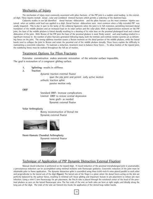
Volar Arthroplasty<br />
Boney reconstruction of Dorsal rim<br />
Dynamic external fixator<br />
Hemi-Hamate Chondral Arthroplasty<br />
Dynamic external fixator<br />
Technique of Application of PIP Dynamic Distraction External Fixation:<br />
Manual closed reduction is performed on the injured digit. If closed reduction of the proximal interphalangeal joint is unattainable,<br />
a percutaneous reduction can be accomplished using minimal incisions with fluroscopic guidance. Concentric reduction of the joint must be<br />
obtainable prior to frame application. The dynamic distraction splint is assembled using three 0.045-inch K-wires placed parallel to each other<br />
and perpendicular to the lateral axis of the digit (figure). The lateral axis of the finger is a plane where the dorsal forces acting on the skin are<br />
perfectly balanced by the palmar forces, resulting in minimal soft tissue gliding and important feature in pin placement to reduce pin tract<br />
infection. Using a mini C-arm fluoroscopy for pin placement, the first K-wire is placed through the rotational center of the head of the proximal<br />
phalanx as seen on the lateral fluoroscopic view. The free ends of the wire on both sides are bent at right angles and distally along the<br />
long axis of the digit. The ends of the wire are formed into hooks for application of the dental loop rubber bands.<br />
39
