

AAHS ASPN ASRM - 2013 Annual Meeting - American Association ...
AAHS ASPN ASRM - 2013 Annual Meeting - American Association ...
AAHS ASPN ASRM - 2013 Annual Meeting - American Association ...

You also want an ePaper? Increase the reach of your titles
YUMPU automatically turns print PDFs into web optimized ePapers that Google loves.
Combining Split Inferior Turbinate (SIT) Mucosal Flaps with Free Flap for Repairing Nasal Cavity in Composite<br />
Palatal and Maxillary Defect Reconstructions<br />
Institution where the work was prepared: Chang Gung memorial hospital, Taipei, Taiwan<br />
C.K. Tsao; Ming-Huei Cheng; Chwei-Chin Chuang; Fu-Chan Wei; Chang Gung Memorial Hospital<br />
Purpose:<br />
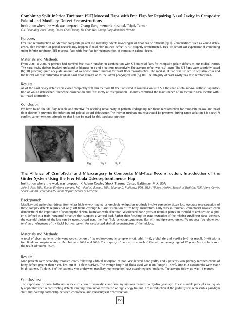
Free flap reconstruction of extensive composite palatal and maxillary defects involving nasal floor can be difficult (fig. I). Complications such as wound dehiscence,<br />
flap infection or partial necrosis may happen if nasal side mucosa defect is not properly reconstructed. Here we report our experience of combining<br />
splint inferior turbinate (SIT) mucosal flaps with free flap for reconstruction of composite palatal defect.<br />
Materials and Methods:<br />
From 2003 to 2006, 9 patients had received free tissue transfers in combination with SIT mucosal flaps for composite palate defects at our medical center.<br />
The nasal cavity defects involved unilateral or bilateral in 4 and 5 patients respectively. The average defect was 4.9*1.8cm. The SIT flaps were superiorly based<br />
(fig. II) providing quite adequate amounts of well-vascularized mucosa for nasal floor reconstruction. The medial SIT flap was sutured to septal mucosa and<br />
the lateral one was sutured to residual nasal floor mucosa or to the lateral pharyngeal wall (fig III). The integrity of nasal cavity was thus reestablished.<br />
Results:<br />
All of the nasal cavity defects were closed completely with this method. 10 free flaps used in combination with SIT flaps had a total survival without flap infection<br />
or wound dehiscence. Fiberscope examination and flow-metry at postoperation 3 months confirmed the maintenance of an adequate nasal meatus without<br />
nasal obstruction.<br />
Conclusion:<br />
We have found the SIT flaps reliable and effective for repairing nasal cavity in patients undergoing free tissue reconstruction for composite palatal and nasal<br />
floor defects. It prevents flap infection and palatal wound dehiscence. The inferior turbinate mucosa should be preserved during tumor ablation if it doesn¡?t<br />
conflict cancer excision principle so that it can be used for this particular purpose<br />
Fig I Fig II Fig III<br />
The Alliance of Craniofacial and Microsurgery in Composite Mid-Face Reconstruction: Introduction of the<br />
Girder System Using the Free Fibula Osteoseptocutaneous Flap<br />
Institution where the work was prepared: R Adams Cowley Shock Trauma Center, Baltimore, MD, USA<br />
Julie E. Park, MD1; Rachel Bluebond-Langner, MD1; Paul N. Manson, MD1; Eduardo D. Rodriguez, DDS, MD2; (1)Johns Hopkins School of Medicine, (2)R Adams Cowley<br />
Shock Trauma Center and the Johns Hopkins School of Medicine<br />
Background:<br />
Maxillary and periorbital defects from either high-energy trauma or oncologic extirpation routinely involve composite tissue loss. Accurate reconstruction of<br />
these complex defects requires not only soft tissue coverage but also restoration of the bony architecture. Early work in traumatic craniofacial reconstruction<br />
demonstrated the importance of restoring the skeletal buttresses with either non-vascularized bone grafts or titanium plates. In the field of architecture, a girder<br />
is defined as a main horizontal structure that supports a vertical load. Rather than focusing on exact recreation of the missing curvilinear facial skeleton,<br />
the essential girders of the face can be reconstructed using the free fibula osteoseptocutaneous flap with multiple osteotomies. We propose “the girder system”<br />
as a refinement of the facial buttress system for vascularized skeletal reconstruction of the midface.<br />
Materials and Methods:<br />
A total of eleven patients underwent reconstruction of the orbitozygomatic complex (n=2), orbit (n=1), orbital rim and maxilla (n=3) or maxilla (n=5) with a<br />
free fibula osteoseptocutaneous flap between 2003 and 2005. The majority of patients were male (73%) with an average age of 37 years. Most defects were<br />
the result of trauma (n=9).<br />
Results:<br />
Nine patients were secondary reconstructions following subtotal resorption of non-vascularized bone grafts, and 2 patients were primary reconstructions of<br />
bony defects greater than 5 cm. Ten out of 11 flaps survived. The average length of fibula used was 8 cm (range 6-15cm). One to 2 osteotomies were made<br />
in all patients. To date, 3 of the patients who underwent maxillary reconstruction have osseointegrated implants. The average follow-up was 18 months.<br />
Conclusions:<br />
The importance of facial buttresses in reconstruction of traumatic craniofacial injuries was realized twenty-five years ago. These valuable principles are equally<br />
applicable when reconstructing defects resulting from tumor extirpation or high energy trauma. The introduction of the girder system represents a paradigm<br />
shift and evolving partnership between craniofacial and microsurgical reconstruction.<br />
155
