

AAHS ASPN ASRM - 2013 Annual Meeting - American Association ...
AAHS ASPN ASRM - 2013 Annual Meeting - American Association ...
AAHS ASPN ASRM - 2013 Annual Meeting - American Association ...

You also want an ePaper? Increase the reach of your titles
YUMPU automatically turns print PDFs into web optimized ePapers that Google loves.
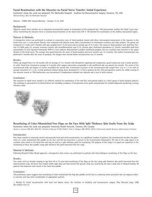
Facial Reanimation with the Masseter-to-Facial Nerve Transfer: Initial Experience<br />
Institution where the work was prepared: The Methodist Hospital - Institute for Reconstructive Surgery, Houston, TX, USA<br />
Michael Klebuc, MD; The Methodist Hospital<br />
Abstract – <strong>ASRM</strong> 2007 <strong>Annual</strong> <strong>Meeting</strong> – January 13-16, 2007<br />
Background:<br />
Adjacent cranial nerve transfers are an important reconstructive option in reanimation of the paralyzed face. This presentation outlines the initial 3 year experience<br />
transferring the masseter nerve to selected buccal branches of the facial nerve (CN V- VII transfer) for reanimation of the midface and perioral region.<br />
Patients & Methods:<br />
A retrospective review was performed to evaluate 6 consecutive cases of facial paralysis treated with direct microsurgical anastomosis of the masseter to the<br />
facial nerve over a 3 year period. Patients were evaluated with physical exam, direct measurement of commissure excursion and video analysis. The group was<br />
composed of 3 males and 3 females with ages ranging from 7 to 84 years and an average age of 41.5 years. The causes of facial paralysis were skull base fracture<br />
(1), Bells palsy (1), acoustic neuroma excision and neurofibromatosis type II (1), petrous apex cholesterol granuloma (1), chronic mastoiditis-skull base<br />
osteomyelitis (1) and ruptured intracranial AVM (1). Four patients demonstrated complete hemifacial paralysis and two retained some function in the upper<br />
division of the facial nerve. The average time period between the onset of facial paralysis and nerve transfer was 14.3 months. The earliest reconstruction was<br />
performed 5 months after the onset of paralysis. The longest time interval before reconstruction was 23 months.<br />
Results:<br />
Follow up ranged from 4 to 30 months with an average of 14.2 months with all patients regaining oral competence, good resting tone and a nearly symmetric<br />
smile. Facial tone returned an average of 4 months after surgery and motion comparable to the unaffected side was present 6.6 months. The vector of the<br />
reconstructed smile and degree of motion resembled the normal side. Commissure excursion of the reconstructed side ranged from 1cm to 1.7 cm with an<br />
average movement of 1.3 cm. A smile produced without biting down developed in 2 of the 6 patients by the 19th postoperative month. No visible wasting of<br />
the masseter muscle or TMJ dysfunction was encountered. Complications included one sialocele and a case of otitis externa.<br />
Conclusion:<br />
The masseter-to-facial nerve transfer is an effective method for reanimation of the mid-face and perioral region in a select group of facial paralysis patients.<br />
The technique is advocated for its limited donor site morbidity, avoidance of interposition nerve grafts and potential for cerebral adaptation producing a strong,<br />
effortless smile.<br />
Resurfacing of Color-Mismatched Free Flaps on the Face With Split Thickness Skin Grafts From the Scalp<br />
Institution where the work was prepared: University Health Network, Toronto, ON, Canada<br />
Declan A. Lannon, MB, BCh, BAO, FR1; Christine B. Novak, PT, MS, PhD(c)1; Peter C. Neligan, MB, FRCSC, FACS2; (1)University Health Network, (2)University of Toronto<br />
Introduction:<br />
Free tissue transfer is commonly used in microvascular head and neck reconstruction. In a significant number of patients, the reconstruction involves the placement<br />
of a color mismatched skin paddle on the face and this has long been a concern in the reconstructive literature(1). The skin in the scalp region is an<br />
excellent color match to the facial skin and may be used as a split thickness graft for resurfacing. The purpose of this study is to report our experience in the<br />
resurfacing of these skin paddles using split thickness skin graft harvested from the scalp.<br />
Material & Methods:<br />
Following Research Ethics Board approval, a retrospective chart review was performed on patients who had undergone resurfacing of free flaps on the face.<br />
Results:<br />
Two males and two females ranging in age from 49 to 72 years had resurfacing of free flaps on the face using split thickness skin grafts harvested from the<br />
shaved parietal scalp. All donor sites healed within eight days and had normal hair growth. Only one resurfacing site had a small area of delayed healing. All<br />
patients had improved color match in the facial skin paddle.<br />
Conclusion:<br />
This preliminary report suggests that resurfacing of color-mismatched free flap skin paddles on the face is a relatively minor procedure that can improve esthetic<br />
outcome and may merit consideration in appropriate patients.<br />
1. Menick FJ. Facial reconstruction with local and distant tissue: the interface of aesthetic and reconstructive surgery. Plast Reconstr Surg. 1998<br />
Oct;102(5):1424-33.<br />
153
