

AAHS ASPN ASRM - 2013 Annual Meeting - American Association ...
AAHS ASPN ASRM - 2013 Annual Meeting - American Association ...
AAHS ASPN ASRM - 2013 Annual Meeting - American Association ...

You also want an ePaper? Increase the reach of your titles
YUMPU automatically turns print PDFs into web optimized ePapers that Google loves.
<strong>AAHS</strong> Concurrent Scientific Paper Session 1A<br />
In-vivo 3-D Distal Radioulnar Joint Arthrokinematic Analysis During Resisted Active Pronation and Supination<br />
Institution where the work was prepared: Mayo Clinic, Rochester, MN, USA<br />
Kazunari Tomita, MD; Shian Chao Tay, MBBS, FRCS, FAMS; Richard A. Berger, MD, PhD; Kimberly Amrami; Kai-Nan An, PhD; Mayo Clinic<br />
Purpose:<br />
Torque production is felt to be a critical function of the human forearm, yet there are no studies that have quantified displacement of the distal radioulnar<br />
joint (DRUJ) resulting from resistance rotation loading of the forearm. The purpose of this study is to quantify displacement of the DRUJ in normal subjects<br />
during resisted rotation loading.<br />
Methods:<br />
Ten normal volunteers without any wrist pathology (age 29.2±7.4 yrs, F:5 M:5) participated in the study. Bilateral 3-D CT scans of the subjects’ distal forearms<br />
were obtained while grasping vertical posts of a custom jig, maintaining a neutral forearm position. Scanning was then performed in three loading conditions:<br />
no load (NL) serving as the control condition, maximum active resisted supination (S), and maximum active resisted pronation (P). Using Matlab and<br />
ANALYZE programs, three different registration methods (manual, automatic voxel, and automatic surface) of CT image were used to quantify relative displacement<br />
of the radius and ulna (designating the radius as the stabilized bone). The ulnar fovea served as the moving reference landmark where a displacement<br />
vector between loading conditions was determined using the registration matrices. Comparisons of 3-D displacement data were performed between no load<br />
and resisted pronation (NL-P), and no load and resisted supination (NL-S).<br />
Results:<br />
The mean magnitudes of displacements in the NL-P condition were 2.51mm (±0.77mm) by manual, 2.76mm (±0.78mm) by voxel and 2.63mm (±1.23mm) by<br />
surface registration methods. The mean magnitudes of displacements in the NL-S condition were 1.65mm (±0.89mm) by manual, 2.19mm (±0.71mm) by voxel<br />
and 1.78mm (±0.73mm) by surface registration methods. No statistically significant differences were detected between the displacements in the NL-P and NL-<br />
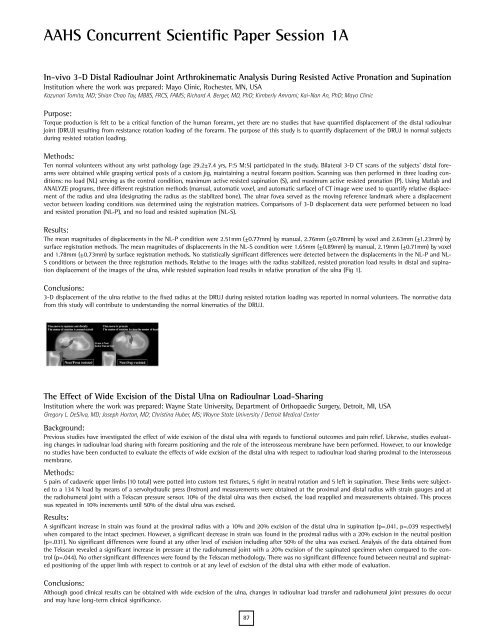
S conditions or between the three registration methods. Relative to the images with the radius stabilized, resisted pronation load results in distal and supination<br />
displacement of the images of the ulna, while resisted supination load results in relative pronation of the ulna (Fig 1).<br />
Conclusions:<br />
3-D displacement of the ulna relative to the fixed radius at the DRUJ during resisted rotation loading was reported in normal volunteers. The normative data<br />
from this study will contribute to understanding the normal kinematics of the DRUJ.<br />
The Effect of Wide Excision of the Distal Ulna on Radioulnar Load-Sharing<br />
Institution where the work was prepared: Wayne State University, Department of Orthopaedic Surgery, Detroit, MI, USA<br />
Gregory L. DeSilva, MD; Joseph Horton, MD; Christina Huber, MS; Wayne State University / Detroit Medical Center<br />
Background:<br />
Previous studies have investigated the effect of wide excision of the distal ulna with regards to functional outcomes and pain relief. Likewise, studies evaluating<br />
changes in radioulnar load sharing with forearm positioning and the role of the interosseous membrane have been performed. However, to our knowledge<br />
no studies have been conducted to evaluate the effects of wide excision of the distal ulna with respect to radioulnar load sharing proximal to the interosseous<br />
membrane.<br />
Methods:<br />
5 pairs of cadaveric upper limbs (10 total) were potted into custom test fixtures, 5 right in neutral rotation and 5 left in supination. These limbs were subjected<br />
to a 134 N load by means of a servohydraulic press (Instron) and measurements were obtained at the proximal and distal radius with strain gauges and at<br />
the radiohumeral joint with a Tekscan pressure sensor. 10% of the distal ulna was then excised, the load reapplied and measurements obtained. This process<br />
was repeated in 10% increments until 50% of the distal ulna was excised.<br />
Results:<br />
A significant increase in strain was found at the proximal radius with a 10% and 20% excision of the distal ulna in supination (p=.041, p=.039 respectively)<br />
when compared to the intact specimen. However, a significant decrease in strain was found in the proximal radius with a 20% excision in the neutral position<br />
(p=.031). No significant differences were found at any other level of excision including after 50% of the ulna was excised. Analysis of the data obtained from<br />
the Tekscan revealed a significant increase in pressure at the radiohumeral joint with a 20% excision of the supinated specimen when compared to the control<br />
(p=.044). No other significant differences were found by the Tekscan methodology. There was no significant difference found between neutral and supinated<br />
positioning of the upper limb with respect to controls or at any level of excision of the distal ulna with either mode of evaluation.<br />
Conclusions:<br />
Although good clinical results can be obtained with wide excision of the ulna, changes in radioulnar load transfer and radiohumeral joint pressures do occur<br />
and may have long-term clinical significance.<br />
87
