

AAHS ASPN ASRM - 2013 Annual Meeting - American Association ...
AAHS ASPN ASRM - 2013 Annual Meeting - American Association ...
AAHS ASPN ASRM - 2013 Annual Meeting - American Association ...

Create successful ePaper yourself
Turn your PDF publications into a flip-book with our unique Google optimized e-Paper software.
A second parallel 0.045-inch k-wire is passed through the distal metaphysis or condlye of the middle phalanx. The free ends of this wire are<br />
also bent at a right angle and directed distally. This wire is parallel to the first wire and its ends are also fashioned into hooks for attachment<br />
of the dental loops. The proximal and distal pins are bent into hook form with a distance between the hooks of 2.5 cm. These two wires bridge<br />
the joint and with the dental loop rubber bands become the engine for continuous distraction to maintain concentric joint reduction.<br />
Ligamentous traction maintains reduction by transferring the forces of distraction to the surrounding soft tissue of the joint, which hold the<br />
fracture alignment. The final K-wire is parallel to other two wires and passes through the mid-diaphysis of the middle phalanx in the mid-axial<br />
line. Its free ends are cut short and bent around the limbs of the first wire to maintain the alignment of the first wire in the same plane with<br />
the digit as it courses through its flexion and extension arc of motion. This wire is provides a palmar translatory moment to aid in balancing<br />
the dorsally-directed forces of displacement. Dental loop rubber bands are then applied between the two hooks in sufficient quantity (usually<br />
three for each pair of hooks) to distract the joint and maintain its reduction throughout full range of motion. Joint reduction through a complete<br />
arc of motion is confirmed with radiographic imaging.<br />
Postoperative Management<br />
Post-operatively, successful restoration of hand function is greatly aided by supervised hand therapy for both active and passive<br />
motion at both interphalangeal joints. Patients are started on an immediate gentle motion protocol within a few days following surgery as<br />
swelling decreases and joint motion increases . Weekly radiographs are obtained to confirm joint reduction . If dorsal subluxation is identified,<br />
applying more dental loop rubber bands until reduction is obtained increases traction. This is a powerful distraction device, and if over distraction<br />
is detected, the number of rubber bands used for traction is reduced. Local wound care to pin tract sites is provided once a day with dilute<br />
hydrogen peroxide and sterile cotton swabs. The use of medicated ointments for pin tract care is not recommended, as these compounds prevent<br />
drainage, and may cause contact dermatitis with prolonged use. It has been shown with this dynamic splint that full recovery of flexion<br />
can be attained . Strict compliance with a supervised hand rehabilitation program is critical if patients are to regain full active extension of the<br />
PIP joint. The device is removed when radiographs demonstrate bony union, usually at five to six weeks. Prior to device removal, lateral radiographs<br />
with the rubber bands removed are obtained in full flexion and extension to confirm that concentric joint reduction is maintained out<br />
of traction . Therapy is continued after splint removal for one to two months, to recover motion and strengthen the hand.<br />
Results<br />
We reported on a series of 9 fracture-dislocations of the PIP joint , treated with this dynamic distraction external fixation device. All<br />
fractures healed without complications recurrent dorsal dislocation or infection. Average flexion measured 94 degrees (range 85 to 115) and<br />
average extension deficit measured 14.5 degrees (range 0 to 35). DIP motion averaged 60 degrees of flexion and 9.5 degrees extension lag.<br />
One patient required a return to the operating room at 5 months for extensor tenolysis, and had restoration of active extension measuring 7<br />
degrees. While all patients recovered functional digital flexion, there was a marked improvement in recovery of extension among the 5 patients<br />
treated with supervised hand therapy when compared with a group of 4 patients treated exclusively with a home exercise program.<br />
Figures<br />
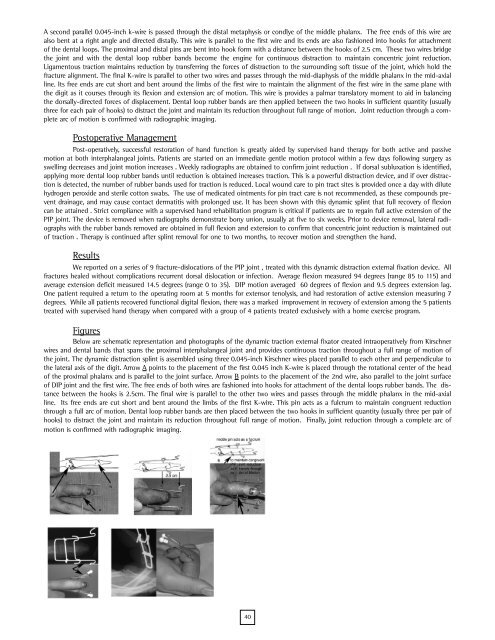
Below are schematic representation and photographs of the dynamic traction external fixator created intraoperatively from Kirschner<br />
wires and dental bands that spans the proximal interphalangeal joint and provides continuous traction throughout a full range of motion of<br />
the joint. The dynamic distraction splint is assembled using three 0.045-inch Kirschner wires placed parallel to each other and perpendicular to<br />
the lateral axis of the digit. Arrow A points to the placement of the first 0.045 inch K-wire is placed through the rotational center of the head<br />
of the proximal phalanx and is parallel to the joint surface. Arrow B points to the placement of the 2nd wire, also parallel to the joint surface<br />
of DIP joint and the first wire. The free ends of both wires are fashioned into hooks for attachment of the dental loops rubber bands. The distance<br />
between the hooks is 2.5cm. The final wire is parallel to the other two wires and passes through the middle phalanx in the mid-axial<br />
line. Its free ends are cut short and bent around the limbs of the first K-wire. This pin acts as a fulcrum to maintain congruent reduction<br />
through a full arc of motion. Dental loop rubber bands are then placed between the two hooks in sufficient quantity (usually three per pair of<br />
hooks) to distract the joint and maintain its reduction throughout full range of motion. Finally, joint reduction through a complete arc of<br />
motion is confirmed with radiographic imaging.<br />
40
