

AAHS ASPN ASRM - 2013 Annual Meeting - American Association ...
AAHS ASPN ASRM - 2013 Annual Meeting - American Association ...
AAHS ASPN ASRM - 2013 Annual Meeting - American Association ...

Create successful ePaper yourself
Turn your PDF publications into a flip-book with our unique Google optimized e-Paper software.
Anatomy and Hystology of the Latissimus Dorsi Subunits for Facial Reanimation<br />
Institution where the work was prepared: Souza Aguiar City Hospital and Federal University of São Paulo, Rio de Janeiro and São Paulo, Brazil<br />
André Salo Buslik Hazan, MD1; Fábio Xerfan Nahas, PhD, MD2; Marcus Vinícius Jardini Barbosa, PhD, MD2; Eugênio Piñeda, PhD, MD3; Lydia Masako Ferreira, PhD, MD2;<br />
(1)Souza Aguiar City Hospital of Rio de janeiro and Federal University of São Paulo, (2)Federal University of São Paulo, (3)Souza Aguiar City Hospital<br />
BACKGROUND:<br />
The use of muscular flaps for the correction of defects after mimetic muscles damage has been described. However, these flaps showed restrictions because of<br />
the large volume of muscle transferred and the vector in a single direction. Recently, perforator flaps has been widely used but its limitation is offer only cutaneous<br />
cover. The goal of this type of reconstruction is to restore both muscular and cutaneous cover with tissues that presents similar size and thickness of<br />
the damage multiple mimetic muscles of the face with different force vectors. The purpose of this study is to present the anatomy and histology of the latissimus<br />
dorsi subunits for facial transplantation and to present a case report with this alternative reconstruction technique.<br />
METHODS:<br />
Ten fresh adult cadavers were dissected. The latissimus dorsi muscle was dissected and the perforators vessels are identified. The vessel that supplies the overlying<br />
skin is individualized. Dissection proceeds into the muscle, sectioning the epimysium and the perimysium, individualizing the lateral segmental bundle or the inferior<br />
one. Then, the vascular and nervous subsegmental bundles penetrating the corresponding muscular groups are observed. The neurovascular subsegmental bundles<br />
and the muscular subunits are isolated by dissection. Each subunit was isolated and was histologically evaluated. A histological search for an artery, a vein and<br />
a nerve was performed 3 mm before and right at the penetration of the pedicle within the muscle. The external diameters of these structures were measured.<br />
RESULTS:<br />
Subunits of independent muscles were created. They are supplied by a subsegmental vessels pedicle connected to a lateral or inferior segmental vessel, which is a<br />
branch of the thoracodorsal vessel. Histological study showed that ninety eight percent of the subunits presented at least one artery, one vein and one nerve.<br />
CONCLUSION:<br />
This study supports the use of subunits of latissimus dorsi flap as a substitute of mimic facial muscles.<br />
The Free Partial Superior Latissimus (PSL) Muscle Flap: Preservation of Donor Site Form and Function<br />
Institution where the work was prepared: The Buncke Clinic, San Francisco, CA, USA<br />
Karen M. Horton, MD, MSc, FRCSC; Rudolf F. Buntic, MD; Darrell Brooks; Charles K. Lee; The Buncke Clinic<br />
PURPOSE:<br />
The latissimus dorsi flap is widely applied, reliable and versatile for microvascular reconstruction. Harvest of the entire muscle results in sacrifice of form, creates a<br />
large donor space, and may create functional loss. Use of the superior portion only decreases donor site morbidity and provides a flap of variable size. We describe<br />
the partial superior latissimus (PSL) muscle flap, its harvest technique, and application as a microvascular transplant for complex defects in thirteen patients.<br />
METHODS:<br />
The superior portion of the latissimus dorsi is isolated on the transverse branch of the thoracodorsal artery though a transverse incision parallel to the upper<br />
muscle border. The pedicle is followed proximally as needed for sufficient length. For functional PSL muscle transfer, the transverse branch of the thoracodorsal<br />
nerve is dissected intraneurally, leaving the branch to the lateral latissimus intact.<br />
RESULTS:<br />
Thirteen patients have undergone PSL flap procedures: 11 were used for extremity salvage or complex wounds, 2 were transplanted for facial reanimation and one sensory<br />
innervated flap was used to achieve a sensate heel. Flap dimensions ranged from 10 x 5 cm to 24 x 12 cm. All flaps survived; one hematoma occurred in a patient<br />
on perioperative heparin. A symmetrical lateral thoracic silhouette was maintained and the remaining latissimus muscle functioned postoperatively in all patients.<br />
DISCUSSION:<br />
Harvest of the superior portion of the latissimus muscle on the transverse branch (TB) of the thoracodorsal vessels (TDA) preserves the entire lateral and inferior<br />
elements of the muscle via the descending branch (DB), together with its nerve supply. This preserves the lateral thoracic form and decreases potential<br />
functional muscle loss. Similar to the partial medial rectus flap, a reliable muscle flap of variable size can be designed while preserving donor site form and<br />
function. “Muscle-sparing” latissimus flaps have been described; however, lateral muscle dissection or complete muscle harvest were used. Innervation of the<br />
PSL flap and neurrorhaphy to a recipient motor nerve enables functional muscle transplantation.<br />
CONCLUSIONS:<br />
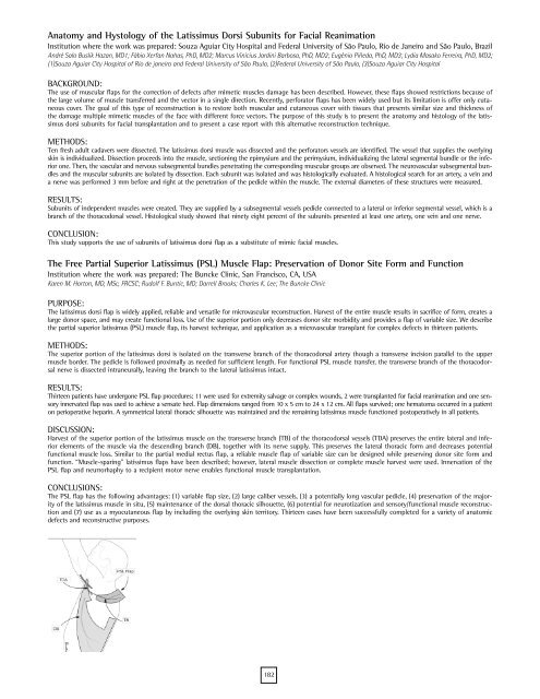
The PSL flap has the following advantages: (1) variable flap size, (2) large caliber vessels, (3) a potentially long vascular pedicle, (4) preservation of the majority<br />
of the latissimus muscle in situ, (5) maintenance of the dorsal thoracic silhouette, (6) potential for neurotization and sensory/functional muscle reconstruction<br />
and (7) use as a myocutaneous flap by including the overlying skin territory. Thirteen cases have been successfully completed for a variety of anatomic<br />
defects and reconstructive purposes.<br />
182
