5-Japanese Puffer Fish - aromatase inhibitor induced masculinization

You also want an ePaper? Increase the reach of your titles
YUMPU automatically turns print PDFs into web optimized ePapers that Google loves.
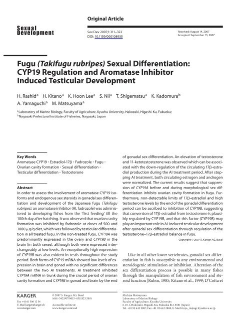
Original Article<br />
Sex Dev 2007;1:311–322<br />
DOI: 10.1159/000108935<br />
Received: August 14, 2007<br />
Accepted: September 13, 2007<br />
Fugu (Takifugu rubripes) Sexual Differentiation:<br />
CYP19 Regulation and Aromatase Inhibitor<br />
Induced Testicular Development<br />
H. Rashid<br />
a<br />
H. Kitano a K. Hoon Lee a S. Nii a T. Shigematsu a K. Kadomura b<br />
A. Yamaguchi a M. Matsuyama a<br />
a<br />
Laboratory of Marine Biology, Faculty of Agriculture, Kyushu University, Hakozaki, Higashi-Ku, Fukuoka ;<br />
b<br />
Nagasaki Prefectural Institute of <strong>Fish</strong>eries, Nagasaki , Japan<br />
Key Words<br />
Aromatase CYP19 Estradiol-17 Fadrozole Fugu <br />
Ovarian cavity formation Sexual differentiation <br />
Testicular differentiation Testosterone<br />
Abstract<br />
In order to assess the involvement of <strong>aromatase</strong> CYP19 isoforms<br />
and endogenous sex steroids in gonadal sex differentiation<br />
and development of the <strong>Japanese</strong> fugu (Takifugu<br />
rubripes) , an <strong>aromatase</strong> <strong>inhibitor</strong> (AI, fadrozole) was administered<br />
to developing fishes from the ‘first feeding’ till the<br />
100th day after hatching. It was observed that ovarian cavity<br />
formation was inhibited by fadrozole at doses of 500 and<br />
1000 g/g diet, which was followed by testicular differentiation<br />
in all treated fugu. In the non-treated fugu, CYP19A was<br />
predominantly expressed in the ovary and CYP19B in the<br />
brain (in both sexes), although both were expressed interchangeably<br />
at low levels. An exceptionally high expression<br />
of CYP19B was also evident in testis throughout the study<br />
period. Both forms of CYP19 mRNA showed low levels of expression<br />
in brain and gonad with no significant differences<br />
between the two AI treatments. AI treatment inhibited<br />
CYP19A mRNA in trunk during the crucial period of ovarian<br />
cavity formation and CYP19B in gonad and brain by the end<br />
of gonadal sex differentiation. An elevation of testosterone<br />
and 11-ketotestosterone was observed which can be associated<br />
with the down-regulation of the circulating 17-estradiol<br />
production during the AI treatment period. After stopping<br />
AI treatment, both circulating estrogen and androgen<br />
were normalized. The current results suggest that suppression<br />
of CYP19A before and during morphological sex differentiation<br />
inhibits ovarian cavity formation in fugu. Furthermore,<br />
non-detectable limits of 17-estradiol and high<br />
testosterone levels by the end of the gonadal differentiation<br />
period can be ascribed to inhibition of CYP19B, suggesting<br />
that conversion of 17-estradiol from testosterone is plausibly<br />
regulated by CYP19B, and that this factor (CYP19B) may<br />
play an important role in AI-<strong>induced</strong> testicular development<br />
after gonadal sex differentiation through regulation of the<br />
testosterone–17-estradiol balance in fugu.<br />
Copyright © 2007 S. Karger AG, Basel<br />
Like in all other lower vertebrates, gonadal sex differentiation<br />
in fish is susceptible to any environmental and<br />
steroidogenic stimulation or inhibition. Alteration of the<br />
sex differentiation process is possible in many fishes<br />
through the manipulation of fish environment and steroid<br />
function [Rubin, 1985; Kitano et al., 1999; D’Cotta et<br />
Fax +41 61 306 12 34<br />
E-Mail karger@karger.ch<br />
www.karger.com<br />
© 2007 S. Karger AG, Basel<br />
1661–5425/07/0015–0311$23.50/0<br />
Accessible online at:<br />
www.karger.com/sxd<br />
Michiya Matsuyama<br />
Laboratory of Marine Biology<br />
Faculty of Agriculture, Kyushu University<br />
6-10-1, Hakozaki, Higashi-Ku, Fukuoka 812-8581 (Japan)<br />
Tel. +81 92 642 2887, Fax +81 92 642 2888, E-Mail rinya_m@agr.kyushu-u.ac.jp
al., 2001; Piferrer et al., 2005; Goto-Kazeto et al., 2006].<br />
Some of the chemicals, which have androgenic or estrogenic<br />
functions, are reported to alter gonadal sex in fish<br />
from female to male or vice versa if administered during<br />
the period of gonadal sex differentiation [Papoulias et al.,<br />
2000; Arslan and Phelps, 2004; Hirai et al., 2006] or even<br />
at maturity [Chang et al., 1995; Yeh et al., 2003]. Other<br />
than the direct androgenic or estrogenic effects, there are<br />
chemicals which are capable of disrupting the enzymatic<br />
pathway of natural androgen-estrogen balance in fish<br />
[McAllister and Kime, 2003; Hallgren et al., 2006; Villeneuve<br />
et al., 2006]. Among the non-steroidal <strong>aromatase</strong><br />
<strong>inhibitor</strong>s (AI), fadrozole is usually used as an effective<br />
human drug in the treatment of estrogen-dependent disease<br />
including breast cancer and was found to be effective<br />
in suppression of all estrogens after oral administration<br />
[Dowsett et al., 1994]. It has been reported that fadrozole<br />
treatment also results in reduced 17 -estradiol (E2) production<br />
in mammals, viz. monkey, hamster, rabbit and rat<br />
[Shetty et al., 1995; Moudgal et al., 1996]. Induction of<br />
male sex determination has also been reported in reptiles<br />
(e.g., lizard and turtle) administered with fadrozole [Wibbels<br />
and Crews, 1994]. In fish, fadrozole is reported to result<br />
in reduced E2 production [Bhandari et al., 2004; Mandiki<br />
et al., 2005], atresia of ovarian follicles in masculinizing<br />
genetic females [Ankley et al., 2002] and complete<br />
irreversible <strong>masculinization</strong> [Kitano et al., 2000; Kwon et<br />
al., 2000; Afonso et al., 2001; Fenske and Segner, 2004;<br />
Tzchori et al. , 2004]. In all cases, estrogen synthesis was<br />
found to be significantly reduced in fadrozole-treated<br />
fish.<br />
The balance between androgen (inducers for maleness)<br />
and estrogen (inducers for femaleness) hormones in<br />
higher and lower vertebrates is regulated via an aromatization<br />
reaction. Cytochrome P450 <strong>aromatase</strong> (P450arom),<br />
a microsomal enzymatic complex, is responsible for this<br />
aromatization reaction which results in irreversible conversion<br />
of androgens into estrogens in most vertebrates<br />
[Simpson et al., 2002]. In most fishes P450arom is a product<br />
of two CYP19 isoforms, predominantly expressed in<br />
ovary (ovarian type <strong>aromatase</strong>, CYP19A) and brain<br />
(brain type <strong>aromatase</strong>, CYP19B). Other than local conversion<br />
of androgen into estrogen, CYP19 isoforms have<br />
been found to play important roles in gonadal differentiation<br />
and development in fish. CYP19A has been found<br />
to contribute to ovarian cavity formation and ovarian development<br />
and CYP19B to testicular development and<br />
germ cell maturation in many fishes [reviewed by Devlin<br />
and Nagahama, 2002]. The multifunctional ability of<br />
<strong>aromatase</strong> CYP19 genes in regulating steroid hormone<br />
balance and gonadal differentiation and development has<br />
made them a key parameter for maintaining sexuality in<br />
fish. Thus, a disruption in <strong>aromatase</strong> gene function using<br />
an AI (fadrozole) can inhibit both the CYP19 isoforms<br />
and suppress E2 production, resulting in oocyte atresia,<br />
formation of intermediate gonads or testicular differentiation<br />
of genetically female fish.<br />
The current experiment was performed on the <strong>Japanese</strong><br />
fugu (Takifugu rubripes) , a gonochoristic tetraodontiform<br />
fish, which lives in the marine waters of Japan,<br />
China and Korea. It is the second vertebrate (the first being<br />
human) whose genome has been sequenced completely<br />
and which is proposed as a useful model for annotating<br />
the human genome due to similarity in the repertoire<br />
of genes [Brenner et al., 1993; Aparicio et al. 2002].<br />
The factors involved in gonadal sex determination and<br />
differentiation of such an important fish species have not<br />
yet been studied well. However, by performing genomewide<br />
linkage analyses, Kikuchi et al. [2007] revealed that<br />
the sex of fugu is determined by a single region on linkage<br />
group 19 in an XX/XY system. As a part of understanding<br />
the mechanism of gonadal sex determination,<br />
differentiation and development of fugu, histological investigation<br />
of the process of gonadal sex differentiation<br />
has been carried out previously in our laboratory and the<br />
course of ovarian and testicular development in this species<br />
was identified [Matsuura et al., 1994; Yamaguchi et<br />
al., 2006]. In a recent study with fugu Dmrt ( Doublesex/<br />
mab3 related transcription factor 1), a transcription factor<br />
that regulates early differentiation of Sertoli cells in<br />
the testes of vertebrates, we have found that it is involved<br />
in gonadal development rather than sex differentiation<br />
and that its expression is correlated with the proliferation<br />
of spermatogonia [Yamaguchi et al., 2006]. So far there is<br />
no detailed information on the effects of different environmental<br />
parameters on the sex determination and differentiation<br />
of fugu. However, a recent study (conducted<br />
in our lab) on the effects of high temperature (29 ° C, environmental<br />
water temperature being 17 ° C) on developing<br />
fugu revealed no effect of high temperature on sexuality<br />
of this fish (unpublished data). Hence, to get a better<br />
understanding of the process of fugu gonadal sex differentiation<br />
together with the role of two CYP19 isoforms in<br />
this process, the current study was aimed at investigating<br />
whether gonadal sex differentiation of this fish is affected<br />
by AI fadrozole which is reported to suppress CYP19 gene<br />
function and disrupt production of E2 in many animals.<br />
312<br />
Sex Dev 2007;1:311–322<br />
Rashid et al.
Materials and Methods<br />
Experimental <strong>Fish</strong><br />
Fertilized eggs of fugu were bought from a local fish hatchery<br />
and kept in a tank at the <strong>Fish</strong>eries Research Laboratory of Kyushu<br />
University situated at Tsuiyazaki, Japan. Most of the eggs hatched<br />
out 12 to 13 days after fertilization. Hatched out larvae were kept<br />
in the hatching tank for 3 days without any supplementary feed.<br />
After the absorption of the yolk sack (3 days after hatching, dah),<br />
they were transferred to a larvae rearing tank. From 4 dah, the<br />
larvae were supplied with live rotifer plus formulated feed (with<br />
or without fadrozole). From 19 dah, decapsulated and nutrified<br />
Artemia were supplied to larvae together with rotifer and formulated<br />
feed. Rotifer supply was stopped on 33 dah and formulated<br />
feed, together with Artemia , was continued until 38 dah. From 39<br />
dah, larvae were supplied only with formulated feed up to the end<br />
of the experiment (9 months after hatching, mah). The fishes were<br />
reared under flow-through seawater and aeration at environmental<br />
temperature throughout the experimental period. The water<br />
temperatures in the experimental tanks were around 14 ° C during<br />
hatching out of fugu larvae, and rose to 24 ° C during the fadrozole<br />
treatment period (up to 100 dah). Formulated feed was supplied<br />
ad libitum.<br />
This experiment was carried out under the guidelines for animal<br />
experiments in the Faculty of Agriculture and Graduate<br />
Course of Kyushu University, and according to the laws (No. 105)<br />
and notifications (No. 6) of the <strong>Japanese</strong> Government.<br />
AI Treatment<br />
Aromatase <strong>inhibitor</strong> used in this experiment was fadrozole (4-<br />
(5,6,7,8-tetrahydroimidazole[1,5- ]pyridin-5-yl)-benzonitrile<br />
monohydrochloride hemihydrate), purchased from Novartis<br />
Pharma K.K. (Japan). After dilution in ethanol (100%), the desired<br />
concentration of fadrozole was sprayed over formulated feed<br />
as uniformly as possible. The fadrozole mixed diets were then<br />
dried properly inside a draft. After complete drying, the diets<br />
were stored at 4 ° C in sealed glass vessels for the entire period of<br />
the experiment. Two doses of fadrozole were used for feeding fugu<br />
larvae, i.e. 500 g/g diet (treatment A) and 1000 g/g diet (treatment<br />
B). Control (treatment C) food was prepared by spraying<br />
only ethanol over formulated diet and drying accordingly.<br />
Sampling Procedure<br />
Both AI-treated and control fish were sampled on 35, 42, 70,<br />
100 dah, and 6 and 9 mah. Brain (or head) and gonad (or trunk)<br />
samples of 20 to 30 fish at every stage were collected from anesthetized<br />
fish. Both brain (or head) and gonad (or trunk) samples<br />
for RNA extraction were either preserved in RNA later TM (QIA-<br />
GEN GmbH, Germany) or quick-frozen in liquid nitrogen, later<br />
preserved at –80 ° C. Gonad (or trunk) samples for histological<br />
investigations were fixed in Bouin’s fluid. After fixation for 24 h,<br />
the gonad samples were run through an ethanol series to dehydrate<br />
the tissue. After dehydration, the gonad samples were preserved<br />
at –20 ° C (keeping in 100% ethanol) until they were embedded<br />
in paraffin. Blood samples were collected on 100 dah and<br />
9 mah from the caudal vein using injection syringes. Soon after<br />
collection, blood samples were centrifuged at 4000 rpm for 25<br />
min to separate the serum. Later the serum samples were preserved<br />
in separate test tubes at –20 ° C until assay.<br />
G o n a d Hi s t o l o g y<br />
Histological observations of fadrozole-treated and non-treated<br />
gonads were done on 42 dah (morphological differentiation of<br />
gonad), 70 dah, 100 dah (termination of fadrozole treatment),<br />
6 mah and 9 mah (end of investigation). In every stage, around 20<br />
to 25 gonad samples from each treatment were studied. For histological<br />
observations, gonad samples in 100% ethanol were first<br />
cleared by a lemosole wash for 25 to 30 min and then embedded<br />
in paraffin. Embedded tissues were sectioned at 5 m thickness<br />
and stained by hematoxylin-eosin for observation under light microscope.<br />
RNA Extraction and Reverse Transcription<br />
Total RNA from frozen or RNA later TM -pre s e r ve d br a i n (or<br />
head) and gonad (or trunk) samples was extracted with Isogen<br />
(Nippon Gene Co., Ltd., Japan) following the manufacturer’s protocol.<br />
First-strand cDNA (10 l reaction volume) for each sample<br />
was synthesized from 1 g of total RNA using SuperScript TM III<br />
Reverse Transcriptase (Invitrogen, USA) according to the manufacturer’s<br />
protocol. Oligo-d(T) 16 primer (5 -GGCCACGCGTC-<br />
GAC TAGTACT(T) 16 -3 ) (50 M ) was used for reverse transcription<br />
of mRNA.<br />
Semi-quantitative PCR: Optimization Experiment<br />
Optimization experiments for PCR conditions for fugu CYP-<br />
19A, CYP19B and -actin primers were initially done. Gene-specific<br />
primers for fugu CYP19A and CYP19B were designed from<br />
the partial (pfCYP19A) and full (pfCYP19B) cDNA sequences that<br />
have been isolated in our laboratory (to be published elsewhere).<br />
Primers for -actin, the housekeeping gene that was used as internal<br />
control, were designed from the gene-specific sequence described<br />
in Venkatesh et al. [1996] (GenBank accession no. U37449).<br />
Standard PCR reactions were performed using these gene-specific<br />
primers ( table 1 ). The PCR reactions were performed in 10- l reaction<br />
mixtures containing 10 m M Tris-HCl (pH 8), 1.5 m M MgCl 2 ,<br />
50 m M KCl, 100 M dNTP, 0.5 U Ampli Taq Gold, and 0.5 M of<br />
each primer. The cycling conditions for PCR reactions were 1 min<br />
at 94 ° C, 1 min at 64 ° C, and 2 min at 72 ° C (for pfCYP19A) and<br />
1 min at 94 ° C, 1 min at 58 ° C, and 2 min at 72 ° C (for CYP19B and<br />
-actin). To get the optimal cycle number for each gene-specific<br />
primer set, PCR was conducted using cDNA collected from 9 mah<br />
fugu brain and ovary tissue (tissues predominantly expressing<br />
CYP19B and CYP19A, respectively). Seven cycle numbers (20, 25,<br />
30, 35, 40, 45 and 50 cycles) were used for PCR with each sample.<br />
Products amplified by CYP19A versus -actin and CYP19B versus<br />
-actin primers were run together on the same gel (1.5% TBE agarose<br />
gel stained with ethidium bromide). Gel photographs were<br />
taken by a CCD camera under UV illumination. The signals for<br />
PCR bands were calculated by NIH Image . The peak scores calculated<br />
by NIH image were plotted for CYP19A/-actin and CYP 19B/<br />
-actin to draw standard curves. PCR cycle number for CYP 19A,<br />
CYP19B and -actin were then chosen from the linear line of the<br />
standard curves to be used for the respective gene throughout the<br />
investigation of all stages.<br />
Semi-quantification of CYP19 mRNA Expression in Fugu<br />
Brain and Gonad<br />
For a single tissue, PCR was conducted in a 10- l reaction volume<br />
for all three sets of primers using their best cycling conditions<br />
obtained from optimization experiments. Five microliters<br />
CYP19 in Fugu Sexual Differentiation Sex Dev 2007;1:311–322 313
Table 1. Primer sets used for semiquantitative<br />
RT-PCR analysis of brain<br />
type and ovarian type <strong>aromatase</strong><br />
(CYP19B and CYP19A) of T. rubripes<br />
using the -actin gene as internal control<br />
Primer Sequence (5]3) Length of amplified<br />
fragment<br />
pfCYP19A F TCCTGGCTTCGGGGTTCTCATGG<br />
pfCYP19A R CTGTCCAGATGCGCCTGAGTGGAG 353 bp<br />
pfCYP19B F GCTCTGGAGGACGACACCATC<br />
pfCYP19B R GGGATGAACCTCATGGCCAG 579 bp<br />
pf-actin F GCCAACAGGGAGAAGATGACCCAGA<br />
pf-actin R CCAGGGAGGAAGAGGAGGCAGC 370 bp<br />
pf: <strong>Puffer</strong>fish (fugu); F: forward; R: reverse; bp: base pairs.<br />
Table 2. Sex ratio of control and<br />
AI-treated T. rubripes at different<br />
developmental stages<br />
Stage of<br />
fish a<br />
Fadrozole<br />
treatment b<br />
No. of samples<br />
studied<br />
Sex composition<br />
- U Intermediate<br />
42 dah A 23 11 (48%) 12 (52%)<br />
B 25 11 (44%) 14 (56%)<br />
C 25 13 (52%) 12 (48%)<br />
70 dah A 23 13 (57%) 10 (43%)<br />
B 20 10 (50%) 10 (50%)<br />
C 25 12 (48%) 13 (52%)<br />
100 dah A 24 14 (58%) 10 (42%)<br />
B 24 15 (63%) 9 (37%)<br />
C 24 11 (46%) 13 (54%)<br />
6 mah A 25 25 (100%)<br />
B 23 25 (100%)<br />
C 24 12 (50%) 12 (50%)<br />
9 mah A 21 25 (100%)<br />
B 22 25 (100%)<br />
C 25 14 (56%) 11 (44%)<br />
a<br />
dah: Days after hatching; mah: months after hatching.<br />
b<br />
A: 500 g/g diet; B: 1000 g/g diet; C: control (no fadrozole treatment).<br />
of products from each of the PCR reactions were run on gel. To<br />
avoid gel to gel variation of results, PCR products from a single<br />
cDNA for CYP19A, CYP19B and -actin were run together on the<br />
same gel. Calculation of PCR band intensity was performed in a<br />
way similar to that described in the previous section. Semi-quantification<br />
of CYP19A and CYP19B for every cDNA sample was<br />
done by calculating the ratio of PCR gel band intensity for CYP-<br />
19A/-actin and CYP19B/-actin. Sample number ranged from<br />
15 to 20 for every tissue at every stage.<br />
Serum Steroid Measurement<br />
Serum steroids were measured using ELISA protocol. Estradiol<br />
enzyme immunoassay kit (Cayman Chemical Company, MI,<br />
USA) was used for measuring serum E2, whereas serum testosterone<br />
(T) and 11-ketotestosterone (11-KT) were measured using the<br />
ELISA protocol previously practiced in our laboratory for measuring<br />
T and E1 [Ohta et al. , 2001]. Blood serum collected on 100<br />
dah and 9 mah was measured for steroids and sample size for every<br />
treatment on each stage was 12 to 15.<br />
D a t a An a ly s i s<br />
All the results for semi-quantitative RT-PCR and serum steroid<br />
measurements were analyzed by one-way ANOVA followed<br />
by Tukey’s multiple comparison test up to 5% level of significance<br />
(p ! 0.05).<br />
Results<br />
Gonad Histology<br />
Histological investigations of AI-treated and control<br />
gonads at different stages of development revealed a gradual<br />
trend of AI-treated gonads towards testicular development.<br />
Detailed descriptions on stage-specific histological<br />
structure of treated and control gonads are presented<br />
in the following section and data on the number of inves-<br />
314<br />
Sex Dev 2007;1:311–322<br />
Rashid et al.
A<br />
B<br />
C<br />
D<br />
Fig. 1. Haematoxylin-eosin stained sections<br />
of fugu gonads from 42 dah ( A – C )<br />
and 70 dah ( D – F ). A , D Control testis (arrowheads).<br />
B , E Control ovary. C , F Intermediate<br />
gonad from fadrozole treated fish.<br />
OC, ovarian cavity; DOC, deformed ovarian<br />
cavity; OG, oogonium; SG, spermatogonium;<br />
G, gonium; EL, empty lumina of<br />
tubule. Scale bars, 20 m; in insets 50<br />
m.<br />
E<br />
F<br />
tigated samples and sex ratios of all the stages are compiled<br />
in table 2 .<br />
42 dah: Morphological gonadal sex differentiation of<br />
T. rubripes occurs on 42 dah as investigated in a previous<br />
study at our laboratory [Yamaguchi et al., 2006]. Nontreated<br />
control fish at this stage were composed of female<br />
and male individuals as identified by ovarian cavity (OC,<br />
ovary) formation and no ovarian cavity (testis) ( fig. 1 A,<br />
B). In both treatments, A and B, some of the gonads contained<br />
deformed ovarian cavities (DOC) ( fig. 1 C) compared<br />
to the well-developed OC in control ovary ( fig. 1 B),<br />
and the rest of the gonads resembled control testis (arrowheads<br />
in fig. 1 A).<br />
CYP19 in Fugu Sexual Differentiation Sex Dev 2007;1:311–322 315
A<br />
B<br />
C<br />
D<br />
Fig. 2. Haematoxylin-eosin stained sections<br />
of fugu gonads from 100 dah ( A – C ),<br />
6 mah ( D , E ) and 9 mah ( F ). A Control testis.<br />
B, E Control ovary. C Intermediate gonad<br />
from fadrozole treated fish. D , F Testis<br />
from fadrozole treated fish. OC, ovarian<br />
cavity; DOC, deformed ovarian cavity;<br />
OG, oogonium; OCT, oocyte; PNO, perinucleolar<br />
oocyte; PVO, previtellogenic oocyte;<br />
SG, spermatogonium; SC, spermatocyte;<br />
ST, spermatid; SZ, spermatozoon.<br />
Scale bars, 20 m; in insets 50 m.<br />
E<br />
F<br />
70 dah: At this stage, the control ovary had a well-developed<br />
OC and proliferating oogonia ( fig. 1 E). Control<br />
testes were composed of mostly spermatogonia ( fig. 1 D).<br />
On the other hand, treated fishes (A and B) had testes similar<br />
to controls or intermediate gonads having DOC and<br />
empty lumina (EL) of tubules ( fig. 1 F). The intermediate<br />
gonads were filled with gonia which were histologically<br />
not distinguishable from spermatogonia and oogonia.<br />
100 dah: Control ovaries contained cavities and were<br />
composed of oogonia and perinucleolar oocytes (PNO)<br />
( fig. 2 B). Control testes, on the other hand, had proliferating<br />
spermatogonia ( fig. 2 A). AI-treated fishes exhibited<br />
testes similar to control and intermediate gonads containing<br />
DOC and remains of oocytes (OCT) ( fig. 2 C).<br />
6 mah: Control ovaries consisted mostly of perinucleolar<br />
oocytes (PNO) and previtellogenic oocytes (PVO)<br />
316<br />
Sex Dev 2007;1:311–322<br />
Rashid et al.
Fig. 3. Standard curves and relevant gel<br />
photographs (1.5% TBE agarose gel stained<br />
with ethidium bromide) for semi-quantitative<br />
RT-PCR of fugu CYP19A ( A ) and<br />
CYP19B ( B ) using -actin as an internal<br />
control. M, molecular marker; numbers<br />
(20 to 50) indicate number of PCR cycles;<br />
bp, b a s e p a i r s .<br />
Peak score<br />
700<br />
600<br />
500<br />
400<br />
300<br />
200<br />
100<br />
0<br />
M 20<br />
A<br />
CYP19A<br />
-Actin<br />
20 25 30 35 40 45 50<br />
PCR cycle number<br />
25 30 35 40 45 50<br />
Peak score<br />
M<br />
M<br />
579 bp<br />
(CYP19A)<br />
370 bp<br />
(-Actin)<br />
B<br />
1,200<br />
1,000<br />
800<br />
600<br />
400<br />
200<br />
0<br />
20<br />
CYP19B<br />
-Actin<br />
20 25 30 35 40 45 50<br />
PCR cycle number<br />
25 30 35 40 45 50<br />
M<br />
353 bp<br />
(CYP19B)<br />
370 bp<br />
(-Actin)<br />
( fig. 2 E). On the other hand, all treated gonads resembled<br />
control testis. All testes (both treatment and control)<br />
were found to be undergoing spermatogenesis. Spermatogonia,<br />
spermatocytes and spermatids were observed in<br />
histological sections ( fig. 2 D).<br />
9 mah: In the control ovary, cellular stages were almost<br />
similar to those observed on 6 mah ovary (figure not<br />
shown). In both control and treated testis, the final stage<br />
of spermatogenesis, spermatozoa, were observed in histological<br />
sections together with spermatogonia and spermatocytes<br />
( fig. 2 F).<br />
Optimization of PCR Conditions for<br />
Semi-Quantitative Experiments<br />
The standard curves for CYP19A and CYP19B show<br />
that PCR products up to 40 cycle numbers fall into the<br />
linear line for CYP19A, up to 35 cycle numbers for CYP19B<br />
and up to 35 cycle numbers for -actin (fig. 3 ). From the<br />
linear line of the standard curve we selected 35, 35 and 30<br />
cycle numbers for CYP19A, CYP19B and -actin, respectively.<br />
These cycle numbers were then used for conducting<br />
PCR reactions throughout the investigation of all the<br />
stages.<br />
Semi-quantification of CYP19 mRNA Expression in<br />
Fugu Brain and Gonad<br />
CYP19A mRNA expression was high in non-treated<br />
female ovary throughout the investigation and the highest<br />
expression was found in 9 mah ovary (composed of<br />
PNO and PVO) ( fig. 4 D). In all the stages studied, CYP19A<br />
showed very low expression in brain (or head) of both<br />
sexes ( fig. 4 ). Its expression was even not detectable (ND)<br />
in treated brain on 100 dah ( fig. 4 C). In addition, ‘malelike’<br />
low expression of CYP19A was observed in treated<br />
gonads (or trunks) throughout the study period ( fig. 4 ).<br />
An ND level of CYP19A expression was observed in treated<br />
trunk on 35 dah (before morphological gonadal differentiation)<br />
( fig. 4 A).<br />
CYP19B mRNA was highly expressed in brain (or<br />
head) of non-treated fish (both male and female) throughout<br />
the study period. Together with high expression of<br />
CYP19B in non-treated brain, an elevated expression was<br />
observed in testis at relatively adult stages, compared to<br />
low level in ovary ( fig. 4 C, D). In AI-treated brain (or<br />
head), CYP19B expression was low compared to control<br />
but was still higher than that of CYP19A. CYP19B was<br />
also low in treated gonads (or trunks) in all stages compared<br />
to control testis but was expressed at higher level<br />
compared to CYP19A. CYP19B was upregulated (towards<br />
normalization) in gonads after withdrawing fadrozole<br />
application (measured on 9 mah) but was maintained at<br />
low level compared to control testis ( fig. 4 ).<br />
Serum Steroid Level<br />
ELISA (E2, 11-KT and T) experiments revealed low<br />
circulating estrogen (E2) and higher androgen (11-KT<br />
and T) levels throughout the experimental period. In<br />
treatment A and B on 100 dah (termination of fadrozole<br />
exposure), a massive suppression of E2 production was<br />
observed evident by non-detectable limit (NDL) of E2 in<br />
this stage compared to detectable levels in control female<br />
and male (27 pg/ml in female and 16 pg/ml in male). After<br />
9 mah, there were no significant differences of E2<br />
levels between control and treatment although the highest<br />
level was detected in control female (23 pg/ml)<br />
( fig. 5 A). By the end of the fadrozole exposure period<br />
CYP19 in Fugu Sexual Differentiation Sex Dev 2007;1:311–322 317
0.6<br />
a<br />
0.6<br />
a<br />
0.5<br />
0.5<br />
x<br />
CYP19/ -actin<br />
A<br />
0.4<br />
0.3<br />
0.2<br />
0.1<br />
0<br />
z<br />
A<br />
x<br />
y<br />
z<br />
ND ND<br />
B C A B C<br />
Head Trunk<br />
CYP19A<br />
b<br />
A<br />
b, c<br />
c<br />
d<br />
d<br />
B C A B C<br />
Head Trunk<br />
CYP19B<br />
CYP19/ -actin<br />
B<br />
0.4<br />
0.3<br />
0.2<br />
0.1<br />
0<br />
y<br />
A<br />
y<br />
y y y<br />
y y<br />
B CM CF A B CM CF<br />
Head Trunk<br />
CYP19A<br />
c<br />
A<br />
b<br />
b, c<br />
c<br />
c<br />
c<br />
c<br />
B CM CF A B CM CF<br />
Head Trunk<br />
CYP19B<br />
CYP19/ -actin<br />
C<br />
0.9<br />
0.8<br />
0.7<br />
0.6<br />
0.5<br />
0.4<br />
0.3<br />
0.2<br />
0.1<br />
0<br />
y, z y, z y<br />
x<br />
ND ND z z<br />
A B CM CF A B CM CF<br />
Brain Gonad<br />
CYP19A<br />
c<br />
A<br />
a<br />
a<br />
b<br />
c<br />
d<br />
B CM CF<br />
d<br />
A<br />
d<br />
B CM CF<br />
Brain Gonad<br />
CYP19B<br />
CYP19/ -actin<br />
D<br />
1.2<br />
1.1<br />
1.0<br />
0.9<br />
0.8<br />
0.7<br />
0.6<br />
0.5<br />
0.4<br />
0.3<br />
0.2<br />
0.1<br />
0<br />
z<br />
A<br />
x<br />
y<br />
y, z<br />
z<br />
z<br />
z<br />
z<br />
B CM CF A B CM CF<br />
Brain Gonad<br />
CYP19A<br />
a<br />
A<br />
a<br />
a<br />
a<br />
a<br />
b<br />
b<br />
c<br />
B CM CF A B CM CF<br />
Brain Gonad<br />
CYP19B<br />
Fig. 4. Relative expression of CYP19A and CYP19B in fugu brain/head and gonad/trunk on 35 dah ( A ), 42 dah<br />
( B ), 100 dah ( C ), and 9 mah ( D ). A , f a d ro z ole t re at me nt A (5 0 0 g/g diet); B, fadrozole treatment B (1000 g/g<br />
diet); C, control; CM, control male; CF, control female; ND, not detected. Values above bars indicate significant<br />
differences (p ! 0.05) compared within the data. Significant values are plotted separately for CYP19A (x, y, z)<br />
and CYP19B (a, b, c, d).<br />
(100 dah), 11-KT levels in treated fishes were almost<br />
twice as much as in control males, the highest level being<br />
in treatment A (494 pg/ml). The 11-KT level decreased in<br />
9 mah AI treatment when it had no significant difference<br />
with control male ( fig. 5 B). The highest level of steroid<br />
detected in treated fish during the AI exposure period<br />
was T, as observed in treatment B (1727 pg/ml) on 100<br />
dah. This T value was almost double the value in control<br />
male at this stage. After withdrawing fadrozole application,<br />
there was a significant drop of T level in both AI<br />
treatments towards normalization (measured on 9 mah)<br />
(fig. 5C).<br />
Discussion<br />
Oocyte atresia, formation of intermediate gonads and<br />
finally testicular development of genetic females have<br />
been observed in both gonochoristic and hermaphroditic<br />
fishes by the effect of AI fadrozole [Kwon et al., 2000;<br />
Afonso et al., 2001; Fenske and Segner, 2004; Suzuki et<br />
al., 2004; Tzchori et al., 2004; Komatsu et al., 2006]. Not<br />
only fadrozole but also other AIs and exogenous androgens<br />
were found to influence testicular differentiation in<br />
fishes such as to lead to <strong>masculinization</strong> [Piferrer et al.,<br />
1994; Feist et al., 1995; Kitano et al., 2000]. The mecha-<br />
318<br />
Sex Dev 2007;1:311–322<br />
Rashid et al.
Serum E2 (pg/ml)<br />
40<br />
30<br />
20<br />
10<br />
b<br />
a<br />
b<br />
b<br />
b<br />
a, b<br />
Serum 11-KT (pg/ml)<br />
600 2,000<br />
a<br />
a<br />
a<br />
a<br />
450 1,500<br />
c, d<br />
150 500<br />
c<br />
d<br />
Serum T (pg/ml)<br />
300 1,000<br />
b<br />
b, c<br />
b, c b, c<br />
b<br />
c<br />
b, c<br />
c, d<br />
d<br />
A<br />
0<br />
NDL NDL<br />
A B CM CF A B CM CF<br />
100 dah 9 mah<br />
B<br />
0 0<br />
A B CM CF A B CM CF<br />
A B CM CF A B CM CF<br />
100 dah 9 mah C<br />
100 dah 9 mah<br />
Fig. 5. Serum steroid levels of fadrozole treated and control fugu on 100 dah and 9 mah. A Serum E2, B serum<br />
11-KT, C s e r u m T. A , f a d ro z ole t re at me nt A (5 0 0 g/g diet); B, fadrozole treatment B (1000 g/g diet); CM, control<br />
male; CF, control female. Values above the bars indicate significant differences (p ! 0.05) compared within<br />
the data.<br />
Fig. 6. Diagrammatic representation of the interaction between<br />
AI, CYP19 and sex steroids in the process of testicular differentiation<br />
of T. rubripes . Top arrow shows different stages of gonadal<br />
sex differentiation, considering 42 dah as the point of morphological<br />
gonadal sex differentiation. ‘Pre-differentiation’ refers to<br />
the stage where no morphological distinction between testis and<br />
ovary is possible; ‘differentiation’ refers to the period of gonadal<br />
sex differentiation and development from 42 dah; and ‘post-differentiation’<br />
refers to the remaining stage after gonadal sex differentiation<br />
is completed (i.e., premature stage). From the highest<br />
to lowest degree of expression, small arrows inside the text boxes<br />
indicate: t upregulated level; r normalized level; w lowered level;<br />
y suppressed level; X inhibited level. dah, days after hatching;<br />
m a h , mont h s a f t e r h at c h i n g .<br />
nism of AI-<strong>induced</strong> testicular differentiation through<br />
disruption of <strong>aromatase</strong> function has been found to be<br />
controlled in different ways in different fishes. In zebrafish,<br />
for example, irreversible <strong>masculinization</strong> was<br />
achieved by manipulation of the <strong>aromatase</strong> system by<br />
fadrozole during the critical period of sexual differentiation<br />
[Fenske and Segner, 2004]. AI-<strong>induced</strong> sex inversion<br />
in red-spotted grouper was attributed to the inhibition of<br />
P450 gene expression and <strong>aromatase</strong> activity and the resultant<br />
decrease in the biosynthesis of endogenous E2<br />
[Li et al., 2006]. In the case of golden rabbitfish, estrogen<br />
was predicted to be involved in ovarian differentiation<br />
and AI-<strong>induced</strong> suppression of estrogen was found to be<br />
an essential prerequisite for testicular differentiation<br />
CYP19 in Fugu Sexual Differentiation Sex Dev 2007;1:311–322 319
[Komatsu et al., 2006]. In fugu, similar to the studies<br />
mentioned above, AI (fadrozole) treatment during the<br />
period of gonadal sex differentiation was found to induce<br />
testicular differentiation towards irreversible <strong>masculinization</strong>.<br />
In the current study, the AI-treated masculinizing<br />
fugu had deformed ovarian cavities (DOC), empty<br />
lumina of tubules (EL) and remains of oocytes (OCT) inside<br />
intermediate gonads at different stages of development.<br />
They were predicted to be sex-changing (from genetic<br />
female to morphological male) fishes and were<br />
clearly distinguished from those containing testis resembling<br />
that of control male, a case similar to that in AItreated<br />
<strong>masculinization</strong> of rabbitfish. Moreover, appearance<br />
of DOC in some of the AI-treated gonads on 42 dah<br />
(time when morphological gonadal sex differentiation is<br />
first evident in fugu) suggests that AI started inhibiting<br />
ovarian cavity (OC) formation in female fugu before gonadal<br />
sex differentiation. The inhibition of OC formation<br />
during this crucial period of gonadal sex differentiation<br />
and OC formation (35 to 42 dah) can be correlated with<br />
a massive suppression of CYP19A mRNA in AI-treated<br />
trunk at this stage (ND level on 35 dah ( fig. 4 A) and suppressed<br />
level on 42 dah ( fig. 4 B) of treated trunk compared<br />
to higher level in relevant female trunk). This correlation<br />
suggests that OC formation in AI-treated fugu<br />
may have been inhibited by the suppression of CYP19A<br />
mRNA due to AI treatment. The involvement of CYP19A<br />
in ovarian differentiation and development in several<br />
fish species has been related to its higher expression in<br />
ovary during the period of gonadal sex differentiation<br />
compared to the low level in testis, and the subsequent<br />
high <strong>aromatase</strong> activity responsible for the synthesis of<br />
estrogen [reviewed by Devlin and Nagahama, 2002]. For<br />
instance, elevation of <strong>aromatase</strong> expression was reported<br />
in rainbow trout ovaries before histological sex differentiation<br />
[Guiguen et al., 1999]. CYP19A expression was<br />
found to be higher in females compared to males in Nile<br />
tilapia and European sea bass during the early stage of sex<br />
differentiation [Kwon et al., 2001; Piferrer et al., 2005]. In<br />
medaka, <strong>aromatase</strong> mRNA was detected in ovary before<br />
and during gonadal sex differentiation and treatment<br />
with AI (fadrozole) during ovarian development was<br />
found to suppress OC formation [Suzuki et al., 2004].<br />
W h i le CYP19A is predominantly expressed in ovary<br />
and predicted to play an important role in ovarian differentiation<br />
of fugu, CYP19B showed high level of expression<br />
in control brain/head (of both sexes) throughout the<br />
study period. Moreover, other than high CYP19B expression<br />
in the brain of fugu, as found in most of the teleosts<br />
[Fukada et al., 1996; Trant et al., 1997; Kishida and Callard,<br />
2001; Fenske and Segner, 2004; Chang et al., 2005],<br />
an exceptionally high expression of CYP19B was also evident<br />
in non-treated fugu testis throughout the study period,<br />
compared to the low level of CYP19A ( fig. 4 ). In addition,<br />
in our recent study (to be published elsewhere) we<br />
have found positive signals (using in situ hybridization<br />
protocol) of pfCYP19B mRNA in Sertoli cells (juvenile<br />
fugu; 1 year old) and spermatids (mature fugu; more than<br />
2 years old) of fugu testis which is very similar to the situation<br />
described for rat testis [Carpino et al., 2001].<br />
CYP19 was also found to be localized in Leydig and Sertoli<br />
cells and maturing spermatocytes of testis in other<br />
mammals [Bourguiba et al., 2003; Carreau et al., 2003].<br />
Until now, there is no report of such a high expression of<br />
CYP19B in the testis of any teleost, nor even any report<br />
on different sites of action at different stages of development<br />
and maturity. Up to now, PCR-based investigations<br />
found only lower levels of CYP19B expression in teleost<br />
testes [Kwon et al., 2001; Fenske and Segner, 2004; Chang<br />
et al., 2005; Choi et al., 2005]. The exceptionally high expression<br />
of CYP19B in the testis of juvenile and adult<br />
fugu, together with specific expression in Sertoli cells of<br />
juvenile and in spermatids of mature testis, indicates a<br />
possible role in regulating endogenous steroid levels (T-<br />
E2 balance) and probable involvement in the development<br />
and maturation of testicular cells like in the case of<br />
mammals.<br />
In AI-treated gonad and brain of fugu, a massive suppression<br />
of CYP19B mRNA was observed by the end of<br />
the gonadal sex differentiation period (measured on 100<br />
dah). This suppression can be correlated with high serum<br />
levels of T and 11-KT and non-detectable limits (NDL) of<br />
E2 at this stage. The highest serum T level at this stage<br />
may have been achieved by little or no conversion of T<br />
into E2, due to the suppressed level of CYP19B mRNA<br />
both in gonad and in brain of AI-treated fish. It is remarkable<br />
here that withdrawal of AI-treatment resulted<br />
in the normalization of CYP19B in gonads (all testes;<br />
measured on 9 mah) and brain associated with the normalization<br />
of serum androgen (T and 11-KT) and E2 levels.<br />
In agreement with our study, a reduced <strong>aromatase</strong><br />
activity in brain associated with a decrease in plasma E2<br />
and preovulatory follicle atresia was observed in adult female<br />
of fathead minnow treated with fadrozole [Ankley<br />
et al., 2002]. And in developing zebrafish [Fenske and<br />
Segner, 2004], treated with fadrozole and methyl-testosterone<br />
(MT), a strong correlation between <strong>aromatase</strong><br />
gene expression, E2 production and sexual differentiation<br />
was observed, suggesting that gonadal differentiation<br />
in zebrafish depends on the androgen-estrogen bal-<br />
320<br />
Sex Dev 2007;1:311–322<br />
Rashid et al.
ance catalyzed by <strong>aromatase</strong>. In our study, the correlated<br />
expression of CYP19B and serum steroids in fugu, similar<br />
to the above exemplified phenomena, suggests that <strong>aromatase</strong><br />
CYP19B isoform may be involved in the regulation<br />
of T-E2 balance in this fish; and the suppression of<br />
CYP19B by AI-treatment at the end of the gonadal sex<br />
differentiation process and resultant imbalance in circulating<br />
sex steroids was probably responsible for AI-<strong>induced</strong><br />
testicular differentiation of developing fugu<br />
(fig. 6 ).<br />
Co n c l u s i o n<br />
The present investigation is the first in vivo study on<br />
a gonochoristic marine teleost to compare and compile<br />
histological evidences, CYP19 mRNA expressions and serum<br />
steroid profiles after administering AI fadrozole. We<br />
have found that treatment of fugu with AI, fadrozole,<br />
from their ‘first feeding’ stage through the period of gonadal<br />
sex differentiation can block OC formation of female<br />
fish followed by testicular differentiation of all<br />
treated fish. During the fadrozole treatment period, suppression<br />
of CYP19A before gonadal sex differentiation resulted<br />
in the inhibition of OC formation. Suppression of<br />
CYP19B by the end of gonadal sex differentiation and the<br />
resulting imbalance in circulating sex steroids (E2, T, 11-<br />
KT) contributed to the testicular differentiation of genetically<br />
female fish. Withdrawal of AI application after<br />
the gonadal sex differentiation period resulted in the normalization<br />
of CYP19 isoforms and serum steroid levels.<br />
These findings suggest that gonadal sex differentiation<br />
and development of fugu are differently regulated by<br />
CYP19 isoforms together with T-E2 balance as catalyzed<br />
by <strong>aromatase</strong> enzyme. Moreover, the molecular and endocrine<br />
mechanism for generation of all-male populations<br />
of fugu, one of the priced aquaculture species of<br />
East Asia, has been uncovered. However, extensive in<br />
vivo and in vitro studies on <strong>aromatase</strong> gene function and<br />
related genetic, molecular and endocrine parameters are<br />
required, to understand detailed mechanisms of gonadal<br />
sex determination and differentiation of this species.<br />
Acknowledgements<br />
The authors are grateful to the students of the Laboratory of<br />
Marine Biology, Kyushu University, for assistance during the experimental<br />
period. Thanks to Dr. Yoshimura at the Fukuoka Prefectural<br />
Institute for <strong>Fish</strong> Stock Enhancement, Fukuoka, for providing<br />
fertilized eggs and technical advise for raising fugu.<br />
This study was supported in part by a Grant-in-Aid (19580207)<br />
for Scientific Research from the Ministry of Education, Science,<br />
Sports and Culture of Japan.<br />
The nucleotide sequence data for pfCYP19A and pfCYP19B<br />
reported in this paper will appear in the DDBJ/EMBL/GenBank<br />
nucleotide sequence databases with the accession numbers<br />
AB330136 and AB330137, respectively.<br />
References<br />
Afonso LOB, Wassermann GJ, Oliveira RTD:<br />
Sex reversal in Nile tilapia (Oreochromis niloticus)<br />
using a nonsteroidal <strong>aromatase</strong> <strong>inhibitor</strong>.<br />
J Exp Zool 290: 177–181 (2001).<br />
Ankley GT, Kahl MD, Jensen KM, Hornung<br />
MW, Korte JJ, et al: Evaluation of the <strong>aromatase</strong><br />
<strong>inhibitor</strong> fadrozole in a short-term reproduction<br />
assay with the fathead minnow<br />
(Pimephales promelas) . Toxicol Sci 67: 121–<br />
130 (2002).<br />
Aparicio S, Chapman J, Stupka E, Putnam N,<br />
Chia JM, et al: Whole-genome shotgun assembly<br />
and analysis of the genome of Fugu<br />
rubripes . Science 297: 1301–1310 (2002).<br />
Arslan T, Phelps RP: Production of monosex<br />
male black crappie, Pomoxis nigromaculatus<br />
, populations by multiple androgen immersion.<br />
Aquaculture 234: 561–573 (2004).<br />
Bhandari RK, Higa M, Nakamura S, Nakamura<br />
M: Aromatase <strong>inhibitor</strong> induces complete<br />
sex change in the protogynous honeycomb<br />
grouper (Epinephelus merra) . Mol Reprod<br />
Dev 67: 303–307 (2004).<br />
Bourguiba S, Genissel C, Lambard S, Bouraima<br />
H, Carreau S: Regulation of <strong>aromatase</strong> gene<br />
expression in Leydig cells and germ cells.<br />
J Steroid Biochem Mol Biol 86: 335–343<br />
(2003).<br />
Brenner S, Elgar G, Sandford R, Macrae A, Venkatesh<br />
B, et al: Characterization of the pufferfish<br />
(Fugu) genome as a compact model<br />
vertebrate genome. Nature 366: 265–268<br />
(1993).<br />
Carpino A, Pezzi V, Rago V, Bilinska B, Ando S:<br />
Immunolocalization of cytochrome P450<br />
<strong>aromatase</strong> in rat testis during postnatal development.<br />
Tissue Cell 33: 349–353 (2001).<br />
Carreau S, Lambard S, Delalande C, Denis-Galeraud<br />
I, Bilinska B, et al: Aromatase expression<br />
and role of estrogens in male gonad: a<br />
review. Reprod Biol Endocrinol 1: 35–40<br />
(2003).<br />
Chang CF, Lau EL, Lin BY: Estradiol-17 suppresses<br />
testicular development and stimulates<br />
sex reversal in protandrous black porgy,<br />
Acanthopagrus schlegeli . <strong>Fish</strong> Physiol Biochem<br />
14: 481–488 (1995).<br />
Chang X, Kobayashi T, Senthilkumaran B, Kobayashi-Kajura<br />
H, Sudhakumari CC, et al:<br />
Two types of <strong>aromatase</strong> with different encoding<br />
genes, tissue distribution and developmental<br />
expression in Nile tilapia (Oreochromis<br />
niloticus) . Gen Comp Endocrinol<br />
141: 101–115 (2005).<br />
Choi JY, Park JG, Jeong HB, Lee YD, Takemura<br />
A, et al: Molecular cloning of cytochrome<br />
P450 <strong>aromatase</strong>s in the protogynous wrasse,<br />
Halichoeres tenuispinis . Comp Biochem<br />
Physiol B 141: 49–59 (2005).<br />
D’Cotta H, Fostier A, Guiguen Y, Govoroun MS,<br />
Baroiller JF: Aromatase plays a key role during<br />
normal and temperature-<strong>induced</strong> sex<br />
differentiation of tilapia Oreochromis niloticus.<br />
Mol Reprod Dev 59: 265–276 (2001).<br />
Devlin RH, Nagahama Y: Sex determination and<br />
sex differentiation in fish: An overview of genetic,<br />
physiological, and environmental influences.<br />
Aquaculture 208: 191–364 (2002).<br />
CYP19 in Fugu Sexual Differentiation Sex Dev 2007;1:311–322 321
Dowsett M, Smithers D, Moore J, Trunet PF,<br />
Coombes RC, et al: Endocrine changes with<br />
the <strong>aromatase</strong> <strong>inhibitor</strong> fadrozole hydrochloride<br />
in breast cancer. Eur J Cancer<br />
30A:1453–1458 (1994).<br />
Feist G, Yeoh CG, Fitzpatrick MS, Schreck CB:<br />
The production of functional sex-reversed<br />
male rainbow trout with 17 -methyltestosterone<br />
and 11-hydroxyandrostenedione.<br />
Aquaculture 131: 1–2 (1995).<br />
Fenske M, Segner H: Aromatase modulation alters<br />
gonadal differentiation in developing<br />
zebrafish (Danio rerio) . Aquat Toxicol 67:<br />
105–126 (2004).<br />
Fukada S, Tanaka M, Matsuyama M, Kobayashi<br />
D, Nagahama Y: Isolation, characterization,<br />
and expression of cDNAs encoding the medaka<br />
(Oryzias latipes) ovarian follicle cytochrome<br />
P-450 <strong>aromatase</strong>. Mol Reprod Dev<br />
45: 285–290 (1996).<br />
Goto-Kazeto R, Abe Y, Masai K, Yamaha E, Adachi<br />
S, et al: Temperature-dependent sex differentiation<br />
in goldfish: Establishing the<br />
temperature-sensitive period and effect of<br />
constant and fluctuating water temperatures.<br />
Aquaculture 254: 617–624 (2006).<br />
Guiguen Y, Baroiller J-F, Ricordel M-J, Iseki K,<br />
McMeel OM, et al: Involvement of estrogens<br />
in the process of sex differentiation in two<br />
fish species: the rainbow trout (Oncorhynchus<br />
mykiss) and a tilapia (Oreochromis niloticus)<br />
. Mol Reprod Dev 54: 154–162<br />
(1999).<br />
Hallgren SLE, Linderoth M, Olsén KH: Inhibition<br />
of cytochrome P450 brain <strong>aromatase</strong> reduces<br />
two male specific sexual behaviours in<br />
the male Endler guppy (Poecilia reticulata) .<br />
Gen Comp Endocrinol 147: 323–328 (2006).<br />
Hirai N, Nanba A, Koshio M, Kondo T, Morita<br />
M, et al: Feminization of <strong>Japanese</strong> medaka<br />
(Oryzias latipes) exposed to 17 -estradiol:<br />
Formation of testis–ova and sex-transformation<br />
during early-ontogeny. Aquat Toxicol<br />
77: 78–86 (2006).<br />
Kikuchi K, Kai W, Hosokawa A, Mizuno N, Suetake<br />
H, et al: The sex-determining locus in<br />
the tiger pufferfish, Takifugu rubripes . Genetics<br />
175: 2039–2042 (2007).<br />
Kishida M, Callard GV: Distinct cytochrome<br />
P450 <strong>aromatase</strong> isoforms in zebrafish (Danio<br />
rerio) brain and ovary are differentially<br />
programmed and estrogen regulated during<br />
early development. Endocrinology 142: 740–<br />
750 (2001).<br />
Kitano T, Takamune K, Kobayashi T, Nagahama<br />
Y, Abe SI: Suppression of P450 <strong>aromatase</strong><br />
gene expression in sex-reversed males produced<br />
by rearing genetically female larvae at<br />
a high water temperature during a period of<br />
sex differentiation in the <strong>Japanese</strong> flounder<br />
(Paralichthys olivaceus) . J Mol Endocrinol<br />
23: 167–176 (1999).<br />
Kitano T, Takamune K, Nagaham Y, Abe S: Aromatase<br />
<strong>inhibitor</strong> and 17 -methyltestosterone<br />
cause sex-reversal from genetical females<br />
to phenotypic males and suppression<br />
of P450 <strong>aromatase</strong> gene expression in <strong>Japanese</strong><br />
flounder (Paralichthys olivaceus) . Mol<br />
Reprod Dev 56: 1–5 (2000).<br />
Komatsu T, Nakamura S, Nakamura M: Masculinization<br />
of female golden rabbitfish Siganus<br />
guttatus using an <strong>aromatase</strong> <strong>inhibitor</strong><br />
treatment during sex differentiation. Comp<br />
Biochem Physiol C 143: 402–409 (2006).<br />
Kwon JY, Haghpanah V, Kogson-Hurtado LM,<br />
McAndrew BJ, Penman DJ: Masculinization<br />
of genetic female Nile tilapia (Oreochromis<br />
niloticus) by dietary administration of an<br />
<strong>aromatase</strong> <strong>inhibitor</strong> during sexual differentiation.<br />
J Exp Zool 287: 46–53 (2000).<br />
Kwon JY, McAndrew BJ, Penman DJ: Cloning of<br />
brain <strong>aromatase</strong> gene and expression of<br />
brain and ovarian <strong>aromatase</strong> genes during<br />
sexual differentiation in genetic male and female<br />
Nile tilapia Oreochromis niloticus . Mol<br />
Reprod Dev 59: 359–370 (2001).<br />
Li G-L, Liu X-C, Zhang Y, Lin H-R: Gonadal development,<br />
<strong>aromatase</strong> activity and P450 <strong>aromatase</strong><br />
gene expression during sex inversion<br />
of protogynous red-spotted grouper Epinephelus<br />
akaara (Temminck and Schlegel)<br />
after implantation of the <strong>aromatase</strong> <strong>inhibitor</strong>,<br />
fadrozole. Aquacult Res 37: 484–491<br />
(2006).<br />
Mandiki SNM, Babiak I, Bopopi JM, Leprieur F,<br />
Kestemont P: Effects of sex steroids and their<br />
<strong>inhibitor</strong>s on endocrine parameters and gender<br />
growth differences in Eurasian perch<br />
(Perca fluviatilis) juveniles. Steroids 70: 85–<br />
94 (2005).<br />
Matsuura S, Naito T, Shincho M, Yoshimura K,<br />
Matsuyama M: Gonadal sex differentiation<br />
in tiger puffer, Takifugu rubripes [in <strong>Japanese</strong>,<br />
with English abstract]. Suisanzoshoku<br />
42: 619–625 (1994).<br />
McAllister BG, Kime DE: Early life exposure to<br />
environmental levels of the <strong>aromatase</strong> <strong>inhibitor</strong><br />
tributyltin causes masculinisation and<br />
irreversible sperm damage in zebrafish (Danio<br />
rerio) . Aquat Toxicol 65: 309–316 (2003).<br />
Moudgal NR, Shetty G, Selvaraj N, Bhatnagar<br />
AS: Use of a specific <strong>aromatase</strong> <strong>inhibitor</strong> for<br />
determining whether there is a role for oestrogen<br />
in follicle/oocyte maturation, ovulation<br />
and preimplantation embryo development.<br />
J Reprod Fertil Suppl 50: 69–81<br />
(1996).<br />
Ohta K, Mine T, Yamaguchi A, Matsuyama M:<br />
Steroidogenic pathway of estradiol-17 synthesis<br />
in the ovarian follicles of the protogynous<br />
wrasse, Pseudolabrus sieboldi . Zool Sci<br />
18: 937–945 (2001).<br />
Papoulias DM, Noltie DB, Tillitt DE: Effects of<br />
methyl testosterone exposure on sexual differentiation<br />
in medaka, Oryzias latipes. Mar<br />
Env Res 50: 181–184 (2000).<br />
Piferrer F, Zanuy S, Carrillo M, Solar II, Devlin<br />
RH, et al: Brief treatment with an <strong>aromatase</strong><br />
<strong>inhibitor</strong> during sex differentiation causes<br />
chromosomally female salmon to develop as<br />
normal, functional males. J Exp Zool 270:<br />
255–262 (1994).<br />
Piferrer F, Blazques M, Navarro L, Gonzalez A:<br />
Genetic, endocrine, and environmental<br />
components of sex determination and differentiation<br />
in the European sea bass ( Dicentrarchus<br />
labrax L.). Gen Comp Endocrinol<br />
142: 102–110 (2005).<br />
Rubin DA: Effect of pH on sex ratio in cichlids<br />
and a poeciliid (Teleostei). Copeia 1: 233–235<br />
(1985).<br />
Shetty G, Bhatnagar AS, Moudgal NR: Blockade<br />
of estrogen synthesis with an <strong>aromatase</strong> <strong>inhibitor</strong><br />
affects luteal function of the pseudopregnant<br />
rat. J Steroid Biochem Mol Biol 55:<br />
347–353 (1995).<br />
Simpson ER, Clyne C, Rubin G, Boon WC, Robrtson<br />
K, et al: Aromatase – a brief overview.<br />
Annu Rev Physiol 64: 93–127 (2002).<br />
Suzuki A, Tanaka M, Shibata N: Expression of<br />
<strong>aromatase</strong> mRNA and effects of <strong>aromatase</strong><br />
<strong>inhibitor</strong> during ovarian development in<br />
the medaka, Oryzias latipes . J Exp Zool<br />
301A:266–273 (2004).<br />
Trant JM, Lehrter J, Gregory T, Nunez S, Wunder<br />
J: Expression of cytochrome P450 <strong>aromatase</strong><br />
in the channel catfish, Ictalurus punctatus<br />
. J Steroid Biochem Mol Biol 61: 393–397<br />
(1997).<br />
Tzchori I, Zak T, Sachs O: Masculinization of genetic<br />
females of the common carp ( Cyprinus<br />
carpio L.) by dietary administration of an<br />
<strong>aromatase</strong> <strong>inhibitor</strong>. Israeli J Aquacult –<br />
Bamidgeh 56: 239–246 (2004) .<br />
Venkatesh B, Tay BH, Elgar G, Brenner S: Isolation,<br />
characterization and evolution of nine<br />
pufferfish (Fugu rubripes) actin genes. J Mol<br />
Biol 259: 655–665 (1996).<br />
Villeneuve DL, Knoebl I, Kahl MD, Jensen KM,<br />
Hammermeister DE, et al: Relationship between<br />
brain and ovary <strong>aromatase</strong> activity<br />
and isoform-specific <strong>aromatase</strong> mRNA expression<br />
in the fathead minnow (Pimephales<br />
promelas) . Aquat Toxicol 76: 353–368 (2006).<br />
Wibbels T, Crews D: Putative <strong>aromatase</strong> <strong>inhibitor</strong><br />
induces male sex determination in a female<br />
unisexual lizard and in a turtle with<br />
temperature-dependent sex determination. J<br />
Endocrinol 141: 295–299 (1994).<br />
Yamaguchi A, Lee KH, Fujimoto H, Kadomura<br />
K, Yasumoto S, et al: Expression of the DMRT<br />
gene and its roles in early gonadal development<br />
of the <strong>Japanese</strong> pufferfish Takifugu rubripes<br />
. Comp Biochem Physiol D 1: 59–68<br />
(2006).<br />
Yamamoto T: Sex differentiation, in Hoar W,<br />
Randall D (eds): <strong>Fish</strong> Physiology, vol. III, pp<br />
117–175 (Academic Press, New York 1969).<br />
Yeh S-L, Kuo C-M, Ting Y-Y, Chang C-F: The effects<br />
of exogenous androgens on ovarian development<br />
and sex change in female orangespotted<br />
protogynous grouper, Epinephelus<br />
coioides . Aquaculture 218: 729–739 (2003).<br />
322<br />
Sex Dev 2007;1:311–322<br />
Rashid et al.