Calcifying Aponeurotic Fibroma of the Elbow - KoreaMed Synapse
Calcifying Aponeurotic Fibroma of the Elbow - KoreaMed Synapse
Calcifying Aponeurotic Fibroma of the Elbow - KoreaMed Synapse

Create successful ePaper yourself
Turn your PDF publications into a flip-book with our unique Google optimized e-Paper software.
The Korean Journal <strong>of</strong> Pathology 2009; 43: 75-8<br />
DOI: 10.4132/KoreanJPathol.2009.43.1.75<br />
Mee-Hye Oh∙Eun Ah Jung<br />
Ji Hye Lee∙Hyun Deuk Cho<br />
Jong Kyu Han 1 ∙Yong-Koo Park 2<br />
Departments <strong>of</strong> Pathology and<br />
1 Radiology, Soonchunhyang University,<br />
Cheonan Hospital, Cheonan;<br />
2 Department <strong>of</strong> Pathology, School <strong>of</strong><br />
Medicine, Kyung Hee University, Seoul,<br />
Korea<br />
Received : August 26, 2008<br />
Accepted : September 26, 2008<br />
Corresponding Author<br />
Jong Kyu Han, M.D.<br />
Department <strong>of</strong> Radiology, Soonchunhyang University,<br />
Cheonan Hospital, 23-20 Bongmyeong-dong,<br />
Cheonan 330-721, Korea<br />
Tel: 041-570-3515<br />
Fax: 041-579-9026<br />
E-mail: mdhjk@schch.co.kr<br />
<strong>Calcifying</strong> <strong>Aponeurotic</strong> <strong>Fibroma</strong> <strong>of</strong> <strong>the</strong> <strong>Elbow</strong><br />
<strong>Calcifying</strong> aponeurotic fibroma is a rare s<strong>of</strong>t tissue tumor that<br />
was first described by Keasbey in 1953 as juvenile aponeurotic<br />
fibroma. 1 This tumor is a slow growing, painless palpable mass<br />
and generally detected on <strong>the</strong> palms and <strong>the</strong> soles <strong>of</strong> young children<br />
and adolescents under 20 years <strong>of</strong> age. This tumor usually<br />
infiltrates into <strong>the</strong> surrounding fascia or muscle, and has a tendency<br />
to recur after surgical resection. To <strong>the</strong> best <strong>of</strong> our knowledge,<br />
six cases <strong>of</strong> calcifying aponeurotic fibroma have been reported<br />
in <strong>the</strong> Korean medical literature 2-7 and <strong>the</strong>re has been no reported<br />
case in which <strong>the</strong> tumor occurred at <strong>the</strong> elbow. We report<br />
here on a case <strong>of</strong> calcifying aponeurotic fibroma that occurred<br />
at <strong>the</strong> right elbow <strong>of</strong> an 8-year-old boy and we review <strong>the</strong> relevant<br />
medical literature.<br />
CASE REPORT<br />
An 8-year-old boy without a history <strong>of</strong> any trauma presented<br />
with a palpable mass on <strong>the</strong> right elbow, and this mass had been<br />
- A Case Report -<br />
<strong>Calcifying</strong> aponeurotic fibroma is a rare s<strong>of</strong>t tissue tumor that mostly occurs in <strong>the</strong> distal extremities<br />
<strong>of</strong> children and adolescents. We report here on a case <strong>of</strong> calcifying aponeurotic fibroma<br />
<strong>of</strong> <strong>the</strong> right elbow in an 8-year-old boy, and <strong>the</strong> tumor was diagnosed by surgical excision.<br />
The patient complained <strong>of</strong> painless swelling and mild limitation <strong>of</strong> <strong>the</strong> range <strong>of</strong> motion <strong>of</strong> <strong>the</strong><br />
elbow joint. Radiologically, <strong>the</strong> mass was ill-defined and showed stippled calcification with<br />
shallow bony erosion. Microscopically, <strong>the</strong> tumor was composed <strong>of</strong> spindle cells with nodular<br />
deposits <strong>of</strong> hyalination and calcification, and <strong>the</strong>se deposits were surrounded by palisading<br />
polygonal plump cells. Immunohistochemically, <strong>the</strong> tumor showed a diffuse positive expression<br />
for CD99 and negativity for smooth muscle actin, S-100 protein and CD34. The patient<br />
has been well with no signs <strong>of</strong> recurrence during <strong>the</strong> 42 months after surgery.<br />
Key Words : <strong>Fibroma</strong>; S<strong>of</strong>t tissue neoplasm<br />
75<br />
incidentally detected <strong>the</strong> previous month. The mass was painless<br />
except for <strong>the</strong> mild discomfort when putting direct pressure on<br />
it. The patient denied any local neurological symptoms and he<br />
was o<strong>the</strong>rwise systemically well: he had no fever or o<strong>the</strong>r constitutional<br />
features. The physical examination showed a firm mass<br />
with equivocal swelling and tenderness, and <strong>the</strong> mass measured<br />
2×2 cm on <strong>the</strong> anteromedial aspect <strong>of</strong> <strong>the</strong> right elbow. The overlying<br />
skin was unremarkable and nonadherent without dimpling.<br />
Mild limitation <strong>of</strong> movement in <strong>the</strong> right elbow joint was noted.<br />
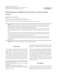
The plain radiographs <strong>of</strong> <strong>the</strong> elbow showed a punctated calcified<br />
s<strong>of</strong>t tissue mass in front <strong>of</strong> <strong>the</strong> medial epicondyle in <strong>the</strong> right<br />
elbow with probable smooth marginated bony erosion (Fig. 1).<br />
The CT scan and three dimensional reformed images confirmed<br />
a calcified mass with bony erosion in <strong>the</strong> right elbow (Fig. 2).<br />
MRI revealed that <strong>the</strong> mass showed intermediate signal intensity<br />
on <strong>the</strong> T1-weighted images and high signal intensity on<br />
<strong>the</strong> T2-weighted fat-suppressed images, and a heterogeneously<br />
well-enhanced mass was seen on <strong>the</strong> gadolinium enhanced fatsuppressed<br />
T1-weighted coronal MRI (Fig. 3). The patient was
76 Mee-Hye Oh∙Eun Ah Jung∙Ji Hye Lee, et al.<br />
Fig. 1. Plain radiograph shows a punctated calcified mass (black<br />
arrows) with smooth marginated bony erosion (white arrow) in<br />
front <strong>of</strong> <strong>the</strong> medial epicondyle in right elbow on lateral view.<br />
Fig. 2. Three dimensional<br />
reformed CT<br />
image shows a punctated<br />
calcified mass<br />
with bony erosion (arrows).<br />
operated on under general anes<strong>the</strong>sia and <strong>the</strong> mass was near-totally<br />
excised except for an intramuscular portion. At surgery, <strong>the</strong><br />
mass was poorly demarcated and it had infiltrated into <strong>the</strong> surrounding<br />
muscle. The microscopic sections revealed a proliferation<br />
<strong>of</strong> spindle cells with nodular deposits <strong>of</strong> hyalination, calcification<br />
and chondroid differentiation that were surrounded by<br />
palisading <strong>of</strong> polygonal plump cells (Fig. 4) and <strong>the</strong> tumor had<br />
infiltrated into <strong>the</strong> adjacent fatty tissue and skeletal muscle (Fig.<br />
Fig. 3. Gadolinium enhanced fat-suppressed T1-weighted coronal<br />
MR image shows a s<strong>of</strong>t tissue mass with heterogeneous well<br />
enhancement and cortical bony erosion in <strong>the</strong> anteromedial aspect<br />
<strong>of</strong> <strong>the</strong> right elbow.<br />
5). Any mitotic figures were not seen. Immunohistochemically,<br />
<strong>the</strong> mass showed a diffuse positive expression for CD99 (1:1,000,<br />
Zymed, San Francisco, CA, USA), and negativity for smooth<br />
muscle actin (1:1,000, Neomarker, Fremont, CA, USA), S-100<br />
protein (1:1,000, Neomarker, Fremont, CA, USA) and CD34<br />
(1:1,000, Neomarker, Fremont, CA, USA). The final pathologic<br />
diagnosis was calcifying aponeurotic fibroma. The patient has<br />
been well with no signs <strong>of</strong> recurrence during <strong>the</strong> 42 months after<br />
surgery.<br />
DISCUSSION<br />
<strong>Calcifying</strong> aponeurotic fibroma was first defined by Keasbey 1<br />
in 1953 as a neoplasm that’s characterized by a proliferation <strong>of</strong><br />
fibroblasts, areas <strong>of</strong> calcification and chondroid differentiation.<br />
Because most <strong>of</strong> <strong>the</strong> patients were children and young adolescents,<br />
it was named juvenile aponeurotic fibroma. Then in 1961,<br />
Keasbey and Fanselau found that <strong>the</strong> tumor presented not only<br />
in children, but in adults too, so <strong>the</strong>y called <strong>the</strong> tumor aponeurotic<br />
fibroma. After that, Lichtenstein and Goldman in 1964<br />
defined a cartilage analogue <strong>of</strong> fibromatosis. At last, Iwasaki and<br />
Enjoji in 1973 named <strong>the</strong>se tumors as calcifying aponeurotic<br />
fibroma. 8,9<br />
Although calcifying aponeurotic fibroma usually appears in
<strong>Calcifying</strong> <strong>Aponeurotic</strong> <strong>Fibroma</strong> 77<br />
Fig. 4. The tumor is composed <strong>of</strong> fibroblastic cells with nodular<br />
deposits <strong>of</strong> calcification and chondroid differentiation that surrounded<br />
by palisading <strong>of</strong> polygonal plump cells.<br />
Table 1. Clinicopathologic characteristics <strong>of</strong> Korean cases<br />
Case<br />
Sex/<br />
age<br />
Site<br />
Location<br />
Size<br />
No. <strong>of</strong><br />
reference<br />
1 F/36 Finger Superficial 1.5×1.0 cm 2<br />
(volar side)<br />
2 M/14 Chin Superficial 2.5×2.5 cm 3<br />
3 F/22 Toe Superficial 2.0×1.5 cm 4<br />
(plantar side)<br />
4 M/14 thumb Superficial 2.0×1.5 cm 5<br />
(ulnar side)<br />
5 F/24 Finger (tip) Superficial 0.5×0.5 cm 6<br />
6 M/11 Foot Superficial 1.5×1.5 cm 7<br />
(plantar side)<br />
7 M/8 <strong>Elbow</strong> Deep 2.0×2.0 cm Our<br />
(anteromedial side) case<br />
<strong>the</strong> first decade <strong>of</strong> life, it can be detected from <strong>the</strong> time <strong>of</strong> birth<br />
to 62 years <strong>of</strong> age. 10,11 The peak incidence is between 8 and 14<br />
years <strong>of</strong> age. 10 Most <strong>of</strong> <strong>the</strong> small published series have not reported<br />
a distinct gender predilection, but 70% <strong>of</strong> <strong>the</strong> patients in <strong>the</strong><br />
series by Allen and Enzinger were male. 12 There is no evidence<br />
<strong>of</strong> a familial or racial prevalence. 12 The pathogenesis <strong>of</strong> calcifying<br />
aponeurotic fibroma is still unknown, but it is thought to be a<br />
tumor with a fibroblastic origin. 10 This tumor was first reported<br />
on <strong>the</strong> palms, fingers and plantar region <strong>of</strong> children, 1 but isolated<br />
tumors have been reported at o<strong>the</strong>r sites, including <strong>the</strong> neck, forearm,<br />
elbow, upper arm, thigh, popliteal fossa, knee, lumbosacral<br />
region, scalp, chin, and abdominal wall. 3,9,12-15 Local recurrence<br />
is common because <strong>of</strong> its infiltrative growth pattern and it <strong>of</strong>ten<br />
Fig. 5. The tumor cells infiltrate to adjacent fatty tissue and skeletal<br />
muscle.<br />
has a sensitive location that mandates performing conservative<br />
surgery. Very few cases <strong>of</strong> malignant transformation have been<br />
reported. 16 On physical examination, calcifying aponeurotic fibroma<br />
presents as a firm, palpable mass that has shown slow growth<br />
for a few months to a few years. 10,11 There is frequently no history<br />
<strong>of</strong> trauma or local irritant factors. Although most <strong>of</strong> <strong>the</strong>se tumors<br />
have presented as a painless nodule, a few cases have been reported<br />
with <strong>the</strong> patients complaining <strong>of</strong> significant pain. 9<br />
The cases in <strong>the</strong> Korean literature and our case are summarized<br />
in Table 1 and <strong>the</strong>se cases have shown no gender predilection.<br />
The mean patient age (range: 8 to 36 years) <strong>of</strong> <strong>the</strong>se Korean<br />
cases was 18.4 years, which was somewhat higher than that<br />
<strong>of</strong> <strong>the</strong> o<strong>the</strong>r previous reports. 10 Most <strong>of</strong> <strong>the</strong> Korean cases occurred<br />
on fingers and toes like that <strong>of</strong> <strong>the</strong> o<strong>the</strong>r previous papers. The<br />
size <strong>of</strong> <strong>the</strong> masses was always under 2.5 cm at <strong>the</strong> greatest dimension<br />
and <strong>the</strong> masses were almost all painless palpable masses<br />
except for case 5. All <strong>the</strong> Korean cases were located in superficial<br />
s<strong>of</strong>t tissue except for our case.<br />
Radiologically, calcifying aponeurotic fibroma may show a s<strong>of</strong>t<br />
tissue mass with fine stippling calcification. Most <strong>of</strong> <strong>the</strong> cases<br />
are not associated with osseous lesion. However, in extremely<br />
rare cases, occasional scalloping <strong>of</strong> <strong>the</strong> cortex and thickening <strong>of</strong><br />
<strong>the</strong> bone have been reported. Shallow bony erosion was noted<br />
in our case, and this required us to differentiate this mass from<br />
o<strong>the</strong>r tumors including malignant tumors.<br />
Histologically, <strong>the</strong>re has been some reports about <strong>the</strong> phases<br />
<strong>of</strong> calcifying aponeurotic fibroma. Keasbey and Fanselau have
78 Mee-Hye Oh∙Eun Ah Jung∙Ji Hye Lee, et al.<br />
defined that <strong>the</strong>re are two phases during <strong>the</strong> growth <strong>of</strong> this tumor.<br />
8-10 In <strong>the</strong> first phase, <strong>the</strong> lesion is a destructive infiltrative<br />
tumor and calcification is usually absent. This phase is named<br />
<strong>the</strong> diffuse or florid phase. In <strong>the</strong> o<strong>the</strong>r phase, <strong>the</strong> lesion forms a<br />
nodular mass with a well-defined border. Therefore, this phase<br />
is named <strong>the</strong> compact or demarcated phase. This phase shows<br />
calcification and cartilage formation. The diffuse phase is mostly<br />
seen in infants and young children, and <strong>the</strong> compact phase<br />
tends to be seen in adolescents. For this reason, Keasbey has<br />
proposed that <strong>the</strong> compact phase is formed by fusion and growth<br />
<strong>of</strong> <strong>the</strong> diffuse phase, and <strong>the</strong> histologic differential diagnosis differs<br />
from case to case depending on <strong>the</strong> age <strong>of</strong> <strong>the</strong> patient at <strong>the</strong><br />
time <strong>the</strong> lesion is excised. In infants and small children, infantile<br />
fibromatosis, palmar and plantar fibromatoses and monophasic<br />
fibrous synovial sarcoma should be considered when making<br />
<strong>the</strong> differential diagnosis. In older patients, s<strong>of</strong>t part chondroma<br />
may rarely be mistaken for calcifying aponeurotic fibroma.<br />
In our case, <strong>the</strong> fibroblastic proliferation was predominant, but<br />
calcification and cartilage formation were relatively scant. From<br />
<strong>the</strong>se findings, we think that our case falls into <strong>the</strong> diffuse phase.<br />
We also reviewed <strong>the</strong> microscopic findings <strong>of</strong> four cases (cases 2<br />
to 5) that were reported in <strong>the</strong> Korean literature. Case 3 and 5<br />
showed scant fibroblastic proliferation and abundant cartilage<br />
formation and calcification. However, cases 2 and 4 revealed relatively<br />
abundant fibroblastic proliferation and scant cartilage<br />
formation and calcification. Theses findings seem to corroborate<br />
that this tumor shows different morphology according to age.<br />
To eliminate a possible misdiagnosis such as sarcoma which<br />
might require amputation, it is important to make an accurate<br />
diagnosis <strong>of</strong> calcifying aponeurotic fibroma, and especially for<br />
<strong>the</strong> case with equivocal radiologic findings showing osseous<br />
involvement like our case.<br />
ACKNOWLEDGEMENTS<br />
I would like to thank Pr<strong>of</strong>. Joon Hyuk Choi (Department <strong>of</strong><br />
Pathology, Yeungnam University), Pr<strong>of</strong>. Eun Ah Shin (Department<br />
<strong>of</strong> Pathology, Sanggye Paik Hospital, Inje University), Pr<strong>of</strong>.<br />
Jee Young Han (Department <strong>of</strong> Pathology, Inha University),<br />
and Pr<strong>of</strong>. Dae-Cheol Kim and Pr<strong>of</strong>. Su-Jin Kim (Department<br />
<strong>of</strong> Pathology, Dong-A University) for allowing me to review<br />
<strong>the</strong>ir microscopic findings.<br />
REFERENCES<br />
1. Keasbey LE. Juvenile aponeurotic fibroma (calcifying fibroma): a<br />
distinctive tumor arising in <strong>the</strong> palms and soles <strong>of</strong> young children.<br />
Cancer 1953; 6: 338-46.<br />
2. Choi SJ, Ahn JH, Kang G, et al. <strong>Calcifying</strong> aponeurotic fibroma with<br />
osseous involvement <strong>of</strong> <strong>the</strong> finger: a case report with radiologic and<br />
US findings. Korean J Radiol 2008; 9: 91-3.<br />
3. Jung YJ, Choi YW, Shin EA. <strong>Calcifying</strong> aponeurotic fibroma occurring<br />
on <strong>the</strong> chin: a case report. J Korean Soc Plast Reconstr Surg<br />
2008; 35: 103-5.<br />
4. Kwon OE, Ku BS, Kim DC, Lee CW, Kim KH. A case <strong>of</strong> calcifying<br />
aponeurotic fibroma with gait disturbance. Korean J Dermatol 2006;<br />
44: 71-4.<br />
5. Choi JH, Seo JS, Cho KH. <strong>Calcifying</strong> aponeurotic fibroma: a case<br />
report. Yeungnam Univ J Med 2003; 20: 223-8.<br />
6. Lim TG, Park JH, Kim YK, Choi GS. A case <strong>of</strong> calcifying aponeurotic<br />
fibroma. Korean J Dermatol 2001; 39: 930-1.<br />
7. Kang JS, Kim TH, Park KB. A case <strong>of</strong> calcifying aponeurotic fibroma.<br />
Korean J Dermatol 1992; 30: 253-7.<br />
8. Lichtenstein L, Goldman RL. The cartilage analogue <strong>of</strong> fibromatosis:<br />
a reinterpretation <strong>of</strong> <strong>the</strong> condition called ‘‘juvenile aponeurotic<br />
fibroma’’. Cancer 1964; 17: 810-6.<br />
. .<br />
9. Oruc M, Uysal A, Kankaya Y, Yildiz K, Aslan G, Sengul D. A case <strong>of</strong><br />
calcifying aponeurotic fibroma <strong>of</strong> <strong>the</strong> scalp: case report and review<br />
<strong>of</strong> <strong>the</strong> literature. Dermatol Surg 2007; 33: 1380-3.<br />
10. Weiss SW, Goldblum JR. S<strong>of</strong>t tissue tumors. 5th ed. St. Louis: Mosby;<br />
2008; 289-93.<br />
11. Goldman RL. The cartilage analogue <strong>of</strong> fibromatosis (aponeurotic<br />
fibroma): fur<strong>the</strong>r observations based on seven new cases. Cancer<br />
1970; 26: 1325-31.<br />
12. Allen PW, Enzinger FM. Juvenile aponeurotic fibroma. Cancer 1970;<br />
26: 857-67.<br />
13. Fetsch JF, Miettinen M. <strong>Calcifying</strong> aponeurotic fibroma: a clinicopathologic<br />
study <strong>of</strong> 22 cases arising in uncommon sites. Human<br />
Pathol 1998; 29: 1504-10.<br />
14. Murphy BA, Kilpatrick SE, Panella MJ, et al. Extra-acral calcifying<br />
aponeurotic fibroma: a distinctive case with 23-year follow-up. J<br />
Cutan Pathol 1996; 23: 369-72.<br />
15. Sharma R, Punia RS, Sharma A, et al. Juvenile (calcifying) aponeurotic<br />
fibroma <strong>of</strong> <strong>the</strong> neck. Pediat Surg Int 1998; 13: 295-6.<br />
16. Amaravati R. Rare malignant transformation <strong>of</strong> a calcifying aponeurotic<br />
fibroma. J Bone Joint Surg 2002; 84: 1889.