quality described microscope invention biomedical environment surfaces

Create successful ePaper yourself
Turn your PDF publications into a flip-book with our unique Google optimized e-Paper software.
urn:nbn:de:gbv:ilm1-2011imeko-085:3<br />
Joint International IMEKO TC1+ TC7+ TC13 Symposium<br />
August 31 st − September 2 nd , 2011, Jena, Germany<br />
urn:nbn:de:gbv:ilm1-2011imeko:2<br />
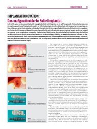
Fig. 9 The implant sample<br />
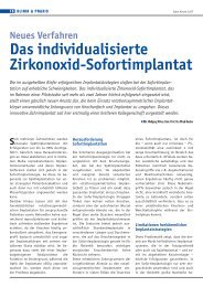
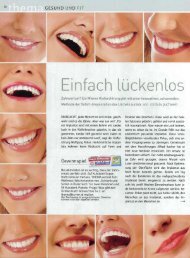
The focused 2D and 3D images of the original<br />
tooth sample are observed by means of the digital<br />
<strong>microscope</strong>. These images were both quantitatively<br />
analyzed and also were evaluated in terms of the surface<br />
roughness using the digital <strong>microscope</strong> software.<br />
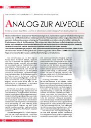
Fig. 10 and Fig. 11 represent the captured 2D image<br />
of the tooth sample and assessed 3D image of the<br />
tooth sample consequently.<br />
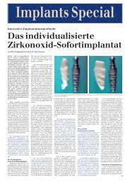
(a)<br />
(b)<br />
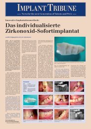
Fig. 12 The scanned 3D models of the original (a)<br />
and implant (b) tooth samples<br />
Fig. 10 The captured 2D image of the tooth sample (x2000)<br />
Fig. 13 The assessment of the difference in original and<br />
implant tooth sample using the scanned 3D image<br />
In this experimental study, roughness measurements<br />
of dental applications were carried out with a<br />
stylus profilometer and a digital <strong>microscope</strong>. Measurements<br />
were repeated for five measurement points<br />
with 1mm intervals on an original tooth, its implant<br />
and other three test implant samples. Each measurement<br />
point taken from both stylus profilometer and<br />
digital <strong>microscope</strong> represent the mean values.<br />
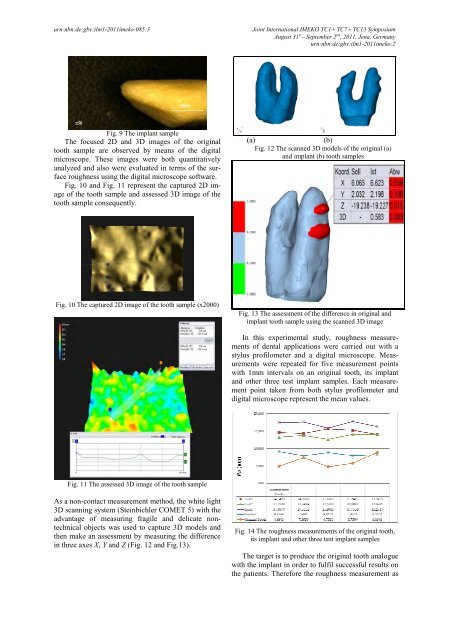
Fig. 11 The assessed 3D image of the tooth sample<br />
As a non-contact measurement method, the white light<br />
3D scanning system (Steinbichler COMET 5) with the<br />
advantage of measuring fragile and delicate nontechnical<br />
objects was used to capture 3D models and<br />
then make an assessment by measuring the difference<br />
in three axes X, Y and Z (Fig. 12 and Fig.13).<br />
Fig. 14 The roughness measurements of the original tooth,<br />
its implant and other three test implant samples<br />
The target is to produce the original tooth analogue<br />
with the implant in order to fulfil successful results on<br />
the patients. Therefore the roughness measurement as