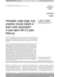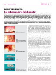
Immediate, non-submerged, root-analogue zirconia ... - ResearchGate

Create successful ePaper yourself
Turn your PDF publications into a flip-book with our unique Google optimized e-Paper software.
Author's personal copy<br />
1128 Pirker and A. Kocher<br />
tissue regression are avoided or significantly<br />
reduced, due to early, albeit limited,<br />
functional load.<br />
One problem associated with immediate<br />
implant placement using conventional<br />
screw- or cylinder-type implants is their<br />
incongruence with the extraction socket<br />
necessitating the use of a barrier membrane<br />
and/or bone augmentation to prevent<br />
down growth of connective tissue or<br />
epithelium in between the implant and the<br />
socket. 5<br />
The concept of replacing teeth with<br />
custom-made <strong>root</strong> <strong>analogue</strong> implants is<br />
not new. The oldest evidence of a dental<br />
implant dates back to around 550 BC. 1,3 In<br />
ancient times, wood, metal, shell or stone<br />
were carved and shaped to form the <strong>root</strong><br />
for the implant. 2 The first literature reference<br />
to a modern style implant came in<br />
1809 when Maggiolo described a tooth<br />
<strong>root</strong>-shaped implant made out of 18-carat<br />
gold. 4 In 1969 the use of a tooth replica<br />
implant was reported, however the autopolymerized<br />
and heat-processed polymethacrylate<br />
utilized to fabricate the<br />
tooth <strong>analogue</strong> was encapsulated by soft<br />
tissue rather than osseointegrated. 7 Meanwhile<br />
placement of dental implants had<br />
become an everyday treatment option for<br />
dental patients missing teeth. All implant<br />
systems involve screw-type threaded<br />
implants or cylindrical implants with no<br />
resemblance to the native <strong>root</strong>.<br />
Hodosh et al. were the first to tackle the<br />
problem of incongruence by employing a<br />
novel approach using custom-made <strong>root</strong><br />
<strong>analogue</strong> implants placed into the extraction<br />
socket. 7 By adapting the <strong>root</strong> to the<br />
extraction socket instead of adapting the<br />
bone to a preformed standardized implant<br />
they reduced the bone and soft tissue<br />
trauma.<br />
Lundgren et al. reintroduced the idea of<br />
<strong>root</strong> <strong>analogue</strong> implants in 1992. 14 Instead<br />
of using polymers, titanium was utilized in<br />
an experimental model of immediate<br />
implant placement leading to bony integration<br />
in 88%. A good fit between<br />
implant and the host bed has been<br />
described as an important factor for<br />
implant success. 8,21,22 For that reason<br />
Kohal et al. further refined the approach<br />
of <strong>root</strong> <strong>analogue</strong> titanium implants by<br />
widening the coronal aspect of the implant<br />
to compensate for the lost periodontium<br />
and to obtain a good congruence between<br />
implant and extraction socket. In several<br />
instances the implant insertion led to fractures<br />
of the thin buccal wall of the alveolar<br />
bone. 5,9<br />
Experimental studies in monkeys gave<br />
favourable results with clear evidence of<br />
osseointegration and clinical stability. 9,10<br />
The ensuing clinical trial by Kohal et al.<br />
resulted in a 100% primary stability at<br />
insertion and 1 month follow-up. Owing<br />
to the high failure rate of 97% over the<br />
short follow-up period of 12 months this<br />
implant system was not been recommended<br />
for clinical use. 12<br />
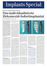
The goal of the present study was to<br />
evaluate a novel approach to <strong>root</strong>-<strong>analogue</strong><br />
dental implants. Zirconia was used for its<br />
excellent biocompatibility, improved<br />
esthetic results by preventing dark discoloration<br />
of the gum and the display of<br />
titanium <strong>root</strong>s in case of gum recession,<br />
compressive strength, bending forces, fracture<br />
toughness and high electrical resistance.<br />
Microretentions were added to the<br />
entire <strong>root</strong> surface. Owing to the high failure<br />
rate, the initial trial was limited to 6<br />
patients and a second series of <strong>root</strong> identical<br />
implants with significant modifications<br />
were started. The first modification was<br />
the addition of macroretentions, strictly<br />
limited to the interdental space, in order<br />
to get beyond primary stability and improve<br />
osseointegration. The second was to reduce<br />
the diameter of the implant next to the thin<br />
cortical bone to avoid fracture and pressure-induced<br />
bone loss. The third was to<br />
choose a single-stage implantation, resulting<br />
in immediate, albeit limited, functional<br />
load via the crown stump to prevent bone<br />
resorption.<br />
Material and methods<br />
18 patients were included in this prospective<br />
study. The 6 patients in group A<br />
received <strong>root</strong> identical replicas with the<br />
implant surface roughened by sandblasting<br />
only. In the 12 patients in group B the<br />
<strong>root</strong> was modified by adding macroretentions,<br />
strictly limited to the interdental<br />
space, and by reducing the buccal and<br />
lingual face by approximately 0.1–<br />
0.2 mm, preventing fractures of the thin<br />
cortical bone layer at insertion. The inclusion<br />
criteria were: patients with a single<br />
tooth gap; with uncompromised periodontal<br />
ligaments in the anterior or premolar<br />
region; informed consent; and willingness<br />
to adhere to the protocol. Indications for<br />
tooth extraction included <strong>root</strong> caries, vertical<br />
or horizontal <strong>root</strong> fracture, endodontic<br />
lesions, and unsuccessful <strong>root</strong> canal<br />
treatment. Patients with dehiscence of<br />
the crestal bone as determined by clinical<br />
examination and with tooth extraction<br />
necessitating surgical intervention leading<br />
to contusion of the bone were excluded<br />
from the study. Chronic apical paradontitis<br />
was not an exclusion criterion, but in<br />
these cases the area of infection was<br />
removed.<br />
The compromised tooth was carefully<br />
extracted under local anaesthesia (Ultracain<br />
DS forte, Aventis), avoiding<br />
damage to the socket and soft tissue.<br />
The extraction socket was cleaned meticulously<br />
by means of curettage and an<br />
iodoform soaked cotton gaze was placed<br />
in the socket. This minimal invasive,<br />
flapless approach was chosen to avoid<br />
trauma to the hard and soft tissue avoiding<br />
swelling and bruising. The <strong>root</strong> was<br />
laser scanned and, in group B, macroretentions<br />
were designed according to<br />
the study protocol, strictly limited to<br />
the interdental space, and the buccal<br />
and lingual face was reduced by 0.1–<br />
0.2 mm. A crown stump was designed<br />
for later connection to the crown. The<br />
<strong>root</strong> was then milled from a medicalgrade<br />
<strong>zirconia</strong> block (more exactly yttria<br />
stabilized tetragonal <strong>zirconia</strong> polycrystal),<br />
the surface roughened by sandblastingandsinteredfor8htoachievethe<br />
desired mechanical properties. The<br />
implant was cleaned in an ultrasonic<br />
bath containing 96% ethanol for<br />
10 min, packaged and steam-sterilized.<br />
1–8 days after extraction the iodoform<br />
cotton gauze was removed, and the<br />
alveolar socket curetted and flushed with<br />
sterile physiologic saline solution. The<br />
custom-made individualized implant was<br />
placed in the socket under finger pressure<br />
and gently tapped into place with a<br />
hammer and a mallet. Primary stability<br />
wasachievedinallinstancesaschecked<br />
by palpation and percussion. Patients<br />
received postoperative analgesics (Parkemed<br />
500 mg, Pfizer) on demand. They<br />
were instructed to chew predominantly<br />
on the contralateral side and to avoid<br />
hard food for 8 weeks on the implant<br />
side. Clinical parameters, such as<br />
implant stability, bleeding on probing,<br />
mucosal margin position, variation of<br />
gingival level, and variation of papilla<br />
position, were ascertained at baseline<br />
and after 1 week, 1 month, 2 months<br />
and thereafter every 6 months post intervention.<br />
The clinical situation was also<br />
photo-documented at the same time<br />
points. Radiographic assessments using<br />
Scanora X-ray images were made at<br />
baseline and every 12 months after<br />
implant placement. Bone level measurement<br />
by radiographic examination was<br />
limited because the implant is radiopaque.<br />
The success of the dental implants<br />
was defined, according to the criteria<br />
suggested for determination of success<br />
with reference to clinical and X-ray control<br />
parameters by Jahn and Buser. Survival<br />
of dental implants was computed<br />
using the Kaplan–Meier method. Data