

What is a fluid challenge?

Create successful ePaper yourself
Turn your PDF publications into a flip-book with our unique Google optimized e-Paper software.
<strong>What</strong> <strong>is</strong> a <strong>fluid</strong> <strong>challenge</strong>?<br />
Maurizio Cecconi, Anthony K. Parsons and Andrew Rhodes<br />
Department of Intensive Care Medicine, St George’s<br />
Healthcare NHS Trust, London, UK<br />
Correspondence to Dr Maurizio Cecconi, MD,<br />
Department of Intensive Care Medicine, St George’s<br />
Healthcare NHS Trust, London SW17 0QT, UK<br />
Tel: +44 208 725 0884; e-mail: m.cecconi@nhs.net<br />
Current Opinion in Critical Care 2011,<br />
17:290–295<br />
Purpose of review<br />
The <strong>fluid</strong> <strong>challenge</strong> <strong>is</strong> used in the <strong>fluid</strong> management of many sick patients. The principle<br />
behind the <strong>fluid</strong> <strong>challenge</strong> technique <strong>is</strong> that by giving a small amount of <strong>fluid</strong> in a<br />
short period of time, the clinician can assess whether the patient has a preload reserve<br />
that can be used to increase the stroke volume with further <strong>fluid</strong>s. The key components<br />
of a <strong>fluid</strong> <strong>challenge</strong> are described.<br />
Recent findings<br />
Dynamic predictors of <strong>fluid</strong> responsiveness are increasingly used in preference to the<br />
central venous and pulmonary artery occlusion pressure. The gold standard to monitor<br />
the response to a <strong>fluid</strong> <strong>challenge</strong> <strong>is</strong> using a continuous cardiac output monitoring. Fluid<br />
therapy guided by flow monitoring has been shown to reduce hospital stay and<br />
postoperative complications.<br />
Summary<br />
A <strong>fluid</strong> <strong>challenge</strong> identifies and simultaneously treats volume depletion, whilst avoiding<br />
deleterious consequences of <strong>fluid</strong> overload through its small volume and targeted<br />
admin<strong>is</strong>tration.<br />
Keywords<br />
cardiac function, colloid, crystalloid, preload response, stroke volume<br />
Curr Opin Crit Care 17:290–295<br />
ß 2011 Wolters Kluwer Health | Lippincott Williams & Wilkins<br />
1070-5295<br />
Introduction<br />
The <strong>fluid</strong> <strong>challenge</strong> <strong>is</strong> a test that allows the clinician to<br />
give <strong>fluid</strong>s and at the same time to test the preload reserve<br />
of the patient. The judicious admin<strong>is</strong>tration of intravenous<br />
<strong>fluid</strong> <strong>is</strong> an essential part of the management of many<br />
sick patients. An inadequate cardiac output (CO) and<br />
systemic arterial pressure reduces the delivery of oxygen<br />
to a level below the necessary requirements, leading to a<br />
cascade of cellular changes that ultimately can result in<br />
organ dysfunction and failure.<br />
Cardiovascular dysfunction may result from a number of<br />
causes that can be simply described as being secondary to<br />
either hypovolaemia, pump dysfunction, altered vascular<br />
tone or more rarely because of an obstructive pathology.<br />
Hypovolaemia may be actual secondary to haemorrhage,<br />
or dehydration or relative due to an increased venous<br />
capacitance. Irrespective of the cause, it has long been<br />
recognized that prolonged hypovolaemia <strong>is</strong> grave, requiring<br />
urgent intervention to improve haemodynamics. We<br />
believe that th<strong>is</strong> <strong>is</strong> best done with a ‘<strong>fluid</strong> <strong>challenge</strong>’ [1].<br />
A <strong>fluid</strong> <strong>challenge</strong> <strong>is</strong> a method of identifying those patients<br />
likely to benefit from an increase in intravenous volume<br />
in order to guide further volume resuscitation. It <strong>is</strong> a<br />
dynamic test of the circulation. The use of a ‘test’ that<br />
uses a small amount of <strong>fluid</strong> to assess the volume responsiveness<br />
may reduce the r<strong>is</strong>k of a too liberal <strong>fluid</strong> strategy<br />
and the possible consequences of <strong>fluid</strong> overload. In 2006,<br />
in a multicentric study design, positive <strong>fluid</strong> balance in<br />
ICU was associated with a worse outcome [2]. It <strong>is</strong><br />
important to stress that a patient who responds to <strong>fluid</strong><br />
may not, however, need <strong>fluid</strong>, with preload dependence<br />
likely to be the normal cardiovascular state. Healthy<br />
volunteers demonstrate significant increases in stroke<br />
volume (SV) in response to head-down tilt (a ‘virtual’<br />
<strong>fluid</strong> <strong>challenge</strong> equivalent to around 500 ml volume) [3].<br />
In health, the peripheral circulation <strong>is</strong> the primary controller<br />
of CO, the heart automatically pumping all the<br />
blood returned to it as described by the Frank–Starling<br />
mechan<strong>is</strong>m. Increased stretch of heart muscle causes<br />
increased contractile strength, and an increased volume<br />
ejected. Th<strong>is</strong> property ex<strong>is</strong>ts in all striated muscle, and <strong>is</strong><br />
consequent to the degree of overlap of actin and myosin<br />
filaments, and in cardiac muscle also to increased sensitivity<br />
of troponin C to calcium [4]. Right atrial stretch also<br />
leads to increased heart rate secondary to sinus node<br />
automaticity and sympathetic stimulation.<br />
The term preload refers to the myocardial sarcomere<br />
length just prior to contraction. SV <strong>is</strong> determined by<br />
preload, afterload, contractility and heart rate. Venous<br />
1070-5295 ß 2011 Wolters Kluwer Health | Lippincott Williams & Wilkins DOI:10.1097/MCC.0b013e32834699cd<br />
Copyright © Lippincott Williams & Wilkins. Unauthorized reproduction of th<strong>is</strong> article <strong>is</strong> prohibited.
<strong>What</strong> <strong>is</strong> a <strong>fluid</strong> <strong>challenge</strong>? Cecconi et al. 291<br />
return <strong>is</strong> the most important determinant of CO and <strong>is</strong><br />
defined by three parameters: the mean systemic filling<br />
pressure, the right atrial pressure and the venous res<strong>is</strong>tance.<br />
The mean systemic filling pressure <strong>is</strong> generated by<br />
the volume in the systemic circulation, driving blood<br />
to the heart. Right atrial pressure opposes blood flow into<br />
the right atrium, decreasing if contractility increases,<br />
whereas the res<strong>is</strong>tance to venous return <strong>is</strong> the intrinsic<br />
res<strong>is</strong>tance to flow within the venous compartment.<br />
These parameters can be described by a venous return<br />
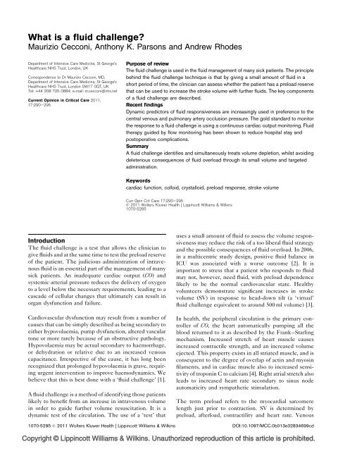
curve, which when combined with the cardiac function<br />
curve <strong>is</strong> useful to explain the response to a <strong>fluid</strong> <strong>challenge</strong><br />
(Fig. 1).<br />
The main point to remember with a <strong>fluid</strong> <strong>challenge</strong> <strong>is</strong> the<br />
fact that the given <strong>fluid</strong> must be of sufficient volume to<br />
stretch the right heart [increasing the right ventricular<br />
end-diastolic volume (RVEDV)]. Only then, can the<br />
Starling’s law be implicated to suggest that right ventricular<br />
(and left ventricular) SV will increase. Different<br />
hearts in different positions of the curve will respond in<br />
different ways, but if the RVEDV has not increased<br />
sufficiently, then there <strong>is</strong> no way that SV will be able<br />
to increase with the r<strong>is</strong>k of a false negative test.<br />
Difference between <strong>fluid</strong> <strong>challenge</strong> and <strong>fluid</strong> loading<br />
It <strong>is</strong> important to remember that the <strong>fluid</strong> <strong>challenge</strong><br />
technique <strong>is</strong> first of all a test of the cardiocirculatory<br />
system. It allows the clinician to test whether the patient<br />
has a preload reserve that can be used to increase the SV<br />
with further <strong>fluid</strong>s. These further <strong>fluid</strong>s can be given after<br />
a positive response to a <strong>fluid</strong> <strong>challenge</strong> or they can be<br />
given in a controlled way by repeating the <strong>fluid</strong> <strong>challenge</strong><br />
as long as there <strong>is</strong> a positive response. Th<strong>is</strong> controlled<br />
Key points<br />
A <strong>fluid</strong> <strong>challenge</strong> identifies and simultaneously<br />
corrects volume depletion in order to optimize<br />
t<strong>is</strong>sue perfusion.<br />
Admin<strong>is</strong>tration of <strong>fluid</strong> using a <strong>fluid</strong> <strong>challenge</strong> protocol<br />
avoids unnecessary <strong>fluid</strong> admin<strong>is</strong>tration and<br />
may improve outcome in critically ill and elective<br />
surgical patients.<br />
Dynamic noninvasive predictors of volume responsiveness<br />
such as SV, PLR and DScVO 2 should be<br />
used in preference to the CVP and PAOP for<br />
guiding <strong>fluid</strong> therapy.<br />
Continuous CO monitors are the best option to<br />
monitor the response to a <strong>fluid</strong> <strong>challenge</strong>.<br />
The physiological properties of the <strong>fluid</strong> and the<br />
clinical picture should be considered when choosing<br />
which <strong>fluid</strong> to use.<br />
approach <strong>is</strong> also called SV maximization and <strong>is</strong> the base of<br />
most goal-directed therapy protocols [5–7]. Th<strong>is</strong> <strong>is</strong> very<br />
different from <strong>fluid</strong> loading in which <strong>fluid</strong>s are given<br />
without monitoring the response in real time. The only<br />
‘excess’ <strong>fluid</strong> that may be given with the <strong>fluid</strong> <strong>challenge</strong><br />
technique <strong>is</strong> the amount of <strong>fluid</strong> used when the patient<br />
fails to respond. Th<strong>is</strong> <strong>is</strong> usually equal to 200 ml or 3 ml/kg.<br />
When and how to give a <strong>fluid</strong> <strong>challenge</strong>?<br />
The primary indication for a <strong>fluid</strong> <strong>challenge</strong> <strong>is</strong> the intention<br />
by the clinician to increase SV and CO. Th<strong>is</strong> <strong>is</strong> usually<br />
the case when there <strong>is</strong> evidence of hypoperfusion [8], but<br />
there may be clinical situations in which a SV increase <strong>is</strong><br />
sought preemptively, as in goal-directed therapy in highr<strong>is</strong>k<br />
surgical patients [9].<br />
Figure 1 Relationships between cardiac output and venous return and stroke volume and <strong>fluid</strong> <strong>challenge</strong> for different levels of<br />
contractility<br />
(a)<br />
CO,<br />
venous<br />
return l/min<br />
10<br />
(b)<br />
SV<br />
ml<br />
B<br />
5<br />
B<br />
D<br />
Fluid <strong>challenge</strong> responder<br />
SV increase > 10%<br />
0<br />
A<br />
C<br />
D<br />
A<br />
C<br />
Fluid <strong>challenge</strong> non-responder<br />
SV increase < 10%<br />
−4 0 4 8 12<br />
Right atrial pressure (mmHg)<br />
Fluid <strong>challenge</strong><br />
(a) In a heart with normal contractility (dashed line), the equilibrium point <strong>is</strong> point A, cardiac output (CO) equals venous return and right atrial pressure <strong>is</strong><br />
0 mmHg. In a heart with impaired contractility (dotted line), the equilibrium point <strong>is</strong> C. A <strong>fluid</strong> <strong>challenge</strong> increases the mean systemic filling pressure. The<br />
venous return curve shifts upward and right, which now intersects the cardiac function curve at point B for the heart with normal contractility, where we<br />
can see a significant r<strong>is</strong>e in CO and right atrial pressure. In the failing heart, the increase in pressure <strong>is</strong> much higher than the effect on CO (from C to D).<br />
(b) The correspondent stroke volume (SV)/<strong>fluid</strong> <strong>challenge</strong> relations for the two hearts. If SV <strong>is</strong> measured, the response to the same <strong>fluid</strong> <strong>challenge</strong> will be<br />
very different depending on the contractility (normal or failing) and on which part of the curve the starting point <strong>is</strong>.<br />
Copyright © Lippincott Williams & Wilkins. Unauthorized reproduction of th<strong>is</strong> article <strong>is</strong> prohibited.
292 Cardiopulmonary monitoring<br />
When the dec<strong>is</strong>ion of increasing the CO <strong>is</strong> made, optimization<br />
of preload <strong>is</strong> usually the first step taken. It goes<br />
without saying, therefore, that the primary target of a<br />
<strong>fluid</strong> <strong>challenge</strong> <strong>is</strong> an increase in SV or CO. An increase<br />
of at least 10–15% <strong>is</strong> considered a positive response [10].<br />
Traditionally, the measurement of SV and CO was<br />
possible only with an invasive device such as the pulmonary<br />
artery catheter (PAC) [11]. The availability of<br />
new less invasive CO monitors has made possible the realtime<br />
assessment of SV changes during a <strong>fluid</strong> <strong>challenge</strong> in<br />
a less invasive fashion [12–14].<br />
If the primary target of a <strong>fluid</strong> <strong>challenge</strong> <strong>is</strong> an increase in<br />
SV, the primary safety limit <strong>is</strong> failure of SV to increase.<br />
When <strong>fluid</strong> loading does not produce any improvement in<br />
haemodynamics, it can increase the r<strong>is</strong>k of <strong>fluid</strong> overload.<br />
Data suggest that only 50% of critically ill patients<br />
respond to a <strong>fluid</strong> <strong>challenge</strong> [10]. For th<strong>is</strong> reasons investigators<br />
have been looking for indices of <strong>fluid</strong> responsiveness<br />
that can be used to predict the response to a <strong>fluid</strong><br />
<strong>challenge</strong> [15,16].<br />
With continuous CO monitors, both the target (SV<br />
increase) and the safety limit (failure of SV to increase)<br />
can be measured in real time. If CO monitors are not used,<br />
clinicians may use different variables in order to identify<br />
surrogate targets that correlate with an increase in SV and<br />
surrogate safety limits that correlate with failure of SV to<br />
increase. On the bas<strong>is</strong> of these assumptions, different<br />
strategies to perform a <strong>fluid</strong> <strong>challenge</strong> have been proposed.<br />
Clinicians report using multiple tools as indicators for<br />
<strong>fluid</strong> admin<strong>is</strong>tration [17] which may also act as predictors,<br />
targets and safety limits. (Table 1). The choice of target<br />
and safety limit depends also on the clinical situation and<br />
availability of monitors. In ICU and operating rooms,<br />
soph<strong>is</strong>ticated CO monitors allow the clinician to focus<br />
accurately on SV. In other circumstances, the mean<br />
arterial pressure (MAP) can be chosen as a target and<br />
the central venous pressure (CVP) as a safety limit [17]. A<br />
r<strong>is</strong>e in MAP can indicate a positive response (the SV has<br />
increased and produced a r<strong>is</strong>e in MAP); the CVP can be<br />
used as a safety limit [if the MAP does not increase<br />
substantially and the CVP r<strong>is</strong>e <strong>is</strong> very marked, it will<br />
suggest that the patient <strong>is</strong> not likely to respond to an<br />
increased intravascular volume and indeed harm (such as<br />
increased peripheral oedema) may be the result].<br />
Fluid <strong>challenge</strong> and CVP<br />
CVP has been used to guide <strong>fluid</strong> <strong>challenge</strong>s for over<br />
40 years [18], and <strong>is</strong> considered an indicator for <strong>fluid</strong><br />
<strong>challenge</strong> by the Surviving Seps<strong>is</strong> Campaign [19]. Th<strong>is</strong><br />
comes from one study only where a CVP of 8–12 cm H 2 O<br />
was considered a target and a CVP less than 8 cm H 2 Oan<br />
indicator. Although that study was successful in improving<br />
outcome, it <strong>is</strong> unlikely based on the most up to date<br />
evidence that a CVP less than 8 cm H 2 O can be recommended<br />
as an indicator [20].<br />
The CVP, which <strong>is</strong> equivalent to right atrial pressure,<br />
does not predict RVEDV, but indicates the relationship<br />
between blood volume and cardiac function. The transmural<br />
right atrial pressure gradient, as opposed to the<br />
‘intracavity’ CVP, and ventricular compliance are more<br />
influential to ventricular filling. Myocardial <strong>is</strong>chaemia<br />
reduces contractility and can increase CVP; improving<br />
cardiac function will decrease CVP. Any cause of<br />
decreased right ventricular compliance, such as pulmonary<br />
hypertension or ra<strong>is</strong>ed pleural pressure, will further<br />
Table 1 Parameters used to guide <strong>fluid</strong> admin<strong>is</strong>tration<br />
Parameter Indicator Predictor Target Safety limit<br />
Clinical judgement Yes Yes Yes Yes<br />
Thirst/dimin<strong>is</strong>hed skin turgor/dry mouth/cool extremities Yes No No No<br />
Perioperative therapy (fasting/maintenance/insensible/evaporative/haemorrhagic losses) Yes No No No<br />
Hypernatraemia Yes No Yes No<br />
Use of vasopressors Yes No Yes No<br />
HR >100 Yes No Yes No<br />
Oliguria
<strong>What</strong> <strong>is</strong> a <strong>fluid</strong> <strong>challenge</strong>? Cecconi et al. 293<br />
elevate the CVP. Myocardial hypertrophy or dilation<br />
can also alter the volume/pressure relationship. A recent<br />
systematic review has demonstrated no association<br />
between CVP and blood volume, with patients with high<br />
or low CVPs equally likely to be volume responsive [20].<br />
During spontaneous respiration, however, a decrease in<br />
CVP during inspiration indicates that the right ventricle<br />
has preload reserve, and <strong>is</strong> therefore on the ascending part<br />
of the cardiac function curve. A fall of 1 mmHg or less<br />
during spontaneous breathing strongly predicts a subsequent<br />
response to a <strong>fluid</strong> <strong>challenge</strong> [21].<br />
A CVP of 8–12 or 12–15 cm H 2 O, if mechanically ventilated,<br />
<strong>is</strong> recommended by the Surviving Seps<strong>is</strong> Campaign<br />
[19]. It <strong>is</strong> important to remember, though, that significant<br />
increases in CO with <strong>fluid</strong> can occur in patients with CVPs<br />
greater than 15 mmHg and many patients with a low CVP<br />
fail to respond to a <strong>fluid</strong> <strong>challenge</strong> [22]. Indeed, neither<br />
CVP nor DCVP can be used as good targets (i.e. preload<br />
indicators) of volume responsiveness.<br />
DCVP as a marker of adequacy of a <strong>fluid</strong> <strong>challenge</strong>?<br />
Recently, Lakhal et al. [23] have demonstrated that<br />
DCVP can be tested to identify in which patients a<br />
passive leg ra<strong>is</strong>ing (PLR) has been successful in<br />
adequately increasing the preload of the ventricles. It<br />
may be that in some patients a <strong>fluid</strong> <strong>challenge</strong> does not<br />
produce a response in SV because insufficient volume has<br />
been given to stretch a ventricle that otherw<strong>is</strong>e may still<br />
have a preload reserve. In th<strong>is</strong> sense, DCVP could be used<br />
to identify an adequate <strong>fluid</strong> <strong>challenge</strong>: for example, a<br />
change in DCVP of 2 cmH 2 O being the evidence of<br />
appropriate stretch in the right ventricle or increases in<br />
the RVEDV. Th<strong>is</strong> approach was described initially by<br />
Weil and Henning [1] and more recently by Vincent and<br />
Weil [17]. To our knowledge, only one study [24] has<br />
successfully used DCVP in th<strong>is</strong> way. It remains to be<br />
proven if th<strong>is</strong> approach can be used coupled with a<br />
CO monitor.<br />
Fluid <strong>challenge</strong> and pulmonary artery<br />
occlusion pressure<br />
Analogous to CVP, the pulmonary artery occlusion pressure<br />
(PAOP) purports to estimate left ventricular enddiastolic<br />
volume (LVEDV), and <strong>is</strong> subject to the same<br />
caveats. PAOP ass<strong>is</strong>ts in diagnosing the cause of pulmonary<br />
oedema and pulmonary hypertension, but does not<br />
predict <strong>fluid</strong> responsiveness better than chance [25,26].<br />
Fluid <strong>challenge</strong> and central venous oxygen<br />
saturation<br />
Central venous oxygen saturation (ScVO 2 ) represents the<br />
relationship between t<strong>is</strong>sue oxygen supply and demand.<br />
An ScVO 2 of less than 70% <strong>is</strong> defined as an indicator and<br />
target for early resuscitation in seps<strong>is</strong>, reducing in-hospital<br />
mortality [27]. A recent RCT of intraoperative <strong>fluid</strong> <strong>challenge</strong>s<br />
to achieve an ScVO 2 of 75% did not show a<br />
reduction in postoperative complications [28]. Interestingly,<br />
the ScVO 2 group received less <strong>fluid</strong>s than the<br />
control group. DScVO2 may be a more useful predictor<br />
and target – a change in ScVO 2 of 4% has been shown to<br />
correlate with response to <strong>fluid</strong> <strong>challenge</strong> [29].<br />
Prediction of <strong>fluid</strong> responsiveness before giving a <strong>fluid</strong><br />
<strong>challenge</strong><br />
There are a number of tests that have been studied to<br />
predict whether a <strong>fluid</strong> <strong>challenge</strong> will lead to an increase<br />
in SV (<strong>fluid</strong> responsiveness).<br />
Heart–lung interaction<br />
The cons<strong>is</strong>tent fluctuation of SV, systolic pressure and<br />
pulse pressure with the mechanically ventilated breath<br />
have cons<strong>is</strong>tently been shown to be sensitive and specific<br />
predictors of volume responsiveness, providing a number<br />
of caveats are followed. These caveats include a tidal<br />
volume of over 8 ml/kg and the absence of either spontaneous<br />
respiratory activity or arrythmogenic activity<br />
[30].<br />
Passive leg ra<strong>is</strong>ing<br />
Elevation of the lower limbs induces an auto-transfusion<br />
haemodynamically equivalent to an exogenous <strong>fluid</strong> <strong>challenge</strong>.<br />
CO and pulse pressure changes in response to PLR<br />
are predictive of <strong>fluid</strong> responsiveness and independent of<br />
mode of ventilation [31].<br />
Corrected flow time<br />
The corrected flow time (FTc), derived from oesophageal<br />
Doppler monitoring, has been described as indicating<br />
<strong>fluid</strong> <strong>challenge</strong> and predicting response in surgical<br />
patients [32]. The FTc should not be considered an<br />
accurate marker of left ventricular preload [33], being<br />
inversely proportional to systemic vascular res<strong>is</strong>tance,<br />
and therefore describing left ventricular afterload.<br />
<strong>What</strong> <strong>fluid</strong>?<br />
Fluid <strong>challenge</strong>s may be performed with crystalloid<br />
(<strong>is</strong>otonic or hypertonic) or colloids. The ability of colloids<br />
to maintain or increase CO should reduce <strong>fluid</strong> extravasation<br />
into the lung, but increased capillary permeability<br />
may negate th<strong>is</strong> advantage. Crystalloid d<strong>is</strong>tributes rapidly<br />
through the extracellular compartment, reducing its<br />
haemodynamic effect. Colloid will stay in the intravascular<br />
compartment for longer, and hyperoncotic <strong>fluid</strong> will<br />
draw <strong>fluid</strong> out of the interstitial space, increasing plasma<br />
volume beyond the admin<strong>is</strong>tered volume. Whether th<strong>is</strong><br />
has clinical relevance for the <strong>fluid</strong> <strong>challenge</strong> <strong>is</strong> unclear. A<br />
Cochrane review demonstrated no mortality difference<br />
between crystalloid and colloid for resuscitation of critically<br />
ill patients [34]. The choice of <strong>fluid</strong> for a <strong>challenge</strong><br />
Copyright © Lippincott Williams & Wilkins. Unauthorized reproduction of th<strong>is</strong> article <strong>is</strong> prohibited.
294 Cardiopulmonary monitoring<br />
may, therefore, be colloid, crystalloid or blood, as guided<br />
by clinical need.<br />
Rate of admin<strong>is</strong>tration<br />
Rate of admin<strong>is</strong>tration <strong>is</strong> probably more important than<br />
the amount of <strong>fluid</strong> and the type of <strong>fluid</strong>. The best<br />
evidence in terms of outcome comes from studies where<br />
the <strong>fluid</strong> <strong>challenge</strong> technique has been used in goaldirected<br />
therapy [5–7,9,24]. In these studies, small<br />
boluses of <strong>fluid</strong> (250 ml, or 3 ml/kg of usually colloids)<br />
were given in a short period of time (5–10 min). A<br />
response in terms of SV with a CO monitor was considered<br />
positive if the SV increased by 10–15%. An algorithm<br />
mandates the clinician to repeat th<strong>is</strong> process until the SV<br />
fails to increase above the chosen threshold. Th<strong>is</strong> process<br />
<strong>is</strong> called SV maximization.<br />
In practice, how to give a <strong>fluid</strong> <strong>challenge</strong>?<br />
Although many of the variables described above have<br />
been used to predict and to guide the admin<strong>is</strong>tration of a<br />
<strong>fluid</strong> <strong>challenge</strong>, the advent of new less invasive technologies<br />
has now allowed us to look directly at SV. Probably,<br />
the most important character<strong>is</strong>tic of a CO monitor in order<br />
to be used to give a <strong>fluid</strong> <strong>challenge</strong> <strong>is</strong> the ability to<br />
measure small changes in SV over real time. Indeed,<br />
the quantity of <strong>fluid</strong> given in a <strong>fluid</strong> <strong>challenge</strong> <strong>is</strong> small<br />
and the rate of admin<strong>is</strong>tration has to be fast enough to<br />
stretch the ventricle and to produce an increase in SV.<br />
After the initial increase in SV, the red<strong>is</strong>tribution of <strong>fluid</strong><br />
in a <strong>fluid</strong> responder may cause the SV to decrease again in<br />
subsequent minutes. A ‘slow’ monitor would not be able<br />
to detect these changes. For instance, the thermodilution<br />
of the PAC (intermittent and semicontinuous) may not<br />
always be fast enough to detect these changes unless they<br />
are very significant. The feature of monitors to detect<br />
changes in a fast way <strong>is</strong> called responsiveness [35]. Most<br />
of the new devices, from Doppler techniques to pulse<br />
pressure analys<strong>is</strong> techniques and bioreactance, have an<br />
adequate responsiveness to monitor changes in SV during<br />
a <strong>fluid</strong> <strong>challenge</strong>.<br />
Conclusion<br />
In summary, the <strong>fluid</strong> <strong>challenge</strong> <strong>is</strong> a controlled way of<br />
admin<strong>is</strong>tering <strong>fluid</strong>s that <strong>is</strong> at the same time a test of the<br />
cardiovascular system. If the response to the test <strong>is</strong><br />
positive, then <strong>fluid</strong> admin<strong>is</strong>tration may be repeated<br />
depending on the response and on the clinical effect that<br />
<strong>is</strong> sought. It <strong>is</strong>, therefore, logical that the smallest feasible<br />
volume should be used. If the volume given <strong>is</strong> too small to<br />
d<strong>is</strong>tend the ventricular wall then the cardiac function curve<br />
has not been ‘<strong>challenge</strong>d’, and the test <strong>is</strong> inadequate. A<br />
volume of 200 ml or 3 ml/kg <strong>is</strong> considered standard, given<br />
in about 5 min. A reasonable way of identifying the<br />
adequate volume of <strong>fluid</strong>s <strong>is</strong> a small r<strong>is</strong>e in the CVP that<br />
can be used as a surrogate of an increase in EDV. Different<br />
targets and safety limits can be used (Table 1). A response<br />
in SV monitored in real-time with a CO monitor <strong>is</strong> the best<br />
way to assess the response to a <strong>fluid</strong> <strong>challenge</strong>.<br />
References<br />
1 Weil MH, Henning RJ. New concepts in the diagnos<strong>is</strong> and <strong>fluid</strong> treatment of<br />
circulatory shock. Thirteenth annual Becton, Dickinson and Company Oscar<br />
Schwidetsky Memorial Lecture. Anesth Analg 1979; 58:124–132.<br />
2 Vincent JL, Sakr Y, Sprung CL, et al. Seps<strong>is</strong> in European intensive care units:<br />
results of the SOAP study. Crit Care Med 2006; 34:344–353.<br />
3 Nixon JV, Murray RG, Leonard PD, et al. Effect of large variations in preload on<br />
left ventricular performance character<strong>is</strong>tics in normal subjects. Circulation<br />
1982; 65:698–703.<br />
4 Hall J. Textbook of medical physiology. 11th ed. Edinburgh/Oxford: Elsevier<br />
Saunders/Elsevier Science [d<strong>is</strong>tributor]; 2006.<br />
5 Gan TJ, Soppitt A, Maroof M, et al. Goal-directed intraoperative <strong>fluid</strong> admin<strong>is</strong>tration<br />
reduces length of hospital stay after major surgery. Anesthesiology<br />
2002; 97:820–826.<br />
6 Pearse R, Dawson D, Fawcett J, et al. Early goal-directed therapy after major<br />
surgery reduces complications and duration of hospital stay. A random<strong>is</strong>ed,<br />
controlled trial [ISRCTN38797445]. Crit Care 2005; 9:R687–R693.<br />
7 Noblett SE, Snowden CP, Shenton BK, Horgan AF. Randomized clinical trial<br />
assessing the effect of Doppler-optimized <strong>fluid</strong> management on outcome after<br />
elective colorectal resection. Br J Surg 2006; 93:1069–1076.<br />
8 Antonelli M, Levy M, Andrews PJ, et al. Hemodynamic monitoring in shock and<br />
implications for management. International Consensus Conference, Par<strong>is</strong>,<br />
France, 27–28 April 2006. Intensive Care Med 2007; 33:575–590.<br />
9 Hamilton MA, Cecconi M, Rhodes A. A systematic review and meta-analys<strong>is</strong><br />
on the use of preemptive hemodynamic intervention to improve postoperative<br />
outcomes in moderate and high-r<strong>is</strong>k surgical patients. Anesth Analg 2010.<br />
[Epub ahead of print]<br />
10 Michard F, Teboul JL. Predicting <strong>fluid</strong> responsiveness in ICU patients: a<br />
critical analys<strong>is</strong> of the evidence. Chest 2002; 121:2000–2008.<br />
11 Harvey SE, Welch CA, Harr<strong>is</strong>on DA, et al. Post hoc insights from PAC-Man:<br />
the U.K. pulmonary artery catheter trial. Crit Care Med 2008; 36:1714–<br />
1721.<br />
12 Hofer CK, Cecconi M, Marx G, Della Rocca G. Minimally invasive haemodynamic<br />
monitoring. Eur J Anaesthesiol 2009; 26:996–1002.<br />
13 Alhashemi JA, Cecconi M, Della Rocca G, et al. Minimally invasive monitoring<br />
of cardiac output in the cardiac surgery intensive care unit. Curr Heart Fail Rep<br />
2010; 7:116–124.<br />
14 Rhodes A, Grounds RM. New technologies for measuring cardiac output: the<br />
future? Curr Opin Crit Care 2005; 11:224–226.<br />
15 Perel A. Assessing <strong>fluid</strong> responsiveness by the systolic pressure variation in<br />
mechanically ventilated patients. Systolic pressure variation as a guide to <strong>fluid</strong><br />
therapy in patients with seps<strong>is</strong>-induced hypotension. Anesthesiology 1998;<br />
89:1309–1310.<br />
16 Pinsky MR, Teboul JL. Assessment of indices of preload and volume responsiveness.<br />
Curr Opin Crit Care 2005; 11:235–239.<br />
17 Vincent JL, Weil MH. Fluid <strong>challenge</strong> rev<strong>is</strong>ited. Crit Care Med 2006;<br />
34:1333–1337.<br />
18 Weil MH, Shubin H. The ‘VIP’ approach to the bedside management of shock.<br />
JAMA 1969; 207:337–340.<br />
19 Dellinger RP, Levy MM, Carlet JM, et al. Surviving Seps<strong>is</strong> Campaign: international<br />
guidelines for management of severe seps<strong>is</strong> and septic shock: 2008.<br />
Crit Care Med 2008; 36:296–327.<br />
20 Marik PE, Baram M, Vahid B. Does central venous pressure predict <strong>fluid</strong><br />
responsiveness? A systematic review of the literature and the tale of seven<br />
mares. Chest 2008; 134:172–178.<br />
21 Magder S, Lagonid<strong>is</strong> D, Erice F. The use of respiratory variations in right atrial<br />
pressure to predict the cardiac output response to PEEP. J Crit Care 2001;<br />
16:108–114.<br />
22 Osman D, Ridel C, Ray P, et al. Cardiac filling pressures are not appropriate to<br />
predict hemodynamic response to volume <strong>challenge</strong>. Crit Care Med 2007;<br />
35:64–68.<br />
23 Lakhal K, Ehrmann S, Runge I, et al. Central venous pressure measurements<br />
improve the accuracy of leg ra<strong>is</strong>ing-induced change in pulse pressure to<br />
predict <strong>fluid</strong> responsiveness. Intensive Care Med 2010; 36:940–948.<br />
Copyright © Lippincott Williams & Wilkins. Unauthorized reproduction of th<strong>is</strong> article <strong>is</strong> prohibited.
<strong>What</strong> <strong>is</strong> a <strong>fluid</strong> <strong>challenge</strong>? Cecconi et al. 295<br />
24 Venn R, Steele A, Richardson P, et al. Randomized controlled trial to investigate<br />
influence of the <strong>fluid</strong> <strong>challenge</strong> on duration of hospital stay and perioperative<br />
morbidity in patients with hip fractures. Br J Anaesth 2002; 88:65–71.<br />
25 Pinsky MR. Clinical significance of pulmonary artery occlusion pressure.<br />
Intensive Care Med 2003; 29:175–178.<br />
26 Kumar A, Anel R, Bunnell E, et al. Pulmonary artery occlusion pressure and<br />
central venous pressure fail to predict ventricular filling volume, cardiac<br />
performance, or the response to volume infusion in normal subjects. Crit<br />
Care Med 2004; 32:691–699.<br />
27 Rivers E, Nguyen B, Havstad S, et al. Early goal-directed therapy in the treatment<br />
of severe seps<strong>is</strong> and septic shock. N Engl J Med 2001; 345:1368–1377.<br />
28 Jammer I, Ulvik A, Erichsen C, et al. Does central venous oxygen saturationdirected<br />
<strong>fluid</strong> therapy affect postoperative morbidity after colorectal surgery?<br />
A randomized assessor-blinded controlled trial. Anesthesiology 2010;<br />
113:1072–1080.<br />
29 Giraud R, Siegenthaler N, Gayet-Ageron A, et al. ScVO 2 as a marker to define<br />
<strong>fluid</strong> responsiveness. J Trauma 2011; 70:802–807.<br />
30 Cannesson M, Aboy M, Hofer CK, Rehman M. Pulse pressure variation: where<br />
are we today? J Clin Monit Comput 2010. [Epub ahead of print]<br />
31 Cavallaro F, Sandroni C, Marano C, et al. Diagnostic accuracy of passive<br />
leg ra<strong>is</strong>ing for prediction of <strong>fluid</strong> responsiveness in adults: systematic review<br />
and meta-analys<strong>is</strong> of clinical studies. Intensive Care Med 2010; 36:1475–<br />
1483.<br />
32 Lee JH, Kim JT, Yoon SZ, et al. Evaluation of corrected flow time in oesophageal<br />
Doppler as a predictor of <strong>fluid</strong> responsiveness. Br J Anaesth 2007;<br />
99:343–348.<br />
33 Singer M. The FTc <strong>is</strong> not an accurate marker of left ventricular preload.<br />
Intensive Care Med 2006; 32:1089–1091.<br />
34 Perel P, Roberts I. Colloids versus crystalloids for <strong>fluid</strong> resuscitation in<br />
critically ill patients. Cochrane Database Syst Rev 2007:CD000567.<br />
35 Squara P, Cecconi M, Rhodes A, et al. Tracking changes in cardiac output:<br />
methodological considerations for the validation of monitoring devices.<br />
Intensive Care Med 2009; 35:1801–1808.<br />
Copyright © Lippincott Williams & Wilkins. Unauthorized reproduction of th<strong>is</strong> article <strong>is</strong> prohibited.