UNCORRECTED PROOF
UNCORRECTED PROOF
UNCORRECTED PROOF

Create successful ePaper yourself
Turn your PDF publications into a flip-book with our unique Google optimized e-Paper software.
sco3:/jobs1/ELSPARIS/pxph/week.43/Ppxph1088y.001ed Nov 15 09:48:05 2000 Page Wed Nov 15 09:<br />
J. Physiol. (Paris) 00 (2000) 00–00<br />
© 2000 Elsevier Science Ltd. Published by Éditions scientifiques et médicales Elsevier SAS. All rights reserved<br />
PII: S0928-4257(00)01088-3/FLA<br />
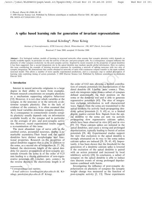
A spike based learning rule for generation of invariant representations<br />
Konrad Körding*, Peter König<br />
Institute of Neuroinformatics, ETH/Uni�ersity Zürich, Winterthurerstr. 190, 8057 Zürich, Switzerland<br />
Received 27 June 2000; accepted 19 October 2000<br />
Abstract – For biological realism, models of learning in neuronal networks often assume that synaptic plasticity solely depends on<br />
locally available signals, in particular on only the activity of the pre- and post-synaptic cells. As a consequence, synapses influence the<br />
plasticity of other synapses exclusively via the post-synaptic activity. Inspired by recent research on the properties of apical dendrites<br />
it has been suggested, that a second integration site in the apical dendrite may mediate specific global information. Here we explore<br />
this issue considering the example of learning invariant responses by examining a network of spiking neurones with two sites of<br />
synaptic integration. We demonstrate that results obtained in networks of units with continuous outputs transfer to the more realistic<br />
neuronal model. This allows a number of more specific experimental predictions, and is a necessary step to unified description of<br />
learning rules exploiting timing of action potentials. © 2000 Elsevier Science Ltd. Published by Éditions scientifiques et médicales<br />
Elsevier SAS<br />
1. Introduction<br />
Interest in neural networks originates to a large<br />
degree in their ability to learn from examples.<br />
Present research concentrates on synaptic plasticity<br />
as a mechanism supporting adaptive behaviour<br />
[32]. However, it is not clear which variables at the<br />
synapse, in the neurones or in the network co-determine<br />
synaptic plasticity. Due to the lack of<br />
evidence of the contrary, it is often assumed that<br />
solely local variables determine synaptic plasticity.<br />
As a consequence in connectionist systems synaptic<br />
plasticity usually depends only on information<br />
available locally at the synapse and in particular<br />
on the averages of pre- and post-synaptic activities.<br />
However, recent experimental results suggest<br />
a re-examination of this issue.<br />
The most abundant type of nerve cells in the<br />
cerebral cortex, pyramidal neurones, display a remarkable<br />
asymmetry.Their basal and the apical<br />
dendrites have distinct morphologies and functional<br />
properties. Research on the properties of<br />
apical dendrites suggests that it acts, in addition to<br />
the soma, as a second site of integration [6, 27, 28].<br />
Indeed, the electrotonic length of the apical dendrite<br />
for passive propagation of post-synaptic potentials<br />
towards the soma is of the order of 0.25<br />
mm leading to a strong attenuation of non-regenerative<br />
potentials (H. Lüscher, pers. comm.). In<br />
the reverse direction the electrotonic length is of<br />
the order of 0.62 mm allowing a limited contribution<br />
of somatic potentials for depolarization of the<br />
distal dendrite (H. Lüscher, pers. comm.). Thus,<br />
each site integrates input from a subset of synapses<br />
defined anatomically via their position on the<br />
soma or the dendritic tree and is able to generate<br />
regenerative potentials [43]. The sites of integration<br />
exchange information in well characterized<br />
ways. Signals from the soma are transmitted to the<br />
apical dendrite via actively back-propagating dendritic<br />
action potentials [1, 9, 45] or, to a limited<br />
degree, passive current flow. Signals from the apical<br />
dendrite to the soma are sent via actively<br />
propagating slow regenerative calcium spikes,<br />
which have been observed in vitro [43] and in vivo<br />
[19, 21]. These calcium spikes are initiated in the<br />
apical dendrites, and cause a strong and prolonged<br />
depolarization, typically leading to bursts of action<br />
potentials [28, 46]. Experimental studies support<br />
the view that excitation to the apical dendrite is<br />
strongly attenuated on its way to the soma unless<br />
calcium spikes are induced [28, 29, 43, 45]. Importantly,<br />
it has been shown that the threshold for the<br />
generation of a dendritic calcium spike is lowered<br />
if the excitation of the apical dendrite is paired<br />
with an action potential and increased by local<br />
inhibitory activity [28]. In conclusion, a subset of<br />
synapses on the apical dendrite is able to induce<br />
rare discrete events of strong prolonged depolarization<br />
combined with bursts.<br />
Investigating the influence of post-synaptic depolarization<br />
on plasticity of synapses the synaptic<br />
weight change was measured as the function of<br />
post-synaptic activity [2, 13]. Those experiments<br />
<strong>UNCORRECTED</strong> <strong>PROOF</strong><br />
* Correspondence and reprints.<br />
E-mail addresses: koerding@ini.phys.ethz.ch (K. Körding),<br />
peterk@ini.phys.ethz.ch (P. König).
sco3:/jobs1/ELSPARIS/pxph/week.43/Ppxph1088y.001ed Nov 15 09:48:05 2000 Page Wed Nov 15 09:<br />
2<br />
showed that at low firing rates/depolarization<br />
LTD occurs whereas at higher activities a switch<br />
to LTP takes place. Thus, without a calcium spike<br />
and, as a consequence, with a limited depolarization,<br />
the cell is in a regime where synapses are<br />
depressed. In contrast, when a calcium spike is<br />
triggered, the high depolarization leads to a potentiation<br />
of active synapses. Thus integrating the<br />
above two lines of research naturally leads to a<br />
plausible biological implementation for the hypothesized<br />
mechanism, where calcium spikes could<br />
correspond to ‘learning events’. Indeed experiments<br />
by Pike et al. [38] give further evidence that<br />
post-synaptic bursting is essential for the induction<br />
of LTP.<br />
The generation of calcium spikes is highly sensitive<br />
to inhibitory activity. Even the activity of one<br />
single inhibitory neurone can significantly raise the<br />
threshold of calcium spike generation [28, 29]. As<br />
axons of inhibitory interneurones branch homogeneously<br />
within the range of a hypercolumn this<br />
implements an effective soft winner take all mechanisms<br />
on the level of calcium spikes. It seems<br />
reasonable to assume that the number of neurones<br />
generating calcium bursts upon presentation of a<br />
stimulus is limited.<br />
To complete the picture we have to consider,<br />
which afferent is targeting the apical and basal<br />
dendritic tree respectively? The anatomy of a cortical<br />
column is complicated; nevertheless, some regular<br />
patterns can be discerned. The apical<br />
dendrites of the considered layer 5 pyramidal cells<br />
receive long range cortico-cortical projections [10,<br />
50]. Furthermore, top-down projections from areas<br />
higher in the hierarchy of the sensory system<br />
usually terminate in layer 1, where many apical<br />
tufts can be observed [41]. This supports the idea<br />
that top-down connections from higher to lower<br />
areas preferentially terminate on the apical dendrites.<br />
The basal dendrites of the considered cells<br />
receive direct subcortical afferents (e.g. the koniocellular<br />
pathway in visual cortex) in addition to<br />
projections from layer 4 spiny stellate cells. These<br />
are the main recipients of afferents from sensory<br />
thalamus or from areas lower in the cortical hierarchy.<br />
Therefore, we use the approximation that<br />
the bottom-up input targets mainly the basal dendritic<br />
tree.<br />
In previous work we proposed that these ingredients<br />
– layer 5 pyramidal neurones possess two<br />
independent sites of integration with separate<br />
threshold processes; the somatic site of integration<br />
receives bottom-up information on stimulus properties<br />
and dominates the level of cellular activity,<br />
K. Körding, P. König / Journal of Physiology 000 (2000) 000–000<br />
i.e. the spike rate; the integration site in the apical<br />
dendrite receives top-down information and gates<br />
synaptic plasticity of synapses in the whole neurone<br />
– amount to a learning rule with interesting<br />
properties and analysed an implementation in a<br />
network of units with continuous outputs [27].<br />
From the experimental results described above, it<br />
is obvious that a network composed of spiking<br />
units is the much more relevant system to investigate,<br />
which is the subject of this article. Furthermore,<br />
we discuss the investigated learning rule as a<br />
possible implementation of algorithmic learning<br />
rules defined on a global level.<br />
2. Methods<br />
The simulated network mimics a cartoon visual<br />
system roughly as outlined in a previous work [27].<br />
It consists of two processing streams, which can be<br />
interpreted either as representing non-overlapping<br />
patches of the visual field or as one stream with<br />
time unfolded into space (figure 1). This allows to<br />
exploit object continuity in space or time as described<br />
below. Each stream contains an input layer<br />
and a hierarchy of two more layers. Each layer<br />
contains excitatory neurones with two separate<br />
compartments (somatic and apical dendritic as<br />
described below) and local inhibition. Bottom-up<br />
projections originate in the input layer and terminate<br />
at the somatic compartment of the excitatory<br />
neurones of layer 1 within each stream. The activity<br />
of these neurones is then relayed to the somatic<br />
compartment of excitatory neurones of layer 2.<br />
Layer 2 neurones send top-down projections to<br />
layer 1 terminating at the apical dendritic compartment<br />
as well as to the apical dendritic compartment<br />
of neurones in layer 2 of the other<br />
stream. Furthermore, each layer contains local<br />
inhibitory mechanisms (figure 2).<br />
A Poisson statistic with a rate set directly by the<br />
topographically mapped stimulus describes activities<br />
of input units. Excitatory cells are implemented<br />
as leaky integrate and fire neurones along<br />
with a mechanism for burst generation. They are<br />
described by two variables corresponding to the<br />
two sites of integration: A is referred to as the<br />
somatic potential, D represents the potential at the<br />
apical dendrite and O the output of the neurone.<br />
When the somatic potential of a neurone reaches<br />
threshold (� s) a spike is emitted and the somatic<br />
potential is reset to its resting value (� r):<br />
<strong>UNCORRECTED</strong> <strong>PROOF</strong><br />
If A i(t)�� s then O i(t)=1
sco3:/jobs1/ELSPARIS/pxph/week.43/Ppxph1088y.001ed Nov 15 09:48:05 2000 Page Wed Nov 15 09:<br />
and<br />
Ai(t)=� r, or else Oi(t)=0 The subthreshold dynamics of A is described in<br />
layer 1 by:<br />
1 �Ai =C1O0 1 � W/A� i ,<br />
1 1 1 with A� i (t)=A� i (t−1)�+Ai (t) � (1−�).<br />
C1=50 and �=0.999, resulting in a running<br />
average with a time-scale of 1000 iterations. The<br />
activity of layer 0 neurones (O0 ) is written as a<br />
vector containing either 1s for active neurones and<br />
0s for inactive neurones. The scalar product ( � )<br />
implies simple summation of all inputs weighted<br />
by their respective synaptic efficacy (W).<br />
In the second layer the subthreshold dynamics<br />
follows:<br />
2 1 2 �Ai =C2 max(Opsp � Wb)/A� i ,<br />
with A� i 2 (t)=A� i 2 (t−1)�+Ai 2 (t) � (1−�).<br />
C 2=200 and �=0.999. In order to mimic the<br />
finite decay of post-synaptic potential, a running<br />
average with an exponential decay of ten iterations<br />
of the output of layer 1 is taken as the effective<br />
input. The synaptic input, however, is not summated<br />
as above, but the maximum determined.<br />
The choice of max as an integration function has<br />
K. Körding, P. König / Journal of Physiology 000 (2000) 000–000 3<br />
been discussed in depth by Riesenhuber and Poggio<br />
[39].<br />
The local inhibition has a multiplicative effect<br />
�A i=−�O iA i<br />
with �=0.001.<br />
The dendritic potential is determined by:<br />
�D i=O pre � W a+C 3A i−C 4D i.<br />
Two factors influence the potential D at the<br />
apical dendrite. Activity of the respective presynaptic<br />
neurones (O 2 pre) increases D proportional<br />
to the corresponding synaptic weights (W a). Following<br />
the experimental finding that the threshold<br />
for generation of a dendritic calcium spike is lowered<br />
when the excitation of the apical dendrite is<br />
paired with a somatic action potential [28, 29], the<br />
activity A (the somatic variable) also increases D<br />
in the case of layer 1 (C 3=1, C 3=0 otherwise). D<br />
decays with a time constant of 100 time steps<br />
(C 4=0.01). Based on the experimental result that<br />
input by a single interneurone may abolish calcium<br />
spikes [28], we assume a strong local competition<br />
within each module. Only in the neurone with the<br />
highest dendritic potential calcium spikes are<br />
triggered.<br />
Figure 1. The network consists of<br />
two streams with three layers each:<br />
input layer, layer 1 and layer 2, each<br />
depicted as a box. In the left part of<br />
a box, a schematic wiring diagram<br />
with three connections to other<br />
boxes is shown. Units receive input<br />
to the apical and basal dendritic tree<br />
(upper and lower connection, respectively).<br />
The output via the axon<br />
is shown in between (middle connection).<br />
Local inhibition is indicated<br />
by the lightly shaded line and influences<br />
the somatic compartment. The<br />
size of each layer is indicated in the<br />
right part of the box. The two<br />
streams communicated by reciprocal<br />
connections onto the apical dendritic<br />
tree on the highest level.<br />
<strong>UNCORRECTED</strong> <strong>PROOF</strong>
sco3:/jobs1/ELSPARIS/pxph/week.43/Ppxph1088y.001ed Nov 15 09:48:05 2000 Page Wed Nov 15 09:<br />
4<br />
Figure 2. The diagram shows a principal with events influencing<br />
synaptic plasticity. An action potential triggered at the axon<br />
hillock (star) and propagates anterogradely along the axon<br />
(down to the right) and retrogradely invades the dendritic tree<br />
(double arrow pointing upwards). Synapses of inhibitory interneurones<br />
are located on the proximal dendrite (filled circles)<br />
and affect the backpropagating action potential. The backpropagating<br />
action potential may reach the distal apical dendrite in<br />
interact with local afferent input (a pre-synaptic action potential<br />
is indicated by a short vertical line riding on an axon) in the<br />
generation of calcium spikes. The calcium spike travels towards<br />
the soma (double arrow pointing downward) where it influences<br />
synaptic plasticity of all active synapses. See text for<br />
details.<br />
Following the experimental results on properties<br />
of LTP and LTD summarized above changes of<br />
synaptic efficacy in our model depend on presynaptic<br />
activity and post-synaptic calcium spike<br />
activity. In those neurones, where input to the<br />
apical dendritic tree triggered calcium spikes all<br />
active synapses of apical as well as basal dendritic<br />
tree are updated:<br />
pre �Wk1=�A� k −Wk1+�(Tc/Nc−0.5), with �=0.0001, A� is a running average with exponential<br />
decay of ten iterations. As in many other<br />
neuronal network simulations we use normalization<br />
of the weight vectors, the second term, to<br />
avoid diverging weights [34]. Actually, there is<br />
some experimental support for such a biological<br />
mechanism. Blocking neural activity in tissue culture<br />
leads to increased miniature excitatory postsynaptic<br />
synaptic currents, whereas blocking<br />
K. Körding, P. König / Journal of Physiology 000 (2000) 000–000<br />
GABA mediated inhibition leads to higher firing<br />
rates and to smaller synaptic currents [48, 49].<br />
Alternatively a BCM-like learning rule to normalize<br />
activity [7] could be combined with the calcium<br />
spike effect. Analysing such effects could help<br />
understand how calcium spikes and ‘normal’ firing<br />
could interact leading to efficient learning. The<br />
cells habituate with respect to calcium spikes, the<br />
third term, and change all their weights by an<br />
amount proportional to the number of iterations<br />
since the last calcium spike (T c, with �=0.000002,<br />
and N c as the total number of units in the layer).<br />
This implies that units, which have not learned for<br />
a long time increase their input.<br />
Initially all weights are chosen randomly in the<br />
interval (0 ...1). The system is simulated for<br />
20 000 iterations.<br />
Stimuli used were elongated Gaussians resembling<br />
‘light bars’ as used in physiological experiments.<br />
Their luminance had a Gaussian profile<br />
orthogonal to the long axis with a length constant<br />
of 1 and along the main axis with a length constant<br />
of 4. The latter was introduced to reduce<br />
boundary effects. They were presented at different<br />
orientations and positions. Most importantly is the<br />
relation of stimuli presented to the two streams.<br />
As in natural images the orientation of contours in<br />
small patches is correlated over time, however, the<br />
precise position of the contours is not correlated<br />
(Weinhäuser, Körding, König, unpubl. data) the<br />
orientation of stimuli presented to both streams is<br />
chosen to be perfectly correlated and the position<br />
of stimuli uncorrelated. For quantitative evaluation<br />
position is parameterized on a one-dimensional<br />
axis orthogonal to the major axis of the<br />
stimulus pattern.<br />
3. Results<br />
As a first step we investigated the dynamics in<br />
the neuronal network. Presenting an oriented stimulus<br />
leads to activity of appropriately tuned neurones.<br />
Due to the two sites of integration, the<br />
temporal structure of activity varies. In figure 3,<br />
spike traces of two neurones in layer 1 are shown,<br />
which receive top-down input from layer 2 (upper<br />
trace) and which do not (lower trace). Although<br />
overall activity is not that different, the grouping<br />
of action potentials to bursts in the activity trace<br />
of the former neurones is obvious. In view of the<br />
learning rule employed, this is a decisive difference.<br />
In neurone c1, the bursts of action potentials<br />
induced by the top-down input gate plasticity<br />
<strong>UNCORRECTED</strong> <strong>PROOF</strong>
sco3:/jobs1/ELSPARIS/pxph/week.43/Ppxph1088y.001ed Nov 15 09:48:05 2000 Page Wed Nov 15 09:<br />
Figure 3. Examples of spike trains showing regular firing and<br />
bursting activity.<br />
of all synapses and allow the neurone to learn the<br />
presented stimulus. This leads to a difference in the<br />
size of representation of a stimulus (many neurones�coarse<br />
coding) and the number of neurones<br />
adapting to a stimulus (few neurones�soft<br />
winner take all).<br />
The network is trained with oriented stimuli<br />
presented at different positions. Importantly, stimuli<br />
to the two streams have correlated orientation,<br />
but uncorrelated positions. After training we investigate<br />
response properties of neurones in layer<br />
1. Spike trains for with different orientation and<br />
position are shown in figure 4. Presenting stimuli<br />
at different orientations (figure 4A, upper panel)<br />
and positions (figure 4A, lower panel) induces different<br />
levels of activity in the neurone considered.<br />
Please note, that the induction of burst occurs at a<br />
delay of a few hundred milliseconds. Although the<br />
precise value of this delay is dependent on the<br />
K. Körding, P. König / Journal of Physiology 000 (2000) 000–000 5<br />
parameters of the simulation, a slight sharpening<br />
in the tuning of the neurones can be observed. For<br />
a further quantification the receptive fields of the<br />
neurones are plotted in visual space (figure 4B,<br />
left) and stimulus space (figure 4B, right). In visual<br />
space receptive fields are elongated with different<br />
orientations and located at different positions. As<br />
a consequence, plotted in stimulus space receptive<br />
fields are structured along both the orientation and<br />
position dimension. This property resembles ‘simple<br />
cells’ in primary visual cortex [22]. The union<br />
of all receptive fields covers evenly the whole stimulus<br />
space (figure 4C).<br />
Neurones in the second layer show a qualitatively<br />
different behaviour after training. Upon<br />
stimulation with oriented bars at different positions<br />
their responses are orientation tuned (figure<br />
5A, upper panel). Comparable to layer 1 tuning of<br />
mean firing rate is somewhat broader than tuning<br />
of burst responses. An important difference can be<br />
seen when presenting stimuli at different positions.<br />
The neurone responds to stimuli presented at any<br />
position and the size of the response drops only at<br />
the extremes, where the stimuli are located near<br />
one corner and few of the units in the input layer<br />
are activated. This property is a result of learned<br />
convergent connections from layer 1 to layer 2.<br />
The linear receptive field, as predicted from the<br />
feed-forward synaptic connections is shown in<br />
figure 5B. The little squares depict receptive fields<br />
in stimulus space of layer 1 neurones connected to<br />
Figure 4. A. Trains of action potentials<br />
of a selected neurone in layer 1<br />
for stimuli of different orientations<br />
and position. The eight orientations<br />
shown cover the whole range of 180<br />
degree available. Position of the<br />
stimulus is parameterized on a onedimensional<br />
axis orthogonal to the<br />
stimulus. B. Receptive fields of four<br />
layer 1 neurones shown in different<br />
rows. The left column depicts the<br />
receptive field in visual space as results<br />
from the synaptic connectivity.<br />
In the right column, strength of response<br />
to a stimulus is shown in<br />
stimulus space. Here all possible<br />
combinations of orientations and<br />
positions are shown in a two-dimensional<br />
diagram. C. Coverage of<br />
stimulus space by the union of all<br />
neurones in layer 1 is shown greylevel<br />
coded.<br />
<strong>UNCORRECTED</strong> <strong>PROOF</strong>
sco3:/jobs1/ELSPARIS/pxph/week.43/Ppxph1088y.001ed Nov 15 09:48:05 2000 Page Wed Nov 15 09:<br />
6<br />
the same layer 2 neurone in sequence of decreasing<br />
synaptic efficacies. The receptive field of the latter<br />
is shown in stimulus space as well in the large<br />
square. As described above, receptive fields of<br />
layer 1 neurones are tuned in the orientation and<br />
position dimensions. As neurones of layer 1 tuned<br />
to similar orientations and dissimilar positions<br />
converge onto a neurone in layer 2, the receptive<br />
fields of the latter is still orientation tuned but<br />
little modulated in the position dimension. The<br />
effective receptive fields, when considering tangential<br />
interactions between the two streams are actually<br />
even smoother. Thus, it has position invariant<br />
responses and resembles in its properties complex<br />
neurones in primary visual cortex.<br />
The union of receptive fields of layer 2 neurones<br />
also covers the complete stimulus space with a<br />
slight drop off at extreme positions. Thus, the<br />
learning rule leads to a homogeneous representation<br />
of the stimulus space combined with translation<br />
invariant responses.<br />
K. Körding, P. König / Journal of Physiology 000 (2000) 000–000<br />
4. Discussion<br />
Figure 5. A. Trains of action potentials<br />
of a selected neurone in layer 2<br />
for stimuli of different orientations<br />
and position. The eight orientations<br />
shown cover the whole range of 180<br />
degree available. Position of the<br />
stimulus is parameterized on a onedimensional<br />
axis orthogonal to the<br />
stimulus. B. Receptive field of the<br />
layer 2 neurone as predicted by the<br />
bottom-up afferent shown in stimulus<br />
space (big square). Receptive<br />
fields of layer 1 neurones strongly<br />
connected to the layer 1 neurone in<br />
stimulus space are shown in the little<br />
squares. Please note that receptive<br />
fields of these layer 1 neurones converging<br />
on the same layer 2 neurone<br />
have similar preferred orientation,<br />
but vary in preferred position. The<br />
resulting receptive field of the layer<br />
2 neurones is therefore orientation<br />
selective and translation invariant.<br />
C. Effective receptive field of layer 2<br />
neurones as determined from the<br />
cellular activity and thus taking the<br />
tangential connectivity between neurones<br />
in different streams into account.<br />
Please note that the response<br />
to stimuli at different positions is<br />
even more homogeneous as predicted<br />
from the feed-forward connectivity<br />
as shown in B. D.<br />
Coverage of stimulus space by the<br />
union of all layer 2 neurones is<br />
shown grey-level coded.<br />
In this work, we demonstrate that properties of<br />
a previously proposed learning rule for learning<br />
invariant representations in a network of neurones<br />
with continuous input/output functions hold up in<br />
a more realistic simulation incorporating spiking<br />
neurones.<br />
Here we studied the learning on invariant representations<br />
using the example of translation invariance<br />
as it occurs in the step from simple cells to<br />
complex cells in primary visual cortex [22]. In the<br />
biological system, the emergence of invariant responses<br />
is observed in many instances. In parallel<br />
to the increasing sophistication of receptive field<br />
properties in higher visual areas, some aspects of<br />
visual stimuli are less and less important. Neurones<br />
in area V4 show some degree of colour<br />
constancy, which is equivalent to invariance with<br />
respect to the spectral composition of illumination.<br />
Furthermore, in inferotemporal cortex translation,<br />
<strong>UNCORRECTED</strong> <strong>PROOF</strong>
sco3:/jobs1/ELSPARIS/pxph/week.43/Ppxph1088y.001ed Nov 15 09:48:05 2000 Page Wed Nov 15 09:<br />
scaling and rotation invariant responses can be<br />
observed [8, 35, 40]. An even more extreme combination<br />
of specificity and invariance can be found<br />
in premotor cortex. Neurones may respond with<br />
high specificity to a stimulus, irrespective of it<br />
being heard, seen or felt [17]. Thus, a highly specific<br />
response is combined with invariance with<br />
respect to modality.<br />
Computing systems obtain invariant properties<br />
by several means [3]. First, appropriate preprocessing<br />
can supply a neuronal network with invariant<br />
input data. Another option is to build the invariance<br />
into the structure of the system so that invariances<br />
gradually increase. Finally, a system may<br />
learn invariances from the presented stimuli following<br />
principles of supervised or unsupervised<br />
learning. The former need labelled training data,<br />
and actually for large networks, a lot of these.<br />
With such data at hand, it is possible to learn<br />
invariant recognition training the network with a<br />
variant of the backpropagation algorithm (e.g.<br />
[20]). To alleviate the problem of getting enough<br />
training data, applying the desired invariance operators<br />
to individual examples can enlarge the<br />
training set. However, then we are back with an a<br />
priori specification of the invariance operation.<br />
Furthermore, these approaches do not supply a<br />
convincing explanation of the development of invariant<br />
response properties in the biological system.<br />
Following this observation the investigation<br />
of unsupervised learning of invariances became a<br />
research topic in itself.<br />
To explain such properties Becker and Hinton<br />
[5] proposed the Imax algorithm. In this learning<br />
algorithm, the mutual information is maximized<br />
between output units which receive separate inputs.<br />
Thus, this algorithm can detect features in<br />
the input which are coherent across the eyes [5]<br />
space, time or modality [4, 12]. However, the<br />
algorithm requires backpropation of derivatives<br />
from output units to hidden units, and the storage<br />
of several variables on each unit. A related approach<br />
for learning with multi-unit local processors<br />
with multivariate binary outputs has been<br />
proposed by Phillips et al. [37] (see also [24]). It is<br />
able to extract a number of coherent features.<br />
These learning rules are similar to the scheme<br />
described here as two separate summations occur.<br />
One of these defines the receptive fields and the<br />
other the context fields [36]. They share several<br />
properties with the basal and apical dendrite in<br />
our studies respectively (see also [44]): The context<br />
gates learning and is simultaneously used for improving<br />
processing of the receptive field input [37].<br />
K. Körding, P. König / Journal of Physiology 000 (2000) 000–000 7<br />
Their learning rule contains a threshold where the<br />
sign of learning changes; it depends on the conditional<br />
probabilities with regard to the context and<br />
the activity of the other output units. Thus, this<br />
signal is local to the cell but not to the synapse.<br />
The learning mechanism proposed here can lend a<br />
straightforward biological interpretation to such<br />
approaches. Although being closely related with<br />
regard to the learning goal, this study also shows a<br />
major computational difference. The context field<br />
represents the covariance matrix of post-synaptic<br />
activity with contextual input. Thus the number of<br />
weights which needs to be stored and updated is<br />
smaller, increasing learning speed at the cost of a<br />
larger statistical error.<br />
4.1. Simplifications<br />
Despite this increased physiological realism in<br />
this simulation, several aspects are simplified.<br />
First, and perhaps most importantly, the detailed<br />
temporal dynamics of activity is not taken into<br />
account. Indeed, it has been proposed previously<br />
that the sign of change of synaptic efficacy depends<br />
on the relative sequence of pre and postsynaptic<br />
action potentials [16]. This hypothesis is<br />
supported by recent in vitro experiments [31]. Such<br />
a dependence would lead to an automatic normalization<br />
of total afferent signals [25, 42]. Indeed, in<br />
vivo experiments provide evidence that optimally<br />
activated neurones tend to fire prior to suboptimally<br />
activated neurones on a millisecond time<br />
scale [26]. Thus, differences in firing rates may be<br />
mapped onto the relative timing of action potentials,<br />
justifying simplifications done in previous<br />
modelling studies. Furthermore, in the present<br />
simulation feed-forward input follows a Poisson<br />
statistics and its temporal dynamics contains no<br />
information. The interesting question of how tangential<br />
interactions leading to a synchronization of<br />
neuronal activity interact with the proposed learning<br />
rule has to be left for future work [30]. Second,<br />
the complex non-linear dendritic properties [23, 33]<br />
are reduced to a threshold mechanism triggering<br />
dendritic calcium spikes. The sole justification for<br />
this approach we can present is the argument of<br />
Occams razor. In view of the large number of<br />
parameters needed for a detailed compartmental<br />
simulation, and our lack of knowledge of these, a<br />
more detailed realistic model would actually include<br />
many more estimates not solidly based on<br />
physiological and anatomical results and thus obscure<br />
the issues investigated. Third, along similar<br />
lines in our simulation the current flow from the<br />
<strong>UNCORRECTED</strong> <strong>PROOF</strong>
sco3:/jobs1/ELSPARIS/pxph/week.43/Ppxph1088y.001ed Nov 15 09:48:05 2000 Page Wed Nov 15 09:<br />
8<br />
apical dendrite to the soma is neglected and calcium<br />
spikes have an effect on learning but not on<br />
firing rates. Introducing the effect on activity could<br />
result in information about the context not only<br />
being used for learning but also for enhancing the<br />
signal itself [37]. The effects of calcium spikes on<br />
the dynamics of the network were analysed in a<br />
previous work [44]. We could demonstrate that<br />
top-down information leads to enhanced processing<br />
of bottom-up signals. We conjecture that both<br />
effects are compatible and act simultaneously, the<br />
top-down signals improving signal processing on a<br />
short time scale and gating learning on a long time<br />
scale.<br />
4.2. Experimental predictions<br />
The simulations described here imply some experimental<br />
predictions.<br />
First, cooling or lesions of higher areas should<br />
significantly reduce the frequency of bursting activity<br />
in lower areas by decreasing the amount of<br />
top-down mediated signals available. This would<br />
reduce synaptic plasticity of all synapses as could<br />
be assessed by paradigms as monocular deprivation<br />
or selective rearing influencing ocular dominance<br />
or orientation tuning of cortical neurones<br />
[14, 15, 18]. Furthermore, as these top-down mediated<br />
signals act on a spatial scale given by the<br />
receptive field size of the neurones in a higher area<br />
the loss of top-down signals should appear as a<br />
reduction of non-classical receptive field effects. In<br />
particular, this effect should be most pronounced<br />
for stimuli matching receptive field properties of<br />
neurones in the higher cortical areas [44].<br />
Second, patching the trunk of the apical dendrite<br />
of pyramidal cortical cells would make it<br />
possible to block the induction of calcium spikes in<br />
the apical dendrites without inflicting strong<br />
changes upon the potential at the soma (except for<br />
the lack of calcium spikes). Simultaneously patching<br />
the soma of that cell in deeper layers and a<br />
pre-synaptic cell would make it possible to assess<br />
synaptic plasticity. The removal of calcium spikes<br />
should lead to strongly reduced LTP not only for<br />
synapses at the apical dendrite, but as well for<br />
synapses at the basal dendrites.<br />
Third, along the same lines the effects of additional<br />
calcium spikes can be investigated. Patching<br />
an apical dendrite in layer 1 in vivo would make it<br />
possible to artificially induce calcium spikes in<br />
analogy to Larkum et al. [28]. Following the predictions<br />
of the proposed model it should be possible<br />
to investigate unsupervised learning in vivo<br />
K. Körding, P. König / Journal of Physiology 000 (2000) 000–000<br />
similar to Debanne et al. [11]. The cell should learn<br />
to represent those stimuli that are paired with<br />
excitation of the apical dendrite. The plasticity<br />
resulting from calcium spikes could thus directly<br />
be compared with the plasticity resulting from<br />
spikes not associated with calcium spikes. Should<br />
some normal spikes have less influence on plasticity<br />
then a calcium spike resulting in a volley of the<br />
same number of spikes?<br />
Fourth, direct interference with the burst generating<br />
mechanism should have a particular strong<br />
effect on top-down mediated signals and also affect<br />
receptive field properties in the described way.<br />
Slice recordings show that the backpropagation of<br />
action potentials into the apical dendrite depends<br />
on muscarinic input, inhibitory input and the<br />
firing rate [9, 47]. Furthermore it has been demonstrated<br />
that the triggering of bursts by correlated<br />
synaptic input at the apical dendrite and backpropagating<br />
action potentials is highly sensitive to<br />
inhibitory input [28]. These findings suggest that<br />
altering the activity of inhibitory or modulatory<br />
systems could be a suitable way to interfere with<br />
the burst generating mechanism.<br />
These experiments are demanding, but within<br />
the reach of state of the art techniques.<br />
Acknowledgements<br />
This work has been supported by the Swiss<br />
National Science Foundation (P.K. 31-51059.97)<br />
and the Boehringer Ingelheim Fond (K.P.K.).<br />
References<br />
[1] Amitai Y., Friedman A., Connors B.W., Gutnick M.J.,<br />
Regenerative activity in apical dendrites of pyramidal cells<br />
in neocortex, Cereb. Cortex 3 (1993) 26–38.<br />
[2] Artola A., Bröcher S., Singer W., Different voltage-dependent<br />
thresholds for inducing long-term depression and<br />
long-term potentiation in slices of rat visual cortex, Nature<br />
347 (1990) 69–72.<br />
[3] Barnard E., Casasent D.P., Invariance and Neural Nets,<br />
IEEE Trans. Neural Networks 2 (1991) 489–508.<br />
[4] Becker S., Models of cortical self-organisation, Network:<br />
Comput. Neural Syst. 7 (1996) 7–31.<br />
[5] Becker S., Hinton G.E., Self-organizing neural network<br />
that discovers surfaces in random-dot stereograms, Nature<br />
355 (1992) 161–163.<br />
<strong>UNCORRECTED</strong> <strong>PROOF</strong><br />
[6] Bernander O., Koch C., Douglas R.J., Amplification and<br />
linearization of distal synaptic input to cortical pyramidal<br />
cells, J. Neurophysiol. 72 (1994) 2743–2753.
sco3:/jobs1/ELSPARIS/pxph/week.43/Ppxph1088y.001ed Nov 15 09:48:05 2000 Page Wed Nov 15 09:<br />
[7] Bienenstock E., Cooper L.N., Munro P.W., Theory for the<br />
development of neuron selectivity: Orientation specificity<br />
and binocular interaction in visual cortex, J. Neurosci. 2<br />
(1982) 32–48.<br />
[8] Booth M.C., Rolls E.T., View-invariant representations of<br />
familiar objects by neurons in the inferior temporal visual<br />
cortex, Cereb. Cortex 8 (1998) 510–523.<br />
[9] Buzsaki G., Kandel A., Somadendritic backpropagation of<br />
action potentials in cortical pyramidal cells of the awake<br />
rat, J. Neurophysiol. 79 (1998) 1587–1591.<br />
[10] Cauller L.J., Connors B.W., Synaptic physiology of horizontal<br />
afferents to layer I in slices of rat SI neocortex, J.<br />
Neurosci. 14 (1994) 751–762.<br />
[11] Debanne D., Shulz D.E., Fregnac Y., Activity-dependent<br />
regulation of ‘on’ and ‘off’ responses in cat visual cortical<br />
receptive fields, J. Physiol. (Lond.) 508 (1998) 523–548.<br />
[12] de Sa V.R., Ballard D.H., Category learning through<br />
multimodality sensing, Neur. Comp. 10 (1998) 1097–1117.<br />
[13] Dudek S.M., Bear M.F., Homosynaptic long term depression<br />
in area CA1 of hippocampus and the effects on<br />
NMDA receptor blockade, Proc. Natl. Acad. Sci. USA 89<br />
(1991) 4363–4367.<br />
[14] Fregnac Y., Shulz D.E., Activity-dependent regulation of<br />
receptive field properties of cat area 17 by supervised<br />
Hebbian learning, J. Neurobiol. 41 (1999) 69–82.<br />
[15] Friedlander M.J., Fregnac Y., Burke J.P., Temporal covariance<br />
of postsynaptic membrane potential and synaptic<br />
input-role in synaptic efficacy in visual cortex, Prog. Brain<br />
Res. 95 (1993) 207–223.<br />
[16] Gerstner W., Kempter R., van Hemmen J.L., Wagner H.,<br />
A neuronal learning rule for sub-millisecond temporal<br />
coding, Nature 383 (1996) 76–78.<br />
[17] Graziano M.S., Gross C.G., Spatial maps for the control<br />
of movement, Curr. Opin. Neurobiol. 8 (1998) 195–201.<br />
[18] Greul J.M., Luhmann H.J., Singer W., Pharmacological<br />
induction of use-dependent receptive field modifications in<br />
visual cortex, Science 242 (1988) 74–77.<br />
[19] Helmchen F., Svoboda K., Denk W., Tank D.W., In vivo<br />
dendritic calcium dynamics in deep-layer cortical pyramidal<br />
neurons, Nat. Neurosci. 2 (1999) 989–996.<br />
[20] Hinton G.E., Learning translation invariant recognition in<br />
a massively parallel network, in: Goos G., Hartmanis J.<br />
(Eds.), PARLE: Parallel Architectures and Languages Europe,<br />
Lecture Notes in Computer Science, Springer-Verlag,<br />
Berlin, 1987, pp. 1–13.<br />
[21] Hirsch J.A., Alonso J.M., Reid R.C., Visually evoked<br />
calcium action potentials in cat striate cortex, Nature 378<br />
(1995) 612–616.<br />
[22] Hubel D.H., Wiesel T.N., Receptive Fields, binocular interaction<br />
and functional architecture in the cat’s visual<br />
cortex, J. Physiol. 160 (1962) 106–154.<br />
[23] Johnston D., Magee J.C., Colbert C.M., Cristie B.R.,<br />
Active properties of neuronal dendrites, Annu. Rev. Neurosci.<br />
19 (1996) 165–186.<br />
[24] Kay J., Floreano D., Phillips W.A., Contextually guided<br />
unsupervised learning using local multivariate binary processors,<br />
Neural Networks 11 (1998) 117–140.<br />
K. Körding, P. König / Journal of Physiology 000 (2000) 000–000 9<br />
[25] Kempter R., Gerstner W., van Hemmen J.L., Hebbian<br />
learning and spiking neurons, Phys. Rev. E 59 (1999)<br />
4498–4514.<br />
[26] König P., Engel A.K., Roelfsema P.R., Singer W., How<br />
precise is neuronal synchronization?, Neural Comp. 7<br />
(1995) 469–485.<br />
[27] Körding K.P., König P., Learning with two sites of synaptic<br />
integration, Network: Comput. Neural Syst. 11 (2000a)<br />
1–15.<br />
[28] Larkum M.E., Zhu J.J., Sakmann B., A new cellular<br />
mechanism for coupling inputs arriving at different cortical<br />
layers, Nature 398 (1999) 338–341.<br />
[29] Larkum M.E., Kaiser K.M., Sakmann B., Calcium electrogenesis<br />
in distal apical dendrites of layer 5 pyramidal<br />
cells at a critical frequency of back-propagating action<br />
potentials, Proc. Natl. Acad. Sci. USA 96 (1999) 14600–<br />
14604.<br />
[30] Lumer E.D., A neural model of binocular integration and<br />
rivalry based on the coordination of action-potential timing<br />
in primary visual cortex, Cereb. Cortex 8 (1998) 553–<br />
561.<br />
[31] Markram H., Lübke J., Frotscher M., Sakmann B., Regulation<br />
of synaptic efficacy by coincidence of postsynaptic<br />
APs and EPSPs, Science 275 (1997) 213–215.<br />
[32] Martin S.J., Grimwood P.D., Morris R.G., Synaptic plasticity<br />
and memory: an evaluation of the hypothesis, Annu.<br />
Rev. Neurosci. 23 (2000) 649–711.<br />
[33] Mel B.W., Synaptic integration in an excitable dendritic<br />
tree, J. Neurophysiol. 70 (1993) 1086–1101.<br />
[34] Miller K.D., MacKay D.J.C., The role of constraints in<br />
Hebbian learning, Neural Comput. 6 (1994) 100–126.<br />
[35] Perrett D.I., Oram M.W., Harries M.H., Bevan R., Hietanen<br />
J.K., Venson P.J., Thomas S., Viewer-centred and<br />
object-centred coding of heads in the macaque temporal<br />
cortex, Exp. Brain. Res. 86 (1991) 159–173.<br />
[36] Phillips W.A., Singer W., In search of common foundations<br />
for cortical computation, Behav. Brain Sci. 20 (1997)<br />
657–683.<br />
[37] Phillips W.A., Kay J., Smyth D., The discovery of structure<br />
by multi-stream networks of local processors with<br />
contextual guidance, Network: Comput. Neural Syst. 6<br />
(1995) 225–246.<br />
[38] Pike F.G., Meredith R.M., Olding A.W.A., Paulsen O.,<br />
Postsynaptic bursting is essential for ‘Hebbian’ induction<br />
of associative long-term potentiation at excitatory<br />
synapses in rat hippocampus, J. Phys. 518 (2) (1999)<br />
571–576.<br />
[39] Riesenhuber M., Poggio T., Hierarchical models of object<br />
recognition in cortex, Nat. Neurosci. 2 (1999) 1019–<br />
1025.<br />
[40] Rolls E., Neurophysiological mechanisms underlying<br />
face processing within and beyond the temporal cortical<br />
visual areas, Phil. Trans. R. Soc. Lond. B 335 (1992)<br />
11–21.<br />
<strong>UNCORRECTED</strong> <strong>PROOF</strong><br />
[41] Salin P.A., Bullier J., Corticocortical connections in the<br />
visual system: Structure and function, Physiol. Rev. 75<br />
(1995) 107–154.
sco3:/jobs1/ELSPARIS/pxph/week.43/Ppxph1088y.001ed Nov 15 09:48:05 2000 Page Wed Nov 15 09:<br />
10<br />
[42] Sanchez-Montanes M.A., Verschure P.F., König P., Local<br />
and global gating of synaptic plasticity, Neural Comput.<br />
12 (2000) 519–529.<br />
[43] Schiller J., Schiller Y., Stuart G., Sakmann B., Calcium<br />
action potentials restricted to distal apical dendrites of rat<br />
neocortical pyramidal neurons, J. Physiol. (Lond.) 505<br />
(1997) 605–616.<br />
[44] Siegel M., Körding K.P., König P., Integrating bottom-up<br />
and top-down sensory processing by somato-dendritic interactions,<br />
J. Comput. Neurosci. 8 (2000) 161–173.<br />
[45] Stuart G.J., Sakmann B., Active propagation of somatic<br />
action potentials into neocortical pyramidal cell dendrites,<br />
Nature 367 (1994) 69–72.<br />
[46] Stuart G.J., Schiller J., Sakmann B., Action potential<br />
K. Körding, P. König / Journal of Physiology 000 (2000) 000–000<br />
initiation and propagation in rat neocortical pyramidal<br />
neurons, J. Physiol. (Lond.) 505 (1997) 617–632.<br />
[47] Tsubokawa H., Ross W.N., IPSPs modulate spike backpropagation<br />
and associated [Ca2+ ]i changes in the dendrites<br />
of hippocampal CA1 pyramidal neurons, J.<br />
Neurophysiol. 76 (1996) 2896–2906.<br />
[48] Turrigiano G.G., Homeostatic plasticity in neuronal networks:<br />
the more things change, the more they stay the<br />
same, Trends Neurosci. 22 (1999) 221–227.<br />
[49] Turrigiano G.G., Leslie K.R., Desai N.S., Rutherford<br />
L.C., Nelson S.B., Activity-dependent scaling of quantal<br />
amplitude in neocortical, Nature 391 (1998) 892–896.<br />
[50] Zeki S., Shipp S., The functional logic of cortical connections,<br />
Nature 335 (1988) 311–317.<br />
<strong>UNCORRECTED</strong> <strong>PROOF</strong><br />
.