Branched DNA Technology in Molecular Diagnostics
Branched DNA Technology in Molecular Diagnostics
Branched DNA Technology in Molecular Diagnostics

Create successful ePaper yourself
Turn your PDF publications into a flip-book with our unique Google optimized e-Paper software.
Microbiology and Infectious Disease / BRANCHED <strong>DNA</strong> DIAGNOSTICS<br />
<strong>Branched</strong> <strong>DNA</strong> <strong>Technology</strong> <strong>in</strong> <strong>Molecular</strong> <strong>Diagnostics</strong><br />
Gregory J. Tsongalis, PhD<br />
Key Words: Microbiology; Virology; <strong>Molecular</strong> diagnostics<br />
DOI: 10.1309/90BU6KDXANFLN4RJ<br />
Abstract<br />
Viral quantification or viral load test<strong>in</strong>g has<br />
become part of the rout<strong>in</strong>e management of patients<br />
<strong>in</strong>fected with HIV-1 or hepatitis C virus (HCV). There<br />
are currently several molecular technologies that are<br />
available for use <strong>in</strong> the cl<strong>in</strong>ical laboratory sett<strong>in</strong>g. Of<br />
these, only the branched <strong>DNA</strong> (b<strong>DNA</strong>) assays are FDAapproved<br />
for HIV-1 and HCV viral load test<strong>in</strong>g. This<br />
signal amplification technology is built on a series of<br />
hybridization reactions that are highly amenable to full<br />
automation and thus lessen the amount of labor<br />
required to perform this type of analysis. This article<br />
provides a historical perspective of b<strong>DNA</strong> and its<br />
cl<strong>in</strong>ical applications.<br />
448 Am J Cl<strong>in</strong> Pathol 2006;126:448-453<br />
448 DOI: 10.1309/90BU6KDXANFLN4RJ<br />
The <strong>in</strong>troduction of new therapeutics <strong>in</strong> the late 1980s<br />
and early 1990s to combat the HIV-1 virus also <strong>in</strong>troduced<br />
the need for viral quantification assays to be performed by<br />
the cl<strong>in</strong>ical laboratory. Therapeutic response then could be<br />
monitored as the virus decreased or <strong>in</strong>creased <strong>in</strong> copy number<br />
ow<strong>in</strong>g to the effectiveness of the treatment. Two HIV-1<br />
viral load assays emerged that used different amplification<br />
technologies. The first was the branched cha<strong>in</strong> <strong>DNA</strong><br />
(b<strong>DNA</strong>) signal amplification technology followed by the<br />
reverse transcriptase–polymerase cha<strong>in</strong> reaction (RT-PCR)<br />
sequence amplification assay. These 2 technologies cont<strong>in</strong>ue<br />
to be the most prom<strong>in</strong>ent for viral quantification test<strong>in</strong>g. This<br />
article provides an overview of b<strong>DNA</strong> technology and cl<strong>in</strong>ical<br />
applications.<br />
<strong>Molecular</strong> diagnostic assays us<strong>in</strong>g b<strong>DNA</strong> technology for<br />
detection of nucleic acid target molecules are sensitive, specific,<br />
and reliable tools <strong>in</strong> the diagnosis of viral and bacterial<br />
<strong>in</strong>fections and for monitor<strong>in</strong>g disease progression dur<strong>in</strong>g the<br />
course of therapy. b<strong>DNA</strong> tests have evolved from developmental<br />
stages <strong>in</strong> the research laboratory to US Food and Drug<br />
Adm<strong>in</strong>istration–approved quantitative assays with valuable<br />
cl<strong>in</strong>ical applications. The b<strong>DNA</strong> assays are less labor-<strong>in</strong>tensive<br />
than many molecular-based procedures because they are<br />
highly amenable to total automation. Us<strong>in</strong>g b<strong>DNA</strong>, amplification<br />
of a target sequence is not required, and, thus, cross-contam<strong>in</strong>ation<br />
between replicate samples due to excessive amplicons<br />
or carryover is less likely <strong>in</strong> b<strong>DNA</strong> assays. In addition,<br />
because b<strong>DNA</strong> is a signal amplification technology, the assay<br />
is able to quantify with less than a 0.5 log or 3-fold variability<br />
for its entire dynamic range.<br />
b<strong>DNA</strong> technology has proved versatile because methods<br />
have been developed for the detection of <strong>in</strong>fection by a wide<br />
© American Society for Cl<strong>in</strong>ical Pathology
ange of microorganisms, <strong>in</strong>clud<strong>in</strong>g the parasite Trypanosoma<br />
brucei, 1 cytomegalovirus, 2 antibiotic-sensitive and antibioticresistant<br />
Staphylococcus bacteria, 3 human papillomavirus, 4<br />
and hepatitis B virus. 5 However, more recent efforts have<br />
focused on the development of b<strong>DNA</strong> assays for the quantification<br />
of HIV-1 and hepatitis C virus (HCV) RNA, lead<strong>in</strong>g to<br />
the rout<strong>in</strong>e application of b<strong>DNA</strong> methods <strong>in</strong> the cl<strong>in</strong>ical<br />
molecular diagnostics laboratory. In describ<strong>in</strong>g the advancement<br />
of b<strong>DNA</strong> methods, this review emphasizes b<strong>DNA</strong><br />
assays for HIV-1 and HCV.<br />
How b<strong>DNA</strong> Works<br />
In contrast with techniques that rely on <strong>in</strong> vitro amplification<br />
of the target sequence (ie, PCR, transcription-mediated<br />
amplification, nucleic acid sequence–based amplification, and<br />
strand displacement amplification), the sensitivity of b<strong>DNA</strong><br />
methods is achieved by signal amplification on the b<strong>DNA</strong><br />
probe after direct b<strong>in</strong>d<strong>in</strong>g of a large hybridization complex to<br />
the target sequence. 6,7 This series of hybridization steps results<br />
<strong>in</strong> a “sandwich” complex of probes and target sequence. These<br />
unusual synthetic oligonucleotides are composed of a primary<br />
sequence and secondary sequences that result <strong>in</strong> a branched<br />
structure extend<strong>in</strong>g from the primary sequence. 8<br />
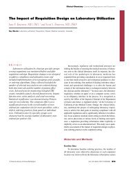
The <strong>in</strong>itial step <strong>in</strong> a b<strong>DNA</strong> assay is to ensure that viral<br />
particles have been disrupted and that viral RNA is present for<br />
analysis ❚Figure 1A❚. In the most recent third-generation<br />
b<strong>DNA</strong> assays, target-specific oligonucleotides (label extenders<br />
and capture extenders) then are hybridized with high str<strong>in</strong>gency<br />
to the target nucleic acid ❚Figure 1B❚. 7 Capture extenders<br />
are designed to hybridize to the target and to capture<br />
probes, which are attached to a microwell plate. Label extenders<br />
are designed to hybridize to contiguous regions on the target<br />
and to provide sequences for hybridization of a preamplifier<br />
oligonucleotide. Signal amplification then beg<strong>in</strong>s with<br />
preamplifier probes hybridiz<strong>in</strong>g to label extenders. The preamplifier<br />
forms a stable hybrid only if it hybridizes to 2 adjacent<br />
label extenders. Other regions on the preamplifier are<br />
designed to hybridize to multiple b<strong>DNA</strong> amplifier molecules<br />
that create a branched structure ❚Figure 1C❚. F<strong>in</strong>ally, alkal<strong>in</strong>ephosphatase<br />
(AP)-labeled oligonucleotides, which are complementary<br />
to b<strong>DNA</strong> amplifier sequences, b<strong>in</strong>d to the b<strong>DNA</strong><br />
molecule by hybridization. The b<strong>DNA</strong> signal is the chemilum<strong>in</strong>escent<br />
product of the AP reaction ❚Figure 1D❚.<br />
The signal <strong>in</strong> the b<strong>DNA</strong> assay is proportional to the number<br />
of AP-labeled probes that hybridize to b<strong>DNA</strong> secondary<br />
sequences. Because b<strong>DNA</strong> assays are based on a series of<br />
hybridization reactions, the <strong>in</strong>hibition of enzymatic steps (AP)<br />
by endogenous or exogenous sources is not a significant<br />
source of analytic error. Also, these assays have not been<br />
shown to be affected by the use of collection devices such as<br />
© American Society for Cl<strong>in</strong>ical Pathology<br />
Microbiology and Infectious Disease / REVIEW ARTICLE<br />
the BD Vacuta<strong>in</strong>er PPT (Becton Dick<strong>in</strong>son, Frankl<strong>in</strong> Lakes,<br />
NJ). Absolute quantification is accomplished by establish<strong>in</strong>g a<br />
standard curve for each run. In addition, negative and positive<br />
control samples are <strong>in</strong>cluded <strong>in</strong> each run.<br />
First-Generation HIV-1 b<strong>DNA</strong> Assays<br />
Accurate, reproducible first-generation b<strong>DNA</strong> assays<br />
were developed for the detection of HIV-1 RNA and HCV<br />
RNA <strong>in</strong> human plasma (Quantiplex HIV-1 or HCV RNA 1.0<br />
assay, Bayer, Tarrytown, NY). 9-11 In one of the first reports of<br />
the Quantiplex b<strong>DNA</strong> assay, no reactivity us<strong>in</strong>g plasma samples<br />
from seronegative donors was observed us<strong>in</strong>g the firstgeneration<br />
b<strong>DNA</strong> assay, <strong>in</strong>dicat<strong>in</strong>g excellent specificity. 9<br />
Positive results <strong>in</strong> the b<strong>DNA</strong> assay were observed <strong>in</strong> 83% of<br />
samples from 348 patients who were seropositive for HIV-1. 9<br />
The dynamic range for quantification us<strong>in</strong>g the Quantiplex<br />
HIV-1 RNA 1.0 assay extended from 10 4 (lower limit of<br />
detection) to more than 10 6 HIV RNA copies/mL. 9,11 Changes<br />
<strong>in</strong> viral load of 2- to 3-fold were statistically significant us<strong>in</strong>g<br />
the b<strong>DNA</strong> test, <strong>in</strong>dicat<strong>in</strong>g that the Quantiplex HIV-1 RNA 1.0<br />
assay was highly accurate and reproducible. 11 By comparison,<br />
changes <strong>in</strong> viral load of at least 3.7- to 5.8-fold were necessary<br />
before results were statistically significant us<strong>in</strong>g the earliest<br />
versions of the RT-PCR assay. 11 Thus, b<strong>DNA</strong> became the<br />
method of choice for most cl<strong>in</strong>ical trials evaluat<strong>in</strong>g viral load<br />
test<strong>in</strong>g and the cl<strong>in</strong>ical efficacy of new HIV-1 reverse transcriptase<br />
and protease <strong>in</strong>hibitors.<br />
The sensitivity of the b<strong>DNA</strong> detection method was<br />
enhanced <strong>in</strong> the second-generation HIV-1 assay (Quantiplex<br />
HIV-1 RNA 2.0 assay, Bayer) by chang<strong>in</strong>g the design of the<br />
target and capture probes and by the addition of preamplifier<br />
oligonucleotides. 12,13 The improved design of the target and<br />
capture probes allowed an <strong>in</strong>crease <strong>in</strong> the str<strong>in</strong>gency of<br />
hybridization, thereby decreas<strong>in</strong>g the assay background.<br />
Preamplifiers dramatically <strong>in</strong>crease signal <strong>in</strong>tensity because<br />
each preamplifier molecule has multiple regions for<br />
hybridization to many b<strong>DNA</strong> molecules. In addition, each<br />
b<strong>DNA</strong> molecule has multiple, repeat sequences for hybridization<br />
of AP-labeled probes. The signal output us<strong>in</strong>g the<br />
Quantiplex HIV-1 RNA 2.0 assay showed l<strong>in</strong>earity from<br />
approximately 500 copies/mL to 1.6 × 10 6 copies/mL (stated<br />
dynamic range was 500 to 8 × 10 5 copies/mL). 11,12 The sensitivity<br />
of the second-generation HIV-1 b<strong>DNA</strong> assay was<br />
<strong>in</strong>creased by 20-fold compared with the first-generation assay<br />
(lower limits of detection were 500 copies/mL and 10 × 10 4<br />
copies/mL, respectively).<br />
In an extensive analysis of precision, <strong>in</strong>itial test results<br />
and retest results <strong>in</strong> the Quantiplex HIV-1 RNA 2.0 assay were<br />
compared. The HIV-1 RNA 2.0 assay was found to be highly<br />
reproducible. 14 Of 174 samples with viral loads of more than<br />
Am J Cl<strong>in</strong> Pathol 2006;126:448-453 449<br />
449 DOI: 10.1309/90BU6KDXANFLN4RJ 449
Tsongalis / BRANCHED <strong>DNA</strong> DIAGNOSTICS<br />
5,000 copies/mL, 96% had differences of less than 0.3 log 10 <strong>in</strong><br />
copy number between <strong>in</strong>itial results and retest results. Of 69<br />
samples with viral loads between 500 and 5,000 copies/mL,<br />
86% had less than 0.3 log 10 differences between <strong>in</strong>itial and<br />
retest results. However, among 5,339 patients who were tested<br />
A B<br />
C<br />
D<br />
Release RNA<br />
Release and<br />
denature RNA<br />
Target RNA<br />
Capture<br />
extender<br />
Target RNA<br />
Capture<br />
extender<br />
450 Am J Cl<strong>in</strong> Pathol 2006;126:448-453<br />
450 DOI: 10.1309/90BU6KDXANFLN4RJ<br />
Microwell<br />
Microwell<br />
<strong>in</strong> rout<strong>in</strong>e cl<strong>in</strong>ical test<strong>in</strong>g dur<strong>in</strong>g a 1-year <strong>in</strong>terval, viral loads<br />
of fewer than 500 copies/mL were observed <strong>in</strong> 41.6% of samples.<br />
Therefore, a b<strong>DNA</strong> assay with higher sensitivity (ie,<br />
lower limit of detection of
The performance characteristics of the Quantiplex HIV-1<br />
RNA 2.0 assay and an RT-PCR test (Amplicor HIV-1 Monitor<br />
1.0 assay, Roche <strong>Diagnostics</strong>, Indianapolis, IN) were compared<br />
us<strong>in</strong>g dilutions of standard samples that had known<br />
HIV-1 virus copy numbers. 13 When dilutions of the same standards<br />
were tested <strong>in</strong> the 2 assays, HIV-1 copy numbers generally<br />
were higher from the RT-PCR assay than from the b<strong>DNA</strong><br />
assay. For example, the ranges of RNA copy numbers were<br />
900 to 7.68 × 10 5 copies/mL <strong>in</strong> the b<strong>DNA</strong> test and 3,360 to<br />
1.88 × 10 6 copies/mL <strong>in</strong> the RT-PCR assay. Both assays were<br />
l<strong>in</strong>ear for the stated dynamic ranges.<br />
Comparison of the slopes of HIV-1 copy number vs signal<br />
output regression l<strong>in</strong>es suggested that the b<strong>DNA</strong> test had<br />
less proportional systematic error. Between-run variability,<br />
us<strong>in</strong>g a standard sample with 1,650 HIV-1 copies/mL, was<br />
lower <strong>in</strong> the b<strong>DNA</strong> test than <strong>in</strong> the RT-PCR assay; coefficients<br />
of variations for the b<strong>DNA</strong> and RT-PCR assays were<br />
24.3% and 34.3%, respectively, <strong>in</strong>dicat<strong>in</strong>g that the b<strong>DNA</strong><br />
assay was slightly more precise at this HIV-1 RNA copy<br />
number. Compared with us<strong>in</strong>g the 1,650-copies/mL standard,<br />
the between-run coefficients of variation were higher for a<br />
sample conta<strong>in</strong><strong>in</strong>g 165 HIV-1 RNA copies/mL (44.0% and<br />
42.7% for the b<strong>DNA</strong> and RT-PCR assays, respectively).<br />
Assay results were similar when equal HIV-1 copy numbers<br />
were compared across HIV subtypes A through F by us<strong>in</strong>g<br />
the b<strong>DNA</strong> test. However, by this version of RT-PCR, subtypes<br />
A, E, and F were detected less efficiently than the B, C,<br />
and D subtypes. The differences between the second-generation<br />
b<strong>DNA</strong> test and the Amplicor Monitor RT-PCR test for<br />
HIV-1 quantification <strong>in</strong>dicated that consistency <strong>in</strong> test<strong>in</strong>g<br />
method was required for each <strong>in</strong>dividual patient throughout<br />
diagnosis and treatment.<br />
HCV b<strong>DNA</strong> Assays<br />
While cont<strong>in</strong>u<strong>in</strong>g to develop an improved third-generation<br />
HIV-1 b<strong>DNA</strong> assay, Bayer recognized the need to develop<br />
an HCV viral load assay with an emerg<strong>in</strong>g cl<strong>in</strong>ical usefulness<br />
similar to that for HIV-1 test<strong>in</strong>g. For the detection of<br />
HCV, the first-generation b<strong>DNA</strong> assay (Quantiplex HCV<br />
RNA 1.0 assay, Bayer) had a dynamic quantification range <strong>in</strong><br />
human plasma from 3.5 × 10 5 to 1.2 × 10 8 HCV RNA<br />
copies/mL. 10,15 Genotypes 1 through 6 were detected by us<strong>in</strong>g<br />
this assay, although the sensitivity was lower for genotypes 2<br />
and 3 (67% detection rate: positive signal for 60 of 89 serum<br />
samples known to conta<strong>in</strong> HCV genotypes 2 or 3) compared<br />
with genotype 1 (97% detection rate: positive signal for 67 of<br />
69 serum samples known to conta<strong>in</strong> the HCV genotype 1). A<br />
comparison of the Quantiplex HCV RNA 1.0 assay and a<br />
research laboratory–developed RT-PCR test for HCV showed<br />
greater sensitivity <strong>in</strong> the RT-PCR test, which had a lower limit<br />
© American Society for Cl<strong>in</strong>ical Pathology<br />
Microbiology and Infectious Disease / REVIEW ARTICLE<br />
of detection of 2.5 × 10 4 HCV RNA copies/mL. However, the<br />
HCV b<strong>DNA</strong> test had greater reproducibility and was less<br />
time-consum<strong>in</strong>g than the laboratory-developed HCV RT-PCR<br />
test. 16<br />
To improve the detection rate for HCV genotypes 2 and<br />
3, a second-generation assay was developed (Quantiplex HCV<br />
RNA 2.0 assay, Bayer). 15 The major design change <strong>in</strong> the second-generation<br />
HCV RNA assay was to use probes to<br />
sequences <strong>in</strong> the HCV genome that were more highly conserved<br />
across genotypes. These conserved regions were 5'<br />
untranslated sequences and sequences <strong>in</strong> the core gene of the<br />
HCV genome. The result of changes to the target and capture<br />
probes was to dramatically reduce the variation <strong>in</strong> detection<br />
rate among HCV genotypes <strong>in</strong> the second-generation assay.<br />
Each of the 6 HCV genotypes had a high detection rate, and<br />
there was marked improvement <strong>in</strong> the detection of HCV genotypes<br />
2 and 3 (detection of 93% vs 67% of samples known to<br />
conta<strong>in</strong> HCV genotypes 2 or 3 <strong>in</strong> the HCV 2.0 and HCV 1.0<br />
assays, respectively). Also, the sensitivity of the second-generation<br />
b<strong>DNA</strong> assay was slightly enhanced compared with the<br />
HCV 1.0 assay (lower limits of quantification were 2.0 × 10 5<br />
[HCV 2.0] versus 3.5 × 10 5 [HCV 1.0]).<br />
Third-Generation b<strong>DNA</strong> Assays for HIV-1<br />
and HCV<br />
In the first- and second-generation b<strong>DNA</strong> assays, nonspecific<br />
hybridization of oligonucleotide probes to nontarget<br />
sequences limited assay sensitivity. The critical technological<br />
improvement <strong>in</strong> the third-generation b<strong>DNA</strong> assay<br />
(Quantiplex [also referred to as VERSANT] HIV-1 RNA 3.0<br />
assay, Bayer) was to use nonnatural bases, 5'-methyl-2'deoxyisoguanos<strong>in</strong>e<br />
(isoG) and 5'-methyl-2'-isodeoxycytid<strong>in</strong>e<br />
(isoC), <strong>in</strong> the synthesis of all probes <strong>in</strong> the b<strong>DNA</strong> system,<br />
with the exception of capture extenders that mediate<br />
capture of the target viral nucleic acid to the plate surface.<br />
Because oligonucleotides conta<strong>in</strong><strong>in</strong>g isoG and isoC are not<br />
present <strong>in</strong> nature, nonspecific hybridization is reduced significantly.<br />
7 Thus, probes modified with the nonnatural bases<br />
do not form stable hybrids with the capture probe <strong>in</strong> the<br />
absence of target RNA. In the <strong>in</strong>itial description of the thirdgeneration<br />
assay, the limit of detection of HIV-1 <strong>in</strong> plasma<br />
samples from 11 patients was 50 copies per mL. 7 This represents<br />
a 10-fold improvement <strong>in</strong> the limit of detection compared<br />
with the second-generation assay. Dur<strong>in</strong>g treatment<br />
with highly active antiretroviral therapy, HIV-1 viral load<br />
decreased to below the limit of detection for all 11 patients.<br />
The VERSANT HIV-1 RNA 3.0 assay has a dynamic range<br />
of 75 to 5 × 10 5 HIV RNA copies/mL. Version 3.0 also has<br />
been approved to a lower limit of detection equal to 50<br />
copies/mL <strong>in</strong> several other countries.<br />
Am J Cl<strong>in</strong> Pathol 2006;126:448-453 451<br />
451 DOI: 10.1309/90BU6KDXANFLN4RJ 451
Tsongalis / BRANCHED <strong>DNA</strong> DIAGNOSTICS<br />
When matched samples were compared <strong>in</strong> the secondgeneration<br />
HIV-1 b<strong>DNA</strong> assay (Quantiplex version 2.0) and<br />
the Amplicor Monitor 1.0 RT-PCR assay, consistently lower<br />
HIV-1 copy numbers were obta<strong>in</strong>ed us<strong>in</strong>g the b<strong>DNA</strong> test. 13,17<br />
However, a close quantitative correlation <strong>in</strong> HIV-1 RNA copy<br />
numbers was observed between the third-generation (version<br />
3.0) assay and the Amplicor Monitor 1.5 RT-PCR test. 18 Viral<br />
load results <strong>in</strong> the version 3.0 b<strong>DNA</strong> assay and <strong>in</strong> the<br />
Amplicor RT-PCR assay were approximately 2-fold higher<br />
than <strong>in</strong> the version 2.0 assay. Quantitatively similar results <strong>in</strong><br />
the version 3.0 b<strong>DNA</strong> test and the RT-PCR test are important<br />
<strong>in</strong> patient care because of the likely possibility that different<br />
methods will be used <strong>in</strong> test<strong>in</strong>g samples from the same patient<br />
for the course of anti-HIV therapies. Recent data suggest that<br />
rebasel<strong>in</strong><strong>in</strong>g for patients tested with an RT-PCR assay may not<br />
be necessary.<br />
Similar to the HIV-1 version 3.0 b<strong>DNA</strong> assay, the thirdgeneration<br />
b<strong>DNA</strong> assay for HCV (VERSANT HCV RNA 3.0,<br />
Bayer) also used isoC- and isoG-substituted oligonucleotides<br />
to reduce nonspecific hybridization. 19 The use of isoC- and<br />
isoG-substituted oligonucleotides <strong>in</strong>creased assay sensitivity<br />
approximately 62-fold. The HCV RNA 3.0 assay lower limit<br />
of detection was 3.2 × 10 3 copies/mL compared with 2 × 10 5<br />
copies/mL <strong>in</strong> the HCV RNA 2.0 assay. The dynamic l<strong>in</strong>ear<br />
quantification range of the HCV RNA 3.0 assay extended<br />
from 3.2 × 10 3 copies/mL (615 IU/mL) to 4 × 10 7 HCV RNA<br />
copies/mL (7.7 × 10 6 IU/mL). The HCV RNA 3.0 assay had<br />
a high specificity (98.2%) and, similar to the second-generation<br />
assay, was equally effective <strong>in</strong> the quantification of HCV<br />
RNA across all genotypes. Between-run and with<strong>in</strong>-run standard<br />
deviations for replicate samples were 0.2 log 10 and 0.14<br />
log 10 , respectively, <strong>in</strong>dicat<strong>in</strong>g that the third-generation assay<br />
was highly reproducible.<br />
In addition to the eradication of HCV <strong>in</strong> serum, an important<br />
goal of anti-HCV therapies is to reduce HCV levels <strong>in</strong> the<br />
liver. The usefulness of the HCV RNA 3.0 assay for the detection<br />
and quantification of HCV RNA <strong>in</strong> liver biopsy specimens<br />
was studied <strong>in</strong> 25 patients co<strong>in</strong>fected with HCV and<br />
HIV. 20 The reproducibility of the third-generation HCV<br />
b<strong>DNA</strong> assay was similar between liver biopsy specimens and<br />
serum samples. Also, detection of HCV RNA <strong>in</strong> liver specimens<br />
from patients <strong>in</strong>fected with genotypes 1, 3, and 4 by the<br />
HCV RNA 3.0 assay was highly specific and sensitive. In this<br />
study of 25 patients, high pretreatment levels of <strong>in</strong>trahepatic<br />
HCV correlated with a low frequency of response to anti-<br />
HCV therapy. Pretreatment <strong>in</strong>trahepatic HCV levels were<br />
highest <strong>in</strong> patients <strong>in</strong>fected with HCV genotype 1. Results of<br />
this study demonstrated that important markers of HCV disease<br />
progression, HCV levels <strong>in</strong> liver and <strong>in</strong> serum, can be<br />
quantitated reliably by b<strong>DNA</strong> analysis dur<strong>in</strong>g treatment.<br />
The methods for third-generation b<strong>DNA</strong> assays <strong>in</strong>clude<br />
sample preparation, hybridization, and signal detection for<br />
452 Am J Cl<strong>in</strong> Pathol 2006;126:448-453<br />
452 DOI: 10.1309/90BU6KDXANFLN4RJ<br />
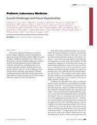
HIV-1 RNA and HCV RNA. All 3 steps are performed <strong>in</strong> the<br />
microwells on the System 340 platform ❚Image 1❚ for the HCV<br />
RNA 3.0 assay that does not require a separate extraction step.<br />
In the version 3.0 HIV-1 RNA assay, sample preparation is different<br />
from the HCV RNA method and is performed outside<br />
the System 340 platform. A recent study evaluated an adaptation<br />
of the version 3.0 HIV-1 RNA method <strong>in</strong> which the HIV-<br />
1 sample process<strong>in</strong>g step was modified to accommodate<br />
simultaneous test<strong>in</strong>g for HCV and HIV-1 on the System 340<br />
platform. 21 The HIV-1 method was modified by omitt<strong>in</strong>g the<br />
2-hour <strong>in</strong>cubation at 63°C for viral lysis. Instead, HIV-1 and<br />
HCV lysis was performed on the System 340 platform. The<br />
HCV b<strong>DNA</strong> test methods were unchanged <strong>in</strong> the comb<strong>in</strong>ed<br />
assay. The specificity and quantification by the comb<strong>in</strong>ed<br />
b<strong>DNA</strong> method were with<strong>in</strong> specifications for the <strong>in</strong>dividual<br />
HIV-1 and HCV assays. Simultaneous test<strong>in</strong>g for HIV-1 and<br />
for HCV improved workflow <strong>in</strong> the cl<strong>in</strong>ical laboratory and<br />
resulted <strong>in</strong> lower costs. Because HIV-1 RNA and HCV RNA<br />
detection and quantification are crucial for diagnosis and for<br />
the evaluation of responses to therapy, the ability to carry out<br />
simultaneous test<strong>in</strong>g for both viruses represents a significant<br />
advance <strong>in</strong> molecular diagnostics.<br />
Summary<br />
b<strong>DNA</strong> methods have progressed from first-generation<br />
assays, which were accurate and reproducible but relatively<br />
<strong>in</strong>sensitive, to third-generation b<strong>DNA</strong> tests that are accurate,<br />
reproducible, highly sensitive, and automated laboratory tests<br />
for more optimal patient management. Essential technological<br />
discoveries that resulted <strong>in</strong> significantly better assay performance<br />
and permitted the evolution from first- to third-generation<br />
b<strong>DNA</strong> assays are as follows: (1) <strong>in</strong>creased hybridization<br />
str<strong>in</strong>gency of target and capture probes, (2) <strong>in</strong>creased signal<br />
amplification by us<strong>in</strong>g preamplifier oligonucleotides, and (3)<br />
❚Image 1❚ Bayer System 340 b<strong>DNA</strong> [branched cha<strong>in</strong> <strong>DNA</strong>]<br />
Analyzer.<br />
© American Society for Cl<strong>in</strong>ical Pathology
greatly reduced nonspecific hybridization to nontarget<br />
sequences by us<strong>in</strong>g isoC- and isoG-substituted oligonucleotides<br />
for all probes <strong>in</strong> the b<strong>DNA</strong> system, with the exception<br />
of probes that mediate capture of the target RNA to the<br />
microwell. The results of this evolution <strong>in</strong> b<strong>DNA</strong> technology<br />
are Food and Drug Adm<strong>in</strong>istration–approved methods for<br />
the detection and quantification of HIV-1 and HCV (VER-<br />
SANT HIV-1 RNA 3.0 assay and the VERSANT HCV RNA<br />
3.0 assay).<br />
From the Department of Pathology, Dartmouth Medical School,<br />
Dartmouth-Hitchcock Medical Center, Lebanon, NH.<br />
F<strong>in</strong>ancial support for preparation of this article was provided<br />
by Bayer Healthcare.<br />
Address repr<strong>in</strong>t requests to Dr Tsongalis: <strong>Molecular</strong><br />
Pathology Laboratory, Dartmouth Medical School, Dartmouth-<br />
Hitchcock Medical Center, One Medical Center Dr, Lebanon, NH<br />
03756.<br />
Acknowledgment: David Sorscher, PhD, is gratefully<br />
acknowledged for prepar<strong>in</strong>g a work<strong>in</strong>g draft of this manuscript.<br />
References<br />
1. Harris E, Detmer J, Dungan J, et al. Detection of Trypanosoma<br />
brucei spp <strong>in</strong> human blood by a nonradioactive branched<br />
<strong>DNA</strong>-based technique. J Cl<strong>in</strong> Microbiol. 1996;34:2401-2407.<br />
2. Chernoff DN, M<strong>in</strong>er RC, Hoo BS, et al. Quantification of<br />
cytomegalovirus <strong>DNA</strong> <strong>in</strong> peripheral blood leukocytes by a<br />
branched-<strong>DNA</strong> signal amplification assay. J Cl<strong>in</strong> Microbiol.<br />
1997;35:2740-2744.<br />
3. Kolbert CP, Arruda J, Varga-Delmore P, et al. <strong>Branched</strong>-<strong>DNA</strong><br />
assay for detection of mecA gene <strong>in</strong> oxacill<strong>in</strong>-resistant and<br />
oxacill<strong>in</strong>-sensitive staphylococci. J Cl<strong>in</strong> Microbiol.<br />
1998;36:2640-2644.<br />
4. Player AN, Shen L-P, Kenny D, et al. S<strong>in</strong>gle-copy gene<br />
detection us<strong>in</strong>g branched <strong>DNA</strong> (b<strong>DNA</strong>) <strong>in</strong> situ hybridization.<br />
J Histochem Cytochem. 2001;49:603-611.<br />
5. Hendricks DA, Stowe BJ, Hoo BS, et al. Quantitation of HBV<br />
<strong>DNA</strong> <strong>in</strong> human serum us<strong>in</strong>g a branched <strong>DNA</strong> (b<strong>DNA</strong>) signal<br />
amplification assay. Am J Cl<strong>in</strong> Pathol. 1995;104;547-546.<br />
6. Horn T, Chang C, Urdea MS. Chemical synthesis and<br />
characterization of branched oligodeoxyribonucleotides<br />
(b<strong>DNA</strong>) for use as signal amplifiers <strong>in</strong> nucleic acid<br />
quantification assays. Nucleic Acids Res. 1997;25:4842-4849.<br />
7. Coll<strong>in</strong>s ML, Irv<strong>in</strong>e B, Tyner D, et al. A branched <strong>DNA</strong> signal<br />
amplification assay for quantification of nucleic acid targets<br />
below 100 molecules/mL. Nucleic Acids Res. 1997;25:2979-<br />
2984.<br />
8. Horn T, Urdea M. Forks and combs and <strong>DNA</strong>: the synthesis of<br />
branched oligodeoxyribonucleotides. Nucleic Acids Res.<br />
1989;17:6959-6967.<br />
© American Society for Cl<strong>in</strong>ical Pathology<br />
Microbiology and Infectious Disease / REVIEW ARTICLE<br />
9. Pachl C, Todd JA, Kern DG, et al. Rapid and precise<br />
quantification of HIV-1 RNA <strong>in</strong> plasma us<strong>in</strong>g a branched<br />
<strong>DNA</strong> (b<strong>DNA</strong>) signal amplification assay. J Acquir Immune<br />
Defic Syndr Hum Retrovirol. 1995;8:446-454.<br />
10. Alter HJ, Sanchez-Pescador R, Urdea MS, et al. Evaluation of<br />
branched <strong>DNA</strong> signal amplification for the detection of<br />
hepatitis C virus RNA. J Viral Hepat. 1995;2:121-132.<br />
11. Todd J, Pachl C, White R, et al. Performance characteristics<br />
for the quantitation of plasma HIV-1 RNA us<strong>in</strong>g branched<br />
<strong>DNA</strong> signal amplification technology. J Acquir Immune Defic<br />
Syndr Hum Retrovirol. 1995;10(suppl 2):S35-S44.<br />
12. Kern D, Coll<strong>in</strong>s M, Fultz T, et al. An enhanced-sensitivity<br />
branched-<strong>DNA</strong> assay for quantification of human<br />
immunodeficiency virus type 1 RNA <strong>in</strong> plasma. J Cl<strong>in</strong><br />
Microbiol. 1996;34:3196-3202.<br />
13. Nolte FS, Boysza J, Thurmond C, et al. Cl<strong>in</strong>ical comparison of<br />
an enhanced-sensitivity branched-<strong>DNA</strong> assay and reverse<br />
transcription–PCR for quantitation of human<br />
immunodeficiency virus type 1 RNA <strong>in</strong> plasma. J Cl<strong>in</strong><br />
Microbiol. 1998;36:716-720.<br />
14. Murphy DG, Gon<strong>in</strong> P, Fauvel M. Reproducibility and<br />
performance of the second-generation branched-<strong>DNA</strong> assay <strong>in</strong><br />
rout<strong>in</strong>e quantification of human immunodeficiency virus type<br />
1 RNA <strong>in</strong> plasma. J Cl<strong>in</strong> Microbiol. 1999;37:812-814.<br />
15. Detmer J, Lagier R, Flynn J, et al. Accurate quantification of<br />
hepatitis C virus (HCV) RNA from all HCV genotypes by<br />
us<strong>in</strong>g branched-<strong>DNA</strong> technology. J Cl<strong>in</strong> Microbiol.<br />
1996;34:901-907.<br />
16. Mayerat C, Burgisser P, Lavanchy D, et al. Comparison of a<br />
competitive comb<strong>in</strong>ed reverse transcription–PCR assay with a<br />
branched-<strong>DNA</strong> assay for hepatitis C virus RNA quantitation.<br />
J Cl<strong>in</strong> Microbiol. 1996;34:2702-2706.<br />
17. Segondy M, Izopet J, Pellegr<strong>in</strong> B, et al. Comparison of the<br />
Quantiplex HIV-1 RNA 2.0 assay with the Amplicor HIV-1<br />
Monitor 1.0 assay for quantitation of levels of human<br />
immunodeficiency virus type 1 RNA <strong>in</strong> plasma of patients<br />
receiv<strong>in</strong>g stavud<strong>in</strong>e-didanos<strong>in</strong>e comb<strong>in</strong>ation therapy. J Cl<strong>in</strong><br />
Microbiol. 1998;36:3392-3395.<br />
18. Elbeik T, Charlesbois E, Nassos P, et al. Quantitative and cost<br />
comparison of ultrasensitive human immunodeficiency virus<br />
type 1 RNA viral load assays: Bayer b<strong>DNA</strong> Quantiplex<br />
Versions 3.0 and 2.0 and Roche PCR Amplicor Monitor<br />
Version 1.5. J Cl<strong>in</strong> Microbiol. 2000;38:1113-1120.<br />
19. Trimoulet P, Halfon P, Pohier E, et al. Evaluation of the<br />
VERSANT HCV RNA 3.0 assay for quantification of hepatitis<br />
C virus RNA <strong>in</strong> serum. J Cl<strong>in</strong> Microbiol. 2002;40;2031-2036.<br />
20. Tedeschi R, Pivetta E, Zanussi S, et al. Quantification of<br />
hepatitis C virus (HCV) <strong>in</strong> liver specimens and sera from<br />
patients with human immunodeficiency virus co<strong>in</strong>fection by<br />
us<strong>in</strong>g the VERSANT HCV RNA 3.0 (branched <strong>DNA</strong>–based)<br />
<strong>DNA</strong> assay. J Cl<strong>in</strong> Microbiol. 2003;41:3046-3050.<br />
21. Elbeik T, Markowitz N, Nassos P, et al. Simultaneous runs of<br />
the Bayer VERSANT HIV-1 version 3.0 and HCV b<strong>DNA</strong><br />
version 3.0 quantitative assays on the system 340 platform<br />
provide reliable quantitation and improved work flow. J Cl<strong>in</strong><br />
Microbiol. 2004;42:3120-3127.<br />
Am J Cl<strong>in</strong> Pathol 2006;126:448-453 453<br />
453 DOI: 10.1309/90BU6KDXANFLN4RJ 453