Masticatory Muscle Pain and Low-Level Laser Therapy
Masticatory Muscle Pain and Low-Level Laser Therapy
Masticatory Muscle Pain and Low-Level Laser Therapy
You also want an ePaper? Increase the reach of your titles
YUMPU automatically turns print PDFs into web optimized ePapers that Google loves.
Original Article / Orijinal Makale<br />
DOI: 10.4274/tftr.57.06<br />
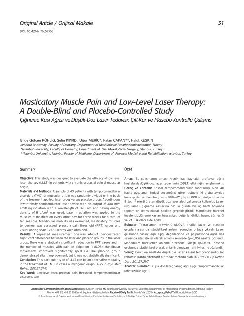
<strong>Masticatory</strong> <strong>Muscle</strong> <strong>Pain</strong> <strong>and</strong> <strong>Low</strong>-<strong>Level</strong> <strong>Laser</strong> <strong>Therapy</strong>:<br />
A Double-Blind <strong>and</strong> Placebo-Controlled Study<br />
Çi¤neme Kas› A¤r›s› ve Düflük-Doz Lazer Tedavisi: Çift-Kör ve Plasebo Kontrollü Çal›flma<br />
Bilge Gökçen RÖHL‹G, Selin KIPIRDI, U¤ur MER‹Ç*, Nalan ÇAPAN**, Haluk KESK‹N<br />
Istanbul University, Faculty of Dentistry, Department of Maxillofacial Prosthodentics Istanbul, Turkey<br />
*Istanbul University, Faculty of Dentistry, Department of Oral Maxillofacial Surgery, Istanbul, Turkey<br />
**Istanbul University, Istanbul Faculty of Medicine, Department of Physical Medicine <strong>and</strong> Rehabilitation, Istanbul, Turkey<br />
Summary<br />
Objective: This study was designed to evaluate the efficacy of low-level<br />
laser therapy (LLLT) in patients with chronic orofacial pain of muscular<br />
origin.<br />
Materials <strong>and</strong> Methods: A sample of 40 patients with temporom<strong>and</strong>ibular<br />
disorders (TMD) of muscular origin was r<strong>and</strong>omly divided on the basis<br />
of the treatment applied: laser group versus placebo group. A continuous<br />
low-intensity semiconductor laser device with an output of 300 mW,<br />
emitting radiation with a wavelength of 820 nm <strong>and</strong> having energy<br />
density of 8 J/cm 2 was used. <strong>Laser</strong> irradiation was applied to the<br />
muscles of mastication every other day for three weeks for a total of<br />
ten sessions. M<strong>and</strong>ibular mobility was examined, masticatory muscles<br />
tenderness was assessed, pressure pain threshold (PPT) values <strong>and</strong><br />
visual analog scale (VAS) scores were obtained.<br />
Results: A repeated measurement one-way ANOVA demonstrated<br />
significant differences between the laser <strong>and</strong> placebo groups. In the laser<br />
group, there was a statically significant reduction in PPT values <strong>and</strong> in<br />
the number of muscles with pain on palpation (p
32<br />
Röhlig et al.<br />
<strong>Low</strong>-<strong>Level</strong> <strong>Laser</strong> <strong>and</strong> <strong>Muscle</strong> <strong>Pain</strong><br />
Introduction<br />
The American Academy of Orofacial <strong>Pain</strong> describes<br />
temporom<strong>and</strong>ibular disorders (TMD) as a collective term<br />
embracing a number of clinical problems related to masticatory<br />
muscles <strong>and</strong> temporom<strong>and</strong>ibular joint (TMJ) associated structures,<br />
or both of them (1). The treatment should be based on possible<br />
underlying etiological factors, signs <strong>and</strong> symptoms of the disorder.<br />
Although TMD has been a subject of study for a long time, many<br />
controversies still remain regarding its etiology <strong>and</strong> treatment. Early<br />
etiological concepts revolved mostly around theories of occlusal<br />
disharmonies, <strong>and</strong> the treatment was based on the model of ideal<br />
biomechanics of TMJ (2). In recent years due to the results of<br />
clinical researches, theories on the etiology of TMD have switched<br />
from the field of occlusion to field of neurophysiologic science (3-7);<br />
this has led to changes in therapeutic approaches. Complex,<br />
conservative treatments rather than aggressive <strong>and</strong> irreversible<br />
ones such as complex occlusal therapies <strong>and</strong> surgeries are preferred<br />
to relieve symptoms, diminish pain <strong>and</strong> reestablish function (2,8).<br />
<strong>Low</strong>-level lasers (LLL) have been used in the treatment of<br />
temporom<strong>and</strong>ibular disorders to alleviate pain <strong>and</strong> in the<br />
reestablishment of normal m<strong>and</strong>ibular function (9,10). Theories<br />
about the mechanism of action have been proposed (11). LLL have<br />
been found to have analgesic, myorelaxant, tissue healing <strong>and</strong><br />
biostimulatory effects (9,12,13). Several clinical studies have<br />
suggested LLL for TMD of articular origin <strong>and</strong> for myofascial pain<br />
syndrome (11,13-16). The interest in this therapy is probably due to its<br />
ease of application, shorter treatment duration, less contraindications<br />
<strong>and</strong> low cost (17-19). However, some other studies such as those done<br />
by Emshoff et al. (20), Venancio et al. (21), Conti (22), Hansen <strong>and</strong><br />
Thoroe (23) revealed no beneficial effect of LLL.<br />
To date, the results regarding the efficacy of LLL on muscle<br />
pain are inconclusive. The protocols used in clinical studies vary<br />
in power intensity, exposure times <strong>and</strong> location of laser application.<br />
The most common wavelength in therapeutic use is probably<br />
810-830 nm (22,24-26). However, different wavelengths: 632.8 nm<br />
neon-hellium laser (20), 660 nm (9), 780 nm (11,21), 904 nm (15),<br />
830 to 904 nm soft laser (10) were also used <strong>and</strong> reported to be<br />
effective in managing TMD. There is insufficient evidence on the<br />
effective dose of low-intensity laser employed in the treatment of<br />
TMD of muscular origin. Therefore, the aim of this study was to<br />
investigate the efficacy of LLL with 820 nm, 3 J/cm2, 300 mW<br />
output power in the treatment of TMD of muscular origin. We<br />
expected that this type of LLL reduce muscle pain <strong>and</strong> improve<br />
m<strong>and</strong>ibular movements better than placebo.<br />
Materials <strong>and</strong> Methods<br />
Subjects<br />
This study was performed at Istanbul University, Faculty of<br />
Dentistry Department of Maxillofacial Prosthodontics. The study<br />
population was selected consecutively among the patients requesting<br />
orofacial pain treatment over a period of eight months. Between<br />
Turk J Phys Med Rehab 2011;57:31-7<br />
Türk Fiz T›p Rehab Derg 2011;57:31-7<br />
March 2009 <strong>and</strong> December 2009, 482 TMD patients were<br />
examined. Of the total 482 TMD patients, 165 fulfilled the inclusion<br />
criteria, 84 accepted to participate in the study, but only 40 of them<br />
(24 female, 16 male, with a mean age of 43.7±1.8 years) who were<br />
gender <strong>and</strong> age-matched were included in the study. The patients<br />
were r<strong>and</strong>omized to laser <strong>and</strong> placebo groups with the help of a<br />
computer program. In the laser group (n=20, 12 female <strong>and</strong> 8 male),<br />
the patients’ age ranged from 22 to 59 (mean age 42.2±3.4) years<br />
<strong>and</strong> in the placebo group (n=20, 12 female-8 male) from 28 to 56<br />
(mean age 42.8±2.1) years. The Ethics Committee of Istanbul<br />
University, Istanbul Faculty of Medicine approved the protocol.<br />
Written informed consent was obtained from each subject after a<br />
full explanation of the study.<br />
The first examination was assigned to evaluate whether the<br />
subjects fulfilled the inclusion criteriae. The inclusion criteria were: 1)<br />
presence of signs <strong>and</strong> symptoms of TMD of myogenic origin according<br />
to the Research Diagnostic Criteriae for Temporom<strong>and</strong>ibular<br />
Disorder (RDC/TMD) (27); 2) orofacial pain lasting for more than 6<br />
months; 3) age between 18 <strong>and</strong> 60 years. Exclusion criteriae were: 1)<br />
disk displacements (disk displacement with reduction, disk<br />
displacement without reduction, with limited opening <strong>and</strong> disk<br />
displacement without reduction, without limited opening) <strong>and</strong><br />
arthralgia, arthritis, arthrosis; 2) general inflammatory connective<br />
tissue diseases (e.g. rheumatoid arthritis); 3) psychiatric disorders;<br />
4) tumors; 5) heart diseases, pacemakers; 7) pregnancy; 8)<br />
symptoms which could be referred to other disorders of the<br />
orofacial region (such as toothache, neuralgia, migraine); 9) any<br />
medication use or treatment for TMD within the last six months;<br />
10) very high baseline pain intensity; 11) local skin infections over the<br />
masseter muscle, temporalis <strong>and</strong>/or sternocleidomastoid muscle.<br />
The patients were asked not to take any analgesics or to have<br />
any pain treatments, starting one week before <strong>and</strong> during the study.<br />
We expressly pointed out that the patients should avoid using<br />
steroids during the treatment, as steroids block the effect of LLL<br />
treatment (28).<br />
Study Design<br />
The study was designed as a r<strong>and</strong>omized, placebo-controlled<br />
double-blind clinical trial. The scientific recommendations of the<br />
World Association for <strong>Laser</strong> <strong>Therapy</strong> (WALT) for conducting<br />
r<strong>and</strong>omized trials were applied to this research (www.walt.nu). The<br />
patients did not know whether they were assigned to laser or<br />
placebo group. The equipment used for the placebo group was the<br />
same as in laser group. However, in the placebo group, the laser<br />
device was only switched-on, not programmed. Assessment of the<br />
participants was conducted by an independent investigator who was<br />
unaware of the study.<br />
<strong>Laser</strong> Exposure<br />
A continuous low-intensity semiconductor (Doris Diode <strong>Laser</strong>,<br />
CTL 1106 MX, Warsaw, Pol<strong>and</strong>) was used for laser irradiation. This<br />
device generates continuous radiation with regulated power. The<br />
single-probe laser device applies a laser diode generating infrared<br />
radiation of 820 nm wavelength. The beam diameter of the device is<br />
6 mm <strong>and</strong> the probe has an angle of 45°. The energy intensity given
Turk J Phys Med Rehab 2011;57:31-7<br />
Türk Fiz T›p Rehab Derg 2011;57:31-7<br />
to each muscle point was adjusted to be 8 J/cm 2 by applying 300<br />
mW output power for 10 seconds. LLL treatment was applied<br />
precisely <strong>and</strong> continuously into five points: three points of the<br />
masseter muscle (superior point, middle point, inferior point), one<br />
point of the temporalis (anterior point) <strong>and</strong> one point of the<br />
sternocleidomastoid muscle (superior SCM). <strong>Laser</strong> was in direct contact<br />
with the skin surface. The patients were exposed to active laser<br />
<strong>and</strong> sham application every other day for three weeks, for a total of<br />
ten sessions. The output of the laser device was tested before <strong>and</strong><br />
after the study.<br />
Procedure<br />
The patients were evaluated two times: half an hour before the<br />
first session <strong>and</strong> half an hour after the last session of the assigned<br />
therapy. Each patient was assessed according to the following<br />
parameters: 1) functional examination; 2) pressure pain threshold<br />
(PPT) measurement, 3) visual analog scale (VAS). All examinations<br />
were performed by the same clinician who was an experienced<br />
prosthodontist trained in the treatment of craniom<strong>and</strong>ibular disorders<br />
<strong>and</strong> was calibrated in using Research Diagnostic Criteria for<br />
Temporom<strong>and</strong>ibular Disorder (RDC/TMD) as the gold st<strong>and</strong>ard. The<br />
clinician was unaware of the study.<br />
11)) FFuunnccttiioonnaall EExxaammiinnaattiioonn: The functional examination was<br />
based on the RDC/TMD (27). The suggested translation of the<br />
RDC/TMD by the International RDC-TMD Consortium was used in<br />
this study. The masticatory muscle tenderness were assessed on<br />
both sides by bilateral palpation. The mobility of the m<strong>and</strong>ible<br />
was measured with a plastic millimeter ruler on opening, lateral<br />
excursions <strong>and</strong> protrusion. The alterations in the opening pathway<br />
were also evaluated. The patients were asked to report any pain<br />
during muscle palpations <strong>and</strong> m<strong>and</strong>ibular movements <strong>and</strong> the<br />
answers were recorded according to the verbal scale. The degree of<br />
pain under palpation was rated as 0-no pain; 1-mild pain; 2-moderate<br />
pain; 3-severe pain.<br />
22)) PPrreessssuurree PPaaiinn TThhrreesshhoolldd ((PPPPTT)) EExxaammiinnaattiioonn:: A dial algometer<br />
(Wagner <strong>Pain</strong> Test Model FPK Algometer, Wagner Instruments,<br />
Post Office Box 1217, Greenwich, CT 06836-1217) was used to<br />
measure the PPT on the masticator muscles. The compressions<br />
were performed via 1 cm 2 rubber tip. The PPT were obtained with<br />
the aid of an algometer by applying pressure to three points in the<br />
Table 1. Demographic features of the study population.<br />
Röhlig et al.<br />
<strong>Low</strong>-<strong>Level</strong> <strong>Laser</strong> <strong>and</strong> Luscle <strong>Pain</strong><br />
33<br />
masseter (superior point, middle point, inferior point), three points in<br />
the temporalis (anterior point, middle point, posterior point) <strong>and</strong> one<br />
in the sternocleidomastoid muscle (superior SCM) before <strong>and</strong><br />
after the treatment. The examiner was calibrated in pressure<br />
measurements with algometer before the study. The measurements<br />
were performed on both sides. Before the procedure was begun, a<br />
few test measurements were performed on the patients’ lower<br />
arms. The patients were instructed to state immediately when the<br />
pressure feeling turned into pain feeling <strong>and</strong> the pressure was then<br />
stopped. After a rest of 30 seconds, the next measurement was<br />
performed. PPT was recorded as kg/cm 2 .<br />
33)) VViissuuaall AAnnaalloogg SSccaallee ((VVAASS)):: In this study, the subjective report<br />
of pain was evaluated using VAS before the treatment was begun, at<br />
the end of the first week, at the end of the second week <strong>and</strong> after<br />
the assigned therapy. <strong>Pain</strong> intensity was recorded in mm on a 100<br />
mm VAS.<br />
Statistical Analysis<br />
Under the assumption of a difference of one st<strong>and</strong>ard deviation<br />
with respect to the primary end point between the groups, an alpha<br />
level of 5 % <strong>and</strong> power goal of 80%, twenty patients per group were<br />
necessary. For all statistical tests, NCSS 2007 <strong>and</strong> PASS 2008<br />
Statistical & Power Analysis Software (NCSS, Kaysville, Utah, USA)<br />
were used. All variables were analyzed descriptively. Descriptive<br />
statistical methods (mean, st<strong>and</strong>ard deviation <strong>and</strong> frequency) were<br />
applied. For the comparison of quantitative data, student t-tests<br />
were performed for between-group comparisons of parameters<br />
showing a normal distribution. The Mann-Whitney U test was<br />
performed for between-group comparisons of parameters showing<br />
a non-normal distribution. For the comparative evaluation of<br />
parameters showing normal distributions within the group, the<br />
paired-samples t-test were used. The results were analyzed at a<br />
95% confidence interval, <strong>and</strong> the significance level was set at p
34<br />
Results<br />
Röhlig et al.<br />
<strong>Low</strong>-<strong>Level</strong> <strong>Laser</strong> <strong>and</strong> <strong>Muscle</strong> <strong>Pain</strong><br />
The current study included forty patients with TMD of muscular<br />
origin (24 female, 16 male, with a mean age of 43.7±1.8 years). The<br />
demographic features of the study participants <strong>and</strong> groups are<br />
shown in Table 1. We did not identify any statistically significant<br />
differences between the laser <strong>and</strong> control groups with regard to age,<br />
gender, education or marital status. M<strong>and</strong>ibular movements <strong>and</strong><br />
associated pain, tenderness upon palpation in masticatory muscles<br />
<strong>and</strong> PPTs were assessed in both groups. There was a statically<br />
significant increase in PPT of the examined muscles in the laser<br />
group, but no statistical significance in the placebo group (Table 2).<br />
The masseter muscle demonstrated the most severe muscle pain in<br />
both groups. As shown in Table 3, in the laser group, there was a<br />
significant decrease in pain at palpation after laser exposure <strong>and</strong> the<br />
number of muscles without any pain on palpation increased<br />
significantly. However, in the placebo group, pain severity decreased<br />
slightly, but it was not statistically significant (Table 3). The laser<br />
group demonstrated statistically significant improvements in<br />
vertical movements, lateral excursions <strong>and</strong> protrusions (Table 4).<br />
The mean difference, st<strong>and</strong>ard deviation <strong>and</strong> p values of VAS<br />
before <strong>and</strong> after the treatment in both groups are given in Table 5.<br />
Table 2. Comparison of PPT (kg/cm 2) values. Prasure pain throrhold.<br />
Turk J Phys Med Rehab 2011;57:31-7<br />
Türk Fiz T›p Rehab Derg 2011;57:31-7<br />
A statistically significant difference in VAS was not identified<br />
between the two groups before treatment (p>0.05), but it was<br />
observed that there was a significant drop in VAS after LLL<br />
treatment (Table 5).<br />
None of the patients reported adverse effects related to laser<br />
application during or after the treatment period.<br />
Discussion<br />
The present study was designed to investigate the clinical effect<br />
of a specific type of LLL treatment (820 nm, 3 J/cm 2 , 300 mW<br />
output power) involving direct application on the painful masticator<br />
muscles. Patients who received laser application did benefit from<br />
the therapy. Significant improvements were obtained in the tested<br />
parameters in the active laser group contrary to the placebo group.<br />
Attempt was made to identify the apparent effectiveness of LLL<br />
in TMJ disorders, but there is insufficient evidence either for or<br />
against the use of LLL treatment. LLL treatment has often been<br />
applied in different types of pain conditions. Simunovic et al. (29)<br />
reported that patients treated with LLL recovered more rapidly <strong>and</strong><br />
demonstrated more pain relief compared to untreated patients. The<br />
mechanisms underlying significant analgesia on muscles after laser<br />
exposure is not exactly known, but rely upon the chances in cell level<br />
<strong>Laser</strong> Group Plasebo Group<br />
Mean±SD (kg/cm2) Mean±SD (kg/cm2) TA Before treatment 18.47±4.08 18.52±4.73<br />
After treatment 30.55±6.68 18.87±4.17<br />
++p 0.027532* 0.27757 **<br />
TM Before treatment 19.70±3.07 14.45±3.29<br />
After treatment 34.02±4.34 15.17±4.93<br />
++p 0.02022 * 0.52936 **<br />
TP Before treatment 14.50±5.72 15.80±5.23<br />
After treatment 32.87±4.48 19.82±4.97<br />
++p 0.00274 * 0.26742 **<br />
MS Before treatment 21.37±6.72 21.87±7.34<br />
After treatment 34.87±5.21 28.60±6.11<br />
++p 0.00956 * 0.55225 **<br />
MM Before treatment 23.30±3.31 22.15±5.19<br />
After treatment 33.62±5.31 23.57±3.22<br />
++p 0.00303 * 0.94166 **<br />
MI Before treatment 21.80±5.53 26,65±6.32<br />
After treatment 32.55±5.64 28.25±5.13<br />
++p 0.00062 * 0.70732 **<br />
SCM Before treatment 15.80±5,93 26.65±6.32<br />
After treatment 22.55±5.64 24.25±5.13<br />
Student t test was used.<br />
++p 0.00061 * 0.95113 **<br />
(SD: St<strong>and</strong>ard deviation, 95 % confidence interval, * p
Turk J Phys Med Rehab 2011;57:31-7<br />
Türk Fiz T›p Rehab Derg 2011;57:31-7<br />
with increased adenosine triphosphate (ATP) production by<br />
mitochondria (30), increase in the electrical potential of the<br />
mitochondria membrane (31) <strong>and</strong> increased serotonin <strong>and</strong><br />
endorphins (32,33). Furthermore, local blood circulation is reported<br />
to be increased, too (34,35). Successful treatment results were<br />
reported for LLL treatment in TMD of arthrogenic origin (36,37).<br />
However, there are contradictory reports that LLL treatment is<br />
ineffective in the treatment of TMJ capsulitis/synovitis <strong>and</strong> painful<br />
Röhlig et al.<br />
<strong>Low</strong>-<strong>Level</strong> <strong>Laser</strong> <strong>and</strong> <strong>Muscle</strong> <strong>Pain</strong><br />
35<br />
disk displacement with reduction (21). The same controversy is valid<br />
for the effect of LLL treatment in muscle pain, too. Many clinical<br />
studies reported favoring results of LLL in the treatment of muscle<br />
pain (9,10,15,25,38). The promising results of this clinical study<br />
support the findings published in the literature that LLL treatment<br />
is better than placebo in myogenic cases. The null hypothesis that<br />
the muscle tenderness does not increase, m<strong>and</strong>ibular mobility <strong>and</strong><br />
pain intensity will not change when patients are exposed to LLL is<br />
Table 3. The results of muscle palpations. It demonstrates the percentage of the pain intensity of the examined muscles before <strong>and</strong> after real laser<br />
exposure <strong>and</strong> placebo laser application.<br />
<strong>Muscle</strong> Treatment <strong>Pain</strong><br />
<strong>Laser</strong> group<br />
n (%)<br />
Plasebo group<br />
n (%)<br />
Temporalis anterior Before No pain 8 (40) 9 (45)<br />
Mild 5 (25) 5 (25)<br />
Moderate 4 (20) 4 (20)<br />
Severe 3 (15 ) 2 (10)<br />
After No pain 13 (65) 10 (50)<br />
Mild 7 (35) 10 (50)<br />
Temporalis middle Before No pain 12 (60) 12 (60)<br />
Mild 5 (25) 5 (25)<br />
Moderate 2 (10) 2 (10)<br />
Severe 2 (10) 1 (5)<br />
After No pain 18 (90) 12 (60)<br />
Mild 2 (10) 8 (40)<br />
Temporalis posterior Before No pain 15 (75) 15 (75)<br />
Mild 1 (5) 1 (5)<br />
Moderate 3 (15) 30 (15)<br />
Severe 1 (5) 10 (5)<br />
After No pain 19 (95) 16 (80)<br />
Mild 1 (5) 4 (20 )<br />
Masseter (superior) Before No pain 1 (5) 1 (5)<br />
Mild 7 (35) 7 (35)<br />
Moderate 6 (30) 6 (30)<br />
Severe 7 (35) 6 (30)<br />
After No pain 13 (65) 2 (10)<br />
Mild 5 (25) 8 (40)<br />
Moderate 2 (10) 10 (50)<br />
Masseter (middle) Before No pain 1 (5) 2 (10)<br />
Mild 9 (45) 9 (45)<br />
Moderate 2 (10) 4 (20)<br />
Severe 8 (40) 5 (25)<br />
After No pain 16 (80) 4 (20)<br />
Mild 4 (20) 16 (80)<br />
Masseter (inferior) Before No pain 1 (5) 7 (35)<br />
Mild 7 (35) 7 (35)<br />
Moderate 7 (35) 5 (25)<br />
Severe 5 (25) 1 (5)<br />
After No pain 10 (50) 10 (50)<br />
Mild 10 (50) 10 (50)
36<br />
Röhlig et al.<br />
<strong>Low</strong>-<strong>Level</strong> <strong>Laser</strong> <strong>and</strong> <strong>Muscle</strong> <strong>Pain</strong><br />
rejected. In this study, objective <strong>and</strong> subjective parameters were<br />
used to evaluate the effect of LLL in patients with myofascial pain.<br />
The PPTs increased significantly <strong>and</strong> other clinical measures showed<br />
improvement <strong>and</strong>, muscle pain on palpation decreased significantly<br />
after the treatment. VAS evaluation revealed similar results.<br />
There are many reasons explaining why the effect of LLL varies<br />
in the treatment of TMD of arthrogenic <strong>and</strong> myogenic origin.<br />
Methodological differences in patient selection <strong>and</strong> LLL treatment<br />
parameters (wavelength, power output, energy intensity <strong>and</strong><br />
exposure duration) may affect the results. The effect of a therapy<br />
can only be identified through a well-designed clinical study.<br />
Beckerman et al. (38) conducted a criteria-based meta-analysis to<br />
assess the efficacy of laser therapy. For this purpose, the authors<br />
included only the r<strong>and</strong>omized clinical trials (RCTs) in the analysis, as<br />
RCTs are the best study designs for obtaining a reliable evaluation of<br />
the treatment effects <strong>and</strong> conclusions can only be drawn from the<br />
best methodological studies. In this meta-analysis, it was concluded<br />
that the studies with positive outcome had generally a higher<br />
methodological quality. Another important point in clinical evaluation<br />
of a therapy effect in chronic pain is the blinding of the researcher.<br />
The blinding of the researcher about the type of the therapy as well as<br />
the unawareness of the patients regarding the type of treatment they<br />
receive (placebo <strong>and</strong>/or active) avoid the interference in the results.<br />
The present study aimed to investigate the effect of LLL treatment<br />
in TMD of myogenic origin. The study was designed as a r<strong>and</strong>omized,<br />
double-blind, placebo-controlled study; neither the patients nor the<br />
observer was aware of the assigned therapy (active or placebo).<br />
The chief complaint of the patients in the present study was<br />
pain. <strong>Pain</strong> levels of the subjects at baseline <strong>and</strong> throughout the study<br />
were evaluated with VAS in order to determine the change over<br />
time. In clinical trials designed to investigate the efficacy of pain<br />
treatments, the primary analysis involves reduction in pain<br />
Turk J Phys Med Rehab 2011;57:31-7<br />
Türk Fiz T›p Rehab Derg 2011;57:31-7<br />
intensity (39). For chronic pain, most measures of treatment<br />
response involve patient-reported outcomes for which the<br />
patient is the most important judge of whether changes are<br />
important or not (40-42). Evaluation of pain is difficult,<br />
because it is not controlled in the clinical research <strong>and</strong> it is a<br />
complex procedure requiring the assessment of biological,<br />
structural, functional as well as emotional pain experience.<br />
Recent chronic pain studies recommend evaluation of pain<br />
through subjective report <strong>and</strong> these should be the primary<br />
measures. In the clinical recordings, objective evaluation of pain<br />
in the masticatory muscles should be performed with a<br />
quantitative method, such as PPT which enhances the clinical<br />
assessment of pain, st<strong>and</strong>ardizes the outcomes <strong>and</strong> makes<br />
comparisons possible. In the present study, the objective<br />
functional measures based on the RDC/TMD <strong>and</strong> PPT values were<br />
obtained with the aid of an algometer in both groups <strong>and</strong> the<br />
patient-reported pain intensity was evaluated with VAS at<br />
different time points. PPT values supported the VAS results <strong>and</strong><br />
demonstrated that LLL is effective in pain release; it does not act<br />
like placebo treatment.<br />
There is a large discrepancy of the wavelength used in trials<br />
investigating the efficacy for muscle disorders. Different<br />
wavelengths: 632.8 nm neon-hellium laser (20), 660 nm (9), 780<br />
nm (11,21), 810 nm (43), 904 nm (15), 830 to 904 nm soft laser (10)<br />
were used <strong>and</strong> reported to be effective in managing TMD. In the<br />
present study, we used a dose (820 nm) that was within the<br />
interval recommended for deep tissues. It is well known that<br />
energy density is also important in the therapeutic effect of LLLs.<br />
In some of the clinical studies that reported negative results,<br />
inadequate power or energy densities were used (20-22).<br />
According to Tuner <strong>and</strong> Hode (17), 6-10 J is suitable for myogenic<br />
conditions. Therefore, in the present study, 8 J/cm 2 was applied<br />
per point over muscle.<br />
Table 4. M<strong>and</strong>ibular movements. Each line demonstrates the statistical analysis of a m<strong>and</strong>ibular movement after LLL treatmant <strong>and</strong> placebo application.<br />
<strong>Laser</strong> Group Plasebo Group<br />
Mean Difference±SD (mm) p Mean Difference±SD (mm) p<br />
OP 18.3±4.60 0.00317* 3.28±0.75 0.34084**<br />
MUO 19.9±2.94 0.00865* 4.32±078 0.26096**<br />
MAO 10.4±5.09 0.05259* 5.22±0.74 0.26551**<br />
LLE 5.9±1.20 0.00071* 0.89±0.19 0.05401**<br />
RLE 6.05±1.50 0.02265* 0.83±0.86 0.08266**<br />
P 5.35±1.38 0.00036* 1.23±0.97 0.53079**<br />
Student t test was used.<br />
(SD: St<strong>and</strong>ard deviation, 95 % confidence interval, * p
Turk J Phys Med Rehab 2011;57:31-7<br />
Türk Fiz T›p Rehab Derg 2011;57:31-7<br />
There is only limited scientific evidence for the effectiveness of<br />
LLL in the treatment of TMD. The present study examined the effect<br />
of specific dose of LLL on pain release <strong>and</strong> m<strong>and</strong>ibular<br />
function <strong>and</strong> found out that active laser treatment is superior to<br />
sham application. However, the present study investigated the<br />
short-term effects. Additional research should be conducted in order<br />
to investigate the long-term effects of LLL in the treatment of TMD.<br />
This study was supported by Research Fund of Istanbul<br />
University (Project Number: UDP-4090/16072009) <strong>and</strong> presented<br />
(as oral presentation) in 33rd Annual Congress of European<br />
Prosthodontic Association 2009, Innsbruck.<br />
References<br />
1. Okeson JP. Orofacial pain: Guidelines for Assessment, Diagnosis<br />
<strong>and</strong> Management. 1.ed. Chigaco: Quintessence Publishing C; 1996.<br />
2. McNeill C. History <strong>and</strong> evolution of TMD concepts. Oral Surg Oral<br />
Med Oral Pathol Oral Radiol Endod 1997;83:51-60. [Abstract] / [PDF]<br />
3. Haskin CL, Milam SB, Cameron IL. Pathogenesis of degenerative<br />
joint disease in human temporom<strong>and</strong>ibular joint. Crit Rev Oral Biol<br />
Med 1995;6:248-77. [Abstract] / [PDF]<br />
4. Israel HA, Diamond BE, Saed-Nejad F, Ratcliffe A. Correlation<br />
between arthoscopic diagnosis of osteoarthritis <strong>and</strong> synovitis of<br />
the human temporom<strong>and</strong>ibular joint <strong>and</strong> keratan sulfate levels in<br />
the synovial fluid. J Oral Maxillofac Surg 1997;55:210-7. [Abstract] /<br />
[PDF]<br />
5. Mense S. Nociception from skeletal muscle in relation to clinical<br />
muscle pain. <strong>Pain</strong> 1993;54:241-89. [Abstract] / [PDF]<br />
6. Stohler CS. <strong>Muscle</strong>-related temporom<strong>and</strong>ibular disorders. J Orofac<br />
<strong>Pain</strong> 1999;13:273-84. [Abstract]<br />
7. Sessle BJ. The neural basis of temporom<strong>and</strong>ibular joint <strong>and</strong><br />
masticatory muscle pain. J Orofac <strong>Pain</strong> 1999;13:238-45. [Abstract]<br />
8. Carlsson GE, Magnusson T. Management of Temporom<strong>and</strong>ibular<br />
Disorders in the General Dental Practice. Chicago: Quintessence<br />
Publishing Co; 1999.<br />
9. Shirani AM, Gutknecht N, Taghizadeh M, Mir M. <strong>Low</strong>-level laser<br />
therapy <strong>and</strong> myofascial pain dysfunction syndrome: a r<strong>and</strong>omized<br />
controlled clinical trial. <strong>Laser</strong>s Med Sci 2009;24:715-20. [Abstract]<br />
/ [Full Text] / [PDF]<br />
10. Kato MT, Kogawa EM, Santos CN, Conti PC. Tens <strong>and</strong> low-level laser<br />
therapy in the management of temporom<strong>and</strong>ibular disorders. J<br />
Appl Oral Sci 2006;14:130-5. [Abstract]<br />
11. Hakgüder A, Birtane M, Gürcan S, Kokino S, Turan FN. Efficacy of<br />
low level laser therapy in myofascial pain syndrome: an algometric<br />
<strong>and</strong> thermographic evaluation. <strong>Laser</strong>s Surg Med 2003;33:339-43.<br />
[Abstract] / [PDF]<br />
12. Fikácková H, Dostalová T, Vosivka R, Peterova V, Navratil L, Lesak J.<br />
Arthralgia of the temporom<strong>and</strong>ibular joint <strong>and</strong> low-level laser<br />
therapy. Photomed <strong>Laser</strong> Surg 2006;24:522-7. [Abstract] / [PDF]<br />
13. Çetiner S, Kahraman SA, Yücetas S. Evaluation of low-level laser<br />
therapy in the treatment of temporom<strong>and</strong>ibular disorders.<br />
Photomed <strong>Laser</strong> Surg 2006;24:637-41. [Abstract] / [PDF]<br />
14. Fikácková H, Dostalová T, Navratil L, Klaschka J. Effectiveness of<br />
low-level laser therapy in temporom<strong>and</strong>ibular joint disorders: a<br />
placebo-controlled study. Photomed <strong>Laser</strong> Surg 2007;25:297-303.<br />
[Abstract] / [PDF]<br />
15. Külekçio¤lu S, Sivrio¤lu K, Özcan O, Parlak M. Effectiveness of<br />
low-level laser therapy in temporom<strong>and</strong>ibular disorder. Sc<strong>and</strong> J<br />
Rheumatol 2003;32:114-8. [Abstract]<br />
16. Mazzetto MO, Carrasco TG, Bidinelo EF, de Andrade Pizzo RC,<br />
Mazzetto RG. <strong>Low</strong> intensity laser application in temporom<strong>and</strong>ibular<br />
disorders: a phase I double-blind study. J Craniom<strong>and</strong>ib Pract<br />
2007;25:186-92. [Abstract]<br />
17. Tunér J, Hode L. <strong>Laser</strong> therapy. Clinical Practice <strong>and</strong> Scientific<br />
Background. Grangesberg: Prima Books; 2002.<br />
18. Kobayashi M, Kubota J. Treatment of TMJ pain with diode laser<br />
therapy. <strong>Laser</strong> Ther 1999;11:11-8. [Abstract]<br />
19. Bradley P, Groth E, Gursoy B. The maxillofacial region: recent<br />
research <strong>and</strong> clinical practice in low-intensity level laser therapy.<br />
In: Simunovic Z, editor. <strong>Laser</strong>s in Medicine <strong>and</strong> Dentistry. DTP<br />
Studio; 2000. p. 385-400.<br />
20. Emshoff R, Bösch R, Pümpel E, Schöning H, Strobl H. <strong>Low</strong>-level laser<br />
therapy for treatment of temporom<strong>and</strong>ibular joint pain: a double<br />
blind <strong>and</strong> placebo-controlled trial. Oral Surg Oral Med Oral Pathol<br />
Oral Radil Endod 2008;105:452-6. [Abstract] / [Full Text] / [PDF]<br />
Röhlig et al.<br />
<strong>Low</strong>-<strong>Level</strong> <strong>Laser</strong> <strong>and</strong> <strong>Muscle</strong> <strong>Pain</strong><br />
37<br />
21. Venancio Rde A, Camparis CM, Lizarelli Rde F. <strong>Low</strong> intensity laser<br />
therapy in the treatment of temporom<strong>and</strong>ibular disorders: a<br />
double-blind study. J Oral Rehabil 2005;32:800-7. [Abstract] / [PDF]<br />
22. Conti PC. <strong>Low</strong> level laser therapy in the treatment of<br />
temporom<strong>and</strong>ibular disorders (TMD): a double-blind pilot study. J<br />
Craniom<strong>and</strong>ib Pract 1997;15:144-9. [Abstract]<br />
23. Hansen HJ, Thoroe U. <strong>Low</strong> power laser biostimulation of chronic<br />
oro-facial pain. A double-blind placebo controlled cross-over study<br />
in 40 patients. <strong>Pain</strong> 1990;43:169-79. [Abstract] / [PDF]<br />
24. Pinheiro Al, Cavalcanti ET, Pinheiro TI, Alves MJ, Mir<strong>and</strong>a ER,<br />
DeQuevedo AS, et al. <strong>Low</strong>-level laser therapy is an important tool to<br />
treat disorders of the maxillofacial region. J Clin <strong>Laser</strong> Med Surg<br />
1998;16:223-6. [Abstract]<br />
25. Simunovic Z. <strong>Low</strong> level laser therapy with trigger points technique:<br />
a clinical study on 243 patients. J Clin <strong>Laser</strong> Med Surg 1996;14:163-7.<br />
[Abstract] / [PDF]<br />
26. Özdemir F, Birtane M, Kokino S. The clinical efficacy of low-level<br />
laser therapy on pain <strong>and</strong> function in cervical osteoarthritis. Clin<br />
Rheumatol 2001;20:181-4. [Abstract] / [PDF]<br />
27. Dworkin SF, LeResche L. Research diagnostic criteria for temporom<strong>and</strong>ibular<br />
disorders: review, criteria, examinations <strong>and</strong> specifications,<br />
critique. J Craniom<strong>and</strong>ib Disord Fac Oral <strong>Pain</strong> 1992;6:301-55. [Abstract]<br />
28. Lopes-Martins RA, Albertini R, Lopes-Martins PS, De Carvalho FA, Neto<br />
HC, Iversen VV, et al. Steroid Receptor Antagonist Mifepristone inhibits<br />
the anti-inflammatory effects of photoradiation. Photomed <strong>Laser</strong> Surg<br />
2006;24:197-201. [Abstract] / [PDF]<br />
29. Simunovic Z, Trobonjaca T, Trobonjaca Z. Treatment of medial <strong>and</strong> lateral<br />
epicondylitis-tennis <strong>and</strong> golfer’s elbow-with low level laser therapy: a multicenter<br />
double blind, placebo-controlled clinical study on 324 patients. J<br />
Clin <strong>Laser</strong> Med Surg 1998;16:145-51. [Abstract]<br />
30. Passarella S. He-Ne laser irradiation of isolated mitochondria. J<br />
Photochem Photobiol 1989;3:642-3. [Abstract]<br />
31. Yu W, Naim JO, Lanzafame RJ. Effects of photostimulation on<br />
wound healing in diabetic mice. <strong>Laser</strong>s Surg Med 1997;20:56-63.<br />
[Abstract] / [PDF]<br />
32. Walker JB. Relief from chronic pain by low-power laser irradiation.<br />
Neuroscience 1983;43:339-44. [Abstract]<br />
33. Yamamoto H, Ozaki A, Iguchi N, Kinochita S. Antinociceptive effects<br />
of laser irradiation of Hoku points in rats. <strong>Pain</strong> Clin 1988;8:43-8.<br />
34. Kemmotsu O, Sato K, Furumido H, Harada K, Takigawa C, Kaseno S, et al.<br />
Efficacy of low reactive-level laser therapy for pain attenuation of postherpetic<br />
neuralgia. <strong>Laser</strong> Ther 1991;3:71-5. [PDF]<br />
35. Schindl A, Schindl M, Schon H, Knobler R, Havelec LO, Schindl L.<br />
<strong>Low</strong> intensity laser improves skin circulation in patients with<br />
diabetic microangiopathy. Diabetes Care 1998;21:390-4. [Abstract] /<br />
[PDF]<br />
36. Carrasco TG, Mazzetto MO, Mazzetto RG, Mestriner W Jr. <strong>Low</strong><br />
intensity laser therapy in temporom<strong>and</strong>ibular disorder: a phase II<br />
double-blind study. Cranio 2008;26:274-81. [Abstract]<br />
37. Bertolucci LE, Grey T. Clinical analysis of mid-laser versus placebo<br />
treatment of arthralgic TMJ degenerative joints. J Craniom<strong>and</strong><br />
Pract 1995;13:26-9. [Abstract]<br />
38. Beckerman H, Bie RA, Bouter LM, De Cuyper HJ, Oostendorp RA.<br />
The efficacy of laser therapy for musculoskeletal <strong>and</strong> skin<br />
disorders: A criteria based meta-analysis of r<strong>and</strong>omized clinical<br />
trials. Phys Ther 1992;72:483-91. [Abstract] / [PDF]<br />
39. Turk DC, Dworkin RH, Allen RR, Bellamy N, Br<strong>and</strong>enburg N, Carr DB,<br />
et al. Core outcome domains for chronic pain clinical trials:<br />
IMMPACT recommendations. <strong>Pain</strong> 2003;106:337-45. [Abstract] /<br />
[Full Text] / [PDF]<br />
40. Dworkin RH, Turk DC, Wyrwich KW, Beaton D, Cleel<strong>and</strong> CS, Farrar<br />
JT, et al. Interpreting the clinical importance of treatment<br />
outcomes in chronic pain clinical trials: IMMPACT recommendations. J<br />
<strong>Pain</strong> 2008;9:105-21. [Abstract]<br />
41. Acquadro C, Berzon R, Dubois D, Leidy NK, Marquis P, Revicki D, et<br />
al. Incorporating the patient’s perspective into drug development<br />
<strong>and</strong> communication: an ad hoc task force report of the Patient-<br />
Reported Outcomes (PRO) Harmonization Group meeting at the<br />
Food <strong>and</strong> Drug Administration, February 16, 2001. Value Health<br />
2003;6:522-31. [Abstract]<br />
42. Turk DC, Dworkin RH, Burke LB, Gershon R, Rothman M, Scott J, et<br />
al. Developing patient-reported outcome measures for pain clinical<br />
trials: IMMPACT recommendations. <strong>Pain</strong> 2006;125:208-15.<br />
[Abstract] / [Full Text] / [PDF]<br />
43. Tulberg M, Alstergen PJ, Ernberg MM. Effects of low-power laser<br />
exposure on masseter muscle pain <strong>and</strong> microcirculation. <strong>Pain</strong><br />
2003;105:89-96. [Abstract] / [Full Text] / [PDF]


