continued from page 1 - Edward Harkness Eye Institute
continued from page 1 - Edward Harkness Eye Institute
continued from page 1 - Edward Harkness Eye Institute

Create successful ePaper yourself
Turn your PDF publications into a flip-book with our unique Google optimized e-Paper software.
Vıewpoınt Vision for the Future:<br />
Jack Cioffi Begins Appointment<br />
as New Chair of Ophthalmology<br />
“Do real and permanent good in<br />
this world.” This famous declaration by<br />
Andrew Carnegie motivates George A. (Jack)<br />
Cioffi, MD, as he assumes his new role as<br />
Chairman of the Department of<br />
Ophthalmology at Columbia University<br />
College of Physicians and Surgeons as well<br />
as Ophthalmologist-in-Chief at New York-<br />
Presbyterian Hospital.<br />
Dr. Cioffi brings to these positions a broad<br />
background of skills and experience in<br />
clinical and research ophthalmology, as well<br />
as a proven academic and administrative background.<br />
His immediate past positions were as<br />
chief medical officer and senior vice president<br />
at Legacy Health, R.G. Chenoweth Endowed<br />
Chair of Ophthalmology and Chairman of<br />
Ophthalmology at Devers <strong>Eye</strong> <strong>Institute</strong>, and<br />
Professor of Ophthalmology at Oregon Health<br />
and Science University in Portland, Oregon.<br />
An active and internationally recognized glaucoma<br />
clinician and researcher, Dr. Cioffi was<br />
responsible for developing the Devers <strong>Eye</strong> <strong>Institute</strong>’s research<br />
department. With his primary interest in glaucoma, Dr. Cioffi<br />
has presented lectures throughout the world. He has been the<br />
recipient of numerous honors, including the Clinician-<br />
Scientist Award <strong>from</strong> the American Glaucoma Society, the<br />
Senior Achievement Award <strong>from</strong> the American Academy of<br />
Ophthalmology, and the 2010 Shaffer-Hetherington-Hoskins<br />
The National <strong>Eye</strong> <strong>Institute</strong><br />
(NEI) has awarded a multimillion<br />
dollar five-year grant<br />
for a collaborative study at five<br />
institutes led by researchers at<br />
Columbia University’s Department<br />
of Ophthalmology to find treatment<br />
options for any or all ABCA4-associated<br />
genetic diseases. Stargardt disease is<br />
a genetic form of juvenile macular<br />
degeneration that causes progressive<br />
vision loss and is the primary disorder<br />
caused by mutations in the ABCA4<br />
gene. This gene also contributes to a<br />
wide variety of other retinal degenerative<br />
disorders, including cone-rod<br />
dystrophy, retinitis pigmentosa, and<br />
age-related macular degeneration.<br />
The Columbia team, led by Rando<br />
Allikmets, PhD, includes Janet Sparrow,<br />
PhD, Peter Gouras, MD, Konstantin<br />
Petrukhin, PhD, and Stephen Tsang,<br />
MD, PhD, co-investigators who have<br />
collaborated together in various combinations<br />
on gene therapy and small molecule<br />
therapy of Stargardt disease for<br />
the past ten years. The NEI grant provides<br />
a substantial increase in research<br />
funding, which had previously been<br />
extremely limited, raising the possibility<br />
of finding treatment options for<br />
Stargardt disease to a whole new level.<br />
The objective of the multicenter grant,<br />
Therapeutic Applications for ABCA4-<br />
Associated Diseases, is to develop therapies<br />
by both gene therapy and small<br />
molecule-mediated approaches.<br />
Award <strong>from</strong> the Glaucoma Research<br />
Foundation in San Francisco. Dr. Cioffi<br />
has also been listed among America’s<br />
Top Doctors since 2002.<br />
In addition to having authored over<br />
150 peer-reviewed articles, three review<br />
articles, one book, 30 book chapters,<br />
and 100 published abstracts, Dr. Cioffi’s<br />
scholarly rigor is kept in sharp focus as<br />
editor-in-chief of the Journal of<br />
Glaucoma. He has served in leadership<br />
positions for many professional societies,<br />
including chairman of the Scientific<br />
Advisory Committee for the Glaucoma<br />
Research Foundation in San Francisco.<br />
Dr. Cioffi excelled scholastically, receiving<br />
his undergraduate degree in zoology<br />
magna cum laude and Phi Beta Kappa<br />
<strong>from</strong> the University of Vermont and his<br />
medical degree cum laude <strong>from</strong> the<br />
University of South Carolina. He completed<br />
his residency at the University of<br />
Maryland, where he was chief resident, a position that attests<br />
to future leadership and calls upon that person to be a teacher,<br />
manager, and problem solver in addition to physician.<br />
A Proven Leader<br />
To direct a department the caliber of the <strong>Harkness</strong> <strong>Eye</strong><br />
<strong>Institute</strong> requires a leader well versed in a multitude of arenas,<br />
<strong>continued</strong> on <strong>page</strong> 6<br />
National <strong>Eye</strong> <strong>Institute</strong> Awards Major Grant<br />
to the Department of Ophthalmology<br />
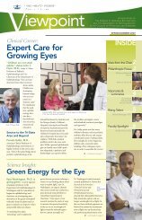
Principal Investigators on the grant<br />
<strong>from</strong> L. to R.: Peter Gouras, MD,<br />
Stephen Tsang, MD, PhD, Janet<br />
Sparrow, PhD, Kostantin Petrukhin,<br />
PhD, Rando Allikmets, PhD.<br />
George A. (Jack) Cioffi, MD<br />
The principal investigator of the NEI<br />
grant, Dr. Allikmets, is also the geneticist<br />
responsible for first identifying and<br />
cloning the ABCA4 gene in 1997. Dr.<br />
Allikmets’ discovery of the role of<br />
ABCA4 in retinal degenerative diseases<br />
has directly led to investigations into<br />
possible treatments for Stargardt<br />
disease. “The goal of every geneticist is<br />
not to find the gene, but through that<br />
discovery, help to find treatment for the<br />
disease,” affirms Dr. Allikmets. And as<br />
our understanding of the genetic basis<br />
of eye disease grows, so does our<br />
capacity to treat these diseases.<br />
Dr. Allikmets and his colleagues at<br />
Columbia will be working in tandem<br />
A PUBLICATION OF<br />
THE EDWARD S. HARKNESS EYE INSTITUTE<br />
AND THE DEPARTMENT OF OPHTHALMOLOGY<br />
IN THE COLLEGE OF PHYSICIANS AND SURGEONS<br />
SPRING/SUMMER 2012<br />
INSIDE<br />
View <strong>from</strong> the Chair 2<br />
Clinical Spotlight 3<br />
Genetic Counselor:<br />
A Critical Connection<br />
Visionaries &<br />
Luminaries 4<br />
Fifteenth Anniversary<br />
of Ollendorff Lectureship<br />
Welcome Reception<br />
for Jack Cioffi<br />
Science Insight 6<br />
Space Telescope Technology<br />
Used to Better See the<br />
Back of the <strong>Eye</strong><br />
with four other centers. They will be<br />
joining forces with Joan Bennett, MD,<br />
PhD, at the University of Pennsylvania,<br />
a pioneer of gene therapy for retinal<br />
diseases; Richard A. Lewis, MD, MS,<br />
and James R. Lupski, MD, PhD, at<br />
Houston’s Baylor College of Medicine.<br />
In 1977 in conjunction with Dr.<br />
Allikmets, they were the first to clone<br />
the ABCA4 gene. The third center is the<br />
Telethon <strong>Institute</strong> and the Federico II<br />
University in Naples, Italy, with Alberto<br />
Auricchio, MD, and Francesca<br />
Simonelli, MD, who had previously<br />
collaborated with Dr. Allikmets on<br />
Stargardt patient characterization and<br />
genetic research. The fourth center is<br />
the University of Illinois, where the<br />
well-respected ophthalmologist,<br />
Gerald A. Fishman, MD, has been<br />
clinically researching Stargardt disease<br />
for decades.<br />
“Rather than competing with each<br />
other and doing things in parallel,”<br />
explains Dr. Allikmets, “I thought the<br />
best way to advance the entire field<br />
was to join forces in a multicenter<br />
study and work together to find the<br />
best therapeutic method to treat<br />
Stargardt disease.”<br />
Stargardt therapy can be conducted by<br />
many different means, including gene<br />
therapy with lentiviral or adeno-associated<br />
vectors, model organism research,<br />
<strong>continued</strong> on <strong>page</strong> 2
View<br />
<strong>from</strong> the Chair<br />
Dear Friends,<br />
This is my first letter to you as the new chair of the Columbia University<br />
Medical Center Department of Ophthalmology, and I could not be more<br />
thrilled. I sincerely thank the <strong>Harkness</strong> <strong>Eye</strong><br />
<strong>Institute</strong>’s physicians, research scientists,<br />
nurses, administrative staff, and patients<br />
for being so incredibly welcoming. In this,<br />
my inaugural issue of the Viewpoint, I am<br />
pleased to share my background with you<br />
and present my vision for the future of<br />
this prestigious institution.<br />
I would like to extend a special note of<br />
gratitude to Stanley Chang who is stepping<br />
down in his role as chairman, but<br />
who will continue to actively see patients,<br />
conduct research, and teach at <strong>Harkness</strong>.<br />
Stanley has led this Department with<br />
exemplary skill and vision, and I look forward<br />
to working closely with him in the<br />
months and years ahead.<br />
We are proud to announce that Columbia researchers have been awarded a<br />
multi-million dollar grant by the National <strong>Eye</strong> <strong>Institute</strong> for a multicenter project<br />
to find treatment options for ABCA4-associated genetic diseases, including<br />
Stargardt disease and retinitis pigmentosa (RP). The grant provides a substantial<br />
increase in our research funding and presents the opportunity to find novel<br />
treatment options for the future.<br />
NEI Grant <strong>continued</strong> <strong>from</strong> <strong>page</strong> 1<br />
or small molecule treatment. With the centers<br />
concurrently working towards the same goal by testing<br />
different methodologies, the scientists can better<br />
determine which treatment, or a combination thereof,<br />
is the most effective.<br />
“I honestly don't care if lentiviral gene therapy, which<br />
is done at Columbia, is better than that based on<br />
adeno-associated viral delivery,” emphasizes Dr.<br />
Allikmets. “What I care about is finding the best<br />
method out there for patients.”<br />
To tackle the large assignment of finding therapeutic<br />
applications for ABCA4-associated<br />
diseases, the grant has been divided into four<br />
modular components:<br />
Module 1 is designed to capture a thorough<br />
and precise clinical and genetic analysis<br />
of Stargardt patients to gain a better understanding<br />
of the disease. The Stargardt gene has<br />
more than 800 known mutations, meaning<br />
only a few affected individuals actually share<br />
the same two disease-causing variations. The<br />
goal of Module 1 is two-fold: first, to gain a<br />
better understanding of Stargardt disease<br />
development and progression, and second, to<br />
define specific patient subpopulations for<br />
most efficient clinical trials.<br />
A clinical evaluation method unique to<br />
Columbia is quantitative fundus autofluorescence,<br />
which was established through collaborative research<br />
with Columbia scientists and François Delori, PhD, at<br />
Harvard Medical School. “This evaluation method,<br />
applied at Columbia, is extremely valuable to<br />
researchers and patients because you can precisely<br />
measure and quantify the disease progression through<br />
an increase or decrease in the autofluorescence,”<br />
emphasizes Dr. Sparrow.<br />
The rate of progression for Stargardt disease is<br />
currently unknown. Ideally in future clinical trials,<br />
researchers will be able to compare the rate of progression<br />
against the disease’s natural history and <strong>from</strong> that<br />
determine how well a specific treatment is working.<br />
Module 2 utilizes animal models as a tool to<br />
evaluate the effectiveness of various treatments.<br />
Vıewpoınt 2 SPRING/SUMMER 2012<br />
Based on the work of Dr. Sparrow, a cell biologist, we<br />
know the ABCA4 gene transports a form of vitamin A<br />
<strong>from</strong> the photoreceptor membranes so that it can<br />
re-enter the visual cycle, and it is the visual cycle that<br />
allows us to see. In Stargardt's, the ABCA4 transporter<br />
is defective, the consequence of which is an accumulation<br />
of a compound called A2E, which is two vitamin<br />
A molecules joined together. A2E accumulation in the<br />
retinal pigment epithelium kills the cells and leads to<br />
many of the characteristics of Stargardt disease.<br />
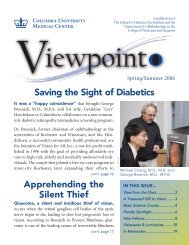
Fundus autofluorescence imaging<br />
in a healthy eye (top) and in an<br />
age-matched patient with recessive<br />
Stargardt disease (bottom).<br />
These images allow measurement<br />
of retinal pigment epithelium<br />
(RPE) lipofuscin accumulation by<br />
an approach (quantitative autofluorescence,<br />
qAF) developed at<br />
Columbia University.<br />
The images on the right are color<br />
coded maps of qAF, with red<br />
being indicative of high AF levels.<br />
The latter reflects elevated lipofuscin<br />
in RPE, a feature of recessive<br />
Stargardt disease.<br />
Researchers at Columbia are on the threshold of unearthing critical information<br />
about the eye’s photoreceptor microstructure by using space telescope technology.<br />
Adaptive optics (AO) allows researchers to noninvasively examine patients<br />
and gain valuable information that previously could only be obtained by looking<br />
at autopsy eyes. AO technology is used at just a handful of sites around the<br />
world, and we are excited to be obtaining such detailed cellular level information<br />
here at Columbia.<br />
With the explosion of new information related to genetic science, we offer our<br />
patients genetic counseling services to offset the confusion and fear a diagnosis<br />
like Stargardt’s or RP might cause by providing much-needed information in its<br />
place. In this issue, we highlight the invaluable role of the genetic counselor.<br />
An amazing story of an ophthalmologist’s journey <strong>from</strong> Nazi Germany to the<br />
halls of Columbia where a lectureship has been named in his honor is a testament<br />
to the spirit of determination a whole generation of immigrants brought<br />
to our shores. We are pleased to share the bittersweet history of Ulrich<br />
Ollendorff, MD, in the <strong>page</strong>s of the Viewpoint.<br />
Finally, it is truly my honor to be asked to guide this celebrated institution. I<br />
look forward to earning your trust and partnership so that we may continue the<br />
fine work of advancing research and preserving vision.<br />
Sincerely,<br />
George A. (Jack) Cioffi, MD<br />
Jean and Richard Deems Professor<br />
<strong>Edward</strong> S. <strong>Harkness</strong> Professor<br />
Chairman, Department of Ophthalmology<br />
The main measure of<br />
Stargardt disease is A2E<br />
accumulation, so a lack of increase or a decrease of<br />
this compound is one measure of a successful therapeutic<br />
outcome. As accumulation of A2E is extremely<br />
similar in both mice and humans, researchers use<br />
mouse models as a way to investigate if a specific treatment<br />
is working or not. Dr. Sparrow and her team are<br />
developing methods to transfer the quantitative fundus<br />
autofluorescence technology, which is used in the<br />
clinic, to their mouse model.<br />
“Currently we need whole eyes to do A2E measurements<br />
by high-performance liquid chromatography,<br />
so we have to sacrifice the mouse. The advantage of<br />
quantitative fundus autofluorescence is that it’s a noninvasive<br />
imaging approach that does not harm the<br />
animal and enables us to follow the mice over time.<br />
In addition, we could model the type of approach<br />
that would actually be used in humans to evaluate<br />
the success of therapy, meaning that through the<br />
mouse model, we would be obtaining information<br />
immediately relevant to the human patient,”<br />
explains Dr. Sparrow.<br />
In the Stargardt mouse model the ABCA4 gene is<br />
“knocked out” or made inactive. These “knockout”<br />
mice are important animal models for studying the<br />
role of genes in normal<br />
physiology and disease.<br />
More importantly for<br />
these studies, the ABCA4deficient<br />
mice are invaluable<br />
models to test therapies<br />
and generate treatments,<br />
necessary steps<br />
prior to establishing<br />
human clinical trials.<br />
For example, ABCA4<br />
"knockout" mice are used<br />
for gene therapy experiments,<br />
which are primarily<br />
done with viruses that<br />
carry the therapeutic or<br />
curative gene to the desired<br />
cells. The genes that cause<br />
disease within the virus are<br />
deleted, so the virus genes instead act as a delivery<br />
vehicle or “vector.” In 2008, Drs. Allikmets, Sparrow,<br />
and Gouras cloned the normal working human<br />
ABCA4 gene into a lentiviral vector and injected it<br />
into mouse eyes. The mice were then followed to see if<br />
their eyes improved, and they did.<br />
Module 3 directly compares two methods of<br />
gene therapy: lentiviral versus adeno-associated viral<br />
delivery, to determine which method is more efficient.<br />
Lentiviral therapy, which has been developed at<br />
Columbia Ophthalmology, entered Phase I clinical<br />
trials last year. Now Dr. Allikmets and colleagues are<br />
looking to further improve the delivery vectors to<br />
<strong>continued</strong> on <strong>page</strong> 8
Clinical Spotlight:<br />
Genetic Counselor: A Critical<br />
Connection Between the<br />
Physician, Patient, and Scientist<br />
Imagine for a moment you were just told that you, your<br />
spouse, or your child had an inherited eye disorder where<br />
vision would seriously worsen over time and for which there<br />
is no known cure. What would you do? Where would you turn? How would<br />
you cope? Receiving such a diagnosis would be understandably overwhelming. Add<br />
to that a genetic component, and questions arise as to how this will impact your<br />
immediate and extended family going forward. In the unchartered waters of genetic<br />
testing, a genetic counselor takes on a critical role in communicating to the patient<br />
and the patient’s family what the diagnosis means and helping them to navigate the<br />
challenging course ahead.<br />
Inherited <strong>Eye</strong> Diseases<br />
Currently 180 retinal disease genes have been identified. Stargardt disease, as one<br />
example, is an inherited eye disorder that causes progressive vision loss. It is<br />
estimated that up to 1:20 people carry a mutation in the ABCA4 gene, the gene<br />
responsible for Stargardt disease. To manifest the disorder a person must have two<br />
mutations by receiving one defective copy of the gene <strong>from</strong> each parent.<br />
Retinitis Pigmentosa (RP) is another hereditary eye disorder that affects approximately<br />
one in 3,000 people worldwide and which also causes incurable vision loss.<br />
Like Stargardt disease, RP often first appears<br />
in childhood, but severe vision problems<br />
usually develop in early adulthood.<br />
BOARD OF ADVISORS<br />
Department of Ophthalmology<br />
Louis V. Gerstner, Jr., Chairman<br />
William Acquavella<br />
Rand Araskog<br />
Dr. Endré Balazs<br />
Debra Black<br />
Shirlee and Bernard Brown<br />
Robert L. Burch III<br />
Howard L. Clark, Jr.<br />
Abraham E. Cohen<br />
Joseph C. Connors<br />
E. Virgil Conway<br />
Alan K. Docter<br />
Gloria and Louis Flanzer<br />
Leslie Hinton<br />
Joel Hoffman<br />
T. C. Hsu<br />
Florence and Herbert Irving<br />
Helen Kimmel<br />
Dr. Henry Kissinger<br />
Ambassador John L. Loeb, Jr.<br />
John Manice<br />
Bjorg and Stephen Ollendorff<br />
Homer McK. Rees<br />
John Robinson<br />
Patricia Rosenwald<br />
Stephen Ross<br />
James Shinn<br />
William Spier<br />
Miranda Wong Tang<br />
David Tobey<br />
Richard Woolworth<br />
Dukes Wooters<br />
IN MEMORIAM<br />
Dorothy Eweson<br />
Martin S. Kimmel<br />
Seymour Milstein<br />
Candace VanAlen<br />
MEDICAL ADVISORS<br />
Rando Allikmets, PhD<br />
James Auran, MD<br />
Stanley Chang, MD<br />
George A. Cioffi, MD<br />
John Espy, MD<br />
John Flynn, MD<br />
Harold Spalter, MD<br />
Janet Sparrow, PhD<br />
Abraham Spector, PhD<br />
Stephen Trokel, MD<br />
VIEWPOINT<br />
Editor-in-Chief<br />
Jane E. Heffner<br />
Writing<br />
Tina-Marie Gauthier<br />
Graphic Design<br />
David Sakla<br />
Photography<br />
Charles E. Manley<br />
Noelle Pensec<br />
(To learn more about Stargardt disease and<br />
the studies being conducted at Columbia<br />
University’s Department of Ophthalmology<br />
to find treatment options for any or all<br />
ABCA4-associated genetic diseases, please<br />
refer to the Stargardt grant article on <strong>page</strong> 1.)<br />
Genetic Counseling<br />
With the explosion of new information related<br />
to genetic science, researchers at Columbia<br />
are aggressively pursuing answers to better<br />
understand hereditary eye<br />
disorders and to ultimately<br />
find treatments, but genetic<br />
eye disease is extremely<br />
complicated both in the<br />
lab and to the patient trying<br />
to comprehend all of<br />
its implications.<br />
Rando Allikmets, PhD,<br />
Columbia University<br />
Medical Center’s Director<br />
of Research for the<br />
Department of<br />
Ophthalmology, and his<br />
team work together with<br />
the Clinical Laboratory<br />
Improvement<br />
Amendments (CLIA)-certified<br />
Molecular Pathology Diagnostics<br />
Laboratory, to offer their patients genetic<br />
counseling services to offset the confusion<br />
and fear a diagnosis like Stargardt’s or RP<br />
might cause by providing much-needed<br />
information in its place.<br />
Carolyn Brown, MSc, has been Columbia’s<br />
on-staff, American Board of Genetic<br />
Counseling-certified genetic counselor for<br />
one and a half years and is a valued asset for<br />
her work with patients and the community.<br />
Due to the medical implications for family<br />
members as well as the affected individual,<br />
counseling is an important part of managing<br />
genetic disorders. Ms. Brown offers an<br />
immediate benefit by providing facts,<br />
support, and resources.<br />
While some physicians may feel they can provide<br />
genetic counseling services themselves,<br />
or may not see the value in it, inclusion of a<br />
genetic counselor is a major added-on benefit<br />
to the patient. In many cases, the<br />
physician might have the best of<br />
intentions but simply doesn’t have<br />
the time or necessary expertise in<br />
human molecular genetics to educate<br />
the patient following diagnosis.<br />
Carolyn Brown, MSc<br />
Dr. Allikmets explains, “It is important to understand<br />
that genetic counseling is conducted by a person with specific training and education.<br />
I direct the molecular genetics lab at Columbia and have conducted basic<br />
genetic research to find the genes that cause eye disease for 15 years now, but even<br />
someone with my background and experience often does not have the proper skills<br />
to be a genetic counselor. It is a specialized and valuable professional service.”<br />
Learning that a disease is genetic often has a practical and psychological impact on<br />
the wellbeing of individuals and their families that extend far beyond the medical<br />
component. When a genetic diagnosis is suspected, Ms. Brown works with the medical<br />
team to determine possible genes involved, inheritance patterns, and logical next<br />
steps, such as examining family members and genetic testing. Perhaps more importantly,<br />
with her training in psychological counseling, Ms. Brown can help the patient<br />
adapt to the diagnosis and provide tangible information, giving patients a greater<br />
sense of control at times that may be highly emotional or filled with uncertainty. Ms.<br />
Brown elaborates, “My goal is to present complicated medical and genetic information<br />
in an understandable way so people can make informed medical decisions, and,<br />
hopefully, to allay what could be a frightening diagnosis. I try to create a space where<br />
people can express concerns and ask questions that might not be brought up during<br />
a typical doctor’s appointment. While each situation is unique, issues often arise like<br />
family planning or how to tell relatives they are at risk for a genetic disease.”<br />
Genetic Testing<br />
Scientists’ knowledge about inherited eye diseases is growing at an unprecedented<br />
rate, and there is great hope that scientists will someday develop a reliable treatment<br />
option. Hence, another key aspect of Ms. Brown’s position has become the recruitment<br />
of patients for ongoing research. By undergoing genetic testing and being<br />
included in a database, patients with hereditary eye disorders can be matched to<br />
those who share the same<br />
combination of genetic<br />
variants and to whom later<br />
treatments would be of<br />
most benefit.<br />
Ms. Brown is counseling family members <strong>from</strong><br />
L. to R.: Tom Wolf, James Wolf, and Mary Russotti.<br />
There are more than 50<br />
known RP genes and<br />
many more unknown that<br />
are clinically similar.<br />
Variations in genes play a<br />
significant role in determining<br />
if gene therapy or a<br />
more general therapy can<br />
be scientifically recommended.<br />
Dependent on the RP-causing gene, for<br />
example, vitamin A supplements might be beneficial<br />
for one patient while detrimental to another.<br />
“With all the variations in the Stargardt gene, ABCA4, it is challenging because we<br />
need a large number of people with the same genetic combination as their disease<br />
progression and response to treatment would likely be similar,” points out Dr.<br />
Allikmets. “We have more than 600 Stargardt study subjects at Columbia in our<br />
database, and we are a center within the National Ophthalmic Disease Genotyping<br />
Network, eyeGENE®, which is the National <strong>Eye</strong> <strong>Institute</strong>’s central depository. We<br />
work with other CLIA-certified centers, providing official diagnostic screening and<br />
results to patients at no cost. Being part of the eyeGENE® is also beneficial scientifically<br />
since we have access to de-identified genetic and clinical results of all patients<br />
enrolled at more than 12 centers around the United States, so the numbers alone<br />
provide a substantially increased statistical power to our studies.”<br />
Gathering information on a patient for inclusion in a database has a long-term<br />
benefit, though not an immediate direct benefit to the patient. Patients’ participation<br />
in genetic studies helps scientists find new disease-causing genes and develop a<br />
better understanding of the disorder. Information can then be used to produce<br />
treatments and ultimately a cure. Studies of family members are also considered<br />
useful in identifying disease-causing genes.<br />
Major strides have been made in gaining an understanding of genetic eye diseases,<br />
which were once thought to be untreatable. While researchers continue their search<br />
for a cure, genetic counselors are the critical connection between the physician,<br />
patient, and scientist: empowering patients through knowledge.<br />
SPRING/SUMMER 2012<br />
Vıewpoınt 3
Visionaries<br />
&<br />
Luminaries<br />
The Department Observes the<br />
Fifteenth Anniversary of the<br />
Ulrich Ollendorff Lectureship<br />
The winds of fate blow a different<br />
course for each traveler. For Ulrich<br />
Ollendorff, MD, they carried a practicing ophthalmologist<br />
and academician <strong>from</strong> Nazi Germany to the<br />
esteemed halls of Columbia University’s Department<br />
L. to R.: Stanley Chang, MD, Jack Cioffi, MD,<br />
Bjorg and Stephen Ollendorff.<br />
of Ophthalmology, where a prestigious lecture is<br />
presented every year in his honor.<br />
Ulrich Ollendorff Lectureship<br />
This March, the Department of Ophthalmology at<br />
Columbia University marked the fifteenth annual<br />
Ulrich Ollendorff Lectureship. Paul L. Kaufman, MD,<br />
professor and chair of ophthalmology and visual sciences<br />
at the University of Wisconsin Clinical Science<br />
Center, presented the talk, “Presbyopia: Up Close.”<br />
The Ulrich Ollendorff Lectureship was established in<br />
1997, the year before Dr. Ollendorff’s death, to honor<br />
his long and renowned career as an eye doctor. The<br />
annual lectureship was endowed by a gift <strong>from</strong> his<br />
son, Stephen and his daughter-in law, Bjorg.<br />
“The Ollendorff Lectureship is a standout for many<br />
reasons,” notes John T. Flynn, MD, special lecturer in<br />
the Department of Ophthalmology who has been<br />
involved with this program since its inception. “It is a<br />
rarity in American ophthalmology because it includes<br />
presentations on every subspecialty, given by experts<br />
on that subject. The lectures focus on innovative<br />
research in the field.”<br />
Open to the entire ophthalmology community, the<br />
Ollendorff Lectureship has an appeal to physicians in<br />
Manhattan and beyond. “We identify individuals who<br />
have made significant contributions to the development<br />
of ophthalmology both clinically and scientifically,”<br />
describes Stanley Chang, MD, former departmental<br />
chair. “As ophthalmic leaders, our presenters<br />
are an inspiration to young trainees, fellows, and residents,<br />
and we want to honor them for their contributions.”<br />
A feature unique to this lectureship is that the<br />
Department’s young ophthalmologists join the guest<br />
speaker for dinner, an occasion that provides an<br />
opportunity for young clinicians to interact with ophthalmologic<br />
luminaries in a more casual atmosphere.<br />
Establishment of the Lectureship<br />
It is impossible to discuss the Ollendorff Lectureship<br />
without talking about the man behind the name. As<br />
Dr. Flynn keenly states, “The Ollendorff Lectureship<br />
has a shine unlike any other because no other lecture<br />
Vıewpoınt 4 SPRING/SUMMER 2012<br />
has such a unique background of triumph<br />
over tragedy.”<br />
Stanley Chang, MD, was the newly minted chair of<br />
the Department of Ophthalmology in 1996 when,<br />
over the long Fourth of July weekend, he successfully<br />
treated Stephen Ollendorff’s eye problem.<br />
In return, Mr. Ollendorff wanted to do<br />
something special for the Department.<br />
“While discussing various options, I mentioned<br />
to Dr. Chang that though my father<br />
had one of the largest ophthalmology<br />
practices in Washington Heights, he was<br />
denied privileges to practice at Columbia’s<br />
Department of Ophthalmology solely<br />
because he was Jewish.”<br />
Mr. Ollendorff was referring to a time in<br />
history when America, in all her infinite<br />
promise, was still a land of quotas and<br />
restrictions. In that era, Columbia held to a<br />
policy of not hiring Jewish physicians. In a<br />
gesture both compassionate and ironic,<br />
Mr. Ollendorff and Dr. Chang decided to<br />
establish a lectureship series in Dr.<br />
Ollendorff’s name at the <strong>Harkness</strong> <strong>Eye</strong><br />
<strong>Institute</strong>. Reflecting that Columbia had once turned its<br />
back on his father, Mr. Ollendorff summarizes<br />
the emotion behind the decision, “It felt<br />
like a redemption of sorts.”<br />
“Steve and I thought an annual lectureship<br />
would greatly honor his father,” explains Dr.<br />
Chang. “We wanted to make people at the eye<br />
institute and in the community aware of Dr.<br />
Ollendorff’s contributions to New York’s<br />
Upper West Side, the neighborhood that<br />
houses Columbia University Medical Center.<br />
I also recognized that this lectureship would<br />
contribute to Columbia’s intellectual environment<br />
by bringing outstanding academic ophthalmologists<br />
<strong>from</strong> throughout the country<br />
to the <strong>Harkness</strong> <strong>Eye</strong> <strong>Institute</strong>.”<br />
The lecture is also an opportunity to reflect<br />
on the entire movement of well-trained professionals,<br />
doctors, scientists, artists, and<br />
musicians who escaped the Holocaust and<br />
successfully integrated into American life.<br />
“Their story is very inspiring,” emphasizes Dr.<br />
Chang. “Dr. Ollendorff, for example, gave up<br />
everything in his home country, yet still built<br />
a successful practice in the United States,<br />
raised a wonderful family, and gave back to<br />
the community.”<br />
The Ollendorffs have also established a<br />
Diagnostic Center for the <strong>Harkness</strong> <strong>Eye</strong><br />
<strong>Institute</strong>. Dr. Chang acknowledges, “The faculty<br />
has come to know Steve and Bjorg. They<br />
are a family interested in helping others, and<br />
they are incredibly supportive of the whole<br />
<strong>Institute</strong>, both as members of the Advisory<br />
Board and as generous donors.”<br />
Ulrich Ollendorff, MD<br />
The marking of the fifteenth anniversary of a<br />
highly respected named lectureship is a proud<br />
achievement, but the journey of the man<br />
behind the name is the true testament to this<br />
accomplishment. It is Dr. Ollendorff’s story,<br />
steeped in history, which creates the special<br />
aura surrounding the lecture program.<br />
Jack Cioffi, MD and Paul L. Kaufman, MD<br />
Ulrich Ollendorff was born in Breslau, Germany in<br />
1906. After graduating <strong>from</strong> medical school at the top<br />
of his class, he became a professor of ophthalmology.<br />
The mood in Europe, however, was darkening. In<br />
November 1938, the day before Kristallnacht (“The<br />
Night of Broken Glass”), Dr. and Mrs. Ollendorff were<br />
cautioned by their landlord that the Nazis were going<br />
to raid their Berlin apartment. Believing the warning<br />
was a harbinger of worse things to come, Ulrich, his<br />
wife Anne, and their four-month old son Stephen<br />
immediately went to stay with Anne’s parents. They<br />
secured visas, and the day following the vicious series<br />
of planned attacks against the Jews, the three safely<br />
arrived in England; shortly thereafter, Anne’s family<br />
relocated to Chile.<br />
Ulrich’s parents Valli and Arthur, his brothers<br />
Wolfgang and Gerhard, and his aunt Ella stayed<br />
behind in Breslau, as Arthur was convinced they were<br />
safe due to his service as a colonel in the German<br />
army during the First World War. To a degree he was<br />
correct, but in 1940, Arthur died of a heart attack and<br />
the family’s security was suddenly less sure. In 1941,<br />
Wolfgang, a member of the resistance, was captured in<br />
Holland, tortured, and ultimately killed while trying to
escape <strong>from</strong> the Mauthausen concentration camp.<br />
Around that same period, Gerhard was abducted and<br />
killed by the Nazi’s for “medical reasons.” On August<br />
24, 1942, the Nazis arrested Valli and her sister Ella.<br />
Certain of their bleak destiny, Valli realized that Ulrich,<br />
safe in New York, would be the only family member to<br />
survive, and she wrote her beloved son a letter of<br />
farewell while on her way to the Theresienstadt concentration<br />
camp. On October 16 Valli died and on<br />
December 2, her sister Ella followed.<br />
On his arrival to the United States, Dr. Ollendorff and<br />
his family began building a new life. He learned<br />
English, re-established his professional credentials, and<br />
opened an ophthalmology practice in the northern<br />
Manhattan neighborhood of Washington Heights, an<br />
enclave of German Jewish immigrants. Dr. Ollendorff<br />
quickly became known throughout the community,<br />
building a wonderful reputation based on his great<br />
skill and care. He is remembered for his compassionate<br />
medical ideals—he would knowingly operate on<br />
patients who could not afford his services in exchange<br />
Welcome Reception<br />
for Jack Cioffi<br />
On Tuesday, April 17, Lee Goldman, MD, dean of the<br />
Faculties of Health Sciences and Medicine, and executive vice<br />
president for Health and Biomedical Sciences at Columbia<br />
University Medical Center (CUMC), hosted a reception at Le<br />
Cirque restaurant to celebrate the appointment of<br />
George (Jack) Cioffi, MD, as the <strong>Edward</strong> S.<br />
<strong>Harkness</strong> professor and chairman of the<br />
Department of Ophthalmology at CUMC.<br />
The evening event was an excellent opportunity to<br />
introduce Dr. Cioffi to our constituency. Invited<br />
guests included ophthalmology faculty, CUMC<br />
departmental chairmen, members of the CUMC<br />
Board of Visitors, members of the ophthalmology<br />
Board of Advisors, and friends of the Department.<br />
The 125 people in attendance enjoyed the Dean’s<br />
welcoming remarks about Dr. Jack Cioffi followed<br />
by Dr. Cioffi’s presentation on his vision for the<br />
Department’s future.<br />
Both the dean and Dr. Cioffi thanked Stanley Chang, MD,<br />
the outgoing chair, for his inspiring leadership for the past<br />
17 years and for his extraordinary counsel facilitating<br />
Dr. Cioffi’s transition to his new position. Everyone had<br />
a wonderful time and left the restaurant with a clear<br />
understanding of Dr. Cioffi’s mission to elevate the<br />
Department to even greater heights.<br />
for an apple pie. He made regular house calls, climbing<br />
countless flights of stairs for a payment of $6 a visit.<br />
After his evening rounds he made sure to stop and<br />
read poetry each night to a patient whose eyes had to<br />
remain bandaged for thirty days, an act of simple<br />
kindness in his daily life.<br />
“Everyone in Washington Heights knew Dr.<br />
Ollendorff, and it was common for people to wait for<br />
three to four hours in order to see him,” recalls his<br />
daughter-in-law Bjorg. “They were willing to wait<br />
because he was a wonderful doctor, a beautiful human<br />
being. He generated enormous respect within the<br />
community.” By the time he retired in 1986 at the age<br />
of 80, Dr. Ollendorff had treated more than 300,000<br />
grateful patients.<br />
The Letter<br />
Although well established in New York, Dr. Ollendorff<br />
had never stopped looking for news <strong>from</strong> his homeland<br />
and ultimately learned the unspeakable fates his<br />
family had suffered. In 1985, Dr. Ollendorff was 79<br />
years old and on the eve of his retirement. In the mail,<br />
lying among the bills and advertisements was a handaddressed<br />
letter to Ulrich <strong>from</strong> South America. It was<br />
the farewell letter his mother Valli had written to him<br />
in 1942, two months before she died.<br />
Translated <strong>from</strong> German, her love letter to her son<br />
begins, “My beloved, my good boy,” and speaks of the<br />
dark future Valli knows she will face. In the most gentle<br />
of goodbyes, she asks her son to carry the knowledge<br />
that through his life he was the source of purest<br />
joy for his parents. The letter ends with Valli acknowledging<br />
her certain destiny, “I was and I am daily even<br />
longing very much for you and your life. However, fate<br />
did not let me go.”<br />
The letter, a beautiful memento of a mother’s undying<br />
love for her son, gives substance to the man himself.<br />
Dr. Ulrich Ollendorff was a true “gentle man” who<br />
served almost 50 years as a beloved ophthalmologist,<br />
honoring the spirit of his family every day in his<br />
devotion and caring of others. (For more information<br />
about Valli Ollendorff's letter to her son, go to:<br />
www.fatedidnotletmego.org.)<br />
Left: Lee Goldman, MD introduces the new Chair.<br />
Clockwise <strong>from</strong> top L. to R.: Stanley Chang, MD, Jack Cioffi, MD, and<br />
Lee Goldman, MD; Bernard and Shirlee Brown; Jane Heffner, Ned<br />
and Emily Sherwood; John and Margot Catsimatidis, Jack Cioffi, MD,<br />
and Stanley Zabar; Dukes and Kay Wooters and Jack Cioffi, MD;<br />
E. Virgil and Elaine Conway and Stanley Chang, MD; Carl Frischling,<br />
Jack and Linda Cioffi; Lama Al-Aswad, MD, Louis Chang, MD, PhD,<br />
Courtney Chang, and Dana Blumberg, MD; Lee Goldman, MD,<br />
Donna Acquavella, Jill Goldman, and William Acquavella; Nicholas<br />
Mezitis, MD and Peter Michalos, MD; David and Julie Tobey; Stephen<br />
Trokel, MD, Cynthia Conigliaro, John Espy, MD and Polly Espy.<br />
SPRING/SUMMER 2012<br />
Vıewpoınt 5
Science Insight:<br />
Space Telescope Technology<br />
Used to Better See the<br />
Back of the <strong>Eye</strong><br />
The same tools used by astronomers to examine the far off<br />
reaches of the universe are being employed by researchers in the<br />
Department of Ophthalmology so that ophthalmologists can better see the back of<br />
the eye. Adaptive optics (AO) is an imaging technology originally developed as an<br />
astronomy tool to adjust for atmospheric turbulence when using ground telescopes.<br />
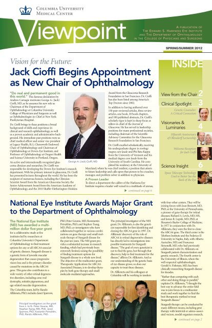
Principal<br />
Investigators of<br />
AO research at<br />
Columbia,<br />
SungPyo Park,<br />
MD, PhD, Stanley<br />
Chang, MD, and<br />
Stephen Tsang,<br />
MD, PhD, are<br />
using this technology<br />
instead to<br />
visualize the retina<br />
at the individual<br />
cell level.<br />
Sungypo Park, MD looking at the<br />
images <strong>from</strong> the AO-SLO machine.<br />
Ophthalmic<br />
Imaging<br />
Ophthalmic<br />
imaging is a fundamental tool in the<br />
ophthalmologist’s arsenal because it<br />
enables a retinal physician to<br />
noninvasively diagnose diseases by observing the retina through the natural optics<br />
of the human eye.<br />
The human eye, however, has an extremely complicated visual system. Due to the<br />
blur caused by natural irregularities in the eye’s optics, the current retinal imaging<br />
methods used in clinical practice are limited in the level of information they are able<br />
to provide. For example, while common imaging modalities are able to show different<br />
trends or patterns in the retina, such as the behavior of the retinal cells, they are<br />
not able to do so at a close distance.<br />
Furthermore, they are unable to provide<br />
details about the photoreceptor<br />
microstructure, which means that possibly<br />
critical information remains hidden.<br />
The retinal specialists at the <strong>Harkness</strong> <strong>Eye</strong><br />
<strong>Institute</strong> are the foremost in the nation and<br />
they use these diagnostic tools to look at<br />
images of the retina. In 2011, in partnership<br />
with the Canon company, Dr. Chang<br />
laid the groundwork for a pioneering imaging<br />
technique by bringing in an AO<br />
machine and combining it with an established<br />
system, scanning laser ophthalmoscopy<br />
(SLO). The two modalities<br />
together create a new imaging system<br />
referred to as AS-SLO, which Columbia’s<br />
Department of Ophthalmology is using to<br />
obtain high-resolution retinal images.<br />
With AO-SLO’s high magnification,<br />
researchers are able to view the retina in<br />
an entirely different way, obtaining<br />
detailed cellular level information about<br />
the retina. The AO-SLO enables physicians<br />
to visualize the shape and morphology of<br />
each cone cell within the retina. They are able to count how many cells are viable, see<br />
how the cells are arranged, note any differences among them, and determine if the<br />
packing arrangement of the cells changes throughout the pathology of the disease.<br />
Dr. Park explains, “This ability to count cells is a significant asset of AO-SLO. Since we<br />
can see every single cone cell, we can count how many exist and then compare that<br />
number to a normal retina. This is a helpful tool, because in some diseases, an<br />
increase in cone numbers indicates many different kinds of problems. We can also<br />
look at the changes in the shape and orientation of the cones. AO-SLO holds promise<br />
of earlier diagnosis because we can observe cone behavior earlier in the disease. In<br />
addition, we can monitor disease progression, <strong>from</strong> when it begins, which will help us<br />
uncover how it manifests itself in the earliest of stages and other subtle changes that<br />
could not be detected otherwise.”<br />
Vıewpoınt 6 SPRING/SUMMER 2012<br />
L. to R.: Sungypo Park, MD, Stanley Chang, MD,<br />
Norihiko Utsunomiya, an engineer with Canon based<br />
in Tokyo, Stephen Tsang, MD, PhD, and Takeshi<br />
Kitamura, an engineer with Canon based in New York.<br />
Features of AO-SLO<br />
The key features of the AO-SLO system include the<br />
incorporation of a dual liquid crystal on silicon spatial light<br />
modulator (LCOS-SLM) as a wavefront compensation<br />
device and sequential processing of aberration measurement/compensation<br />
and SLO imaging.<br />
In an AO system, two important implementations are used<br />
to compensate for natural irregularities, or aberration, of the<br />
eye’s optics: a deformable mirror and a LCOS-SLM.<br />
Although deformable mirrors are widely used for this purpose,<br />
their performance alone is not enough to correct the<br />
large amounts of aberration in the human eye, and<br />
deformable mirrors are also susceptible to static electricity.<br />
The LCOS-SLM is able to compensate for large amounts of natural aberration by a<br />
phase-wrapping method utilizing the continuity of the wavefront of a laser beam, but it<br />
corrects only a particular polarization component. To overcome these deficiencies and<br />
to counterbalance the two orthogonal polarization components and sequential processing<br />
taking the SLO image, Columbia researchers have adopted dual LCOS-SLMs.<br />
The Columbia AO-SLO system sufficiently compensates for optical irregularities in<br />
the eye, improves the contrast of the SLO image, and allows for efficient high-speed<br />
imaging—a convenient attribute for busy clinics. These improvements pioneered by<br />
Columbia’s particular system have furthered AO-SLO technology and have provided<br />
ophthalmologists, especially retina specialists, a new and effective way of producing<br />
high-resolution, cellular level images of retinas in a clinical setting.<br />
AO-SLO in the Clinic<br />
To examine diseased retinas at the cellular level prior to the advent of AO-SLO<br />
technology, researchers have been limited to looking at autopsy eyes that have been<br />
enucleated <strong>from</strong> the body. Although a great deal of information has been learned<br />
through these types of histologic studies, the stains used during the pathology<br />
procedure and the artifacts introduced during preparation of the retina further<br />
complicates the interpretation of disease processes at different stages.<br />
In contrast, AO-SLO allows noninvasive, direct observation of the retina at a microscopic<br />
resolution comparable to that of histology, but happily for the patient, they are<br />
alive and well in the clinic! While the procedure is comfortable, it does require the<br />
patient to fixate on a target. Each procedure takes about 20 minutes per eye, and<br />
patients are normally in and out in usually less than an hour.<br />
Research Studies<br />
AO technology is currently only used at a handful of sites. For example, only one<br />
AO-SLO system is available in all of Europe. As the AO-SLO system is still relatively<br />
primitive at this time, it is currently only used in research studies.<br />
For the Department of Ophthalmology’s first AO-SLO study, researchers recruited<br />
approximately 200 patients of all different age groups, ethnicities, and demographics.<br />
Study subjects were scanned by the AO-SLO and a comparative database of normative<br />
patients was created. Dr. Tsang comments, “We explain to the patients that by<br />
using the AO-SLO system, we hope to<br />
learn more about disease mechanisms,<br />
including why they have the<br />
disease and their rate of progression.<br />
It's up to the patient if they're interested<br />
in participating as a study subject<br />
and learning more. Once we image<br />
the study subject, we have a baseline.<br />
It is our hope that AO-SLO can offer<br />
earlier detection of disease progression.<br />
In addition, when a treatment<br />
comes along, this technology will let<br />
us know very early on if the treatment<br />
works, knowledge which could possibly<br />
shorten human clinical treatment<br />
trials <strong>from</strong> five years to one year.”<br />
Using the AO-SLO, scientists at<br />
Columbia have also looked at<br />
enhanced S-cone cells. S-cone syndrome<br />
is characterized by S-type<br />
cones overpopulating the retina at the<br />
expense of other cone cells. It causes patients to lose vision<br />
that they would have otherwise obtained <strong>from</strong> the rod cells.<br />
Through AO, researchers can obtain a detailed visual picture<br />
of what is actually occurring in this syndrome.<br />
The Department is considering other possible studies using the AO-SLO. This technology,<br />
for instance, could be informative in gaining greater understanding of retinitis<br />
pigmentosa (RP). In RP, the visual field constricts <strong>from</strong> the periphery of the retina.<br />
The AO-SLO could ideally be used for looking at the cone cells in the center of the<br />
constricting ring to determine if they are normal or not. Dr. Park is also especially<br />
interested in learning more about end-stage glaucoma, while Dr. Chang is intrigued<br />
by using AO to see the effect of drusen, the small accumulations of hyaline bodies<br />
underneath the retina. AO could also possibly provide information related to diabetic<br />
retinopathy or age-related macular degeneration.<br />
Researchers at Columbia hope that eventually AO-SLO will become another useful<br />
tool in the arsenal of ophthalmic imaging techniques, providing ophthalmologists<br />
with greater insight into the cellular workings of the retina.
Jack Cioffi Appointment<br />
<strong>continued</strong> <strong>from</strong> <strong>page</strong> 1<br />
and Dr. Cioffi has extensive experience in the laboratory,<br />
clinic, operating room, and boardroom. In his<br />
role as chief medical officer at Legacy Health, Dr.<br />
Cioffi oversaw five hospitals and a children’s hospital,<br />
which provided an array of health care services.<br />
A cornerstone of Columbia University’s Department<br />
of Ophthalmology is a medical team made up of<br />
globally renowned physicians, who are at the forefront<br />
of medical eye research, which is an area familiar to<br />
Dr. Cioffi. “I have an established history of having<br />
expanded a large research department and an eye<br />
institute. As chief medical officer, I was responsible for<br />
recruiting hundreds of gifted physicians and<br />
researchers with cutting-edge and complementary<br />
skill sets to manage and prevent eye disease.”<br />
Dr. Cioffi takes over the chairmanship <strong>from</strong> Stanley<br />
Chang, MD. During his time as chairman, Dr. Chang<br />
attracted a top-notch team of researchers and clinicians<br />
to the department, developed new faculty practices<br />
in New York and New Jersey, more than doubled<br />
the number of surgical cases in the <strong>Harkness</strong> <strong>Eye</strong><br />
<strong>Institute</strong>, and dramatically increased research funding.<br />
Although Dr. Chang is stepping down as chair, he will<br />
continue his patient care, research, and teaching commitments.<br />
“I am so pleased and happy that Stanley is<br />
remaining fully active at Columbia,” affirms Dr. Cioffi.<br />
“He is a vital member of the faculty and his value here<br />
cannot be overstated.”<br />
Dr. Cioffi’s personality is warm and forthcoming, and<br />
he is quick to acknowledge that through the great<br />
work of the department’s previous chairs, he stands on<br />
the shoulders of giants, proclaiming, “Under their<br />
leadership, Columbia ophthalmologists and scientists<br />
accomplished a series of ‘firsts,’ including the first use<br />
of lasers in medicine, the first human corneal transplantation<br />
in the United States, the first human retinal<br />
cell transplantation, and the first artificial cornea.”<br />
Grateful especially for the legacy left by Dr. Chang, Dr.<br />
Cioffi affirms, “A perfect groundwork has been laid for<br />
our department to lead ophthalmology training and<br />
vision research into the future. Dr. Chang has recruited<br />
great talent, both in research and clinical ophthalmology,<br />
and he developed a strong philanthropic base,<br />
which better secures us going forward.”<br />
Dr. Cioffi and Dr. Chang have been working closely<br />
together over the past months. “Dr. Chang has been<br />
extremely candid throughout this transition, providing<br />
insights so I can better interpret the complexities<br />
of the department. Thus, I am more effectively able to<br />
see the potential for building upon Dr. Chang’s<br />
achievements and achieving my vision. I look forward<br />
to working alongside Dr. Chang to realize these goals.”<br />
Dr. Chang is confident in his successor’s ability. “Jack is<br />
a terrific individual with a great vision for further<br />
growth of this department, especially in the challenging<br />
times medicine is facing today.”<br />
Vision for the Future<br />
Dr. Cioffi’s overarching goal is for the <strong>Harkness</strong> <strong>Eye</strong><br />
<strong>Institute</strong> to become the foremost department of ophthalmology<br />
in the country. “It's not often you get<br />
offered the opportunity to guide a celebrated<br />
<strong>Institute</strong>,” Dr. Cioffi explains. “I plan to build upon the<br />
great foundation that is already in place and then add<br />
my vision, which is for us to be the top ophthalmology<br />
program in the country. We have that potential.”<br />
Dr. Cioffi’s vision for the <strong>Harkness</strong> <strong>Eye</strong> <strong>Institute</strong> focuses<br />
on four primary areas he plans to address in the<br />
upcoming months and years: Clinical Services,<br />
Residency Training, Vision Research Growth, and the<br />
Aging Infrastructure of the Department.<br />
Clinical Services<br />
Dr. Cioffi supports the “Hub and Spoke” model as a<br />
vehicle to provide the highest quality and most efficient<br />
health care to more people. For the hub and<br />
spoke archetype, one site acts as a centralized principal<br />
base supporting connected satellite sites. To expand<br />
access to basic clinical services and diagnostics to more<br />
patients, Dr. Cioffi sees the opportunity for outreach<br />
offices, while high-end services–such as specialized<br />
surgery–would remain on the Columbia campus. He<br />
will also be exploring the possibility of developing<br />
ambulatory surgical centers (ASC) at other sites. In his<br />
previous position, Dr. Cioffi opened a highly efficient<br />
and successful ASC with outreach to a six-state region<br />
and the Pacific Rim. Dr. Cioffi elaborates, “By recruiting<br />
the best physicians and cultivating both high-end<br />
and efficient clinical services, I believe the outreach<br />
potential for Columbia’s Department of<br />
Ophthalmology is great, and I look forward to<br />
making it a reality.”<br />
Residency Training<br />
Agreeing with the words of his mentor, “You will be<br />
known by those you train,” Dr. Cioffi emphatically<br />
believes that one of the Department of<br />
Ophthalmology’s core missions is to teach the medical<br />
leaders of tomorrow. “We have the unique ability to<br />
train practicing ophthalmologists, technicians, nurses,<br />
and researchers. Skilled clinicians and scientists come<br />
to us <strong>from</strong> around the world seeking the latest ophthalmic<br />
education, and our residency program is a<br />
focal point for training these future leaders. In order to<br />
be in the most elite group of training programs, we<br />
need to grow and enhance our educational capabilities,<br />
and I will work diligently to enhance the<br />
Department of Ophthalmology’s residency training<br />
program to ensure it is one of the top tier educational<br />
platforms in the country.”<br />
Vision Research Growth<br />
With his experience and proficiency in research,<br />
including having been continuously funded for 18<br />
years by the National <strong>Institute</strong>s<br />
of Health (NIH), Dr. Cioffi<br />
wants to build upon<br />
Columbia’s proud vision<br />
research history. “Our collaborations<br />
with scientists in other<br />
departments, such as neurosciences,<br />
biochemistry, and<br />
pathology, are critical to our<br />
future success, and I would like<br />
for us to strengthen and<br />
extend these collaborations<br />
going forward.”<br />
Creating a successful research<br />
program requires several key<br />
elements, according to Dr.<br />
Cioffi. “First, research programs<br />
that aim to answer vexing<br />
clinical problems are often<br />
more successful as they tend to<br />
be funded by the NIH, donors<br />
contribute to these programs,<br />
and patients directly benefit<br />
<strong>from</strong> these programs. Second,<br />
by following a ‘Clinician-<br />
Scientist’ model, both clinicians<br />
and scientists bring<br />
unique perspectives to a project,<br />
which is a great advantage<br />
and works hand-in-hand with<br />
a third important element, collaboration among scientists<br />
at Columbia. Finally, I believe in programmatic<br />
focus: I would rather the research program be world<br />
class in a select number of areas, than be broad based<br />
in too many areas. Columbia has been a research<br />
leader in retinal disorders and retinal genetics. At a<br />
minimum, I would like to add an increased glaucoma<br />
research endeavor to Columbia’s portfolio, as retinal<br />
research and glaucoma research are very complementary,<br />
and the addition of glaucoma scientists would be<br />
extremely positive.”<br />
Aging Infrastructure<br />
The aging infrastructure of the <strong>Harkness</strong> <strong>Eye</strong> <strong>Institute</strong><br />
is a concern voiced by many, including Dr. Cioffi. “In<br />
order to attract and retain the best faculty, we must<br />
address the infrastructure issue. Due to the amazing<br />
fundraising success of the department in the past<br />
decade, I am confident that we can raise funds to<br />
revive the facility.” New York-Presbyterian Hospital<br />
and Columbia University Medical Center have committed<br />
resources to Dr. Cioffi to renovate space within<br />
the <strong>Harkness</strong> <strong>Eye</strong> <strong>Institute</strong>, and they will jointly develop<br />
a short-term and long-term facilities plan. Dr.<br />
Cioffi has begun a formal space assessment, including<br />
current and projected space needs for research, faculty<br />
practices, resident clinics, eye surgery, procedural<br />
units, meeting and educational facilities, and administrative<br />
space. An informed decision too will be made<br />
about the advisability of renovating rather than building<br />
new space.<br />
Moving Dr. Cioffi’s vision forward will take dedicated<br />
leadership, and he plans to be as straightforward and<br />
transparent as possible. A tenet of Dr. Cioffi’s is that a<br />
good leader listens and learns. Since his arrival, he has<br />
been interviewing faculty members one-on-one to<br />
gain an understanding of their desires and aspirations.<br />
He is also conducting a series called “Lunch with the<br />
Chair” that enables staff members a chance to meet in<br />
a smaller setting and ask any questions that may be on<br />
their minds.<br />
Background<br />
At a young age, Dr. Cioffi was given the nickname of<br />
Jack, the acronym of his initials, G.A.C. He grew up<br />
in New England, and it was his undergraduate study<br />
of zoology that first piqued his curiosity in science.<br />
“I was intrigued by science, and particularly by<br />
genetics during my undergraduate years, and I<br />
realized early on that a medical degree would open<br />
up a world of possibilities, which I could then explore<br />
to the furthest reaches.”<br />
He was drawn to ophthalmology and the area of glaucoma<br />
for the surgical specialty it offered and for the<br />
patient relationships that develop due to the chronic<br />
nature of the disease. Dr. Cioffi elaborates, “Glaucoma<br />
impacts the entire age spectrum, and you get to know<br />
your patients over long periods of time. Treatment<br />
may begin in infancy and you are with that patient<br />
until they go off to college. In other cases, you treat<br />
someone as they are about to retire and stay involved<br />
Department Ranks High<br />
in Research Funding<br />
The Columbia Department of Ophthalmology ranked seven out of 65<br />
Departments of Ophthalmology nationwide in dollars awarded by the<br />
National <strong>Institute</strong>s of Health (NIH) for vision research in 2011. This<br />
continues a forward trend by our Department. In 2010, Columbia was<br />
ranked 16, which was a significant jump <strong>from</strong> earlier years when our<br />
Department of Ophthalmology was ranked in the mid-twenties.<br />
Total NIH funding was $7,455,200. Additional grant applications are<br />
being submitted this year that will further expand our program depth<br />
in the research portfolio.<br />
The credit for this substantial increase in ranking goes to our group of<br />
brilliant scientists led by Rando Allikmets, PhD, Director of Research. The<br />
generosity of our patients provides much of the seed funding for projects<br />
and allows our senior scientists, such as Janet Sparrow, PhD, and Carol<br />
Mason, PhD, to mentor our younger investigators to success by creating<br />
the preliminary body of work necessary to apply and receive research<br />
funding. We congratulate all those who made this wonderful news<br />
possible, and we look forward to advancing even higher in the future!<br />
in their lives through their golden years. Physicians<br />
play a privileged role. There aren’t many specialties in<br />
ophthalmology, nor in medicine for that matter,<br />
where you can contribute in all phases of a person’s<br />
life and interact closely with their family. I feel truly<br />
blessed to be entrusted with the long-term care of my<br />
patients.” Dr. Cioffi’s role as a physician will continue<br />
at the <strong>Harkness</strong> <strong>Eye</strong> <strong>Institute</strong>, where he will be interacting<br />
with glaucoma patients clinically and surgically.<br />
For Dr. Cioffi, several personal factors drew him to<br />
Columbia University’s Department of<br />
Ophthalmology. “First it was the storied history of the<br />
<strong>Harkness</strong> <strong>Eye</strong> <strong>Institute</strong> itself. It holds a special place in<br />
American and worldwide ophthalmology in terms of<br />
the greats who have walked these halls. Second,<br />
Columbia University is an academic institution<br />
beyond compare. Finally, there is an undeniable combined<br />
appeal of being part of a world-class eye institute,<br />
at a world-class university, in a world-class city.”<br />
Dr. Cioffi, his wife Linda, and their two children,<br />
William and Elizabeth, are excited about moving<br />
<strong>from</strong> the Pacific Coast and living in New York. With<br />
their extended families scattered up and down the<br />
Eastern Seaboard, they are looking forward to being<br />
closer to home.<br />
With the roadmap he has laid out, Dr. Cioffi reflects<br />
on the upcoming months and years ahead, “We truly<br />
have the opportunity here to do real and permanent<br />
good, which is an awe inspiring and humbling<br />
thought. I am honored to be at this celebrated institution,<br />
and I look forward to working together with this<br />
fine community to accomplish many great things.”<br />
SPRING/SUMMER 2012<br />
Vıewpoınt 7
Columbia Ophthalmology<br />
Consultants Opens New<br />
Satellite Office in New Jersey<br />
Bryan J. Winn, MD, has opened a satellite<br />
office in Ridgewood, New Jersey, to offer a<br />
convenient alternative to patients. This move signifies<br />
a new presence for Columbia Ophthalmology<br />
Consultants outside of Manhattan.<br />
Specializing in ophthalmic plastic and reconstructive<br />
surgery, Dr. Winn performs critical reconstructive<br />
procedures that positively impact on both the<br />
physical and emotional wellbeing<br />
of a patient, such as<br />
rebuilding areas of the face after<br />
trauma or skin cancer removal,<br />
eyelid surgery to resolve congenital<br />
or age-related changes, and<br />
tear duct and orbital surgery. In<br />
addition, Dr. Winn offers a wide<br />
array of aesthetic procedures,<br />
including brow, cheek, and lid<br />
lifts in addition to Botox and<br />
dermal fillers.<br />
Having grown up in northern<br />
New Jersey, Dr. Winn is excited<br />
Bryan J. Winn, MD<br />
to work in a location that he<br />
describes as close to his heart. He also feels that his<br />
specialty and choice of location will be of benefit to<br />
his patients. “It turns out that a third of my patients<br />
who see me in Manhattan actually come in <strong>from</strong><br />
New Jersey, so it's a natural place to go. The area has<br />
also been underserved for the particular treatments<br />
I offer,” notes Dr. Winn.<br />
V ıewpoınt<br />
THE EDWARD S. HARKNESS EYE INSTITUTE AND<br />
THE DEPARTMENT OF OPHTHALMOLOGY<br />
635 WEST 165TH STREET<br />
NEW YORK, NY 10032-3797<br />
RETURN SERVICE REQUESTED<br />
Dr. Winn is an Assistant Professor of Clinical<br />
Ophthalmology at Columbia University Medical<br />
Center, board certified in ophthalmology, fellowship<br />
trained in oculofacial plastic surgery, and a fellow of<br />
the American Society of Ophthalmic Plastic and<br />
Reconstructive Surgery (ASOPRS). Dr. Winn graduated<br />
summa cum laude <strong>from</strong> Amherst College where<br />
he was elected to the Phi Beta Kappa honor society.<br />
He received his medical education at the Columbia<br />
University College of Physicians and<br />
Surgeons, where he graduated at the top of<br />
his class and was elected to the Alpha<br />
Omega Alpha honor society.<br />
In addition to tending to his practice, Dr.<br />
Winn is the Associate Residency Program<br />
Director and Director of Medical Student<br />
Education for the Department of<br />
Ophthalmology at Columbia University<br />
College of Physicians and Surgeons, and he<br />
is responsible for teaching the ophthalmology<br />
residents oculofacial plastic surgery.<br />
Dr. Winn’s New Jersey office is located at<br />
119 Prospect Street, Suite 1, Ridgewood,<br />
New Jersey, 07450; the office telephone number<br />
201-445-0444. Dr. Winn continues to see patients at<br />
the <strong>Harkness</strong> <strong>Eye</strong> <strong>Institute</strong> and Columbia’s midtown<br />
Manhattan east side office located at 880 Third<br />
Street. For more information about Dr. Winn,<br />
please go to www.Columbia<strong>Eye</strong>lidSurgery.com.<br />
Important Patient Care Information<br />
Specialties: Cornea/External Ocular Disease<br />
Glaucoma<br />
Pediatric Ophthalmology and Strabismus<br />
Refractive Surgery/LASIK<br />
Vitreoretinal and Uveitis<br />
NEI Grant<br />
<strong>continued</strong> <strong>from</strong> <strong>page</strong> 2<br />
provide more efficient treatment to patients. The<br />
University of Pennsylvania and the Telethon <strong>Institute</strong><br />
centers are working on the adeno-associated virus<br />
delivery systems.<br />
“Gene therapy is likely the cure for recessive diseases,”<br />
notes Dr. Allikmets. “In recessive diseases there is not<br />
enough gene activity, so the gene is not working properly.<br />
If you give the patient the working gene, that should, theoretically,<br />
alleviate the problem and resolve the disease.”<br />
Gene therapy works especially well in early stages of a<br />
disease where a large number of cells are still alive. In<br />
severe late cases, gene therapy would likely not be successful<br />
because there are fewer cells or possibly no cells<br />
available in which to insert the normal gene.<br />
Module 4 addresses small molecule therapy, which<br />
is performed primarily by Dr. Petrukhin in collaboration<br />
with Drs. Sparrow and Allikmets. Dr. Petrukhin, who<br />
brings 10 years of drug development <strong>from</strong> Merck & Co.,<br />
Inc., and colleagues are working on compounds that can<br />
modulate the visual cycle by slowing down the uptake of<br />
vitamin A or by interacting with key proteins in the cycle.<br />
These molecules are identified by three complementary<br />
strategies: high throughput screening of large libraries of<br />
small molecules, optimization of known leads derived<br />
<strong>from</strong> chemical structures, and in silico screening of<br />
virtual compound libraries.<br />
While small molecule therapy is not a cure for<br />
Stargardt’s, its critical role would be in slowing down<br />
the progression of disease. Theoretically, gene therapy<br />
and small molecule therapy could be used concurrently.<br />
Small molecule therapy would be used to slow the<br />
disease progression so gene therapy could be introduced<br />
to treat the disease even at later stages.<br />
Through the focused work of the many gifted researchers<br />
at Columbia University, and their talented and dedicated<br />
partners involved in this multicenter study, treatment for<br />
Stargardt disease and other genetic retinal disorders,<br />
which was once thought to be unattainable, is now closer<br />
at hand than ever before.<br />
For inquiries and appointments, please call 212.305.9535