continued from page 1 - Edward Harkness Eye Institute
continued from page 1 - Edward Harkness Eye Institute
continued from page 1 - Edward Harkness Eye Institute

You also want an ePaper? Increase the reach of your titles
YUMPU automatically turns print PDFs into web optimized ePapers that Google loves.
Science Insight:<br />
Space Telescope Technology<br />
Used to Better See the<br />
Back of the <strong>Eye</strong><br />
The same tools used by astronomers to examine the far off<br />
reaches of the universe are being employed by researchers in the<br />
Department of Ophthalmology so that ophthalmologists can better see the back of<br />
the eye. Adaptive optics (AO) is an imaging technology originally developed as an<br />
astronomy tool to adjust for atmospheric turbulence when using ground telescopes.<br />
Principal<br />
Investigators of<br />
AO research at<br />
Columbia,<br />
SungPyo Park,<br />
MD, PhD, Stanley<br />
Chang, MD, and<br />
Stephen Tsang,<br />
MD, PhD, are<br />
using this technology<br />
instead to<br />
visualize the retina<br />
at the individual<br />
cell level.<br />
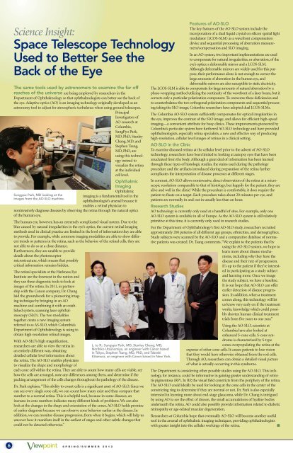
Sungypo Park, MD looking at the<br />
images <strong>from</strong> the AO-SLO machine.<br />
Ophthalmic<br />
Imaging<br />
Ophthalmic<br />
imaging is a fundamental tool in the<br />
ophthalmologist’s arsenal because it<br />
enables a retinal physician to<br />
noninvasively diagnose diseases by observing the retina through the natural optics<br />
of the human eye.<br />
The human eye, however, has an extremely complicated visual system. Due to the<br />
blur caused by natural irregularities in the eye’s optics, the current retinal imaging<br />
methods used in clinical practice are limited in the level of information they are able<br />
to provide. For example, while common imaging modalities are able to show different<br />
trends or patterns in the retina, such as the behavior of the retinal cells, they are<br />
not able to do so at a close distance.<br />
Furthermore, they are unable to provide<br />
details about the photoreceptor<br />
microstructure, which means that possibly<br />
critical information remains hidden.<br />
The retinal specialists at the <strong>Harkness</strong> <strong>Eye</strong><br />
<strong>Institute</strong> are the foremost in the nation and<br />
they use these diagnostic tools to look at<br />
images of the retina. In 2011, in partnership<br />
with the Canon company, Dr. Chang<br />
laid the groundwork for a pioneering imaging<br />
technique by bringing in an AO<br />
machine and combining it with an established<br />
system, scanning laser ophthalmoscopy<br />
(SLO). The two modalities<br />
together create a new imaging system<br />
referred to as AS-SLO, which Columbia’s<br />
Department of Ophthalmology is using to<br />
obtain high-resolution retinal images.<br />
With AO-SLO’s high magnification,<br />
researchers are able to view the retina in<br />
an entirely different way, obtaining<br />
detailed cellular level information about<br />
the retina. The AO-SLO enables physicians<br />
to visualize the shape and morphology of<br />
each cone cell within the retina. They are able to count how many cells are viable, see<br />
how the cells are arranged, note any differences among them, and determine if the<br />
packing arrangement of the cells changes throughout the pathology of the disease.<br />
Dr. Park explains, “This ability to count cells is a significant asset of AO-SLO. Since we<br />
can see every single cone cell, we can count how many exist and then compare that<br />
number to a normal retina. This is a helpful tool, because in some diseases, an<br />
increase in cone numbers indicates many different kinds of problems. We can also<br />
look at the changes in the shape and orientation of the cones. AO-SLO holds promise<br />
of earlier diagnosis because we can observe cone behavior earlier in the disease. In<br />
addition, we can monitor disease progression, <strong>from</strong> when it begins, which will help us<br />
uncover how it manifests itself in the earliest of stages and other subtle changes that<br />
could not be detected otherwise.”<br />
Vıewpoınt 6 SPRING/SUMMER 2012<br />
L. to R.: Sungypo Park, MD, Stanley Chang, MD,<br />
Norihiko Utsunomiya, an engineer with Canon based<br />
in Tokyo, Stephen Tsang, MD, PhD, and Takeshi<br />
Kitamura, an engineer with Canon based in New York.<br />
Features of AO-SLO<br />
The key features of the AO-SLO system include the<br />
incorporation of a dual liquid crystal on silicon spatial light<br />
modulator (LCOS-SLM) as a wavefront compensation<br />
device and sequential processing of aberration measurement/compensation<br />
and SLO imaging.<br />
In an AO system, two important implementations are used<br />
to compensate for natural irregularities, or aberration, of the<br />
eye’s optics: a deformable mirror and a LCOS-SLM.<br />
Although deformable mirrors are widely used for this purpose,<br />
their performance alone is not enough to correct the<br />
large amounts of aberration in the human eye, and<br />
deformable mirrors are also susceptible to static electricity.<br />
The LCOS-SLM is able to compensate for large amounts of natural aberration by a<br />
phase-wrapping method utilizing the continuity of the wavefront of a laser beam, but it<br />
corrects only a particular polarization component. To overcome these deficiencies and<br />
to counterbalance the two orthogonal polarization components and sequential processing<br />
taking the SLO image, Columbia researchers have adopted dual LCOS-SLMs.<br />
The Columbia AO-SLO system sufficiently compensates for optical irregularities in<br />
the eye, improves the contrast of the SLO image, and allows for efficient high-speed<br />
imaging—a convenient attribute for busy clinics. These improvements pioneered by<br />
Columbia’s particular system have furthered AO-SLO technology and have provided<br />
ophthalmologists, especially retina specialists, a new and effective way of producing<br />
high-resolution, cellular level images of retinas in a clinical setting.<br />
AO-SLO in the Clinic<br />
To examine diseased retinas at the cellular level prior to the advent of AO-SLO<br />
technology, researchers have been limited to looking at autopsy eyes that have been<br />
enucleated <strong>from</strong> the body. Although a great deal of information has been learned<br />
through these types of histologic studies, the stains used during the pathology<br />
procedure and the artifacts introduced during preparation of the retina further<br />
complicates the interpretation of disease processes at different stages.<br />
In contrast, AO-SLO allows noninvasive, direct observation of the retina at a microscopic<br />
resolution comparable to that of histology, but happily for the patient, they are<br />
alive and well in the clinic! While the procedure is comfortable, it does require the<br />
patient to fixate on a target. Each procedure takes about 20 minutes per eye, and<br />
patients are normally in and out in usually less than an hour.<br />
Research Studies<br />
AO technology is currently only used at a handful of sites. For example, only one<br />
AO-SLO system is available in all of Europe. As the AO-SLO system is still relatively<br />
primitive at this time, it is currently only used in research studies.<br />
For the Department of Ophthalmology’s first AO-SLO study, researchers recruited<br />
approximately 200 patients of all different age groups, ethnicities, and demographics.<br />
Study subjects were scanned by the AO-SLO and a comparative database of normative<br />
patients was created. Dr. Tsang comments, “We explain to the patients that by<br />
using the AO-SLO system, we hope to<br />
learn more about disease mechanisms,<br />
including why they have the<br />
disease and their rate of progression.<br />
It's up to the patient if they're interested<br />
in participating as a study subject<br />
and learning more. Once we image<br />
the study subject, we have a baseline.<br />
It is our hope that AO-SLO can offer<br />
earlier detection of disease progression.<br />
In addition, when a treatment<br />
comes along, this technology will let<br />
us know very early on if the treatment<br />
works, knowledge which could possibly<br />
shorten human clinical treatment<br />
trials <strong>from</strong> five years to one year.”<br />
Using the AO-SLO, scientists at<br />
Columbia have also looked at<br />
enhanced S-cone cells. S-cone syndrome<br />
is characterized by S-type<br />
cones overpopulating the retina at the<br />
expense of other cone cells. It causes patients to lose vision<br />
that they would have otherwise obtained <strong>from</strong> the rod cells.<br />
Through AO, researchers can obtain a detailed visual picture<br />
of what is actually occurring in this syndrome.<br />
The Department is considering other possible studies using the AO-SLO. This technology,<br />
for instance, could be informative in gaining greater understanding of retinitis<br />
pigmentosa (RP). In RP, the visual field constricts <strong>from</strong> the periphery of the retina.<br />
The AO-SLO could ideally be used for looking at the cone cells in the center of the<br />
constricting ring to determine if they are normal or not. Dr. Park is also especially<br />
interested in learning more about end-stage glaucoma, while Dr. Chang is intrigued<br />
by using AO to see the effect of drusen, the small accumulations of hyaline bodies<br />
underneath the retina. AO could also possibly provide information related to diabetic<br />
retinopathy or age-related macular degeneration.<br />
Researchers at Columbia hope that eventually AO-SLO will become another useful<br />
tool in the arsenal of ophthalmic imaging techniques, providing ophthalmologists<br />
with greater insight into the cellular workings of the retina.