Oral Medicine and Pathology Quiz – Case 18
Oral Medicine and Pathology Quiz – Case 18
Oral Medicine and Pathology Quiz – Case 18

Create successful ePaper yourself
Turn your PDF publications into a flip-book with our unique Google optimized e-Paper software.
Copyright © Athens Medical Society<br />
714 www.mednet.gr/archives<br />
N.G. NIKITAKIS et al<br />
ARCHIVES OF HELLENIC MEDICINE: ISSN 11-05-3992<br />
CONTINUING MEDICAL EDUCATION<br />
ΣΥΝΕΧΙΖΟΜΕΝΗ ΙΑΤΡΙΚΗ ΕΚΠΑΙ∆ΕΥΣΗ<br />
<strong>Oral</strong> <strong>Medicine</strong> <strong>and</strong> <strong>Pathology</strong> <strong>Quiz</strong> <strong>–</strong><br />
<strong>Case</strong> <strong>18</strong><br />
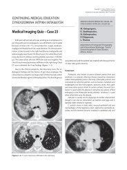
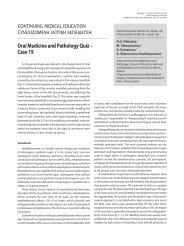
A 51-year-old male was referred to our clinic for investigation<br />
of multiple symptomatic oral mucosal lesions of several weeks<br />
duration. The patient reported chronic hepatitis C infection <strong>and</strong><br />
was a heavy smoker; he had been recently tested negative for<br />
HIV. On clinical examination, multiple irregular lesions of various<br />
sizes were noticed on the labial mucosa, the palate <strong>and</strong> the<br />
dorsal surface of the tongue (figures 1−3). The lesions appeared<br />
as whitish plaques, focally assuming a snail track pattern, with<br />
partial sloughing <strong>and</strong> surrounding erythema. The patient had<br />
used topical antifungal medication (miconazole oral gel) for one<br />
week without improvement. On careful questioning, the patient<br />
admitted recurrent skin rash, as well as ulcers in the genital<br />
mucosa for the past two months. On the basis of the clinical<br />
diagnosis, specific serologic tests were ordered.<br />
Comment<br />
Syphilis is a chronic infection caused by Treponema pallidum.<br />
The microorganism is primarily transmitted by sexual intercourse <strong>and</strong><br />
from mother to fetus (congenital syphilis). Although the incidence of<br />
syphilis had steadily decreased from the 1940s to 2000, a significant<br />
worldwide increase has been documented during the last decade.<br />
The main reasons for the recent rise include the decreasing use of<br />
barrier protection (i.e. condoms), the false sense of security that<br />
sexually transmitted diseases are curable <strong>and</strong> the widespread use of<br />
alternative oral practices such as oral sex, which is falsely considered<br />
to be safer than vaginal or anal sex. Today, the majority of the new<br />
cases of syphilis occur in men who have sex with men (MSM) <strong>and</strong><br />
are strongly associated with HIV coinfection.<br />
Syphilis proceeds through three clinical stages (primary, secondary<br />
<strong>and</strong> tertiary); a latent stage of several years duration intervenes<br />
Figure 1<br />
Figure 2 Figure 3<br />
ARCHIVES OF HELLENIC MEDICINE 2011, 28(5):714-715<br />
ÁÑ×ÅÉÁ ÅËËÇÍÉÊÇÓ ÉÁÔÑÉÊÇÓ 2011, 28(5):714-715<br />
...............................................<br />
N.G. Nikitakis,<br />
G. Kamperos,<br />
P. Argyris,<br />
A. Sklavounou-Andrikopoulou<br />
...............................................<br />
Department of <strong>Oral</strong> <strong>Medicine</strong> <strong>and</strong><br />
<strong>Pathology</strong>, School of Dentistry, National<br />
<strong>and</strong> Kapodistrian University of Athens,<br />
Athens, Greece<br />
between the secondary <strong>and</strong> tertiary stages. The patients are highly<br />
infectious during the first two stages. Nevertheless, pregnant women<br />
can also transmit the disease to the fetus during the latent stage.<br />
<strong>Oral</strong> lesions are uncommon but may occur at any stage.<br />
Primary syphilis is characterized by the development of a chancre,<br />
an ulcer located at the site of inoculation, 3−90 days after the<br />
exposure. The lesion is commonly solitary but multiple lesions may<br />
be also observed. The majority of extragenital chancres occur in the<br />
mouth (40<strong>–</strong>75%), usually located in the lips, presenting as painless,<br />
clean-based ulceration. Regional lymphadenopathy, which may be<br />
bilateral, occurs in up to 80% of cases about 7<strong>–</strong>10 days following<br />
the development of the chancre. If left untreated, the initial lesion<br />
heals within 3 to 8 weeks.<br />
The secondary stage develops 1−6 months after the first contact<br />
with the organism. This stage results from the hematogenous<br />
dissemination of T. pallidum giving systemic <strong>and</strong> mucocutaneous<br />
manifestations. Lesions of primary <strong>and</strong> secondary syphilis may also<br />
coexist. The most common signs <strong>and</strong> symptoms of the secondary stage<br />
are painless lymphadenopathy, diffuse maculopapular cutaneous<br />
rash, malaise, fever, headache, weight loss <strong>and</strong> musculoskeletal<br />
pain. About 5<strong>–</strong>6% of patients may develop patchy hair loss of the<br />
beard <strong>and</strong> eyebrows <strong>and</strong> scalp-localized alopecia. Approximately<br />
30% of the patients develop oral mucous patches, in the form of<br />
whitish plaques with partial sloughing, occurring at any oral site.<br />
Papillary lesions also appear (condyloma lata) mainly in the genital
ORAL MEDICINE AND PATHOLOGY QUIZ <strong>–</strong> CASE <strong>18</strong> 715<br />
or anal area in 5<strong>–</strong>22% of patients. <strong>Oral</strong> condyloma lata, as well as<br />
oral snail track lesions, are also occasionally observed. Atypical <strong>and</strong><br />
more aggressive necrotic lesions of secondary syphilis may be seen<br />
in HIV positive patients. The lesions of secondary syphilis typically<br />
resolve within 1−2 months.<br />
After the secondary stage, there is a latent period (1−30 years)<br />
during which the patient does not show any clinical sign of infection.<br />
Tertiary syphilis develops in 30% of patients, with serious complications<br />
in the cardiovascular <strong>and</strong> central nervous system (CNS). Scattered foci<br />
of granulomatous inflammation are observed in the skin, CNS, liver,<br />
spleen, bones <strong>and</strong> other organs. These lesions, known as gummas,<br />
range in size from tiny deposits to large masses. Intraorally, they<br />
usually affect the tongue, which appears large, lobulated or even<br />
atrophic, <strong>and</strong> the palate, which may even be perforated through to<br />
the nasal cavity. The premalignant nature of syphilitic atrophic (luetic)<br />
glossitis, a lesion rarely observed nowadays, is contradictory.<br />
As for congenital syphilis, infection of the fetus may result in<br />
abortion, stillbirth, neonatal death or disease, depending on the<br />
time of infection <strong>and</strong> the severity of mother’s disease. The clinical<br />
features involve the classic Hutchinson’s triad (Hutchinson’s teeth,<br />
ocular interstitial keratitis, eighth nerve deafness), as well as other<br />
signs such as growth retardation, anemia, hepatosplenomegaly,<br />
skin diseases, bone alterations <strong>and</strong> CNS problems.<br />
Syphilis’ diagnosis is based on laboratory tests. The presence of<br />
T. pallidum can be confirmed by dark-field examination of a smear<br />
of the exudate of an active lesion. False positive results may occur<br />
from oral lesions due to other spirochetal microorganisms which<br />
are part of the microflora. The serologic investigation involves<br />
nontreponemal screening tests (such as VDRL), which are nonspecific<br />
<strong>and</strong> are positive 3 weeks after the infection <strong>and</strong> throughout the<br />
primary <strong>and</strong> secondary syphilis, <strong>and</strong> treponemal tests (such as FTA-<br />
ABS), which are specific <strong>and</strong> highly sensitive <strong>and</strong> remain positive<br />
for life. The histopathologic features of oral primary or secondary<br />
syphilis are nonspecific <strong>and</strong> involve intense deep, perivascular<br />
chronic, mainly plasmacytic, inflammation. The presence of T.<br />
pallidum may be confirmed by the use of special stains such as<br />
Warthin-Starry silver stain. On the other h<strong>and</strong>, in tertiary syphilis,<br />
granulomatous inflammation is evident <strong>and</strong> the microorganisms<br />
are hard to demonstrate even with special stains.<br />
The oral lesions are expected to heal completely in a few weeks.<br />
Treatment usually involves penicillin administration. The medication’s<br />
dose <strong>and</strong> schedule depend on the stage, neurologic involvement<br />
<strong>and</strong> immune status of the patient. It should be stressed that patients<br />
diagnosed with syphilis should be tested for HIV as well. In the case<br />
presented here, specific serologic tests confirmed the diagnosis of<br />
syphilis <strong>and</strong> the patient was placed on IV penicillin treatment with<br />
prompt resolution of the oral lesions.<br />
References<br />
1. VIÑALS-IGLESIAS H, CHIMENOS-KÜSTNER E. The reappearance of<br />
a forgotten disease in the oral cavity: Syphilis. Med <strong>Oral</strong> Patol<br />
<strong>Oral</strong> Cir Bucal 2009, 14:e416−e420<br />
2. FICARRA G, CARLOS R. Syphilis: The renaissance of an old disease<br />
with oral implications. Head Neck Pathol 2009, 3:195<strong>–</strong><br />
206<br />
Corresponding author:<br />
N.G. Nikitakis, Department of <strong>Oral</strong> <strong>Pathology</strong> <strong>and</strong> <strong>Medicine</strong>,<br />
School of Dentistry, National <strong>and</strong> Kapodistrian University of<br />
Athens, 2 Thivon street, GR-115 27 Athens, Greece,<br />
tel.: +30 210 74 61 003, fax: +30 210 7461220<br />
e-mail: nnikitakis1@yahoo.com<br />
Diagnosis: <strong>Oral</strong> manifestations of secondary syphilis (oral mucous patches)<br />
..............................................................................................................................