Subcutaneous and visceral adipose tissue - Beck Natural Medicine ...
Subcutaneous and visceral adipose tissue - Beck Natural Medicine ...
Subcutaneous and visceral adipose tissue - Beck Natural Medicine ...

You also want an ePaper? Increase the reach of your titles
YUMPU automatically turns print PDFs into web optimized ePapers that Google loves.
obesity reviews doi: 10.1111/j.1467-789X.2009.00623.x<br />
Etiology <strong>and</strong> Pathophysiology<br />
<strong>Subcutaneous</strong> <strong>and</strong> <strong>visceral</strong> <strong>adipose</strong> <strong>tissue</strong>:<br />
structural <strong>and</strong> functional differencesobr_623 11..18<br />
M. Mohsen Ibrahim<br />
Cardiology Department, Cairo University,<br />
Cairo, Egypt<br />
Received 14 January 2009; revised 10 May<br />
2009; accepted 15 May 2009<br />
Address for correspondence: MM Ibrahim,<br />
Cardiology Department, Cairo University, 1<br />
El-Sherifein Street, Abdeen, Cairo 11111,<br />
Egypt. E-mail: ehs@link.net<br />
Aim of this review<br />
Summary<br />
Obesity is a heterogeneous disorder. Obese individuals vary in their body fat<br />
distribution, their metabolic profile <strong>and</strong> degree of associated cardiovascular <strong>and</strong><br />
metabolic risk. Abdominal obesity carries greater risk of developing diabetes <strong>and</strong><br />
future cardiovascular events than peripheral or gluteofemoral obesity. There<br />
are differences between <strong>adipose</strong> <strong>tissue</strong> present in subcutaneous areas (SCAT) <strong>and</strong><br />
<strong>visceral</strong> <strong>adipose</strong> <strong>tissue</strong> (VAT) present in the abdominal cavity. These include<br />
anatomical, cellular, molecular, physiological, clinical <strong>and</strong> prognostic differences.<br />
Anatomically, VAT is present mainly in the mesentery <strong>and</strong> omentum, <strong>and</strong> drains<br />
directly through the portal circulaion to the liver. VAT compared with SCAT is<br />
more cellular, vascular, innervated <strong>and</strong> contains a larger number of inflammatory<br />
<strong>and</strong> immune cells, lesser preadipocyte differentiating capacity <strong>and</strong> a greater percentage<br />
of large adipocytes. There are more glucocorticoid <strong>and</strong> <strong>and</strong>rogen receptors<br />
in VAT than in SCAT. VAT adipocytes are more metabolically active, more<br />
sensitive to lipolysis <strong>and</strong> more insulin-resistant than SCAT adipocytes. VAT has a<br />
greater capacity to generate free fatty acids <strong>and</strong> to uptake glucose than SCAT <strong>and</strong><br />
is more sensitive to adrenergic stimulation, while SCAT is more avid in absorption<br />
of circulating free fatty acids <strong>and</strong> triglycerides. VAT carries a greater prediction of<br />
mortality than SCAT.<br />
Keywords: Adipokines, <strong>adipose</strong> <strong>tissue</strong>, fatty acids, obesity.<br />
obesity reviews (2010) 11, 11–18<br />
• provides a better underst<strong>and</strong>ing of the structure <strong>and</strong><br />
function of <strong>adipose</strong> <strong>tissue</strong> as it relates to its distribution in<br />
subcutaneous <strong>and</strong> <strong>visceral</strong> areas;<br />
• describe the heterogenous nature of obesity <strong>and</strong> the<br />
important differences between <strong>visceral</strong> <strong>and</strong> subcutaneous<br />
adiposity;<br />
• introduces the reader to modern basic science information<br />
related to adipocyte physiology such as molecular<br />
biology of <strong>adipose</strong> <strong>tissue</strong>, insulin resistance <strong>and</strong> regional fat<br />
metabolism.<br />
Introduction<br />
© 2009 The Author<br />
Journal compilation © 2009 International Association for the Study of Obesity. obesity reviews 11, 11–18<br />
It has been recognized for more than 60 years (1) that the<br />
cardiovascular risk of obesity <strong>and</strong> increased body weight<br />
are related more to body fat distribution rather than total<br />
body fat. Individuals with upper abdominal, central or<br />
<strong>and</strong>roid obesity are at a greater risk than those with<br />
gluteofemoral, peripheral or gynoid obesity. Fat present<br />
around abdominal viscera in mesentery <strong>and</strong> omentum,<br />
known as <strong>visceral</strong> fat, is different from that present in<br />
subcutaneous areas (subcutaneous fat). The type of fat cells<br />
(adipocytes), their endocrine function, lipolytic activity,<br />
11
12 <strong>Subcutaneous</strong> <strong>and</strong> <strong>visceral</strong> <strong>adipose</strong> <strong>tissue</strong> M. M. Ibrahim obesity reviews<br />
response to insulin <strong>and</strong> other hormones differ between<br />
subcutaneous <strong>adipose</strong> <strong>tissue</strong> (SCAT) <strong>and</strong> <strong>visceral</strong> <strong>adipose</strong><br />
<strong>tissue</strong> (VAT). Inflammatory cells (macrophages) are more<br />
prevalent in <strong>visceral</strong> compared with subcutaneous fat (2,3).<br />
However this was not found by all investigators (4).<br />
<strong>Subcutaneous</strong> fat accumulation represents the normal<br />
physiological buffer for excess energy intake (highcaloric<br />
diet) with limited energy expenditure (physical inactivity).<br />
It acts as a metabolic sink where excess free fatty<br />
acids (FFAs) <strong>and</strong> glycerol are stored as triglycerides (TGs)<br />
in adipocytes (5). When the storage capacity of SCAT is<br />
exceeded or its ability to generate new adipocytes is<br />
impaired because of either genetic predisposition or stresses<br />
(physiological <strong>and</strong> mental stress), fat begins to accumulate<br />
in areas outside the subcutaneous <strong>tissue</strong> – the natural store<br />
house for energy. Chronic stress leads to elevated cortesol<br />
levels that may lead to accumulation of VAT (6).<br />
The anatomical <strong>and</strong> physiological differences between<br />
VAT <strong>and</strong> SCAT help explain the increased metabolic <strong>and</strong><br />
cardiovascular risks associated with abdominal obesity. It<br />
is important to mention that the sequences proposed are<br />
being hypothetical.<br />
Anatomical differences<br />
The main areas for subcutaneous fat deposition are the<br />
femerogluteal regions, back <strong>and</strong> anterior abdominal wall.<br />
About 80% of all body fat is in the subcutaneous area<br />
(7,8). The abdominal fat is present in two main depots:<br />
subcutaneous <strong>and</strong> intra-abdominal.<br />
Intra-abdominal fat<br />
Visceral fat accounts for up to 10–20% of total fat in men<br />
<strong>and</strong> 5–8% in women (7). The amount of <strong>visceral</strong> fat<br />
increases with age in both genders (7).<br />
Portal drainage<br />
Because of its anatomical position, <strong>visceral</strong> fat venous<br />
blood is drained directly to the liver through the portal<br />
vein. This contrasts with subcutaneous fat where venous<br />
drainage is through systemic veins. The portal drainage<br />
of <strong>visceral</strong> fat provides direct hepatic access to FFAs <strong>and</strong><br />
adipokines secreted by <strong>visceral</strong> adipocytes. Adipokines<br />
activate hepatic immune mechanisms with production of<br />
inflammatory mediators such as C-reactive protein (CRP)<br />
(9,10).<br />
Cellular differences<br />
Structure of <strong>adipose</strong> <strong>tissue</strong><br />
The <strong>adipose</strong> <strong>tissue</strong> is made of a large number of adipocytes,<br />
other non-fat cells, connective <strong>tissue</strong> matrix, vascular <strong>and</strong><br />
neural <strong>tissue</strong>s. The non-adipocytes cellular component<br />
includes inflammatory cells (macrophages), immune cells,<br />
preadipocytes <strong>and</strong> fibroblasts.<br />
Adipocytes<br />
Adipocytes constitute the main cellular component of<br />
<strong>adipose</strong> <strong>tissue</strong> <strong>and</strong> are the chief storage depots of the energy<br />
in form of TG droplets. New smaller adipocytes act as a<br />
sink or powerful buffers, which avidly absorb FFAs <strong>and</strong><br />
TGs in the postpr<strong>and</strong>ial period. As adipocytes grow larger,<br />
they become dysfunctional. Large adipocytes are insulinresistant,<br />
hyperlipolytic <strong>and</strong> resistant to anti-lipolytic effect<br />
of insulin. VAT contains greater number of large adipocytes<br />
in contrast to SCAT, which contains the small adipocytes.<br />
Small adipocytes are more insulin-sensitive <strong>and</strong> have high<br />
avidity for FFAs <strong>and</strong> TGs uptake, preventing their deposition<br />
in non-<strong>adipose</strong> <strong>tissue</strong> (10,11).<br />
Vascularity <strong>and</strong> innervation<br />
Visceral <strong>adipose</strong> <strong>tissue</strong> is characterized by being more vascular,<br />
rich in blood supply <strong>and</strong> more heavily innervated<br />
than SCAT.<br />
Molecular differences<br />
• Receptors: <strong>adipose</strong> <strong>tissue</strong> cells are provided by receptors<br />
that are activated by three types of signals (i) Chemical<br />
signals in form of the circulating endocrine hormones<br />
that reach adipocytes through bloodstream; (ii) Chemical<br />
signals of biologically active molecules (adipokines) that<br />
are generated locally in <strong>adipose</strong> <strong>tissue</strong> <strong>and</strong> activate the<br />
neighbouring fat <strong>tissue</strong> cells through paracrine mechanisms<br />
<strong>and</strong> (iii) Nervous signals in form of the nerve impulses<br />
originating in the central nervous system <strong>and</strong> activating<br />
specific adrenergic receptors in fat <strong>tissue</strong> e.g. b3-adrenergic<br />
receptors <strong>and</strong> a2-adrenergic receptors.<br />
• Adipokines: <strong>adipose</strong> <strong>tissue</strong> is capable of synthesizing a<br />
number of peptides, proteins <strong>and</strong> cytokines. These biologically<br />
active molecules are known as adipokines. Over 50<br />
adipokines have already been identified (12).<br />
Adipose <strong>tissue</strong> receptors<br />
There are regional variations in the <strong>adipose</strong> <strong>tissue</strong> receptors<br />
density, affinity <strong>and</strong> signal transduction.<br />
Glucocorticoid receptors<br />
Glucocorticoid receptors are involved in metabolic regulation<br />
<strong>and</strong> distribution of body fat (13). They show regional<br />
variation in density with elevated concentrations in VAT<br />
(14).<br />
© 2009 The Author<br />
Journal compilation © 2009 International Association for the Study of Obesity. obesity reviews 11, 11–18
obesity reviews <strong>Subcutaneous</strong> <strong>and</strong> <strong>visceral</strong> <strong>adipose</strong> <strong>tissue</strong> M. M. Ibrahim 13<br />
Androgen receptors<br />
Androgen receptors have higher density in VAT adipocytes<br />
than in adipocytes isolated form SCAT (5). After middle<br />
age in men, with decline in testosterone, more fat is deposited<br />
in VAT stores, <strong>and</strong> SCAT tends to decrease after age of<br />
50 years (15).<br />
Oestrogen receptors<br />
Oestrogen receptors are expressed in human <strong>adipose</strong> <strong>tissue</strong><br />
(16) <strong>and</strong> show regional variation in density with gender<br />
differences <strong>and</strong> greater binding capacity in SCAT (17).<br />
Oestrogen promotes the accumulation of peripheral gluteofemoral<br />
SCAT, which may be protective. Deficiency of<br />
oestrogens contribute to increase in VAT in postmenopausal<br />
women (5,15).<br />
Adrenergic receptors<br />
Abdominal <strong>visceral</strong> adipocytes, compared with subcutaneous<br />
abdominal or femoral <strong>adipose</strong> cells, are more sensitive<br />
to catecholamine-induced lipolysis <strong>and</strong> less sensitive to<br />
a2-adrenergic receptor-dependent inhibition of lipolysis<br />
(18,19).<br />
Increased b3-adrenoreceptor sensitivity <strong>and</strong> a2adrenergic<br />
receptor sensitivity to catecholamine stimulation<br />
is present in VAT compared with SCAT (20). Level of<br />
b3-adrenoreceptors is higher in VAT (21).<br />
Adipokines <strong>and</strong> modern adipocyte physiology<br />
Mature adipocytes act as an active endocrine <strong>and</strong> paracrine<br />
organ <strong>and</strong> through a communication network with other<br />
<strong>tissue</strong>, sympathetic nervous system <strong>and</strong> brain can influence<br />
appetite, energy balance, immunity, insulin sensitivity,<br />
angiogenesis, blood pressure, lipid metabolism <strong>and</strong><br />
homeostasis. Adipocytes contribute to the raised proinflammatory<br />
state in obesity <strong>and</strong> diabetes. They are<br />
capable of synthesizing pro-inflammatory <strong>and</strong> antiinflammatory<br />
proteins. They secrete monocyte chemoattract<br />
protein-1 that can induce macrophages infiltration<br />
<strong>and</strong> activation in <strong>adipose</strong> <strong>tissue</strong>. Macrophages are important<br />
source of inflammatory cytokines such as tumour<br />
necrosis factor (TNF)-a <strong>and</strong> IL-6. New discoveries have<br />
demonstrated the diversity of adipokines (12,22–24)<br />
including classical cytokines, growth factors, protein<br />
involved in vascular haemostasis, glucose haemostasis,<br />
angiogenesis <strong>and</strong> acute phase responses. Six adipokines<br />
were newly identified as secretary products of omental<br />
<strong>adipose</strong> <strong>tissue</strong> (25): three chemokines (growth-related<br />
oncogen factor, RANTES, macrophage inflammatory<br />
protein-1B), one interlukin (IL-7), one <strong>tissue</strong> inhibitor of<br />
metalloproteinases (TIMP-), <strong>and</strong> one growth factor<br />
(thrombopoietin). The following is a summary of functions<br />
of some important adipokines.<br />
Leptin<br />
• Signals the status of energy stores <strong>and</strong> its secretion can<br />
reduce appetite <strong>and</strong> increase energy expenditure.<br />
• Development of vasculature (angiogenesis).<br />
• Production of red blood cells (haemotopoisesis).<br />
• Immunity.<br />
• Induce proliferation <strong>and</strong> migration of vascular smooth<br />
muscle cells.<br />
• Enhance platelet aggregation <strong>and</strong> arterial thrombosis.<br />
• Obesity is associated with elevated leptin levels. Leptin<br />
is a sensitive marker for predicting cardiovascular risk <strong>and</strong><br />
metabolic syndrome.<br />
Adiponectin<br />
• Anti-atherogenic: inhibits expression of adhesion molecules<br />
<strong>and</strong> vascular smooth muscle cells proliferation <strong>and</strong><br />
suppresses transformation of macrophages to foam cells.<br />
It induces the production of important anti-inflammatory<br />
factors such as IL-10.<br />
• Anti-diabetic: increases insulin sensitivity <strong>and</strong><br />
decreases hepatic glucose output. It increases fatty acid<br />
oxidation.<br />
• Adiponectin is decreased in abdominal obesity.<br />
IL-6<br />
• Proatherogenic: increase vascular inflammation.<br />
• Prodiabetic: decrease insulin signalling.<br />
• Major regulator of hepatic CRP production.<br />
• IL-6 is increased is abdominal obesity; 30% of circulating<br />
IL-6 originates from <strong>adipose</strong> <strong>tissue</strong>.<br />
TNF-a<br />
• Proatherogenic: increase vascular inflammation (activates<br />
transcription factor nuclear factor-k B).<br />
• Prodiabetic: decrease insulin sensitivity <strong>and</strong> insulin<br />
signalling.<br />
•TNF-a is increased in abdominal obesity.<br />
CRP<br />
© 2009 The Author<br />
Journal compilation © 2009 International Association for the Study of Obesity. obesity reviews 11, 11–18<br />
• Proatherogenic: increase vascular inflammation.<br />
• Correlates with metabolic syndrome.<br />
• Prodiabetic: predict development of diabetes.<br />
• CRP is increased in abdominal obesity.<br />
Plasminogen activator inhibitor-1 (PAI-1)<br />
• Proatherogenic: increase atherothrombotic risk,<br />
increase thrombosis function.<br />
Interactions between adipokines<br />
Tumour necrosis factor-a <strong>and</strong> interlukins increase leptin<br />
gene expression <strong>and</strong> circulating leptin levels (26,27).<br />
TNF-a contributes to obesity-related hyperliptonemia by<br />
regulating leptin release from adipocytes (26). PAI-1 production<br />
was significantly correlated with that of TNF-a,<br />
emphasizing a possible local contribution of TNF-a in the
14 <strong>Subcutaneous</strong> <strong>and</strong> <strong>visceral</strong> <strong>adipose</strong> <strong>tissue</strong> M. M. Ibrahim obesity reviews<br />
regulation of PAI-1 production by human <strong>adipose</strong> <strong>tissue</strong><br />
(28). Adiponectin has been shown to inhibit the TNF-ainduced<br />
changes in monocyte adhesion molecule expression<br />
<strong>and</strong> in the endothelial inflammatory response (29).<br />
Interlukins <strong>and</strong> TNF-a are potent inhibitors of adiponectin<br />
expression <strong>and</strong> secretion in human white <strong>adipose</strong> <strong>tissue</strong><br />
(30).<br />
There are differences between VAT <strong>and</strong> SCAT regarding<br />
the capacity to synthesize <strong>and</strong> release adipokines.<br />
Leptin. Leptin expression <strong>and</strong> levels increase as the size of<br />
<strong>adipose</strong> <strong>tissue</strong> TG stores increase (24,26). <strong>Subcutaneous</strong> fat<br />
depot is the major source of leptin (7).<br />
Adiponectin. Adiponectin is expressed more in VAT<br />
than SCAT (5,31). There is significant negative correlation<br />
between body weight <strong>and</strong> plasma adiponectin level.<br />
Pro-inflammatory cytokines – TNF-a, CRP <strong>and</strong> IL-6. VAT<br />
is more infiltrated with inflammatory cells <strong>and</strong> is more<br />
capable of generating those proteins than SCAT (4,32,33).<br />
Abdominal obesity increases levels of inflammatory<br />
markers. CRP levels were significantly related to waist<br />
circumference (WC) <strong>and</strong> VAT (34,35). Compared with<br />
SCAT, VAT was more highly associated with monocyte<br />
chemoattract protein-1 (35).<br />
Angiotensinogen. Adipose <strong>tissue</strong> constitutes the most<br />
important source of angiotensinogen after the liver (7).<br />
Angiotensinogen is expressed more in VAT than SCAT<br />
(36,37).<br />
Plasminogen activator inhibitors-1. In obesity, PAI-1 levels<br />
are increased. PAI-1 is expressed more in VAT than SCAT.<br />
Omental <strong>adipose</strong> <strong>tissue</strong> secretes PAI-1 than does SCAT<br />
(38).<br />
Physiological <strong>and</strong> metabolic differences<br />
Insulin resistance<br />
Adipocytes from VAT are more insulin-resistant than SCAT<br />
adipocytes (39,40). Smaller adipocytes tend to be more<br />
insulin-sensitive; large adipocytes become insulin-resistant<br />
(41,42). Amount of <strong>visceral</strong> fat is an important factor associated<br />
with variations in insulin sensitivity (10,11,43).<br />
Insulin resistance prevents glucose <strong>and</strong> more fat from<br />
entering the cell <strong>and</strong> becoming preferentially oxidized. Subjects<br />
with <strong>visceral</strong> abdominal obesity, when compared with<br />
those with peripheral obesity, had lower glucose disposal,<br />
glucose oxidation <strong>and</strong> greater lipid oxidation.<br />
Insulin resistance may be one of the most important<br />
factors linking abdominal <strong>visceral</strong> adiposity to cardiovascular<br />
risk.<br />
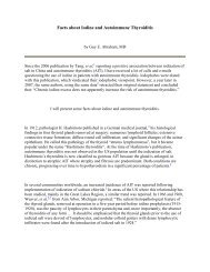
Figure 1 Adverse effects of increased lipolysis <strong>and</strong> FFA mobilization.<br />
FFAs, free fatty acids; TG, triglyceride.<br />
Rate of lipolysis – free fatty acids <strong>and</strong><br />
glycerol release<br />
Visceral adipocytes are more metabolically active <strong>and</strong> have<br />
a greater lipolytic activity than SCAT adipocytes (44,45).<br />
VAT is more susceptible to the catecholamine-induced<br />
lipolysis <strong>and</strong> less to the anti-lipolytic action of insulin.<br />
Free fatty acids induce insulin resistance. In the liver,<br />
insulin inhibits gluconeogenesis <strong>and</strong> glycogenlysis <strong>and</strong><br />
stimulates glycogen formation. Actions that limit hepatic<br />
glucose production are shown in Fig. 1.<br />
The degree of FFA suppression following meal ingestion<br />
differs between abdominally <strong>and</strong> peripherally obese<br />
persons. FFAs release is greater in the abdominally obese<br />
individuals (5).<br />
Glucose uptake<br />
Visceral <strong>adipose</strong> <strong>tissue</strong> has higher rate of insulin-stimulated<br />
glucose uptake compared with SCAT adipocytes.<br />
Absorption of circulating free fatty acids<br />
<strong>and</strong> triglycerides<br />
Small adipocytes in SCAT have a high avidity for FFAs <strong>and</strong><br />
TG uptake. The new, small, more insulin-sensitive adipocytes<br />
act as a sink or powerful ‘buffers’, avidly absorbing<br />
circulating FFAs <strong>and</strong> TGs in the postpr<strong>and</strong>ial period (5,8).<br />
SCAT cells may act as a buffer or sink for circulating FFAs<br />
<strong>and</strong> TGs, but once they reach their capacity they loose their<br />
protective benefit, fat begins to accumulate in <strong>tissue</strong>s not<br />
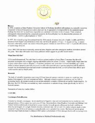
suited for lipid storage (5) (Fig. 2). SCAT in abdominal wall<br />
has higher uptake of TGs <strong>and</strong> larger FFA release per kilograms<br />
than does femoral fat (10,43).<br />
© 2009 The Author<br />
Journal compilation © 2009 International Association for the Study of Obesity. obesity reviews 11, 11–18
obesity reviews <strong>Subcutaneous</strong> <strong>and</strong> <strong>visceral</strong> <strong>adipose</strong> <strong>tissue</strong> M. M. Ibrahim 15<br />
Figure 2 Energy surplus results in accumulation of triglycerides in<br />
adipocytes at subcutaneous <strong>adipose</strong> <strong>tissue</strong>, which acts as a metabolic<br />
sink. When capacity of subcutaneous fat is exceeded or if it is<br />
impaired, fat will accumulate in areas outside the subcutaneous<br />
compartment.<br />
Clinical <strong>and</strong> prognostic differences<br />
Metabolic risks<br />
Visceral fat accumulation is associated with tendency to<br />
hyperglycaemia, hyperinsulinemia, hypertriglyceridemia,<br />
impaired glucose tolerance, increased apolipoproteins<br />
B-rich lipoproteins, which are features of the insulin resistance<br />
syndrome. Increased risk of developing diabetes is<br />
greater in individuals with excess VAT (42,45,46). Individuals<br />
with high levels of VAT area had higher mean<br />
plasma cholesterol <strong>and</strong> TG levels <strong>and</strong> lower high-density<br />
lipoprotein cholesterol values (47).<br />
Metabolic syndrome<br />
An increased body WC is considered now to be a prerequisite<br />
of the metabolic syndrome.<br />
Visceral obesity, like hyperinsulinemia <strong>and</strong> insulin resistance,<br />
not only accompanies but antedates the components<br />
of the metabolic syndrome (45,48).<br />
Elevated arterial blood pressure that is one of the components<br />
of the metabolic syndrome was explained by<br />
insulin resistance <strong>and</strong> compensating hyperinsulinemia in<br />
<strong>visceral</strong>ly obese individuals (49–51). Central obesity can<br />
induce the development of hypertension through increased<br />
activity of <strong>adipose</strong> <strong>tissue</strong> renin-angiotensin-aldosterone<br />
system, sympathetic activation <strong>and</strong> other mechanisms<br />
closely connected with insulin resistance.<br />
Vascular risk <strong>and</strong> cardiovascular events<br />
Visceral fat quantified as waist size has been identified as an<br />
independent risk factor for cardiovascular disease, hypertension<br />
<strong>and</strong> stroke (46,52). Excess VAT has the potential to<br />
cause hypercoagulability because of increased secretion of<br />
PAI-1. Increased WC when accompanied by increased TG<br />
leads to increased risk of coronary heart disease (53,54).<br />
Abdominal obesity correlates closely with other measures<br />
of atherosclerosis such as intima–media thickness<br />
(55). Peripheral arterial disease also has been correlated to<br />
VAT, but not to total body fat in elderly subjects (56).<br />
Subjects with abdominal obesity were reported to have<br />
greater risk of having an abnormal albumin excretion rate<br />
(57). Microalbuminuria signifies enhanced cardiovascular<br />
risk. Hyperinsulinemia, associated with <strong>visceral</strong> obesity, is<br />
a predictor of coronary artery disease (58).<br />
Increase in circulating FFAs in abdominal obesity is associated<br />
with increase in cardiovascular risk. Elevations in<br />
FFA levels promote endothelial dysfunction (50).<br />
Assessment of cardiovascular risk in obese patients from<br />
the measurement of body weight may be misleading (59).<br />
Only obese individuals characterized by increased VAT<br />
show the complications predictive of type 2 diabetes <strong>and</strong><br />
cardiovascular disease (51). Women generally display a<br />
more favourable risk profile than men, <strong>and</strong> generally lower<br />
level of VAT than men. Adjustments for differences in <strong>visceral</strong><br />
fat between men <strong>and</strong> women eliminated most of the<br />
sex differences in cardiovascular risk factors (60). Peripheral<br />
or gluteofemoral fat distribution seems to be protective<br />
against atherosclerosis (61–63).<br />
Prediction of mortality<br />
Obesity is associated with increased cardiovascular disease<br />
mortality (64,65). Cardiovascular disease death rates are<br />
directly related to body mass index in both men <strong>and</strong><br />
women. Obesity in adulthood is also associated with a<br />
striking reduction in life expectancy (66). Abdominal adiposity<br />
as measured by WC is a significant predictor of<br />
mortality independently of body mass index (67). Visceral<br />
fat is a strong, independent predictor of all-cause mortality<br />
in men (68).<br />
Effects of weight reduction<br />
© 2009 The Author<br />
Journal compilation © 2009 International Association for the Study of Obesity. obesity reviews 11, 11–18<br />
Visceral <strong>adipose</strong> <strong>tissue</strong> is more sensitive to weight reduction<br />
than SCAT (percentage wise) (69). All forms of weight<br />
loss affect <strong>visceral</strong> fat more than subcutaneous fat (70).<br />
However, recently Hall <strong>and</strong> Hallgreen (71) using simple<br />
allometric model predicts that increasing weight loss<br />
attenuates the preferential loss of VAT vs. SCAT. They<br />
concluded that greater weight loss will cause a greater<br />
absolute reduction of VAT mass.<br />
Obesity being a chronic low-grade inflammatory state is<br />
associated with increased plasma levels of inflammatory<br />
markers such CRP. A recent study shows that weight reduction<br />
is associated with decrease in CRP level. For every 1 kg<br />
of weight loss, CRP levels dip by 0.13 mg L -1 (72). Other<br />
adipokines also change with weight loss. Extreme weight<br />
reduction in obese individuals was associated with an<br />
increase in plasma adiponectin concentrations (73).
16 <strong>Subcutaneous</strong> <strong>and</strong> <strong>visceral</strong> <strong>adipose</strong> <strong>tissue</strong> M. M. Ibrahim obesity reviews<br />
Future directions<br />
Research is needed on the molecular <strong>and</strong> cellular mechanisms<br />
<strong>and</strong> signals for <strong>adipose</strong> <strong>tissue</strong>. Determination as to<br />
which adipocyte factors are most important in promoting<br />
metabolic disease will be an important focus for future<br />
studies.<br />
Manipulation of adipocyte biology might be a useful<br />
therapeutic strategy in metabolic disease. There is a lot of<br />
work that remains to be done in the area of adopocytes <strong>and</strong><br />
adipokines physiology. There are large gaps in our knowledge<br />
about the mechanism of action of many adipokines.<br />
The existence of additional, yet-unidentified adipocytespecific<br />
factors is highly likely. Prospective cohort trials that<br />
use imaging procedures to estimate abdominal <strong>and</strong> various<br />
<strong>adipose</strong> <strong>tissue</strong> depots in a precise manner, assess biochemical<br />
<strong>and</strong> metabolic parameters in relation to cardiovascular<br />
endpoints are needed.<br />
Racial differences in the accumulation of <strong>adipose</strong> <strong>tissue</strong><br />
depots <strong>and</strong> resulting cardiovascular risk need further<br />
investigation.<br />
Abdominal obesity is very common in Middle Eastern<br />
countries (74). Women in Egypt <strong>and</strong> Turkey have the highest<br />
proportion of overweight as well as the highest proportion<br />
of obesity (75). Ongoing studies are aiming to identify the<br />
WC cut-off points that define the threshold of abdominal<br />
obesity <strong>and</strong> an abdominal waist girth. There are racial <strong>and</strong><br />
genetic variations in body fat distribution that dictates the<br />
needs for different thresholds of abdominal obesity among<br />
different populations. We are currently correlating in a<br />
cross-sectional study (MM Ibrahim et al. unpublished<br />
observations) the prevalence of a number of cardiovascular<br />
risk factors at different levels of WC. The cut-off point based<br />
upon this approach is different from the threshold among<br />
US, European <strong>and</strong> Asian men <strong>and</strong> women.<br />
Conflict of Interest Statement<br />
No conflict of interest was declared.<br />
References<br />
1. Vague J. The degree of masculine differentiation of obesities: a<br />
factor determining predisposition to diabetes, atherosclerosis, gout<br />
<strong>and</strong> uri-calculus disease. Am J Clin Nutr 1956; 4: 20–29.<br />
2. Bruun JM, Lihn AS, Pedersen SB, Richelsen B. Monocyte<br />
chemoattractant Protein-1 release is higher in <strong>visceral</strong> than subcutaneous<br />
human <strong>adipose</strong> <strong>tissue</strong> (AT): implication of macrophages<br />
resident in the AT. J Clin Endocrinol Metab 2005; 90: 2282–2289.<br />
3. Curat CA, Wegner V, Sengenès C, Miranville A, Tonus C, Busse<br />
R, Bouloumié A. Macrophages in human <strong>visceral</strong> <strong>adipose</strong> <strong>tissue</strong>:<br />
increased accumulation in obesity <strong>and</strong> a source of resistin <strong>and</strong><br />
visfatin. Diabetologia 2006; 49: 744–747.<br />
4. Weisberg SP, McCann D, Desai M, Rosenbaum M, Leibel RL,<br />
Ferrante AW. Obesity is associated with macrophage accumulation<br />
in <strong>adipose</strong> <strong>tissue</strong>. J Clin Invest 2003; 112: 1796–1808.<br />
5. Freedl<strong>and</strong> ES. Role of critical <strong>visceral</strong> <strong>adipose</strong> <strong>tissue</strong> threshold<br />
in metabolic syndrome: implications for controlling dietary carbohydrates:<br />
a review. Nutr Metab 2004; 1: 12.<br />
6. Bjorntrop P. Do stress reactions cause abdominal obesity <strong>and</strong><br />
comorbidities. Obes Rev 2001; 2: 73–86.<br />
7. Wajchenberg BL. <strong>Subcutaneous</strong> <strong>and</strong> <strong>visceral</strong> <strong>adipose</strong> <strong>tissue</strong>:<br />
their relation to the metabolic syndrome. Endocr Rev 2000; 21:<br />
679–738.<br />
8. Arner P. Obesity <strong>and</strong> the adipocyte. Regional adipocity in man.<br />
J Endocrinol 1997; 155: 191–192.<br />
9. Heinrich PC, Castell JV, Andus T. Interlukin-6 <strong>and</strong> the acute<br />
phase response. Biochem J 1990; 265: 621–636.<br />
10. Mårin P, Andersson B, Ottosson M, Olbe L, Chowdhury B,<br />
Kvist H, Holm G, Sjöström L, Björntorp P. The morphology <strong>and</strong><br />
metabolism of intra-abdominal <strong>adipose</strong> <strong>tissue</strong> in men. Metabolism<br />
1992; 41: 1241–1248.<br />
11. Hisra A, Vikram NK. Clinical <strong>and</strong> pathophysiological consequences<br />
of abdominal adiposity <strong>and</strong> abdominal <strong>adipose</strong> <strong>tissue</strong><br />
depots. Nutrition 2003; 19: 457–466.<br />
12. Trayhurn P, Wood IS. Adipokines: inflammation <strong>and</strong> pleiotropic<br />
role of white <strong>adipose</strong> <strong>tissue</strong>. Br J Nutr 2004; 92: 347–<br />
355.<br />
13. Joyner JM, Hutley LJ, Cameron DP. Glucocorticoid receptors<br />
in human preadipocytes: regional <strong>and</strong> gender variations. J Endocrinol<br />
2000; 166: 145.<br />
14. Rebuffe’-Scrive M, Lundholm K, Björntorp P. Glucocorticoid<br />
hormone binding to human <strong>adipose</strong> <strong>tissue</strong>. Eur J Clin Invest 1985;<br />
15: 267–271.<br />
15. Bjorntorp P. Endocrine abnormalities in obesity. Diabetes Rev<br />
1997; 5: 52–68.<br />
16. Mitzutani T, Nishikawa Y, Adachi H, Enomoto T, Ikegami H,<br />
Kurachi H, Nomura T, Miyake A. Identification of estrogen receptor<br />
in human <strong>adipose</strong> <strong>tissue</strong> <strong>and</strong> adipocytes. J Clin Endocrinol<br />
Metab 1994; 78: 950–954.<br />
17. Pedersen SB, Hansen PS, Lund S, Andersen PH, Odgaard A,<br />
Richelsen B. Identification of oestrogen receptors <strong>and</strong> oestrogen<br />
receptor mRNA in human <strong>adipose</strong> <strong>tissue</strong>. Eur J Clin Invest 1996;<br />
26: 259.<br />
18. Hellmer J, Marcus C, Sonnefeld T, Arner P. Mechanisms for<br />
differences in lipolysis between human subcutaneous <strong>and</strong> omental<br />
fat cells. J Clin Endocrinol Metab 1992; 75: 15–20.<br />
19. Arner P, Hellstrom L, Wahrenberg H, Bronnegard M.<br />
b-adrenoceptor expression in human fat cells from different<br />
regions. J Clin Invest 1990; 86: 1595–1600.<br />
20. Imbeault P, Couillard C, Tremblay A, Després J-P, Mauriège<br />
P. Reduced alpha (2)-adrenergic sensitivity of subcutaneous<br />
abdominal adipocytes as a modulator of fasting <strong>and</strong> post-pr<strong>and</strong>ial<br />
triglyceride levels in men. J Lipid Res 2000; 41: 1367.<br />
21. Krief S, Lönnqvist F, Raimbault S, Baude B, Van Spronsen A,<br />
Arner P, Strosberg AD, Ricquier D, Emorine LJ. Tissue distribution<br />
of b3-adrenergic receptor MRNA in man. J Clin Invest 1993;<br />
91: 344–349.<br />
22. Matsuzawa Y. Therapy insight: adipocytokines in metabolic<br />
syndrome <strong>and</strong> related cardiovascular disease. Nat Clin Pract<br />
Cardiovasc Med 2006; 3: 35–42.<br />
23. David CW, Dhillon B, Yan H, Szmitko PE, Verma S. Adipokines:<br />
molecular links between obesity <strong>and</strong> atherosclerosis. Am J<br />
Physiol Heart Circ Physiol 2005; 288: H2031–H2041.<br />
24. Tritos NA, Mantzoros CS. Leptin: its role in obesity <strong>and</strong><br />
beyond. Diabetologica 1997; 40: 1371–1379.<br />
25. Maury E, Ehala-Aleksejev K, Guiot Y, Detry R, V<strong>and</strong>enhooft<br />
A, Brichard SM. Adipokines oversecreted by omental <strong>adipose</strong><br />
<strong>tissue</strong> in human obesity. Am J Physiol Endocrinol Metab 2007;<br />
293: E656–E665.<br />
© 2009 The Author<br />
Journal compilation © 2009 International Association for the Study of Obesity. obesity reviews 11, 11–18
obesity reviews <strong>Subcutaneous</strong> <strong>and</strong> <strong>visceral</strong> <strong>adipose</strong> <strong>tissue</strong> M. M. Ibrahim 17<br />
26. Mantzoros CS, Moschos S, Avramopoulos I, Kaklamani V,<br />
Liolios A, Doulgerakis DE, Griveas I, Katsilambros N, Flier JS.<br />
Leptin concentrations in relation to body mass index <strong>and</strong> the<br />
tumor necrosis factor-a system in humans. J Clin Endocrinol<br />
Metab 1997; 82: 3408–3413.<br />
27. Kirchgessner TG, Uysal T, Wiesbrock SM, Marino MW,<br />
Hotamisligil G. Tumour necrosis factor-alpha contributes to<br />
obesity related hypoleptinemia by regulating leptin release from<br />
adipocytes. J Clin Invest 1997; 100: 2777–2782.<br />
28. Morange P, Alessi MC, Ventura N, Casanova D, Magalon G,<br />
Juhan-Vague I. PAI-1 antigen production by human adipode <strong>tissue</strong><br />
is correlated with that of TNF-a. Eighth international congress on<br />
obesity (Paris, France, August 29–September 3, 1998). Int J Obes<br />
1998; 22(Suppl. 3): P31–PS103.<br />
29. Ouchi N, Kihara S, Arita Y, Maeda K, Kuriyama H, Okamoto<br />
Y, Hotta K, Nishida M, Takahashi M, Nakamura T, Yamashita S,<br />
Funahashi T, Matsuzawa Y. Novel modulator for endothelial<br />
adhesion molecules: adipocyte-derived plasma protein adiponectin.<br />
Circulation 1999; 100: 2473–2476.<br />
30. Brunn JM, Lihn AS, Verdich C, Pedersen SB, Toubro S, Astrup<br />
A, Richelsen B. Regulation of adiponectin by <strong>adipose</strong> <strong>tissue</strong>derived<br />
cytokines: in vivo an in vitro investigations in human. Am<br />
J Physiol Endocrinol Metab 2003; 285: E527–E533.<br />
31. Motoshima H, Wu X, Sinha M, Hardy E, Rosato EL, Barbot<br />
DJ, Rosato FE, Goldstein BJ. Differential regulation of adiponectin<br />
secretion from cultured human omental <strong>and</strong> subcutaneous adipocytes:<br />
effects of insulin <strong>and</strong> rosiglitazone. J Clin Endocrinol Metab<br />
2002; 87: 5662–5667.<br />
32. Lemieux I, Pascot A, Prud’homme D, Alméras N, Bogaty P,<br />
Nadeau A, Bergeron J, Després JP. Elevated C-reactive protein:<br />
another component of the atherothrombotic profile of abdominal<br />
obesity. Arterioscler Thromb Vasc Biol 2001; 21: 961–967.<br />
33. Pepys MB, Hirschfield C-M. C-reactive protein: a critical<br />
update. J Clin Invest 2003; 111: 1805–1812.<br />
34. Forouhi NG, Sattar N, McKeigue PM. Relation of C-reactive<br />
protein to body fat distribution <strong>and</strong> features of the metabolic<br />
syndrome in Europeans <strong>and</strong> South Asians. Int J Obs Relat Metab<br />
Discord 2001; 25: 1327–1331.<br />
35. Pou KM, Massaro JM, Hoffmann U, Vasan RS, Maurovich-<br />
Horvat P, Larson MG, Keaney JF, Meigs JB, Lipinska I, Kathiresan<br />
S, Murabito JM, O’Donnell CJ, Benjamin EJ, Fox CS, Adipose S.<br />
Visceral <strong>and</strong> subcutaneous <strong>adipose</strong> <strong>tissue</strong> volumes are crosssectionally<br />
related to markers of inflammation <strong>and</strong> oxidative stress:<br />
the framingham heart study. Circulation 2007; 116: 1234–1241.<br />
36. Karlsson C, Lindell K, Ottoson M, Sjostrom L, Carlsson B,<br />
Carlson LMS. Human <strong>adipose</strong> <strong>tissue</strong> expresses angiotensinogen<br />
<strong>and</strong> enzymes required for its conversion to angiotensin II. J Clin<br />
Endocrinol Metab 1998; 83: 3925–3929.<br />
37. Dusserre E, Moulin P, Vidal H. Differences in mRNA<br />
expression of proteins secreted by adipocytes in human subcutaneous<br />
<strong>and</strong> <strong>visceral</strong> <strong>adipose</strong> <strong>tissue</strong>. Biochim Biophy Acta 2000;<br />
1500: 88.<br />
38. Alessi MC, Peirretti F, Morange P, Henry M, Nalbone G,<br />
Juhan-Vague I. Production of plasminogen activator inhibitor 1 by<br />
human <strong>adipose</strong> <strong>tissue</strong> – possible link between <strong>visceral</strong> fat accumulation<br />
<strong>and</strong> vascular disease. Diabetes 1997; 46: 860.<br />
39. Abate N, Garg A, Peshock RM, Stray-Gundersen J, Grundy<br />
SM. Relationship of generalized <strong>and</strong> regional adiposity to insulin<br />
sensitivity in man. J Clin Invest 1995; 96: 88–98.<br />
40. Frayn KN. Visceral fat <strong>and</strong> insulin resistance: causative a<br />
correlative. Br J Nutr 2000; 83(Suppl. 1): S71–S77.<br />
41. Salans LB, Cushman SW, Weismann RE. Studies of human<br />
<strong>adipose</strong> <strong>tissue</strong>. Adipose all size <strong>and</strong> number in nonobese <strong>and</strong> obese<br />
patients. J Clin Invest 1973; 52: 929–941.<br />
© 2009 The Author<br />
Journal compilation © 2009 International Association for the Study of Obesity. obesity reviews 11, 11–18<br />
42. Bjorntorp P. Metabolic difference between <strong>visceral</strong> fat <strong>and</strong><br />
subcutaneous abdominal fat. Diabetes Metab 2000; 26(Suppl. 3):<br />
10–12.<br />
43. Kadswaki T, Hara K, Yamauchi T, Terauchi Y, Tobe K, Nagai<br />
R. Molecular mechanisms of insulin resistance <strong>and</strong> obesity. Exp<br />
Biol Med 2003; 228: 1111–1117.<br />
44. Amer P. Differences in lipolysis between human subcutaneous<br />
<strong>and</strong> omental <strong>adipose</strong> <strong>tissue</strong>s. Ann Med 1995; 27: 435–438.<br />
45. Lemieux S, Despres SP. Metabolic complications of <strong>visceral</strong><br />
obesity: contribution to the etiology of life of type 2 diabetes <strong>and</strong><br />
implications for prevention <strong>and</strong> treatment. Diabetes Metab 1994;<br />
20: 375–393.<br />
46. Dobbelsteyn CJ, Joffres MR, MacLean DR, Flowerdew G. A<br />
comparative evaluation of waist circumference. Waist-to-hip ratio<br />
<strong>and</strong> body mass index as indicators of cardiovascular risk factors.<br />
The Canadian Heart Health Surveys. Int J Obs Relat Metab<br />
Disord 2001; 25: 652.<br />
47. Despres JP, Allard C, Tremblay A, Talbot J, Bouchard C.<br />
Evidence for a regional component of body fatness in association<br />
with serum in men <strong>and</strong> women. Metabolism 1985; 34: 967–973.<br />
48. Bergstrom RW, Newell-Morris LL, Leonetti DL, Shuman WP,<br />
Wahl PW, Fujimoto WY. Association of elevated fasting C-peptide<br />
level <strong>and</strong> increased intra-abdominal fat distribution with development<br />
of MID in Japanese-American men. Diabetes 1990; 39: 104–<br />
111.<br />
49. Rocchini A. Obesity hypertension. Am J Hypertens 2002; 15:<br />
505–525.<br />
50. Sharma AM, Engeli S, Pischon T. New developments in<br />
mechansims of obesity-induced hypertension: role of <strong>adipose</strong><br />
<strong>tissue</strong>. Curr Hypertens Rep 2001; 3: 152–156.<br />
51. Mc Farlane SI, Banerji M, Sowers JR. Insulin resistance <strong>and</strong><br />
cardiovascular disease. J Clin Endocrinol Metabol 2001; 86: 713–<br />
718.<br />
52. Despres JP. The atherothrombotic <strong>and</strong> anti-inflammatory<br />
profile of <strong>visceral</strong> obesity. Int Congr Ser 2003; 23: 27–34.<br />
53. Tanko LB, Bagger YZ, Qin G, Alex<strong>and</strong>ersen P, Larsen PJ,<br />
Christiansen C. Enlarged waist combined with elevated triglycerides<br />
is a strong predictor of accelerated atherogenesis <strong>and</strong> related<br />
cardiovascular mortality in postmenopausal women. Circulation<br />
2005; 111: 1883–1890.<br />
54. Lemieux I, Pascot A, Couillard C, Lamarche B, Tchernof A,<br />
Alméras N, Bergeron J, Gaudet D, Tremblay G, Prud’homme D,<br />
Nadeau A, Després JP. Hypertriglyceridemic waist: A marker of<br />
the atherogenic metabolic triad (hyperinsulinemia, hyperapolipoprotein<br />
B, small dense LDL) in men. Circulation 2000; 102: 179–<br />
184.<br />
55. Harris MM, Stevens J, Thomas N, Schreiner P, Folsom AR.<br />
Association of fat distribution <strong>and</strong> obesity with hypertension in a<br />
bi-ethnic population: the ARIC study. Atherosclerosis Risk in<br />
Communities Study. Obes Res 2000; 8: 516–524.<br />
56. Planas A, Clará A, Pou J-M, Vidal-Barraquer F, Gasol A, de<br />
Moner A, Contreras C, Marrugat J. Relationship of obesity distribution<br />
to peripheral arterial occlusive disease in elderly men. Int J<br />
Obes Relat Metab Disord 2001; 25: 1068–1070.<br />
57. Mulyadi L, Stevens C, Munro S, Lingard J, Bermingham M.<br />
Body fat distribution <strong>and</strong> total fat as risk factors for microalbuminuria<br />
in the obese. Ann Nutr Metab 2000; 45: 6–71.<br />
58. Fontbonne A, Charles MA, Thibult N, Richard JL, Claude JR,<br />
Warnet JM, Rosselin GE, Eschwège E. Hyperinsulinemia as a<br />
predictor of coronary heart disease mortality in a healthy population:<br />
the Paris Prospective Study, 15-year follow-up. Diabetologica<br />
1991; 34: 356–361.<br />
59. Kissebah AH. Central obesity: measurement <strong>and</strong> metabolic<br />
effects. Diabet Rev 1997; 5: 8–20.
18 <strong>Subcutaneous</strong> <strong>and</strong> <strong>visceral</strong> <strong>adipose</strong> <strong>tissue</strong> M. M. Ibrahim obesity reviews<br />
60. Lemieax S, Després JP, Moorjani S, Nadeau A, Thériault G,<br />
Prud’homme D, Tremblay A, Bouchard C, Lupien PJ. Are gender<br />
differences of cardiovascular disease risk factors explained by the<br />
level of <strong>visceral</strong> <strong>adipose</strong> <strong>tissue</strong>. Diabetologica 1994; 37: 757–764.<br />
61. Tanko LB, Bagger YZ, Alex<strong>and</strong>ersen P, Larsen PJ, Christiansen<br />
C. Central <strong>and</strong> peripheral fat mass have contrasting effect<br />
on the progression of aortic calcification in post menopausal<br />
women. Eur Heart J 2003; 24: 1531–1537.<br />
62. Tanko LB, Bagger YZ, Alex<strong>and</strong>ersen P, Larsen PJ, Christiansen<br />
C. Peripheral adiposity exhibits an independent dominant<br />
antiatherogeneic effect in elderly women. Circulation 2003; 107:<br />
1626–1631.<br />
63. Lassner L, Bjokelund C, Heitmann BL, Seidell JC, Bengtssan C.<br />
Larger hip circumference independently predicts health <strong>and</strong> longitivity<br />
in a Swedish female cohort. Obes Res 2001; 9: 644–646.<br />
64. Dagenais GR, Yi Q, Mann JF, Bosch J, Pogue J, Yusuf S.<br />
Prognostic impact of body weight <strong>and</strong> abdominal obesity in<br />
women <strong>and</strong> men with cardiovascular disease. Am Heart J 2005;<br />
149: 54–60.<br />
65. Allison DB, Fontaine KR, Manson JE, Stevens J, VanItallie TB.<br />
Annual death attributable to obesity in the United States. JAMA<br />
1999; 282: 1530–1538.<br />
66. Lee IM, Blair SN, Allison DB, Folsom AR, Harris TB, Manson<br />
JE, Wing RR. Epidemiologic data on the relationships of calori<br />
intake, energy balance, <strong>and</strong> weight gain over the lifespan <strong>and</strong><br />
longevity <strong>and</strong> morbidity. J Gerontol A Biol Sci Med Sci 2001; 56:<br />
7–19.<br />
67. Zhang X, Shu XO, Yang G, Li H, Cai H, Gao UT, Zheng W.<br />
Abdominal obesity <strong>and</strong> mortality in Chinese women. Arch Intern<br />
Med 2007; 167: 886–892.<br />
68. Kuk JL, Katzmarzyk PT, Nichaman MZ, Church TS, Blair<br />
SN, Ross R. Visceral fat is an independent predictor of all-cause<br />
mortality in men. Obesity 2006; 14: 336–341.<br />
69. Bjorntorp P. Regional obesity. In: Bjorntorp P, Brodoff BN<br />
(eds). Obesity. JB Lippincott Co.: Philadelphica, PA, 1992, pp.<br />
S79–S86.<br />
70. Armellini F, Zamboni M, Rigo L, Bergamo-Andries IA,<br />
Robbi R, De Marchi M, Bosello O. Sonography detection of<br />
small intra abdominal fat variation. Int J Obes 1991; 15: 847–<br />
852.<br />
71. Hall KD, Hallgreen CE. Increasing weigh loss attenuates the<br />
preferential loss of <strong>visceral</strong> vs. subcutaneous fat: a predicted result<br />
of an allometric model. Int J Obes 2008; 32: 722.<br />
72. Selvin E, Paynter NP, Erlinger TP. The effect of weight loss on<br />
C-reactive protein: a systematic review. Arch Intern Med 2007;<br />
167: 31–39.<br />
73. Corpeleijn E, Feskens EJM, Jansen EH, Mensink M, Saris<br />
WHM, Blaak EE. Lifestyle intervention <strong>and</strong> adipokine levels in<br />
subjects at high risk for type 2 diabetes: the study on lifestyle<br />
intervention <strong>and</strong> impaired glucose tolerance Maastricht (SLIM).<br />
Diabetes Care 2007; 30: 3125–3127.<br />
74. WHO Eastern Mediterranean Region: Annual Report of<br />
the Regional Director. The work of WHO in the Eastern<br />
Mediterranean Region (2006): Annual Report of the Regional<br />
Director. [WWW document]. URL http://www.emro.who.int/rd/<br />
annualreports/2006/chapter1_6.htm (accessed 22 March 2009).<br />
75. Martorell R, Khan LK, Hughes ML, Grummer-Strawn LM.<br />
Obesity in women from developing countries. Eur J Clin Nutr<br />
2000; 54: 247–252.<br />
© 2009 The Author<br />
Journal compilation © 2009 International Association for the Study of Obesity. obesity reviews 11, 11–18