Low Spigelian hernia in a 6-year-old boy presenting as an ...
Low Spigelian hernia in a 6-year-old boy presenting as an ...
Low Spigelian hernia in a 6-year-old boy presenting as an ...
Create successful ePaper yourself
Turn your PDF publications into a flip-book with our unique Google optimized e-Paper software.
Journal of Medical C<strong>as</strong>e Reports<br />
C<strong>as</strong>e report<br />
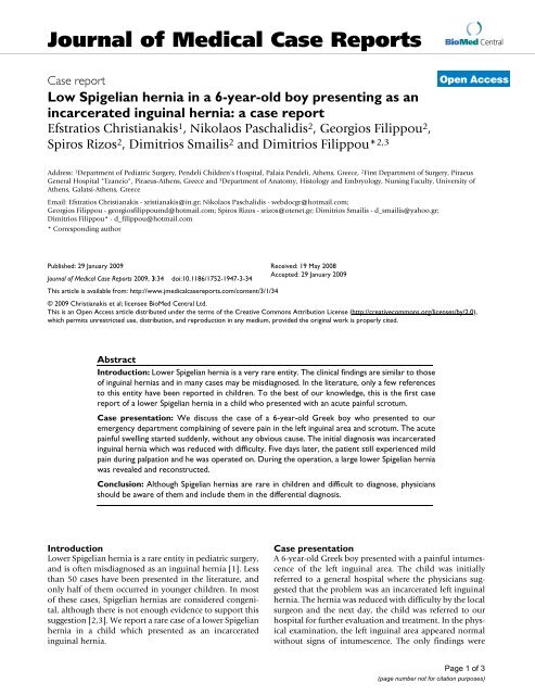
<strong>Low</strong> <strong>Spigeli<strong>an</strong></strong> <strong>hernia</strong> <strong>in</strong> a 6-<strong>year</strong>-<strong>old</strong> <strong>boy</strong> present<strong>in</strong>g <strong>as</strong> <strong>an</strong><br />
<strong>in</strong>carcerated <strong>in</strong>gu<strong>in</strong>al <strong>hernia</strong>: a c<strong>as</strong>e report<br />
Efstratios Christi<strong>an</strong>akis 1 , Nikolaos P<strong>as</strong>chalidis 2 , Georgios Filippou 2 ,<br />
Spiros Rizos 2 , Dimitrios Smailis 2 <strong>an</strong>d Dimitrios Filippou* 2,3<br />
BioMed Central<br />
Open Access<br />
Address: 1Department of Pediatric Surgery, Pendeli Children's Hospital, Palaia Pendeli, Athens, Greece, 2First Department of Surgery, Piraeus<br />
General Hospital "Tz<strong>an</strong>eio", Piraeus-Athens, Greece <strong>an</strong>d 3Department of Anatomy, Histology <strong>an</strong>d Embryology, Nurs<strong>in</strong>g Faculty, University of<br />
Athens, Galatsi-Athens, Greece<br />
Email: Efstratios Christi<strong>an</strong>akis - xristi<strong>an</strong>akis@<strong>in</strong>.gr; Nikolaos P<strong>as</strong>chalidis - webdocgr@hotmail.com;<br />
Georgios Filippou - georgiosfilippoumd@hotmail.com; Spiros Rizos - srizos@otenet.gr; Dimitrios Smailis - d_smailis@yahoo.gr;<br />
Dimitrios Filippou* - d_filippou@hotmail.com<br />
* Correspond<strong>in</strong>g author<br />
Published: 29 J<strong>an</strong>uary 2009<br />
Received: 19 May 2008<br />
Journal of Medical C<strong>as</strong>e Reports 2009, 3:34 doi:10.1186/1752-1947-3-34<br />
Accepted: 29 J<strong>an</strong>uary 2009<br />
This article is available from: http://www.jmedicalc<strong>as</strong>ereports.com/content/3/1/34<br />
© 2009 Christi<strong>an</strong>akis et al; licensee BioMed Central Ltd.<br />
This is <strong>an</strong> Open Access article distributed under the terms of the Creative Commons Attribution License (http://creativecommons.org/licenses/by/2.0),<br />
which permits unrestricted use, distribution, <strong>an</strong>d reproduction <strong>in</strong> <strong>an</strong>y medium, provided the orig<strong>in</strong>al work is properly cited.<br />
Abstract<br />
Introduction: <strong>Low</strong>er <strong>Spigeli<strong>an</strong></strong> <strong>hernia</strong> is a very rare entity. The cl<strong>in</strong>ical f<strong>in</strong>d<strong>in</strong>gs are similar to those<br />
of <strong>in</strong>gu<strong>in</strong>al <strong>hernia</strong>s <strong>an</strong>d <strong>in</strong> m<strong>an</strong>y c<strong>as</strong>es may be misdiagnosed. In the literature, only a few references<br />
to this entity have been reported <strong>in</strong> children. To the best of our knowledge, this is the first c<strong>as</strong>e<br />
report of a lower <strong>Spigeli<strong>an</strong></strong> <strong>hernia</strong> <strong>in</strong> a child who presented with <strong>an</strong> acute pa<strong>in</strong>ful scrotum.<br />
C<strong>as</strong>e presentation: We discuss the c<strong>as</strong>e of a 6-<strong>year</strong>-<strong>old</strong> Greek <strong>boy</strong> who presented to our<br />
emergency department compla<strong>in</strong><strong>in</strong>g of severe pa<strong>in</strong> <strong>in</strong> the left <strong>in</strong>gu<strong>in</strong>al area <strong>an</strong>d scrotum. The acute<br />
pa<strong>in</strong>ful swell<strong>in</strong>g started suddenly, without <strong>an</strong>y obvious cause. The <strong>in</strong>itial diagnosis w<strong>as</strong> <strong>in</strong>carcerated<br />
<strong>in</strong>gu<strong>in</strong>al <strong>hernia</strong> which w<strong>as</strong> reduced with difficulty. Five days later, the patient still experienced mild<br />
pa<strong>in</strong> dur<strong>in</strong>g palpation <strong>an</strong>d he w<strong>as</strong> operated on. Dur<strong>in</strong>g the operation, a large lower <strong>Spigeli<strong>an</strong></strong> <strong>hernia</strong><br />
w<strong>as</strong> revealed <strong>an</strong>d reconstructed.<br />
Conclusion: Although <strong>Spigeli<strong>an</strong></strong> <strong>hernia</strong>s are rare <strong>in</strong> children <strong>an</strong>d difficult to diagnose, physici<strong>an</strong>s<br />
should be aware of them <strong>an</strong>d <strong>in</strong>clude them <strong>in</strong> the differential diagnosis.<br />
Introduction<br />
<strong>Low</strong>er <strong>Spigeli<strong>an</strong></strong> <strong>hernia</strong> is a rare entity <strong>in</strong> pediatric surgery,<br />
<strong>an</strong>d is often misdiagnosed <strong>as</strong> <strong>an</strong> <strong>in</strong>gu<strong>in</strong>al <strong>hernia</strong> [1]. Less<br />
th<strong>an</strong> 50 c<strong>as</strong>es have been presented <strong>in</strong> the literature, <strong>an</strong>d<br />
only half of them occurred <strong>in</strong> younger children. In most<br />
of these c<strong>as</strong>es, <strong>Spigeli<strong>an</strong></strong> <strong>hernia</strong>s are considered congenital,<br />
although there is not enough evidence to support this<br />
suggestion [2,3]. We report a rare c<strong>as</strong>e of a lower <strong>Spigeli<strong>an</strong></strong><br />
<strong>hernia</strong> <strong>in</strong> a child which presented <strong>as</strong> <strong>an</strong> <strong>in</strong>carcerated<br />
<strong>in</strong>gu<strong>in</strong>al <strong>hernia</strong>.<br />
C<strong>as</strong>e presentation<br />
A 6-<strong>year</strong>-<strong>old</strong> Greek <strong>boy</strong> presented with a pa<strong>in</strong>ful <strong>in</strong>tumescence<br />
of the left <strong>in</strong>gu<strong>in</strong>al area. The child w<strong>as</strong> <strong>in</strong>itially<br />
referred to a general hospital where the physici<strong>an</strong>s suggested<br />
that the problem w<strong>as</strong> <strong>an</strong> <strong>in</strong>carcerated left <strong>in</strong>gu<strong>in</strong>al<br />
<strong>hernia</strong>. The <strong>hernia</strong> w<strong>as</strong> reduced with difficulty by the local<br />
surgeon <strong>an</strong>d the next day, the child w<strong>as</strong> referred to our<br />
hospital for further evaluation <strong>an</strong>d treatment. In the physical<br />
exam<strong>in</strong>ation, the left <strong>in</strong>gu<strong>in</strong>al area appeared normal<br />
without signs of <strong>in</strong>tumescence. The only f<strong>in</strong>d<strong>in</strong>gs were<br />
Page 1 of 3<br />
(page number not for citation purposes)
Journal of Medical C<strong>as</strong>e Reports 2009, 3:34 http://www.jmedicalc<strong>as</strong>ereports.com/content/3/1/34<br />
subcut<strong>an</strong>eous edema <strong>an</strong>d a hematoma, probably due to<br />
the m<strong>an</strong>ipulations for the reduction. Dur<strong>in</strong>g palpation,<br />
the child presented with mild discomfort <strong>an</strong>d pa<strong>in</strong> <strong>in</strong> the<br />
left testicle without cl<strong>in</strong>ical evidence of testicular torsion.<br />
These symptoms persisted for 3 days <strong>an</strong>d f<strong>in</strong>ally we<br />
decided to operate on the patient. A lower <strong>Spigeli<strong>an</strong></strong> <strong>hernia</strong><br />
w<strong>as</strong> identified dur<strong>in</strong>g the operation. The <strong>hernia</strong> sac<br />
w<strong>as</strong> found to penetrate from a small defect (with <strong>an</strong> estimated<br />
diameter of about 1.5 cm) <strong>in</strong> the <strong>Spigeli<strong>an</strong></strong> f<strong>as</strong>cia<br />
with<strong>in</strong> Hesselbach's tri<strong>an</strong>gle (Figure 1).<br />
The lower <strong>Spigeli<strong>an</strong></strong> <strong>hernia</strong> orifice is usually located <strong>in</strong> a<br />
well-def<strong>in</strong>ed area. The defect develops <strong>as</strong> a r<strong>in</strong>g-like open<strong>in</strong>g<br />
through the fibers of the tr<strong>an</strong>sversalis f<strong>as</strong>cia <strong>an</strong>d the<br />
f<strong>as</strong>cia of the tr<strong>an</strong>sversalis abdom<strong>in</strong>us <strong>an</strong>d <strong>in</strong>ternal oblique<br />
abdom<strong>in</strong>us muscles. <strong>Spigeli<strong>an</strong></strong> <strong>hernia</strong>s usually present just<br />
lateral to the rectus muscle <strong>in</strong> the lower left quadr<strong>an</strong>t.<br />
In our patient, the <strong>hernia</strong> sac w<strong>as</strong> that of a typical <strong>Spigeli<strong>an</strong></strong><br />
<strong>hernia</strong>. The orifice diameter w<strong>as</strong> about 1.5 cm, while<br />
the sac extended up to 7 cm surrounded by preperitoneal<br />
fat (Figure 1). The <strong>hernia</strong> sac conta<strong>in</strong>ed a small part of the<br />
Intra-operative photograph of the <strong>hernia</strong> show<strong>in</strong>g the opened sac<br />
Figure 1<br />
Intra-operative photograph of the <strong>hernia</strong> show<strong>in</strong>g the opened sac.<br />
large omentum which w<strong>as</strong> reduced. Reconstruction of the<br />
<strong>hernia</strong> w<strong>as</strong> performed with non-absorbable sutures. Eight<br />
<strong>year</strong>s after the operation, the patient rema<strong>in</strong>s free of symptoms<br />
<strong>an</strong>d recurrence.<br />
Discussion<br />
<strong>Spigeli<strong>an</strong></strong> <strong>hernia</strong>s occur through a congenital or usually<br />
acquired defect of the <strong>Spigeli<strong>an</strong></strong> f<strong>as</strong>cia, lateral to the rectus<br />
muscle sheath [1]. In adults, almost 90% of acquired <strong>hernia</strong>s<br />
occur with<strong>in</strong> the <strong>an</strong>terior superior iliac sp<strong>in</strong>e <strong>an</strong>d the<br />
umbilicus, while the majority of congenital <strong>hernia</strong>s occur<br />
at the level of the arcuate l<strong>in</strong>e (f<strong>old</strong> of Dougl<strong>as</strong>) [1-3]. Herni<strong>as</strong><br />
that are protrud<strong>in</strong>g through the <strong>Spigeli<strong>an</strong></strong> f<strong>as</strong>cia<br />
with<strong>in</strong> Hesselbach's tri<strong>an</strong>gle, caudally <strong>an</strong>d medially to the<br />
<strong>in</strong>ferior epig<strong>as</strong>tric vessels, are called low or lower <strong>Spigeli<strong>an</strong></strong><br />
<strong>hernia</strong>s [4]. Direct <strong>in</strong>gu<strong>in</strong>al <strong>hernia</strong>s c<strong>an</strong> also occur <strong>in</strong><br />
this area, although <strong>in</strong> most of these c<strong>as</strong>es, the <strong>hernia</strong>'s orifice<br />
is difficult to identify. This is the re<strong>as</strong>on why m<strong>an</strong>y<br />
<strong>Spigeli<strong>an</strong></strong> <strong>hernia</strong>s are misdiagnosed <strong>an</strong>d usually considered<br />
<strong>as</strong> direct <strong>in</strong>gu<strong>in</strong>al <strong>hernia</strong>s, para-<strong>in</strong>gu<strong>in</strong>al <strong>hernia</strong>s or<br />
<strong>hernia</strong>s through the conjo<strong>in</strong>ed tendon [5,6]. The diagnosis<br />
is usually made on the b<strong>as</strong>is of the location of the her-<br />
Page 2 of 3<br />
(page number not for citation purposes)
Journal of Medical C<strong>as</strong>e Reports 2009, 3:34 http://www.jmedicalc<strong>as</strong>ereports.com/content/3/1/34<br />
nia's orifice. The orifice of the <strong>Spigeli<strong>an</strong></strong> <strong>hernia</strong> is usually<br />
located <strong>in</strong>side the fibers of the tr<strong>an</strong>sversalis f<strong>as</strong>cia. The orifice<br />
is sharp <strong>an</strong>d rigid <strong>in</strong> palpation <strong>an</strong>d its diameter r<strong>an</strong>ges<br />
from 2 to 3 cm [2,3,6]. It extends through this defect <strong>in</strong>terstitially<br />
or penetrates the entire abdom<strong>in</strong>al wall [7,8]. The<br />
sac of these <strong>hernia</strong>s usually conta<strong>in</strong>s preperitoneal fatty<br />
tissue, <strong>an</strong>d rarely omentum or <strong>in</strong>test<strong>in</strong>e. Some authors<br />
have suggested that, <strong>in</strong> <strong>in</strong>f<strong>an</strong>ts, <strong>Spigeli<strong>an</strong></strong> <strong>hernia</strong>s may<br />
coexist with undescended testes.<br />
The small size of the <strong>hernia</strong> neck may be a possible expl<strong>an</strong>ation<br />
for the <strong>in</strong>cre<strong>as</strong>ed <strong>in</strong>cidence of <strong>in</strong>carceration. The<br />
<strong>in</strong>cidence of <strong>in</strong>carceration varies signific<strong>an</strong>tly between<br />
various series <strong>an</strong>d authors. Some authors have suggested<br />
that <strong>Spigeli<strong>an</strong></strong> <strong>hernia</strong> <strong>in</strong>carceration is a rare entity while<br />
others have suggested that it is very common with <strong>an</strong> estimated<br />
<strong>in</strong>cidence of approximately 22% [9,10].<br />
The pre-operative diagnosis of lower <strong>Spigeli<strong>an</strong></strong> <strong>hernia</strong>s is<br />
difficult, especially <strong>in</strong> non-<strong>in</strong>carcerated c<strong>as</strong>es. In most of<br />
the c<strong>as</strong>es, diagnosis c<strong>an</strong>not be achieved by physical exam<strong>in</strong>ation<br />
<strong>an</strong>d the <strong>hernia</strong> c<strong>an</strong> be e<strong>as</strong>ily misdiagnosed <strong>as</strong> congenital<br />
<strong>in</strong>gu<strong>in</strong>al <strong>hernia</strong>. Recent studies have reported that<br />
the use of ultr<strong>as</strong>ound <strong>an</strong>d computed tomography (CT)<br />
may contribute to the pre-operative diagnosis <strong>in</strong> selected<br />
c<strong>as</strong>es. A CT sc<strong>an</strong> may be useful <strong>in</strong> the diagnosis of <strong>an</strong> <strong>in</strong>carcerated<br />
<strong>Spigeli<strong>an</strong></strong> <strong>hernia</strong> ma<strong>in</strong>ly due to its ability to demonstrate<br />
the layers of the <strong>an</strong>terior abdom<strong>in</strong>al wall [11,12].<br />
Reduction of the content with excision of the sac <strong>an</strong>d closure<br />
of the f<strong>as</strong>cia defect with non-absorbable sutures is the<br />
most common <strong>an</strong>d widely accepted surgical approach,<br />
present<strong>in</strong>g the lowest recurrence rates [1,3,7]. Recent studies<br />
support the possible role of laparoscopy <strong>in</strong> the diagnosis<br />
<strong>an</strong>d treatment of <strong>Spigeli<strong>an</strong></strong> <strong>hernia</strong> <strong>in</strong> children,<br />
suggest<strong>in</strong>g that it may represent <strong>an</strong> acceptable therapeutic<br />
alternative [4,5].<br />
Conclusion<br />
<strong>Spigeli<strong>an</strong></strong> <strong>hernia</strong> occurs through a congenital or, more<br />
often, <strong>an</strong> acquired defect <strong>in</strong> the <strong>Spigeli<strong>an</strong></strong> f<strong>as</strong>cia, lateral to<br />
the rectus muscle sheath. It is very rare <strong>in</strong> children. The<br />
c<strong>as</strong>e that we present is unique because it is probably the<br />
first report of a lower <strong>Spigeli<strong>an</strong></strong> <strong>hernia</strong> present<strong>in</strong>g <strong>as</strong> acute<br />
scrotum. No similar c<strong>as</strong>es have been reported <strong>in</strong> the literature.<br />
The aim of the present c<strong>as</strong>e report is to alert the physici<strong>an</strong><br />
of this rare entity because, although it is difficult to<br />
diagnose pre-operatively, accurate diagnosis <strong>an</strong>d appropriate<br />
treatment are essential to avoid future recurrences.<br />
Abbreviations<br />
SH: <strong>Spigeli<strong>an</strong></strong> <strong>hernia</strong>; CT: computed tomography.<br />
Compet<strong>in</strong>g <strong>in</strong>terests<br />
The authors declare that they have no compet<strong>in</strong>g <strong>in</strong>terests.<br />
Authors' contributions<br />
All authors contributed equally to the treatment of the<br />
patient (EC, NP, GF, DS, SR, DF). EC, NP <strong>an</strong>d GF wrote<br />
the draft. SR <strong>an</strong>d DF approved it. GF, DS <strong>an</strong>d DF carried<br />
out the revision.<br />
Consent<br />
Written <strong>in</strong>formed consent w<strong>as</strong> obta<strong>in</strong>ed from the patient's<br />
parents for publication of this c<strong>as</strong>e report <strong>an</strong>d <strong>an</strong>y accomp<strong>an</strong>y<strong>in</strong>g<br />
images. A copy of the written consent is available<br />
for review by the Editor-<strong>in</strong>-Chief of this journal.<br />
Acknowledgements<br />
The authors would like to th<strong>an</strong>k Dr Stolkidis Dimitris from Kor<strong>in</strong>thos General<br />
Hospital for his support <strong>in</strong> the treatment of this rare type of <strong>in</strong>gu<strong>in</strong>al<br />
<strong>hernia</strong>.<br />
References<br />
1. Komura JL, Y<strong>an</strong>o H, Uchida M, Sh<strong>in</strong>a I: Pediatric <strong>Spigeli<strong>an</strong></strong> <strong>hernia</strong>:<br />
reports of three c<strong>as</strong>es. Surg Today 1994, 24:1081-1084.<br />
2. Al-Salem AH: Congenital spigeli<strong>an</strong> <strong>hernia</strong> <strong>an</strong>d cryptorchidism:<br />
cause or co<strong>in</strong>cidence? Pediatr Surg Int 2000, 16:433-436.<br />
3. White JJ: Concomit<strong>an</strong>t spigeli<strong>an</strong> <strong>an</strong>d <strong>in</strong>gu<strong>in</strong>al <strong>hernia</strong>s <strong>in</strong> a<br />
neonate. J Pediatr Surg 2002, 37:659-660.<br />
4. Rodgers BM, McGahren ED, Burns R: Pediatric <strong>hernia</strong>s. In Abdom<strong>in</strong>al<br />
Wall Herni<strong>as</strong> Edited by: Bendavid R, Abrahamson I, Arregui E, et<br />
al. New York: Spr<strong>in</strong>ger-Verlag; 2001:591-609.<br />
5. Sp<strong>an</strong>gen L: <strong>Spigeli<strong>an</strong></strong> <strong>hernia</strong>. In Hernia Edited by: Nyhus LM, Condon<br />
RE. Philadelphia, PA: Lipp<strong>in</strong>cott Co; 1995:381-392.<br />
6. Donnell<strong>an</strong> WL: Umbilical <strong>hernia</strong>s, <strong>Spigeli<strong>an</strong></strong> <strong>an</strong>d other unusual<br />
abdom<strong>in</strong>al <strong>hernia</strong>s. In Abdom<strong>in</strong>al Surgery of Inf<strong>an</strong>cy <strong>an</strong>d Childhood<br />
Volume 2. Edited by: Donnell<strong>an</strong> WL. NY, USA: Harwood Academic<br />
Publishers; 1996:1-7.<br />
7. Iswariah H, Metcalfe M, Morrison CP, Maddern GJ: Facilitation of<br />
open spigeli<strong>an</strong> <strong>hernia</strong> repair by laparoscopic location of the<br />
<strong>hernia</strong> defect. Surg Endosc 2003, 17:832.<br />
8. Los<strong>an</strong>off J, Richm<strong>an</strong> B, Jones J: <strong>Spigeli<strong>an</strong></strong> <strong>hernia</strong> <strong>in</strong> a child: c<strong>as</strong>e<br />
report <strong>an</strong>d review of the literature. Hernia 2004, 6:191-193.<br />
9. Kirby RM: Str<strong>an</strong>gulated <strong>Spigeli<strong>an</strong></strong> <strong>hernia</strong>. Postgrad Med J 1987,<br />
63:51-52.<br />
10. Sp<strong>an</strong>gen L: <strong>Spigeli<strong>an</strong></strong> <strong>hernia</strong>. World J Surg 1989, 13:573-580.<br />
11. Levy G, Nagar H, Blachar A, Ben-Sira L, Kessler A: Preoperative<br />
sonographic diagnosis of <strong>in</strong>carcerated neonatal <strong>Spigeli<strong>an</strong></strong><br />
<strong>hernia</strong> conta<strong>in</strong><strong>in</strong>g the testis. Pediatr Radiol 2003, 33:307-409.<br />
12. Toms AP, Dixon AK, Murphy JMP, Jameson NV: Illustrated review<br />
of new imag<strong>in</strong>g techniques <strong>in</strong> the diagnosis of abdom<strong>in</strong>al wall<br />
<strong>hernia</strong>s. Br J Surg 1999, 86:1243-1249.<br />
Publish with BioMed Central <strong>an</strong>d every<br />
scientist c<strong>an</strong> read your work free of charge<br />
"BioMed Central will be the most signific<strong>an</strong>t development for<br />
dissem<strong>in</strong>at<strong>in</strong>g the results of biomedical research <strong>in</strong> our lifetime."<br />
Sir Paul Nurse, C<strong>an</strong>cer Research UK<br />
Your research papers will be:<br />
available free of charge to the entire biomedical community<br />
peer reviewed <strong>an</strong>d published immediately upon accept<strong>an</strong>ce<br />
cited <strong>in</strong> PubMed <strong>an</strong>d archived on PubMed Central<br />
yours — you keep the copyright<br />
BioMedcentral<br />
Submit your m<strong>an</strong>uscript here:<br />
http://www.biomedcentral.com/<strong>in</strong>fo/publish<strong>in</strong>g_adv.<strong>as</strong>p<br />
Page 3 of 3<br />
(page number not for citation purposes)


