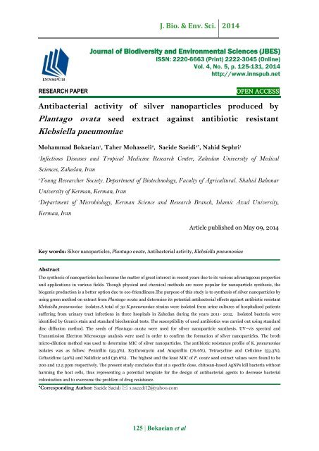
Antibacterial activity of silver nanoparticles produced by Plantago ovata seed extract against antibiotic resistant Klebsiella pneumoniae
Abstract The synthesis of nanoparticles has become the matter of great interest in recent years due to its various advantageous properties and applications in various fields. Though physical and chemical methods are more popular for nanoparticle synthesis, the biogenic production is a better option due to eco-friendliness.The purpose of this study is to synthesis of silver nanoparticles by using green method on extract from Plantago ovata and determine its potential antibacterial effects against antibiotic resistant Klebsiella pneumoniae isolates.A total of 30 K.pneumoniae strains were isolated from urine cultures of hospitalized patients suffering from urinary tract infections in three hospitals in Zahedan during the years 2011- 2012. Isolated bacteria were identified by Gram's stain and standard biochemical tests. The susceptibility of used antibiotics was carried out using standard disc diffusion method. The seeds of Plantago ovata were used for silver nanoparticle sunthesis. UV–vis spectral and Transmission Electron Microscopy analysis were used in order to confirm the formation of silver nanoparticles. The broth micro-dilution method was used to determine MIC of silver nanoparticles. The antibiotic resistance profile of K. pneumoniae isolates was as follow: Penicillin (93.3%), Erythromycin and Ampicillin (76.6%), Tetracycline and Cefixime (53.3%), Ceftazidime (40%) and Nalidixic acid (36.6%). The highest and the least MIC of P. ovate seed extract values were found to be 200 and 12.5 ppm respectively. The present study concludes that at a specific dose, chitosan-based AgNPs kill bacteria without harming the host cells, thus representing a potential template for the design of antibacterial agents to decrease bacterial colonization and to overcome the problem of drug resistance.
Abstract
The synthesis of nanoparticles has become the matter of great interest in recent years due to its various advantageous properties and applications in various fields. Though physical and chemical methods are more popular for nanoparticle synthesis, the biogenic production is a better option due to eco-friendliness.The purpose of this study is to synthesis of silver nanoparticles by using green method on extract from Plantago ovata and determine its potential antibacterial effects against antibiotic resistant Klebsiella pneumoniae isolates.A total of 30 K.pneumoniae strains were isolated from urine cultures of hospitalized patients suffering from urinary tract infections in three hospitals in Zahedan during the years 2011- 2012. Isolated bacteria were identified by Gram's stain and standard biochemical tests. The susceptibility of used antibiotics was carried out using standard disc diffusion method. The seeds of Plantago ovata were used for silver nanoparticle sunthesis. UV–vis spectral and Transmission Electron Microscopy analysis were used in order to confirm the formation of silver nanoparticles. The broth micro-dilution method was used to determine MIC of silver nanoparticles. The antibiotic resistance profile of K. pneumoniae isolates was as follow: Penicillin (93.3%), Erythromycin and Ampicillin (76.6%), Tetracycline and Cefixime (53.3%), Ceftazidime (40%) and Nalidixic acid (36.6%). The highest and the least MIC of P. ovate seed extract values were found to be 200 and 12.5 ppm respectively. The present study concludes that at a specific dose, chitosan-based AgNPs kill bacteria without harming the host cells, thus representing a potential template for the design of antibacterial agents to decrease bacterial colonization and to overcome the problem of drug resistance.

Create successful ePaper yourself
Turn your PDF publications into a flip-book with our unique Google optimized e-Paper software.
J. Bio. & Env. Sci. 2014<br />
Journal <strong>of</strong> Biodiversity and Environmental Sciences (JBES)<br />
ISSN: 2220-6663 (Print) 2222-3045 (Online)<br />
Vol. 4, No. 5, p. 125-131, 2014<br />
http://www.innspub.net<br />
RESEARCH PAPER<br />
OPEN ACCESS<br />
<strong>Antibacterial</strong> <strong>activity</strong> <strong>of</strong> <strong>silver</strong> <strong>nanoparticles</strong> <strong>produced</strong> <strong>by</strong><br />
<strong>Plantago</strong> <strong>ovata</strong> <strong>seed</strong> <strong>extract</strong> <strong>against</strong> <strong>antibiotic</strong> <strong>resistant</strong><br />
<strong>Klebsiella</strong> <strong>pneumoniae</strong><br />
Mohammad Bokaeian 1 , Taher Mohasseli 2 , Saeide Saeidi 3* , Nahid Sephri 1<br />
1<br />
Infectious Diseases and Tropical Medicine Research Center, Zahedan University <strong>of</strong> Medical<br />
Sciences, Zahedan, Iran<br />
2<br />
Young Researcher Society. Department <strong>of</strong> Biotechnology, Faculty <strong>of</strong> Agricultural. Shahid Bahonar<br />
University <strong>of</strong> Kerman, Kerman, Iran<br />
3<br />
Department <strong>of</strong> Microbiology, Kerman Science and Research Branch, Islamic Azad University,<br />
Kerman, Iran<br />
Article published on May 09, 2014<br />
Key words: Silver <strong>nanoparticles</strong>, <strong>Plantago</strong> ovate, <strong>Antibacterial</strong> <strong>activity</strong>, <strong>Klebsiella</strong> <strong>pneumoniae</strong><br />
Abstract<br />
The synthesis <strong>of</strong> <strong>nanoparticles</strong> has become the matter <strong>of</strong> great interest in recent years due to its various advantageous properties<br />
and applications in various fields. Though physical and chemical methods are more popular for nanoparticle synthesis, the<br />
biogenic production is a better option due to eco-friendliness.The purpose <strong>of</strong> this study is to synthesis <strong>of</strong> <strong>silver</strong> <strong>nanoparticles</strong> <strong>by</strong><br />
using green method on <strong>extract</strong> from <strong>Plantago</strong> <strong>ovata</strong> and determine its potential antibacterial effects <strong>against</strong> <strong>antibiotic</strong> <strong>resistant</strong><br />
<strong>Klebsiella</strong> <strong>pneumoniae</strong> isolates.A total <strong>of</strong> 30 K.<strong>pneumoniae</strong> strains were isolated from urine cultures <strong>of</strong> hospitalized patients<br />
suffering from urinary tract infections in three hospitals in Zahedan during the years 2011- 2012. Isolated bacteria were<br />
identified <strong>by</strong> Gram's stain and standard biochemical tests. The susceptibility <strong>of</strong> used <strong>antibiotic</strong>s was carried out using standard<br />
disc diffusion method. The <strong>seed</strong>s <strong>of</strong> <strong>Plantago</strong> <strong>ovata</strong> were used for <strong>silver</strong> nanoparticle sunthesis. UV–vis spectral and<br />
Transmission Electron Microscopy analysis were used in order to confirm the formation <strong>of</strong> <strong>silver</strong> <strong>nanoparticles</strong>. The broth<br />
micro-dilution method was used to determine MIC <strong>of</strong> <strong>silver</strong> <strong>nanoparticles</strong>. The <strong>antibiotic</strong> resistance pr<strong>of</strong>ile <strong>of</strong> K. <strong>pneumoniae</strong><br />
isolates was as follow: Penicillin (93.3%), Erythromycin and Ampicillin (76.6%), Tetracycline and Cefixime (53.3%),<br />
Ceftazidime (40%) and Nalidixic acid (36.6%). The highest and the least MIC <strong>of</strong> P. ovate <strong>seed</strong> <strong>extract</strong> values were found to be<br />
200 and 12.5 ppm respectively. The present study concludes that at a specific dose, chitosan-based AgNPs kill bacteria without<br />
harming the host cells, thus representing a potential template for the design <strong>of</strong> antibacterial agents to decrease bacterial<br />
colonization and to overcome the problem <strong>of</strong> drug resistance.<br />
* Corresponding Author: Saeide Saeidi s.saeedi12@yahoo.com<br />
125 | Bokaeian et al
J. Bio. & Env. Sci. 2014<br />
Introduction<br />
Human beings are <strong>of</strong>ten infected <strong>by</strong> micro-organisms<br />
such as bacteria, molds, yeasts, and viruses present in<br />
their living environments. Because <strong>of</strong> the emergence<br />
and increase in the number <strong>of</strong> multiple <strong>antibiotic</strong><strong>resistant</strong><br />
bacteria and the continuing emphasis on<br />
health-care costs, many scientists have researched<br />
methods to develop new effective antimicrobial<br />
agents that overcome the resistances <strong>of</strong> these<br />
organisms. The <strong>silver</strong> metal has a great toxicity<br />
<strong>against</strong> a wide range <strong>of</strong> micro-organisms, particularly<br />
gram negative bacteria. Silver <strong>nanoparticles</strong> (Ag-NPs)<br />
are found to be effective as anti-inflammatory, antiangiogenesis,<br />
anti-platelet <strong>activity</strong> and <strong>against</strong> cancer<br />
cells which makes them vital (Sotiriou, 2010). Besides<br />
that, Ag-NPs were also being reported in the<br />
literature to exhibit a strong cyto-protective <strong>activity</strong><br />
towards human immunodeficiency virus (HIV)<br />
infections (Sun et al., 2005). Considering the welldocumented<br />
crucial importance <strong>of</strong> the<br />
transmembrane proton gradient in overall microbial<br />
metabolism, it seems inevitable that the elimination<br />
<strong>of</strong> proton motive force should result in cell death<br />
(Dibrov et al., 2002). Ag+ also forms complexes with<br />
bases contained in DNA and is a potent inhibitor <strong>of</strong><br />
fungal DNases (Ahearn et al., 1995; Ghandour et al.,<br />
1988).<br />
However, green synthesis approaches <strong>of</strong> producing Ag<br />
NPs are an alternative source <strong>of</strong> conventional method<br />
and possess excellent antimicrobial <strong>activity</strong> (Sharma<br />
et al., 2009). Researchers showed that <strong>Plantago</strong><br />
<strong>ovata</strong> have hypo-cholesterolemic (Salas-Salvado et<br />
al., 2007), anti-diarrheal (Washington et al., 1999),<br />
anti-diabetic (Hannan et al., 2006) and low antioxidant<br />
(Souri et al., 2008) effects.<br />
The aim <strong>of</strong> present study was synthesis <strong>of</strong> <strong>silver</strong><br />
nano-particles <strong>by</strong> using green method on <strong>seed</strong> <strong>extract</strong><br />
from <strong>Plantago</strong> <strong>ovata</strong> and determination <strong>of</strong> its<br />
potential antibacterial effects <strong>against</strong> <strong>antibiotic</strong><br />
<strong>resistant</strong> <strong>Klebsiella</strong> <strong>pneumoniae</strong>.<br />
Material and methods<br />
Isolation <strong>of</strong> bacteria<br />
A total <strong>of</strong> 30 K.<strong>pneumoniae</strong> strains were isolated<br />
from urine cultures <strong>of</strong> hospitalized patients suffering<br />
from urinary tract infections in three hospitals in<br />
Zahedan (south-eastern Iran) during 2011- 2012.<br />
Isolated bacteria were identified <strong>by</strong> Gram's stain and<br />
standard biochemical tests (Forbes et al., 2007).<br />
Agar disk diffusion assay<br />
The susceptibility <strong>of</strong> all <strong>antibiotic</strong>s was carried out<br />
using standard disc diffusion method on Mueller-<br />
Hinton agar as recommended <strong>by</strong> CLSI. Briefly,<br />
K.<strong>pneumoniae</strong> isolated plates were grown overnight<br />
on blood agar, and colony suspension was prepared<br />
using the sterile saline water equivalent to a 0.5<br />
McFarland standard. Suspension (10 μl) was spread<br />
over the Mueller-Hinton plates and <strong>antibiotic</strong> discs<br />
were transferred aseptically on the surface <strong>of</strong><br />
inoculated media plates. The <strong>antibiotic</strong>s and their<br />
potencies were as follow: Ceftazidime (30 μg),<br />
Tetracycline (30 μg), Erythromycin (15 μg), Cefixime<br />
(30 μg), Penicillin (10 μg), Ampicillin (25 μg),<br />
Nalidixic acid (30 μg).<br />
Plant materials<br />
The <strong>seed</strong>s <strong>of</strong> <strong>Plantago</strong> <strong>ovata</strong> were collected in the<br />
region <strong>of</strong> Iran (Zabol, south-eastern, Iran) and were<br />
identified <strong>by</strong> Zabol university herbarium. The <strong>seed</strong>s<br />
were dried at room temperature and transferred into<br />
glass containers and preserved until <strong>extract</strong>ion<br />
procedure was performed in the laboratory.<br />
Preparation <strong>of</strong> <strong>seed</strong> <strong>extract</strong><br />
Seed samples (50g) were sterilized using 30% sodium<br />
hypochlorite for 5 minutes and then rinsed three<br />
times with sterile distilled water. The process was<br />
followed <strong>by</strong> soaking in 70% alcohol for two minutes<br />
and then rinsed five times with sterile distilled water.<br />
Sterile water was added to disinfected <strong>seed</strong>s (2:1 V/V)<br />
and incubated 25°C temperature for 7 days. The<br />
prepared <strong>seed</strong> <strong>extract</strong> was filtered through 40<br />
whattman filter paper and was kept in refrigerator for<br />
further studies.<br />
126 | Bokaeian et al
J. Bio. & Env. Sci. 2014<br />
Synthesis <strong>of</strong> <strong>silver</strong> <strong>nanoparticles</strong><br />
Silver nitrate (AgNO3) was used as the source for<br />
synthesis <strong>of</strong> <strong>silver</strong> <strong>nanoparticles</strong>. Briefly, 5ml <strong>of</strong> the<br />
obtained <strong>seed</strong> <strong>extract</strong> was diluted <strong>by</strong> 15ml sterile<br />
water and was added to a concentration <strong>of</strong> 2mM <strong>silver</strong><br />
nitrate for the reduction <strong>of</strong> Ag+ to Ag0.<br />
distributions with average diameter <strong>of</strong> 13 nm with<br />
some deviations.<br />
The biosynthesis <strong>of</strong> <strong>silver</strong> <strong>nanoparticles</strong> using the<br />
<strong>extract</strong> <strong>of</strong> P. <strong>ovata</strong> was preliminary confirmed <strong>by</strong> the<br />
change <strong>of</strong> the color <strong>of</strong> the solution from yellow to<br />
brown. The synthesis was further confirmed <strong>by</strong> the<br />
absorption peak between 400-450 nm due to surface<br />
plasma resonance (Manikprabhu and Lingappa,<br />
2013).<br />
Chart 1. UV-vis spectrum <strong>of</strong> Ag <strong>nanoparticles</strong><br />
<strong>produced</strong> <strong>by</strong> <strong>Plantago</strong> <strong>ovata</strong> <strong>seed</strong> <strong>extract</strong><br />
MIC determination <strong>of</strong> <strong>silver</strong> <strong>nanoparticles</strong><br />
The MIC (Minimum Inhibitory Concentration) is<br />
defined as the lowest concentration <strong>of</strong> the <strong>extract</strong> at<br />
which the microorganism does not demonstrate the<br />
visible growth. The broth micro-dilution method was<br />
used to determine MIC. Briefly, serial doubling<br />
dilutions <strong>of</strong> the <strong>silver</strong> <strong>nanoparticles</strong> <strong>produced</strong> in the<br />
plant P. <strong>ovata</strong> <strong>seed</strong> <strong>extract</strong> were prepared in a 96-well<br />
micro-titer plate ranged from 12.5ppm to 200ppm. To<br />
each well, 10 μl <strong>of</strong> indicator solution and 10 μl <strong>of</strong><br />
Mueller Hinton Broth were added. Finally, 10 μl <strong>of</strong><br />
bacterial suspension (10 6 CFU/ml) was added to each<br />
well to achieve a concentration <strong>of</strong> 10 4 CFU/ml. The<br />
plates were wrapped loosely with cling film to ensure<br />
that the bacteria did not get dehydrated. The plates<br />
were prepared in triplicates, and then they were<br />
placed in an incubator at 37ºC for 18-24 hours. The<br />
color change was then assessed visually. The<br />
microorganism growth was indicated <strong>by</strong> turbidity.<br />
A<br />
Results<br />
UV–vis spectral and Transmission Electron<br />
Microscopy (TEM) analysis were used in order to<br />
confirm the formation <strong>of</strong> <strong>silver</strong> <strong>nanoparticles</strong> from<br />
2mM solution <strong>of</strong> <strong>silver</strong> nitrate (Chart no.1). Fig. 1 is<br />
TEM image <strong>of</strong> P. <strong>ovata</strong> <strong>seed</strong> <strong>extract</strong> containing 2mM<br />
AgNO3 solution at 30 °C. P. <strong>ovata</strong> <strong>seed</strong> <strong>extract</strong><br />
<strong>produced</strong> <strong>silver</strong> <strong>nanoparticles</strong> which were <strong>of</strong>ten semi<br />
spherical. The <strong>silver</strong> <strong>nanoparticles</strong> showed Gaussian<br />
B<br />
Fig. 1. (A) TEM image and (B) particles size<br />
distribution <strong>of</strong> Ag <strong>nanoparticles</strong> synthesized <strong>by</strong><br />
<strong>Plantago</strong> <strong>ovata</strong> <strong>seed</strong> <strong>extract</strong>.<br />
127 | Bokaeian et al
J. Bio. & Env. Sci. 2014<br />
The <strong>antibiotic</strong> resistance pr<strong>of</strong>ile <strong>of</strong> K. <strong>pneumoniae</strong><br />
isolates were as follow: Penicillin (93.3%),<br />
Erythromycin and Ampicillin (76.6%), Tetracycline<br />
and Cefixime (53.3%), Ceftazidime (40%) and<br />
Nalidixic acid (36.6%) (Table1). The highest and the<br />
least MIC <strong>of</strong> P. <strong>ovata</strong> <strong>seed</strong> <strong>extract</strong> values were found<br />
to be 200 and 12.5 ppm respectively (Table 2).<br />
Table 1. Percentage <strong>of</strong> antimicrobial resistance <strong>of</strong> 30<br />
isolatess <strong>of</strong> <strong>Klebsiella</strong> <strong>pneumoniae</strong><br />
CAZ CN E TE P AM NA<br />
S 53.3 36.6 6.6 40 0 0 50<br />
I 6.6 10 16.6 6.6 6.6 23.3 13.3<br />
R 40 53.3 76.6 53.3 93.3 76.6 36.6<br />
S= Sensitive, I= Intermediate, R= Resistant, CAZ=<br />
Ceftazidime, TE= Tetracycline, E= Erythromycin,<br />
CN= Cefixime, P=Penicillin, AM=Ampicillin,<br />
NA=Nalidixic acid<br />
Table 2. Antimicrobial susceptibility and MIC<br />
Bacterial code MIC(ppm) CAZ CN E TE P AM NA<br />
1 50 I R R R R R R<br />
2 50 S R R I I R R<br />
3 50 S S R R R I I<br />
4 25 S S I S R R R<br />
5 50 I I R I R R S<br />
6 100 R R R R R I S<br />
7 200 R R R R R R I<br />
8 25 R R R R R R R<br />
9 25 S S I S R R R<br />
10 100 R R R R R R R<br />
11 50 S S R R R I S<br />
12 100 S R S S R R S<br />
13 100 S S R S R I R<br />
14 200 S I R R R R S<br />
15 25 R R R R R R I<br />
16 25 R R R R R R R<br />
17 12.5 S I R R R R R<br />
18 25 R R R S R R S<br />
19 25 R R R R R R S<br />
20 100 S S R S R R S<br />
21 Any grow R R R R R R S<br />
22 Any grow S S R S R R S<br />
23 25 S S R S R R S<br />
24 Any grow S S R S I I R<br />
25 Any grow R R R R R R R<br />
26 25 S R I S R I R<br />
27 100 S S S S R R S<br />
28 25 R R I R R R S<br />
29 12.5 R R R R R R I<br />
30 25 S S I S R R S<br />
CAZ= Ceftazidime, TE= Tetracycline, E= Erythromycin, CN= Cefixime , P=Penicillin, AM=Ampicillin,<br />
NA=Nalidixic acid<br />
128 | Bokaeian et al
J. Bio. & Env. Sci. 2014<br />
Discussion<br />
In the present study, K.<strong>pneumoniae</strong> isolates were<br />
resistanct to penicillin (93.3%), erythromycin and<br />
ampicillin (76.6%), tetracycline and cefixime (53.3%),<br />
ceftazidime (40%) and nalidixic acid (36.6%). As our<br />
pervious study, overall K. <strong>pneumoniae</strong> were resistanct<br />
to ampicillin (65%), gentamicin (30%), trimethoprimsulfamethoxazol<br />
(25%), cipr<strong>of</strong>loxacin (20%),<br />
nitr<strong>of</strong>urantoin (15%) and nalidixic acid (15%) (Saeidi<br />
et al., 2014). In contrast, Zamani et al. showed that<br />
the most effective <strong>antibiotic</strong>s <strong>against</strong> the bacterial<br />
isolates are tobramycin and ceftazidime (79%),<br />
ceftizoxime (78%), cipr<strong>of</strong>loxacin (76.1%), ceftriaxone<br />
(76.2%) and amikacin (74.2%) (Zamani et al., 2013).<br />
Results <strong>of</strong> another study showed that 28 to 76 % <strong>of</strong><br />
isolates are <strong>resistant</strong> to ceftizoxime and cefotaxime<br />
(Sikarwar and Batra, 2011). The study <strong>of</strong> Feizabadi<br />
on <strong>Klebsiella</strong> <strong>pneumoniae</strong> isolates showed the highest<br />
resistance to amoxicillin-clavulanic acid (81.8%),<br />
cefixime and ceftazidime (72.7%) (Feizabadi et al.,<br />
2007).<br />
In our study, the highest and the least MIC <strong>of</strong> P.<br />
<strong>ovata</strong> <strong>seed</strong> <strong>extract</strong> values were found to be 200 and<br />
12.5 ppm respectively. In the study <strong>of</strong> Soo-Hwan the<br />
results show that S. aureus and E. coli were<br />
substantially inhibited <strong>by</strong> Ag-NPs (Soo-Hwan et al.,<br />
2011). Sondi and Salopek-Sondi reported that <strong>silver</strong><br />
<strong>nanoparticles</strong> have excellent antibacterial <strong>activity</strong><br />
<strong>against</strong> E. coli (Sondi and Salopek-Sondi, 2004). As<br />
the results <strong>of</strong> Guzman et al study, the <strong>nanoparticles</strong> <strong>of</strong><br />
<strong>silver</strong> showed high antimicrobial and bactericidal<br />
<strong>activity</strong> <strong>against</strong> gram positive bacteria such as<br />
Escherichia coli, Pseudomonas aeruginosa and<br />
Staphylococcus aureus which is a highly methicillin<br />
<strong>resistant</strong> strain (Guzman et al., 2009). Silver<br />
<strong>nanoparticles</strong> have been shown to be effective<br />
biocides <strong>against</strong> different bacteria such as Escherichia<br />
coli, Staphylococcus aureus, Staphylococcus<br />
epidermis, Leuconostoc mesenteroides, Bacillus<br />
subtilis, and <strong>Klebsiella</strong> <strong>pneumoniae</strong> among others<br />
(Benn and Westerh<strong>of</strong>f, 2008; Chen and Chiang,<br />
2008; Falletta et al., 2008; Hernandez-Sierra et al.,<br />
2008; Ingle et al., 2008; Jung et al., 2008; Kim,<br />
2007; Kim et al., 2008; Kvitek et al., 2008). Gopinath<br />
et al reported that Staphylococcus aureus and<br />
Streptococcus <strong>pneumoniae</strong> exhibit similar zone <strong>of</strong><br />
inhibition for all three <strong>silver</strong> nanoparticle<br />
concentrations. They concluded that the most<br />
significant effect <strong>of</strong> <strong>silver</strong> <strong>nanoparticles</strong> at low<br />
concentration <strong>of</strong> 10 μL per disc <strong>against</strong> Shigella<br />
dysenteriae produces a 2-mm inhibition zone for<br />
gram-negative bacteria, and at the same<br />
concentration, the <strong>nanoparticles</strong> did not show any<br />
significant effect on Bacillus subtilis, Pseudomonas<br />
aeruginosa, and Proteus vulgaris (Gopinath et al.,<br />
2013). The study <strong>of</strong> Das showed that these<br />
<strong>nanoparticles</strong> can be used as effective growth<br />
inhibitors <strong>against</strong> Staphylococcus, Basillus and<br />
Pseudimonas species (Das et al., 2011).<br />
The present study concludes that at a specific dose,<br />
chitosan-based AgNPs kill bacteria without harming<br />
the host cells, thus representing a potential template<br />
for the design <strong>of</strong> antibacterial agents to decrease<br />
bacterial colonization and to overcome the problem <strong>of</strong><br />
drug resistance. Silver <strong>nanoparticles</strong> have a potent<br />
antimicrobial <strong>activity</strong> <strong>against</strong> <strong>antibiotic</strong> <strong>resistant</strong><br />
K.<strong>pneumoniae</strong> strains. However, further research is<br />
required to evaluate the practical value <strong>of</strong> these<br />
particles before therapeutic usage.<br />
References<br />
Sotiriou GA, Pratsinis SE. 2010. <strong>Antibacterial</strong><br />
Activity <strong>of</strong> Nano<strong>silver</strong> Ions and Particles. Environmental<br />
Science & Technology 44, 5649-5654.<br />
http://dx.doi.org/ 10.1021/es101072s<br />
Sun RW, Chen R, Chung NP, Ho CM, Lin CL,<br />
Che CM. 2005. Silver <strong>nanoparticles</strong> fabricated in<br />
Hepes buffer exhibit cytoprotective activities toward<br />
HIV-1 infected cells. Chemical Communications 40,<br />
5059–5061.<br />
http://dx.doi.org/ 10.1039/b510984a<br />
Dibrov P, Dzioba J, Gosink KK, Häse CC. 2002.<br />
Chemiosmotic mechanism <strong>of</strong> antimicrobial <strong>activity</strong> <strong>of</strong><br />
129 | Bokaeian et al
J. Bio. & Env. Sci. 2014<br />
Ag+ in Vibrio cholerae. Antimicrobial Agents and<br />
Chemother apy 46, 2668–2670.<br />
http://dx.doi.org/10.1128/AAC.46.8.2668-2670.2002<br />
Forbes BA, Sahm DF, Weissfeld AS. 2007.<br />
Bailey & Scott`s diagnostic microbiology. 12th ed.<br />
Missouri: Mos<strong>by</strong> Co; 323-333.<br />
Ahearn DG, May LL, Gabriel MM. 1995.<br />
Adherence <strong>of</strong> organisms to <strong>silver</strong>-coated surfaces.<br />
Journal <strong>of</strong> Industrial Microbiology 15, 372–376.<br />
http://dx.doi.org/ 10.1007/BF01569993<br />
Ghandour W, Hubbard JA, Deistung J,<br />
HughesMN, Poole RK. 1988. The uptake <strong>of</strong> <strong>silver</strong><br />
ions <strong>by</strong> Escherichia coli K12: toxic effects and<br />
interaction with copper ion. Applied<br />
Microbiology and Biotechnology 28, 559–565.<br />
Sharma VK, Yngard RA, Lin Y. 2009. Silver<br />
<strong>nanoparticles</strong>: green synthesis and their antimicrobial<br />
activities. Advances in Colloid and Interface Science<br />
145, 83–96.<br />
http://dx.doi.org/10.1016/j.cis.2008.09.002<br />
Salas-Salvado J, Farres X, Luque X, Narejos S,<br />
Borren M, Blanza R. 2007. Effect <strong>of</strong> two doses <strong>of</strong> a<br />
mixture <strong>of</strong> soluble fibers on body weight and metabolic<br />
variables in over-weight or obese patients: A randomized<br />
trial. British Journal <strong>of</strong> Nutrition 99, 1380-1387.<br />
http://dx.doi.org/10.1017/S0007114507868528<br />
Manikprabhu D, Lingappa K. 2013. Microwave<br />
assisted rapid and green synthesis <strong>of</strong> <strong>silver</strong><br />
<strong>nanoparticles</strong> using a pigment <strong>produced</strong> <strong>by</strong><br />
Streptomyces coelicolor KImp 33. Bioinorganic<br />
Chemistry and Applications 2013:341798, 1-8.<br />
Saeidi S, Alavi-Naini R, Shayan S. 2014.<br />
Antimicrobial susceptibility and distribution <strong>of</strong> TEM<br />
and CTX-M genes among ESBL-producing <strong>Klebsiella</strong><br />
<strong>pneumoniae</strong> and Pseudomonas aeruginosa causing<br />
urinary tract infection. Zahedan Journal <strong>of</strong> Research<br />
in Medical Sciences 16(4), 1-5.<br />
Zamani A, Yousefi Mashouf R, Ebrahimzadeh<br />
NamvarA, Alikhani M.Y. 2013. Detection <strong>of</strong> magA<br />
Gene in <strong>Klebsiella</strong> spp. Isolated from Clinical<br />
Samples. Iranian Journal <strong>of</strong> Basic Medical Sciences<br />
16, 173-76.<br />
Sikarwar SA and Batra HV. 2011. Prevalence <strong>of</strong><br />
Antimicrobial Drug Resistance <strong>of</strong> <strong>Klebsiella</strong><br />
<strong>pneumoniae</strong> in India. International Journal <strong>of</strong><br />
Bioscience, Biochemistry and Bioinformatics 3(1).<br />
Washington N, Harris M, Mussellwithe A,<br />
Spiller RC. 1999. Moderation <strong>of</strong> lactulose–induced<br />
diarrhea <strong>by</strong> Psyllium: Effects on motility and<br />
fermentation. The American Journal <strong>of</strong><br />
Clinical Nutrition 67, 317-321.<br />
Hannan JMA, Ali L, Khaleqe J, Akhter M, Flatt<br />
PR, Abdel-Wahab YHA. 2006. Aqueous <strong>extract</strong>s <strong>of</strong><br />
husks <strong>of</strong> <strong>Plantago</strong> ovate reduce hyperglycaemia<br />
glucose absorption. British Journal <strong>of</strong> Nutrition<br />
96,131-137.<br />
http://dx.doi.org/10.1079/BJN20061819<br />
Souri E, Amin G, Farsam H, Tehrani MB. 2008.<br />
Screening <strong>of</strong> antioxidant <strong>activity</strong> and phenolic content<br />
<strong>of</strong> 24 medicinal plant <strong>extract</strong>s. DARU 16, 83-87.<br />
Feizabadi MM, Etemadi G, Rahmati M,<br />
Mohammadi-Yeganeh S, Shabanpoor S, Asadi<br />
S. 2007. Antibiotic Resistance Patterns and Genetic<br />
Analysis <strong>of</strong> <strong>Klebsiella</strong> Pneumoniae Isolates from the<br />
Respiratory Tract. National Research Institute <strong>of</strong><br />
Tuberculosis and Lung Disease. Journal <strong>of</strong><br />
Respiratory Disease, Thoracic Surgery, Intensive care<br />
and Tuberculosis 6(3), 20-25.<br />
Soo-Hwan K, Lee HS, Ryu DS, Choi SG, and<br />
Lee DS. 2011. <strong>Antibacterial</strong> Activity <strong>of</strong> Silver<strong>nanoparticles</strong><br />
Against Staphylococcus aureus and<br />
Escherichia coli. Korean Journal <strong>of</strong> Microbiology and<br />
Biotechnology 39(1), 77–85.<br />
130 | Bokaeian et al
J. Bio. & Env. Sci. 2014<br />
Sondi I, Salopek-Sondi B. 2004. Silver<br />
<strong>nanoparticles</strong> as antimicrobial agent: a case study on<br />
E. coli as a model for Gram-negative bacteria. Journal<br />
<strong>of</strong> Colloid and Interface Science 275, 177–182.<br />
http://dx.doi.org/10.1016/j.jcis.2004.02.012<br />
Guzman M, Dille J, Godet S. 2009. Synthesis <strong>of</strong><br />
<strong>silver</strong> <strong>nanoparticles</strong> <strong>by</strong> chemical reduction method and<br />
their antibacterial <strong>activity</strong>. International Journal <strong>of</strong><br />
Chemical and Biomolecular Engineering 2(3), 104- 111.<br />
Benn T, Westerh<strong>of</strong>f P. 2008. Nanoparticle <strong>silver</strong> released<br />
into water from commercially available sock fabrics.<br />
Environmental Science & Technology 42, 4133–4139.<br />
http://dx.doi.org/10.1021/es7032718<br />
Chen C, Chiang C. 2008. Preparation <strong>of</strong> cotton<br />
fibers with antibacterial <strong>silver</strong> <strong>nanoparticles</strong>. Journal<br />
<strong>of</strong> Materials Science Letters 62, 3607–3609.<br />
http://dx.doi.org/ 10.1016/j.matlet.2008.04.008<br />
Falletta E, Bonini M, Fratini E, Lo Nostro A,<br />
Pesavento G, Becheri A, Lo Nostro P, Canton<br />
P, Baglioni P. 2008. Clusters <strong>of</strong> poly (acrylates) and<br />
<strong>silver</strong> <strong>nanoparticles</strong>: structure and applications for<br />
antimicrobial fabrics. The Journal <strong>of</strong> Physical<br />
Chemistry 112, 11758–11766.<br />
http://dx.doi.org/ 10.1021/jp8035814<br />
Hernandez-Sierra J, Ruiz F, Pena D,<br />
Martinez-Gutierrez F, Martinez A, Guillen A,<br />
Tapia-Perez H, Castanon G. 2008. The<br />
antimicrobial sensitivity <strong>of</strong> Streptococcus mutants to<br />
<strong>nanoparticles</strong> <strong>of</strong> <strong>silver</strong>, zinc oxide, and gold.<br />
Nanomed Nanotechnology. 4,237–240.<br />
http://dx.doi.org/ 10.1016/j.nano.2008.04.005<br />
Ingle A, Gade A, Pierrat S, Sonnichsen C, Rai M.<br />
2008. Mycosynthesis <strong>of</strong> <strong>silver</strong> <strong>nanoparticles</strong> using the<br />
fungus Fusarium acuminatum and its <strong>activity</strong> <strong>against</strong><br />
some human pathogenic bacteria. Dekker Encyclopedia<br />
<strong>of</strong> Nanoscience and Nanotechnology 4, 141–144.<br />
http://dx.doi.org/ 10.2174/157341308784340804<br />
Jung W, Koo H, Kim K, Shin S, Kim S, Park Y.<br />
2008. <strong>Antibacterial</strong> <strong>activity</strong> and mechanism <strong>of</strong> action<br />
<strong>of</strong> the <strong>silver</strong> ion in Staphylococcus aureus and<br />
Escherichia coli. Applied and Environmental Microbiology<br />
74,2171–2178.<br />
http://dx.doi.org/ 10.1128/AEM.02001-07<br />
Kim J. 2007. <strong>Antibacterial</strong> <strong>activity</strong> <strong>of</strong> Ag+ ioncontaining<br />
<strong>silver</strong> <strong>nanoparticles</strong> prepared using the<br />
alcohol reduction method. Journal <strong>of</strong> Industrial<br />
and Engineering Chemistry 13,718–722.<br />
Kim Y, Kim J, Cho H, Rha D, Kim J, Park J,<br />
Choi B, Lim R, Chang H, Chung Y, Kwon I,<br />
Jeong J, Han B, Yu I. 2008.Twenty-eight-day oral<br />
toxicity, genotoxicity, and gender related tissue<br />
distribution <strong>of</strong> <strong>silver</strong> <strong>nanoparticles</strong> in Sprague-<br />
Dawley rats. Inhalation Toxicology 20, 575–583.<br />
http://dx.doi.org/ 10.1080/08958370701874663<br />
Kim K, Sung W, Moon S, Choi J, Kim J, Lee D.<br />
2008. Antifungal effect <strong>of</strong> <strong>silver</strong> <strong>nanoparticles</strong> on<br />
dermatophytes. Journal <strong>of</strong> Microbiology and Biotechnology<br />
18, 1482–1484.<br />
Kvitek L, Panacek A, Soukupova J, Kolar M,<br />
Vecerova R, Prucek R, Holecova M, Zboril R.<br />
2008. Effect <strong>of</strong> surfactants and polymers on stability<br />
and antibacterial <strong>activity</strong> <strong>of</strong> <strong>silver</strong> <strong>nanoparticles</strong> (NPs).<br />
The Journal <strong>of</strong> Physical Chemistry 112, 5825–34.<br />
Gopinath K, Gowri S, Arumugam A. 2013.<br />
Phytosynthesis <strong>of</strong> <strong>silver</strong> <strong>nanoparticles</strong> using<br />
Pterocarpus santalinus leaf <strong>extract</strong> and their<br />
antibacterial properties. Journal <strong>of</strong> Nanostructure in<br />
Chemistry 3, 63- 68.<br />
http://dx.doi.org/ 10.1186/2193-8865-3-68<br />
Das R, Gang S, Nath SS. 2011. Preparation and<br />
<strong>Antibacterial</strong> Activity <strong>of</strong> Silver Nanoparticles. Journal<br />
<strong>of</strong> Biomaterials and Nanobiotechnology 2, 472-475.<br />
131 | Bokaeian et al

![Review on: impact of seed rates and method of sowing on yield and yield related traits of Teff [Eragrostis teff (Zucc.) Trotter] | IJAAR @yumpu](https://documents.yumpu.com/000/066/025/853/c0a2f1eefa2ed71422e741fbc2b37a5fd6200cb1/6b7767675149533469736965546e4c6a4e57325054773d3d/4f6e6531383245617a537a49397878747846574858513d3d.jpg?AWSAccessKeyId=AKIAICNEWSPSEKTJ5M3Q&Expires=1714096800&Signature=hqf2cEMoGAk0sAG%2FPG8%2Bfo82Dt8%3D)