Performance of Immobilized Zymomonas mobilis 31821 ... - CiteSeer
Performance of Immobilized Zymomonas mobilis 31821 ... - CiteSeer
Performance of Immobilized Zymomonas mobilis 31821 ... - CiteSeer

You also want an ePaper? Increase the reach of your titles
YUMPU automatically turns print PDFs into web optimized ePapers that Google loves.
902 Yamada et al.<br />
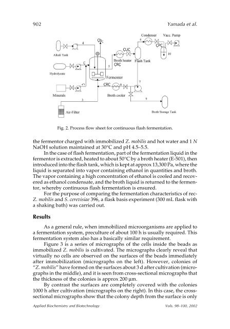
the fermentor charged with immobilized Z. <strong>mobilis</strong> and hot water and 1 N<br />
NaOH solution maintained at 30°C and pH 4.5–5.5.<br />
In the case <strong>of</strong> flash fermentation, part <strong>of</strong> the fermentation liquid in the<br />
fermentor is extracted, heated to about 50°C by a broth heater (E-501), then<br />
introduced into the flash tank, which is kept at approx 13,300 Pa, where the<br />
liquid is separated into vapor containing ethanol in quantities and broth.<br />
The vapor containing a high concentration <strong>of</strong> ethanol is cooled and recovered<br />
as ethanol condensate, and the broth liquid is returned to the fermentor,<br />
whereby continuous flash fermentation is ensured.<br />
For the purpose <strong>of</strong> comparing the fermentation characteristics <strong>of</strong> rec-<br />
Z. <strong>mobilis</strong> and S. cerevisiae 396, a flask basis experiment (300 mL flask with<br />
a shaking bath) was carried out.<br />
Results<br />
Fig. 2. Process flow sheet for continuous flash fermentation.<br />
As a general rule, when immobilized microorganisms are applied to<br />
a fermentation system, preculture <strong>of</strong> about 100 h is usually required. This<br />
fermentation system also has a basically similar requirement.<br />
Figure 3 is a series <strong>of</strong> micrographs <strong>of</strong> the cells inside the beads as<br />
immobilized Z. <strong>mobilis</strong> is cultivated. The micrographs clearly reveal that<br />
virtually no cells are observed on the surfaces <strong>of</strong> the beads immediately<br />
after immobilization (micrographs on the left). However, colonies <strong>of</strong><br />
“Z. <strong>mobilis</strong>” have formed on the surfaces about 3 d after cultivation (micrographs<br />
in the middle), and it is seen from cross-sectional micrographs that<br />
the thickness <strong>of</strong> the colonies is approx 200 µm.<br />
By contrast the surfaces are completely covered with the colonies<br />
1000 h after cultivation (micrographs on the right). In this case, the crosssectional<br />
micrographs show that the colony depth from the surface is only<br />
Applied Biochemistry and Biotechnology Vols. 98–100, 2002