

the coupled system L-glutamate dehydrogenase/NADH oxidase
the coupled system L-glutamate dehydrogenase/NADH oxidase
the coupled system L-glutamate dehydrogenase/NADH oxidase

Create successful ePaper yourself
Turn your PDF publications into a flip-book with our unique Google optimized e-Paper software.
An enzymatic process to a-ketoglutarate from L-<strong>glutamate</strong>:<br />
<strong>the</strong> <strong>coupled</strong> <strong>system</strong> L-<strong>glutamate</strong> <strong>dehydrogenase</strong>/<strong>NADH</strong> <strong>oxidase</strong><br />
Peter Ödman, a,b William B. Wellborn b and Andreas S. Bommarius b, *<br />
a Department of Chemical Engineering, Lund University, S-22100 Lund, Sweden<br />
b School of Chemical and Biomolecular Engineering, Parker H. Petit Institute of Bioengineering and Bioscience,<br />
Georgia Institute of Technology, 315 Ferst Drive, Atlanta, GA 30332-0363, USA<br />
Received 10 June 2004;accepted 7 July 2004<br />
Available online 11 September 2004<br />
Abstract—a-Ketoglutarate, employed to treat mild chronic renal insufficiency, was obtained through enzymatic oxidation of monosodium<br />
<strong>glutamate</strong> (MSG) catalyzed by L-<strong>glutamate</strong> <strong>dehydrogenase</strong> (L-gluDH) <strong>coupled</strong> with <strong>NADH</strong> <strong>oxidase</strong> for <strong>the</strong> regeneration of<br />
<strong>NADH</strong> back to NAD + . The irreversible reduction of molecular oxygen to water by <strong>NADH</strong> <strong>oxidase</strong> is demonstrated to drive oxidation<br />
of MSG to a-ketoglutarate to completion. L-gluDH was found to be inhibited by all three oxidative deamination products, aketoglutarate,<br />
<strong>NADH</strong>, and ammonia. As <strong>the</strong> pH in <strong>the</strong> current <strong>system</strong> was balanced by sodium, not ammonia, and <strong>NADH</strong> was<br />
recycled to NAD + , inhibition of L-gluDH by a-ketoglutarate is believed to present <strong>the</strong> biggest challenge to an efficient process.<br />
In a batch experiment, we achieved a volumetric productivity of 1g/(LÆd).<br />
Ó 2004 Elsevier Ltd. All rights reserved.<br />
1. Introduction<br />
Keto acids are <strong>the</strong> nitrogen-free analogs of amino acids,<br />
and are transaminated to form <strong>the</strong> respective amino acid<br />
in <strong>the</strong> body. This improves nitrogen balance at a lower<br />
nitrogen intake and corresponds with relief of <strong>the</strong> symptoms<br />
of uremia while maintaining good nutrition. 1<br />
Thus, a-ketoglutarate is beneficial as a component of<br />
endoperitoneal solutions for <strong>the</strong> conservative treatment<br />
of mild chronic renal insufficiency in hemodialysis<br />
patients and, in combination with calcium carbonate,<br />
of hyperphosphatemia. 2–4 Long-term co-administration<br />
of keto acids, erythropoietin, and low-protein diet was<br />
also associated with a delay in progression of renal<br />
insufficiency and a reduction in proteinuria.<br />
a-Ketoglutarate is most advantageously produced by<br />
oxidation of inexpensive L-<strong>glutamate</strong> (MSG), a flavor<br />
enhancer available at huge scale. The current process<br />
involves oxidation of MSG in air with a copper complex.<br />
The oxidation of MSG, however, can be afforded<br />
enzymatically, by catalysis with L-<strong>glutamate</strong> <strong>dehydrogenase</strong><br />
(L-gluDH), with simultaneous reduction of NAD +<br />
* Corresponding author. Tel.: +1 404 385 1334;fax: +1 404 894<br />
2291;e-mail: andreas.bommarius@chbe.gatech.edu<br />
0957-4166/$ - see front matter Ó 2004 Elsevier Ltd. All rights reserved.<br />
doi:10.1016/j.tetasy.2004.07.055<br />
Tetrahedron: Asymmetry 15 (2004) 2933–2937<br />
to <strong>NADH</strong>. Herein, we demonstrate <strong>the</strong> feasibility to<br />
couple L-gluDH with <strong>NADH</strong> <strong>oxidase</strong> to regenerate <strong>the</strong><br />
co-factor <strong>NADH</strong> back to NAD + and to drive <strong>the</strong> <strong>the</strong>rmodynamically<br />
unfavorable equilibrium from L-<strong>glutamate</strong><br />
to a-ketoglutarate. This is accompanied by <strong>the</strong><br />
irreversible four-electron reduction of molecular oxygen<br />
to water. Recently, we published <strong>the</strong> characterization of<br />
a novel water-forming <strong>NADH</strong> <strong>oxidase</strong> from Lactobacillus<br />
sanfranciscensis, which accomplishes this task. 5,6<br />
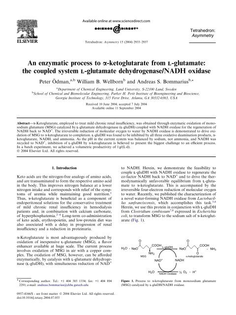
Herein, we use this protein in conjunction with L-gluDH<br />
from Clostridium symbiosum 7,8 expressed in Escherichia<br />
coli, to transform MSG to <strong>the</strong> sodium salt of a-ketoglutarate<br />
(Fig. 1).<br />
O<br />
NaO<br />
COOH<br />
NH3 O<br />
NAD <strong>NADH</strong><br />
+<br />
O<br />
H2O + NaO<br />
COOH<br />
L-gluDH<br />
+<br />
NH2<br />
MSG α-ketoglutarate<br />
H 2O<br />
O<br />
<strong>NADH</strong> Oxidase ½ 2 +<br />
Tetrahedron:<br />
Asymmetry<br />
Figure 1. Process to a-ketoglutarate from monosodium <strong>glutamate</strong><br />
(MSG) catalyzed by L-gluDH/<strong>NADH</strong> <strong>oxidase</strong>.<br />
H +
2934 P. Ödman et al. / Tetrahedron: Asymmetry 15 (2004) 2933–2937<br />
Recently, in humans, L-<strong>glutamate</strong> <strong>dehydrogenase</strong> was<br />
implicated to also play a role in glucose homeostasis,<br />
linked to gain-of-function mutations in L-gluDH in connection<br />
with <strong>the</strong>ir function of oxidizing L-<strong>glutamate</strong> to<br />
a-ketoglutarate. 9<br />
While <strong>the</strong> <strong>the</strong>rmodynamics of <strong>the</strong> oxidation of L-<strong>glutamate</strong><br />
to a-ketoglutarate are very unfavorable, <strong>the</strong> irreversible<br />
reduction of molecular oxygen drives <strong>the</strong><br />
oxidative biotransformation to completion, unlike<br />
regeneration schemes of <strong>NADH</strong> to NAD + based on acetone<br />
and an alcohol <strong>dehydrogenase</strong> functionality. 10,11<br />
Previous work utilizing <strong>NADH</strong> <strong>oxidase</strong> for driving<br />
transformations with amino acid <strong>dehydrogenase</strong>s<br />
include <strong>the</strong> recent separation of D,L-tert-leucine to trimethylpyruvate<br />
and D-tert-leucine catalyzed by L-leucine<br />
<strong>dehydrogenase</strong> (L-leuDH), 12 in which complete<br />
conversion is critical to achieve high enantiomeric excess<br />
of <strong>the</strong> remaining enantiomer.<br />
A second advantage of cofactor regeneration is cost<br />
reduction of potentially expensive pyridine dinucleotide<br />
cofactor, if successful, to insignificant levels. Cycle<br />
numbers of up to 600,000 have been achieved for reductive<br />
direction (regeneration of NAD + back to<br />
<strong>NADH</strong>). 13 Assuming a typical price of NAD + (MW<br />
663.4) of $10/g, <strong>the</strong> cost contribution per mole of product<br />
is $6.6 at a cycle number of 1000 and 1.1cents at a<br />
number of 600,000.<br />
2.1. Materials<br />
2. Material and methods<br />
L-Glutamate <strong>dehydrogenase</strong> (L-gluDH) from C. symbiosum,<br />
expressed in E. coli, was a kind gift of Roche Diagnostics<br />
(Mannheim/Germany). It was used as received<br />
and featured a specific activity of 12.5U/mg protein at<br />
pH7.5 and 30°C (for assay, see below).<br />
L-GluDH from bovine liver was ei<strong>the</strong>r obtained from<br />
Sigma or was a kind gift of Biocatalytics (Pasadena,<br />
CA). Each was used as received and featured a specific<br />
activity of 1.1–1.2U/mg protein at pH 9.0 and 30°C.<br />
<strong>NADH</strong> <strong>oxidase</strong> from L. sanfranciscensis was obtained<br />
from our own stock 5,6 (for preparation and assay, see<br />
below).<br />
Monosodium <strong>glutamate</strong> (MSG), a-ketoglutarate, Tris,<br />
and o<strong>the</strong>r components were obtained from Sigma–<br />
Aldrich–Fluka (Milwaukee, WI) and used as received.<br />
2.1.1. GluDH activity assay. GluDH activity was<br />
measured by monitoring <strong>the</strong> production of <strong>NADH</strong> in<br />
a UV/vis spectrophotometer at 340nm, with temperature<br />
control set at 30°C. The assay solution was<br />
150mM Tris buffer pH7.5 with 250mM MSG and<br />
4mM NAD + . After allowing 990lL of <strong>the</strong> solution to<br />
reach temperature, 3lg L-gluDH (10lL of 300 lg/mL<br />
stock) was added to yield a final volume of 1mL, <strong>the</strong><br />
solution was mixed and data collection was started.<br />
2.1.2. Determination of protein concentration. Protein<br />
concentration was determined by <strong>the</strong> Bradford method<br />
utilizing Coomassie Plus Protein assay reagent, prediluted<br />
protein assay standards-BSA (Pierce Chemical,<br />
Rockford, IL).<br />
2.2. Purification of <strong>NADH</strong> <strong>oxidase</strong><br />
Enzyme preparation (growth, expression, purification,<br />
and activity assay) was performed with a modified version<br />
of <strong>the</strong> literature methods. 5,6<br />
2.2.1. Cell growth and protein expression. Cells of<br />
E. coli JM101 containing <strong>the</strong> sfnox2 gene in <strong>the</strong><br />
pKK223-3 vector were grown in 5mL cultures at 37°C<br />
and 250rpm in 15mL disposable culture tubes to<br />
1.0OD 600 nm in LB media + 100lg/mL ampicillin.<br />
One-liter cultures of LB medium were seeded with a<br />
5 mL starter culture and grown at 30°C and 200rpm<br />
in baffled 2.8L Fernbach shake flasks. At OD600 = 0.7,<br />
protein production was induced by addition of 1.0mM<br />
IPTG final concentration and cells allowed to grow for<br />
an additional 3h. Additional ampicillin, 200lg/mL,<br />
was added at induction and 1.5h later to maintain selection<br />
pressure on <strong>the</strong> culture. Cultures were harvested by<br />
centrifugation at 5000rpm in 1L centrifuge containers<br />
(Beckman J2-M) for 15min at 4°C and <strong>the</strong> resulting cell<br />
pellet was frozen at 80°C.<br />
2.2.2. Enzyme purification. Frozen cell pellets, 31.5g<br />
WCP, were thawed and resuspended in 30mL of<br />
100mM 1-methylpiperazine buffer pH5.0 + 1 mM ED-<br />
TA + 5mM DTT + 20mM Spermine. The resulting cell<br />
slurry was sonicated with a Fisher Scientific 60 Sonic<br />
dismembrator for 6 · 2 min while floating <strong>the</strong> tube in<br />
ice/water for cooling. The resulting lysate was centrifuged<br />
at 16,000 rpm in a Beckman J2-21 M for 45min<br />
at 4°C. For acid precipitation, <strong>the</strong> clarified lysate was<br />
<strong>the</strong>n loaded into Specto/Por Ò regenerated cellulose<br />
dialysis membrane tubing (60 K MWCO) and dialyzed<br />
with 1.5L of 20mM 1-methylpiperazine pH 5.0 at<br />
30°C + 1mM EDTA + 5mM DTT. The sample was<br />
dialyzed versus 1.5L of buffer for 2h at 30C and<br />
200rpm stirring before exchanging <strong>the</strong> dialysis buffer<br />
and dialyzing for two more hours under <strong>the</strong> same conditions.<br />
Temperature and stirring conditions were<br />
maintained by digital stir plate with an external temperature<br />
probe. The sample was <strong>the</strong>n transferred and centrifuged<br />
at 16,000 rpm for 45min at 4°C. The resulting<br />
clarified solution was <strong>the</strong>n loaded onto an Amersham<br />
Pharmacia Hiprep 16/10 Source TM 30Q column on an<br />
AKTA explorer <strong>system</strong> at 4°C. After sample loading,<br />
<strong>the</strong> column was washed with 10 column volumes of<br />
20mM 1-methylpiperazine pH5.0 at 4°C+5mM<br />
DTT. The protein was <strong>the</strong>n eluted with a linear gradient<br />
from 0–100% of 1M NaCl in <strong>the</strong> same buffer. Fractions<br />
(5mL) were collected at a flow rate of 5mL/min. The<br />
most concentrated fraction had a specific activity of<br />
221U/mg with 10kU total activity.<br />
2.2.3. <strong>NADH</strong> <strong>oxidase</strong> activity assay. <strong>NADH</strong> <strong>oxidase</strong><br />
activity was assayed at 30 °C in 0.1M triethanolamine<br />
(TEA), pH7.5 in a total volume of 1 mL at 340nm using
a final concentration of 0.2mM <strong>NADH</strong> and adding<br />
10lL enzyme solution. Enzyme reaction was followed<br />
for 1min and activity was calculated using an extinction<br />
coefficient e of <strong>NADH</strong> of 6.22L/(mmol Æcm).<br />
2.3. Cofactor regenerating assay<br />
Runs with <strong>the</strong> <strong>coupled</strong> L-gluDH/<strong>NADH</strong> <strong>oxidase</strong> reaction<br />
<strong>system</strong> were conducted in 2mL Eppendorf tubes<br />
at 30°C and pH7.5 in air-saturated 150mM Tris buffer.<br />
The initial concentrations of MSG and NAD + were 5<br />
and 0.5mM, respectively. Enzyme levels in <strong>the</strong> reaction<br />
solution were 0.25mg/mL L-gluDH (3.2U/mL) and<br />
0.1mg/mL (22U/mL) <strong>NADH</strong> <strong>oxidase</strong>;5mM DTT were<br />
added to stabilize <strong>NADH</strong> <strong>oxidase</strong>.<br />
Samples were taken periodically, and one sample volume<br />
of acetonitrile was added to stop <strong>the</strong> reaction.<br />
The mixture separated by HPLC (HP3300) on a RP18<br />
reversed phase column with acetonitrile–water–1% formic<br />
acid mobile phase. Retention time of a-ketoglutarate<br />
(UV detection, 260nm) was 6.8min.<br />
3. Results<br />
3.1. Characterization of GluDH<br />
First, we attempted to utilize L-gluDH from bovine liver<br />
for <strong>the</strong> task of oxidizing monosodium <strong>glutamate</strong> (MSG)<br />
to a-ketoglutarate. However, we found <strong>the</strong> activity level<br />
in <strong>the</strong> direction of oxidative deamination to be insufficient<br />
and <strong>the</strong> pH profile of <strong>the</strong> enzyme to be unfavorable.<br />
In contrast to <strong>the</strong> specified 104U/mg in <strong>the</strong><br />
reductive amination direction, we measured only a specific<br />
activity level of 1.2U/mg in 0.1M NaHCO 3-buffer<br />
at 30°C and pH9.0, a pH value favoring oxidative<br />
deamination. Even at pH8.0, close to <strong>the</strong> reported pH<br />
optimum of 8.3, 14 we found a low specific activity of just<br />
1.5U/mg. Such a low activity level was deemed to be too<br />
low to sustain a <strong>coupled</strong> <strong>system</strong> with cofactor<br />
regeneration.<br />
We <strong>the</strong>n proceeded to test <strong>the</strong> L-gluDH from C. symbiosum<br />
at <strong>the</strong> same conditions. At pH8.0, <strong>the</strong> enzymeÕspH<br />
optimum, with 250mM MSG and 4mM NAD + , we<br />
measured a specific vmax of 12.5U/mg, suitable for <strong>coupled</strong><br />
cofactor regeneration with <strong>NADH</strong> <strong>oxidase</strong>. The<br />
K M values were found to be 22.5mM with respect to<br />
L-<strong>glutamate</strong> (MSG) and 0.39mM with respect to<br />
NAD + . Both values indicate fairly weak binding of <strong>the</strong><br />
native substrates L-<strong>glutamate</strong> and NAD + to L-gluDH.<br />
In comparison, <strong>the</strong> KM value for <strong>NADH</strong> on <strong>NADH</strong> <strong>oxidase</strong><br />
is 6.1lM. 5<br />
In accordance with <strong>the</strong> literature, 7,8,15,16 we found<br />
L-gluDH to be inhibited by all three products of <strong>the</strong><br />
oxidative deamination reaction, ammonium ion, 15,16<br />
a-ketoglutarate, and <strong>NADH</strong>.<br />
3.1.1. Inhibition by ammonia. When comparing NaH-<br />
CO 3/Na 2CO 3- and (NH 4)HCO 3/(NH 4) 2CO 3-containing<br />
buffers at a concentration level of 0.1M, <strong>the</strong> specific<br />
P. Ödman et al. / Tetrahedron: Asymmetry 15 (2004) 2933–2937 2935<br />
activity of <strong>the</strong> former was about twice as high as <strong>the</strong> latter.<br />
As L-<strong>glutamate</strong> advantageously is added as monosodium<br />
<strong>glutamate</strong> (MSG), <strong>the</strong> only potential disadvantage<br />
of Na + is its more difficult isolation in comparison with<br />
NH4 + .<br />
3.1.2. Inhibition by a-ketoglutarate. a-Ketoglutarate<br />
inhibits L-gluDH nearly competitively (Fig. 2) with a<br />
K IP-value of 2.8mM;with <strong>the</strong> K M value of 22.5mM<br />
for MSG, <strong>the</strong> inhibition ratio KM/KIP equals 8.0.<br />
1/v (mg/U)<br />
3<br />
2.5<br />
2<br />
1.5<br />
1<br />
0.5<br />
0 mM α-KG<br />
1 mM α-KG<br />
3.3 mM α-KG<br />
4.7 mM α-KG<br />
0<br />
0 0.050.1 0.150.2 0.250.3 0.350.4 0.450.5<br />
1/[MSG] (mM -1 )<br />
Figure 2. Competitive inhibition of L-gluDH by a-ketoglutarate<br />
(Lineweaver–Burk plot, KIP = 2.8mM).<br />
3.1.3. Inhibition by <strong>NADH</strong>. Within an <strong>NADH</strong> concentration<br />
range of 0–0.5 mM, <strong>the</strong> Lineweaver–Burk plot<br />
provides evidence that <strong>NADH</strong> also inhibits L-gluDH<br />
competitively (Fig. 3), as does a-ketoglutarate, but with<br />
a KIP value of 8.5lM;<strong>the</strong> inhibition ratio KM/KIP<br />
equals 45.6, given <strong>the</strong> K M value of 0.39mM.<br />
1/[V] (mg/U)<br />
45<br />
40<br />
35<br />
30<br />
25<br />
20<br />
15<br />
10<br />
5<br />
0 mM <strong>NADH</strong><br />
0.1 mM <strong>NADH</strong><br />
0.2 mM <strong>NADH</strong><br />
0.3 mM <strong>NADH</strong><br />
0<br />
0 0.5 1 1.5 2 2.5 3 3.5<br />
1/[S] (µmol -1 )<br />
Figure 3. Competitive inhibition of L-gluDH by <strong>NADH</strong> (Lineweaver–<br />
Burk plot, KIP = 8.5lM).<br />
Lineweaver–Burk and Eadie–Hofstee plots yielded similar<br />
KM and KI values, usually within a 10% range.<br />
3.2. Characterization of <strong>NADH</strong> <strong>oxidase</strong><br />
Characterization of <strong>NADH</strong> <strong>oxidase</strong> from L. sanfranciscensis<br />
has been described in <strong>the</strong> literature. 5,6 The enzyme<br />
is active between pH7.0 and 8.5. The specific
2936 P. Ödman et al. / Tetrahedron: Asymmetry 15 (2004) 2933–2937<br />
activity according to an improved purification protocol<br />
is 221 U/mg (at pH 7.0).<br />
We tested two features that may influence performance<br />
in a <strong>coupled</strong> <strong>system</strong> of cofactor regeneration with LgluDH,<br />
inhibition patterns by any compound present<br />
in such a <strong>system</strong>, and operational stability of <strong>NADH</strong><br />
<strong>oxidase</strong> at <strong>the</strong> reaction conditions of such a <strong>coupled</strong><br />
<strong>system</strong>.<br />
3.2.1. Inhibition patterns. We tested <strong>NADH</strong> <strong>oxidase</strong><br />
for inhibition in <strong>the</strong> presence of <strong>NADH</strong> and NAD + as<br />
well as MSG and a-ketoglutarate. No inhibition was<br />
found up to 0.25mM <strong>NADH</strong>, 0.5mM NAD + , or<br />
250mM MSG and 25mM a-ketoglutarate.<br />
3.2.2. Operational stability of <strong>NADH</strong> <strong>oxidase</strong>. When<br />
testing <strong>the</strong> operational stability of <strong>NADH</strong> <strong>oxidase</strong>, we<br />
found that <strong>the</strong> enzyme under reaction conditions<br />
(30°C, pH7.5, 0.2mM <strong>NADH</strong>) is not deactivated by<br />
temperature but is limited by catalytic turnover. We<br />
tested <strong>the</strong> degree of conversion, as calculated from <strong>the</strong><br />
decrease of absorption of <strong>NADH</strong> at 340nm, with various<br />
concentrations of <strong>NADH</strong> <strong>oxidase</strong>. Especially at<br />
low concentrations of <strong>NADH</strong> <strong>oxidase</strong>, up to 40nM,<br />
we often observed <strong>the</strong> reaction come to a standstill with<br />
incomplete conversion of <strong>NADH</strong> (Fig. 4). We verified<br />
inactivation of <strong>the</strong> enzyme by spiking again with<br />
<strong>NADH</strong>, but no fur<strong>the</strong>r conversion of <strong>NADH</strong> occurred.<br />
As concentration of <strong>NADH</strong> <strong>oxidase</strong> and degree of conversion<br />
of <strong>NADH</strong> seem to be related, we calculated <strong>the</strong><br />
number of turnovers of <strong>the</strong> enzyme at each of its concentration<br />
levels: we divided <strong>the</strong> number of moles of converted<br />
<strong>NADH</strong> substrate by <strong>the</strong> molar concentration of<br />
active sites at various enzyme concentration levels.<br />
Given one site per subunit of 48.8kDa 5 and an <strong>NADH</strong><br />
<strong>oxidase</strong> purity level of 95%, 6 we found a total turnover<br />
number of 5000 ± 1500.<br />
AU (340nm)<br />
1.6<br />
1.4<br />
1.2<br />
1<br />
0.8<br />
0.6<br />
0.4<br />
0.2<br />
0<br />
8 nM NOX<br />
24 nM NOX<br />
40 nM NOX<br />
80 nM NOX<br />
0 100 200 300 400 500 600<br />
Time (s)<br />
Figure 4. Turnover-limited stability of <strong>NADH</strong> <strong>oxidase</strong> from L.<br />
sanfranciscensis;calculation: (A340/e)/[E], 1 active site/subunit.<br />
The data above were obtained in <strong>the</strong> absence of any<br />
thiol-protecting agent such as b-mercaptoethanol or<br />
dithiothreitol (DTT). We also found that in presence<br />
of 5mM DTT, <strong>the</strong> number of turnovers increased more<br />
than 20-fold to >112,500;<strong>the</strong> enzyme was so stable that<br />
we could not observe complete deactivation after more<br />
than 24h.<br />
3.3. Coupled GluDH/<strong>NADH</strong> <strong>oxidase</strong> experiments<br />
The high value of 45.6 for <strong>the</strong> inhibition ratio, KM/KIP,<br />
of L-gluDH with respect to NAD + /<strong>NADH</strong> causes a<br />
marked progressive decrease in reaction rate at high<br />
degrees of conversion as product accumulates. 17 We<br />
attempted to avoid <strong>the</strong> accumulation of <strong>NADH</strong> in <strong>the</strong><br />
<strong>coupled</strong> reaction of L-gluDH and <strong>NADH</strong> <strong>oxidase</strong> by<br />
supplying sufficiently high levels of <strong>the</strong> latter, such as<br />
to keep <strong>the</strong> steady-state level of <strong>NADH</strong> low. To check<br />
<strong>the</strong> steady-state level of <strong>NADH</strong>, we measured <strong>the</strong> time<br />
course of absorption at 340nm over <strong>the</strong> course of <strong>the</strong><br />
<strong>coupled</strong> reaction run. The data in Figure 5 represent<br />
runs with different amounts and different ratios of <strong>the</strong><br />
two enzymes;<strong>the</strong>y were collected under <strong>the</strong> conditions<br />
as in <strong>the</strong> <strong>coupled</strong> <strong>system</strong>. The top curve, however, was<br />
measured with 50mM MSG ra<strong>the</strong>r than 5mM. Figure<br />
5 demonstrates that <strong>the</strong> steady-state level of <strong>NADH</strong> indeed<br />
is low: <strong>the</strong> absorption of 0.004 units corresponds to<br />
[<strong>NADH</strong>] of 0.7lM, that is, about 0.15% of <strong>the</strong> total<br />
cofactor concentration of 0.5mM.<br />
Absorbance(340 nm)<br />
0.8<br />
0.7<br />
0.6<br />
0.5<br />
0.4<br />
0.3<br />
0.2<br />
0.1<br />
0<br />
0.1 mg/ml NOX, 0.25mg/ml GDH<br />
0.1 mg/ml NOX, 0.25mg/ml GDH<br />
0.01 mg/ml NOX, 0.06 mg/ml GDH<br />
0.1 mg/ml NOX, 1.25mg/ml GDH<br />
0.05 mg/ml NOX, 0.25mg/ml GDH<br />
0.1 mg/ml NOX, 0.25mg/ml GDH<br />
0 0.51 1.52 2.5<br />
Time (hrs)<br />
Figure 5. Approach of <strong>NADH</strong> absorption to steady-state level.<br />
After demonstrating that a concentration level of<br />
0.5mM of cofactor should not unduly inhibit <strong>the</strong> <strong>coupled</strong><br />
L-gluDH/<strong>NADH</strong> <strong>oxidase</strong> reaction, we conducted<br />
a batch experiment to recycle <strong>the</strong> cofactor NAD(H)<br />
while oxidizing MSG to a-ketoglutarate with concomitant<br />
reduction of molecular oxygen to water. Figure 6<br />
depicts <strong>the</strong> conversion–time course of L-<strong>glutamate</strong> oxidation<br />
to a-ketoglutarate;we measured formation of<br />
a-ketoglutarate product via HPLC.<br />
From <strong>the</strong> slope of <strong>the</strong> conversion–time plot, we calculate<br />
an initial rate of 0.33mmol/(LÆh) and an average rate of<br />
0.26mmol/(LÆh). Given <strong>the</strong> duration of run with 18h<br />
and <strong>the</strong> molecular mass of MSG of 169.1g/mol, we obtain<br />
an average volumetric productivity (space–time–<br />
yield) of 1.07g/(LÆd). However, in a <strong>coupled</strong> reaction<br />
run at 25mM MSG, conversion after 18h was just beyond<br />
25% instead of near 100% as at 5mM MSG. This
% Conversion<br />
120<br />
100<br />
80<br />
60<br />
40<br />
20<br />
0<br />
0 2 4 6 8 10 12 14 16 18 20<br />
Time (hrs)<br />
Figure 6. Conversion–time course of a-ketoglutarate formation from<br />
MSG catalyzed by L-gluDH/<strong>NADH</strong> <strong>oxidase</strong> (Conditions: 5mM MSG,<br />
0.5mM NAD + , 3.2U/mL L-gluDH, 22U/mL <strong>NADH</strong> <strong>oxidase</strong>, 30°C,<br />
pH8.0, 0.1M Tris buffer).<br />
finding is consistent with inhibition of L-gluDH by a-ketoglutarate,<br />
as found by our inhibition studies (Figure 2).<br />
4.1. Kinetics of L-gluDH<br />
4. Discussion<br />
Inhibition results on L-gluDH strongly depend on <strong>the</strong><br />
origin of <strong>the</strong> enzyme. In addition, no clear picture<br />
emerges from <strong>the</strong> literature about <strong>the</strong> mechanism of<br />
L-gluDH. 7,8,15,16 The mechanism of L-<strong>glutamate</strong> oxidation<br />
by bacterial L-gluDH has been found to be best<br />
represented by a random mechanism, 7 and not to<br />
depend on allosteric activation by compounds such as<br />
ADP. 15,16 As we employed concentrations of<br />
NAD + >40 lM, our observations of linear Lineweaver–Burk<br />
plots is consistent with <strong>the</strong> literature. 18<br />
4.2. Stability of <strong>NADH</strong> <strong>oxidase</strong><br />
Stability limitations caused by catalytic turnover are frequently<br />
encountered in enzymes processing molecular<br />
oxygen. Proteins possessing two neighboring cysteines<br />
instead of one, as does <strong>NADH</strong> <strong>oxidase</strong>, are prone to<br />
similar limitations. 19<br />
4.3. Coupled enzyme <strong>system</strong> L-gluDH/<strong>NADH</strong> <strong>oxidase</strong><br />
When conducting <strong>the</strong> <strong>coupled</strong> enzyme reaction of<br />
L-gluDH/<strong>NADH</strong> <strong>oxidase</strong> with regeneration of <strong>NADH</strong><br />
cofactor back to NAD + , <strong>the</strong> conversion–time profile of<br />
MSG to a-ketoglutarate is consistent with predictions<br />
from inhibition studies. The observation that we could<br />
achieve complete conversion justifies our treatment of<br />
<strong>the</strong> a-ketoglutarate and <strong>NADH</strong> inhibitions on L-gluDH<br />
as competitive.<br />
5. Conclusion<br />
In conclusion, we demonstrated <strong>the</strong> feasibility of an<br />
enzymatic process to a-ketoglutarate from monosodium<br />
<strong>glutamate</strong> (MSG) with catalysis by a <strong>coupled</strong> <strong>system</strong> of<br />
P. Ödman et al. / Tetrahedron: Asymmetry 15 (2004) 2933–2937 2937<br />
6<br />
5<br />
4<br />
3<br />
2<br />
1<br />
0<br />
AKG (mM)<br />
L-gluDH and <strong>NADH</strong> <strong>oxidase</strong>. However, before implementation,<br />
<strong>the</strong> space–time–yield of <strong>the</strong> <strong>system</strong> has to<br />
be improved. The main challenge seems to be inhibitions<br />
of L-gluDH by <strong>the</strong> products a-ketoglutarate and<br />
<strong>NADH</strong>. Provided DTT or ano<strong>the</strong>r thiol-protecting<br />
agent is present, stability of <strong>NADH</strong> <strong>oxidase</strong> against<br />
turnover-dependent deactivation seems sufficient.<br />
Acknowledgements<br />
The authors gratefully acknowledge kind gifts of L<strong>glutamate</strong><br />
<strong>dehydrogenase</strong> from C. symbiosum, expressed<br />
in E. coli, from Roche Molecular Diagnostics (Mannheim,<br />
Germany), and from bovine liver from Biocatalytics<br />
(Pasadena, CA). We thank Dr. Bettina R. Riebel for<br />
help with cloning and o<strong>the</strong>r molecular biology work.<br />
Support with HPLC–MS by Drs. Jie Lu and Pamela<br />
Pollet also is gratefully acknowledged. We also thank<br />
Paul C. Engel (University College, Dublin, Eire) for<br />
helpful discussions. Support from NSF-MRI #<br />
0320786 is gratefully acknowledged. We thank Perry<br />
Mars for help.<br />
References<br />
1. Druml, W. Wiener Klin. Wochenschr 2001, 113, 638–640.<br />
2. Riedel, E.;Nundel, M.;Hampl, H. Nephron 1996, 74, 261–<br />
265.<br />
3. Teplan, V.;Schuck, O.;Votruba, M.;Poledne, R.;<br />
Kazdova, L.;Skibova, J.;Maly, J. Wiener Klin.<br />
Wochenschr 2001, 113, 661–669.<br />
4. Franzone, J. S.;Omini, C.;Zuccari, G. PCT Int. Appl.<br />
2004, WO 2004012706.<br />
5. Riebel, B. R.;Gibbs, P. R.;Wellborn, W. B.;Bommarius,<br />
A. S. Adv. Synth. Catal. 2002, 344, 1156–1169.<br />
6. Riebel, B. R.;Gibbs, P. R.;Wellborn, W. B.;Bommarius,<br />
A. S. Adv. Synth. Catal. 2003, 345, 707–712.<br />
7. Syed, S. E. H.;Engel, P. C.;Parker, D. M. Biochim.<br />
Biophys. Acta 1991, 1115, 123–130.<br />
8. Basso, L. A.;Engel, P. C.;Walmsley, A. R. Eur. J.<br />
Biochem. 1993, 213, 935–945.<br />
9. Anno, T.;Uehara, S.;Katagiri, H.;Ohta, Y.;Ueda, K.;<br />
Mizuguchi, H.;Moriyama, Y.;Oka, Y.;Tanizawa, Y. Am.<br />
J. Physiol. 2004, 286, E280–E285.<br />
10. Stampfer, W.;Kosjek, B.;Moitzi, C.;Kroutil, W.;Faber,<br />
K. Angew. Chem., Int. Ed. 2002, 41, 1014–1017.<br />
11. Kosjek, B.;Stampfer, W.;Pogorevc, M.;Goessler, W.;<br />
Faber, K.;Kroutil, W. Biotechnol. Bioeng. 2004, 86, 55–<br />
62.<br />
12. Hummel, W.;Kuzu, M.;Geueke, B. Org. Lett. 2003, 5,<br />
3649–3650.<br />
13. Wandrey, C. In Neijssel, O. M.;van der Meer, R. R.;<br />
Luyben, K. Ch. A. M., Eds.;Proceedings of <strong>the</strong> Fourth<br />
European Congress on Biotechnology;Amsterdam, 1987;<br />
Vol. 4, pp 171–188.<br />
14. Brenda homesite: www.brenda.uni-koeln.de.<br />
15. Brown, A.;Colen, A. H.;Fisher, H. F. Biochemistry 1978,<br />
17, 2031–2034.<br />
16. Brown, A.;Colen, A. H.;Fisher, H. F. Biochemistry 1979,<br />
18, 5924–5928.<br />
17. Lee, L. G.;Whitesides, G. M. J. Org. Chem. 1986, 51, 25–<br />
36.<br />
18. Syed, S. E. H.;Engel, P. C. Biochem. Soc. Trans. 1987, 15,<br />
237–238.<br />
19. Jiang, R.;Bommarius, A. S. Tetrahedron: Asymmetry<br />
2004, in this issue. doi:10.1016/j.tetasy.2004.07.057.