

the coupled system L-glutamate dehydrogenase/NADH oxidase
the coupled system L-glutamate dehydrogenase/NADH oxidase
the coupled system L-glutamate dehydrogenase/NADH oxidase

You also want an ePaper? Increase the reach of your titles
YUMPU automatically turns print PDFs into web optimized ePapers that Google loves.
a final concentration of 0.2mM <strong>NADH</strong> and adding<br />
10lL enzyme solution. Enzyme reaction was followed<br />
for 1min and activity was calculated using an extinction<br />
coefficient e of <strong>NADH</strong> of 6.22L/(mmol Æcm).<br />
2.3. Cofactor regenerating assay<br />
Runs with <strong>the</strong> <strong>coupled</strong> L-gluDH/<strong>NADH</strong> <strong>oxidase</strong> reaction<br />
<strong>system</strong> were conducted in 2mL Eppendorf tubes<br />
at 30°C and pH7.5 in air-saturated 150mM Tris buffer.<br />
The initial concentrations of MSG and NAD + were 5<br />
and 0.5mM, respectively. Enzyme levels in <strong>the</strong> reaction<br />
solution were 0.25mg/mL L-gluDH (3.2U/mL) and<br />
0.1mg/mL (22U/mL) <strong>NADH</strong> <strong>oxidase</strong>;5mM DTT were<br />
added to stabilize <strong>NADH</strong> <strong>oxidase</strong>.<br />
Samples were taken periodically, and one sample volume<br />
of acetonitrile was added to stop <strong>the</strong> reaction.<br />
The mixture separated by HPLC (HP3300) on a RP18<br />
reversed phase column with acetonitrile–water–1% formic<br />
acid mobile phase. Retention time of a-ketoglutarate<br />
(UV detection, 260nm) was 6.8min.<br />
3. Results<br />
3.1. Characterization of GluDH<br />
First, we attempted to utilize L-gluDH from bovine liver<br />
for <strong>the</strong> task of oxidizing monosodium <strong>glutamate</strong> (MSG)<br />
to a-ketoglutarate. However, we found <strong>the</strong> activity level<br />
in <strong>the</strong> direction of oxidative deamination to be insufficient<br />
and <strong>the</strong> pH profile of <strong>the</strong> enzyme to be unfavorable.<br />
In contrast to <strong>the</strong> specified 104U/mg in <strong>the</strong><br />
reductive amination direction, we measured only a specific<br />
activity level of 1.2U/mg in 0.1M NaHCO 3-buffer<br />
at 30°C and pH9.0, a pH value favoring oxidative<br />
deamination. Even at pH8.0, close to <strong>the</strong> reported pH<br />
optimum of 8.3, 14 we found a low specific activity of just<br />
1.5U/mg. Such a low activity level was deemed to be too<br />
low to sustain a <strong>coupled</strong> <strong>system</strong> with cofactor<br />
regeneration.<br />
We <strong>the</strong>n proceeded to test <strong>the</strong> L-gluDH from C. symbiosum<br />
at <strong>the</strong> same conditions. At pH8.0, <strong>the</strong> enzymeÕspH<br />
optimum, with 250mM MSG and 4mM NAD + , we<br />
measured a specific vmax of 12.5U/mg, suitable for <strong>coupled</strong><br />
cofactor regeneration with <strong>NADH</strong> <strong>oxidase</strong>. The<br />
K M values were found to be 22.5mM with respect to<br />
L-<strong>glutamate</strong> (MSG) and 0.39mM with respect to<br />
NAD + . Both values indicate fairly weak binding of <strong>the</strong><br />
native substrates L-<strong>glutamate</strong> and NAD + to L-gluDH.<br />
In comparison, <strong>the</strong> KM value for <strong>NADH</strong> on <strong>NADH</strong> <strong>oxidase</strong><br />
is 6.1lM. 5<br />
In accordance with <strong>the</strong> literature, 7,8,15,16 we found<br />
L-gluDH to be inhibited by all three products of <strong>the</strong><br />
oxidative deamination reaction, ammonium ion, 15,16<br />
a-ketoglutarate, and <strong>NADH</strong>.<br />
3.1.1. Inhibition by ammonia. When comparing NaH-<br />
CO 3/Na 2CO 3- and (NH 4)HCO 3/(NH 4) 2CO 3-containing<br />
buffers at a concentration level of 0.1M, <strong>the</strong> specific<br />
P. Ödman et al. / Tetrahedron: Asymmetry 15 (2004) 2933–2937 2935<br />
activity of <strong>the</strong> former was about twice as high as <strong>the</strong> latter.<br />
As L-<strong>glutamate</strong> advantageously is added as monosodium<br />
<strong>glutamate</strong> (MSG), <strong>the</strong> only potential disadvantage<br />
of Na + is its more difficult isolation in comparison with<br />
NH4 + .<br />
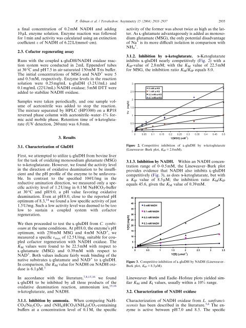
3.1.2. Inhibition by a-ketoglutarate. a-Ketoglutarate<br />
inhibits L-gluDH nearly competitively (Fig. 2) with a<br />
K IP-value of 2.8mM;with <strong>the</strong> K M value of 22.5mM<br />
for MSG, <strong>the</strong> inhibition ratio KM/KIP equals 8.0.<br />
1/v (mg/U)<br />
3<br />
2.5<br />
2<br />
1.5<br />
1<br />
0.5<br />
0 mM α-KG<br />
1 mM α-KG<br />
3.3 mM α-KG<br />
4.7 mM α-KG<br />
0<br />
0 0.050.1 0.150.2 0.250.3 0.350.4 0.450.5<br />
1/[MSG] (mM -1 )<br />
Figure 2. Competitive inhibition of L-gluDH by a-ketoglutarate<br />
(Lineweaver–Burk plot, KIP = 2.8mM).<br />
3.1.3. Inhibition by <strong>NADH</strong>. Within an <strong>NADH</strong> concentration<br />
range of 0–0.5 mM, <strong>the</strong> Lineweaver–Burk plot<br />
provides evidence that <strong>NADH</strong> also inhibits L-gluDH<br />
competitively (Fig. 3), as does a-ketoglutarate, but with<br />
a KIP value of 8.5lM;<strong>the</strong> inhibition ratio KM/KIP<br />
equals 45.6, given <strong>the</strong> K M value of 0.39mM.<br />
1/[V] (mg/U)<br />
45<br />
40<br />
35<br />
30<br />
25<br />
20<br />
15<br />
10<br />
5<br />
0 mM <strong>NADH</strong><br />
0.1 mM <strong>NADH</strong><br />
0.2 mM <strong>NADH</strong><br />
0.3 mM <strong>NADH</strong><br />
0<br />
0 0.5 1 1.5 2 2.5 3 3.5<br />
1/[S] (µmol -1 )<br />
Figure 3. Competitive inhibition of L-gluDH by <strong>NADH</strong> (Lineweaver–<br />
Burk plot, KIP = 8.5lM).<br />
Lineweaver–Burk and Eadie–Hofstee plots yielded similar<br />
KM and KI values, usually within a 10% range.<br />
3.2. Characterization of <strong>NADH</strong> <strong>oxidase</strong><br />
Characterization of <strong>NADH</strong> <strong>oxidase</strong> from L. sanfranciscensis<br />
has been described in <strong>the</strong> literature. 5,6 The enzyme<br />
is active between pH7.0 and 8.5. The specific