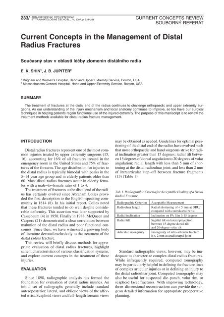
Current Concepts in the Management of Distal Radius Fractures
Current Concepts in the Management of Distal Radius Fractures
Current Concepts in the Management of Distal Radius Fractures

You also want an ePaper? Increase the reach of your titles
YUMPU automatically turns print PDFs into web optimized ePapers that Google loves.
233/<br />
ACTA CHIRURGIAE ORTHOPAEDICAE<br />
ET TRAUMATOLOGIAE ČECHOSL., 74, 2007, p. 233–246<br />
CURRENT CONCEPTS REVIEW<br />
SOUBORNÝ REFERÁT<br />
<strong>Current</strong> <strong>Concepts</strong> <strong>in</strong> <strong>the</strong> <strong>Management</strong> <strong>of</strong> <strong>Distal</strong><br />
<strong>Radius</strong> <strong>Fractures</strong><br />
Současný stav v oblasti léčby zlomen<strong>in</strong> distálního radia<br />
E. K. SHIN 1 ,J.B.JUPITER 2<br />
1 Brigham and Women’s Hospital, Hand and Upper Extremity Service, Boston, USA<br />
2 Massachusetts General Hospital, Hand and Upper Extremity Service, Boston, USA<br />
SUMMARY<br />
The treatment <strong>of</strong> fractures at <strong>the</strong> distal end <strong>of</strong> <strong>the</strong> radius cont<strong>in</strong>ues to challenge orthopaedic and upper extremity surgeons.<br />
As our understand<strong>in</strong>g <strong>of</strong> <strong>the</strong> <strong>in</strong>jury mechanism and local anatomy cont<strong>in</strong>ues to improve, so too have our surgical<br />
techniques <strong>in</strong> help<strong>in</strong>g patients rega<strong>in</strong> functional use <strong>of</strong> <strong>the</strong> <strong>in</strong>jured extremity. The purpose <strong>of</strong> this maniscript is to review <strong>the</strong><br />
treatment methods available for distal radius fracture management.<br />
INTRODUCTION<br />
<strong>Distal</strong> radius fractures represent one <strong>of</strong> <strong>the</strong> most common<br />
<strong>in</strong>juries treated by upper extremity surgeons (15,<br />
16), account<strong>in</strong>g for 16% <strong>of</strong> all fractures treated <strong>in</strong> <strong>the</strong><br />
emergency room <strong>in</strong> <strong>the</strong> United States and 75% <strong>of</strong> fractures<br />
<strong>of</strong> <strong>the</strong> forearm. The age distribution for <strong>in</strong>juries to<br />
<strong>the</strong> distal radius is typically bimodal with peaks <strong>in</strong> <strong>the</strong><br />
5–14 year age group and <strong>in</strong> elderly patients older than<br />
60. Most distal radius fractures occur <strong>in</strong> elderly females<br />
with a male–to–female ratio <strong>of</strong> 1 to 4.<br />
The treatment <strong>of</strong> fractures at <strong>the</strong> distal end <strong>of</strong> <strong>the</strong> radius<br />
has certa<strong>in</strong>ly evolved s<strong>in</strong>ce Abraham Colles provided<br />
<strong>the</strong> first description to <strong>the</strong> English–speak<strong>in</strong>g community<br />
<strong>in</strong> 1814 (8). In his <strong>in</strong>itial report, Colles noted<br />
that <strong>the</strong>se fractures tended to do well despite considerable<br />
deformity. This assertion was later supported by<br />
Cassebaum (4) <strong>in</strong> 1950. F<strong>in</strong>ally <strong>in</strong> 1988, McQueen and<br />
Caspers (21) demonstrated a clear correlation between<br />
malunion <strong>of</strong> <strong>the</strong> distal radius and poor functional outcomes.<br />
S<strong>in</strong>ce <strong>the</strong>n, we have witnessed a grow<strong>in</strong>g body<br />
<strong>of</strong> literature devoted exclusively to <strong>the</strong> treatment <strong>of</strong> <strong>the</strong><br />
distal radius fracture.<br />
This review will briefly discuss methods for appropriate<br />
evaluation <strong>of</strong> distal radius fractures, highlight<br />
salient characteristics <strong>of</strong> various classification systems,<br />
and explore current concepts <strong>in</strong> <strong>the</strong> treatment <strong>of</strong> <strong>the</strong>se<br />
<strong>in</strong>juries.<br />
EVALUATION<br />
S<strong>in</strong>ce 1898, radiographic analysis has formed <strong>the</strong><br />
foundation for evaluation <strong>of</strong> distal radius <strong>in</strong>juries. An<br />
<strong>in</strong>itial set <strong>of</strong> radiographs generally <strong>in</strong>clude standard<br />
anteroposterior, lateral, and oblique views <strong>of</strong> <strong>the</strong> affected<br />
wrist. Scaphoid views and full–length forearm views<br />
may be obta<strong>in</strong>ed as needed. Guidel<strong>in</strong>es for optimal position<strong>in</strong>g<br />
<strong>of</strong> <strong>the</strong> distal end <strong>of</strong> <strong>the</strong> radius have evolved such<br />
that most orthopaedic and hand surgeons strive for radial<br />
<strong>in</strong>cl<strong>in</strong>ation greater than 15 degrees; radial tilt between<br />
15 degrees <strong>of</strong> dorsal angulation to 20 degrees <strong>of</strong> volar<br />
angulation; radial length with less than 5 mm <strong>of</strong> shorten<strong>in</strong>g<br />
at <strong>the</strong> distal radioulnar jo<strong>in</strong>t; and less than 2 mm<br />
<strong>of</strong> <strong>in</strong>traarticular step–<strong>of</strong>f between fracture fragments<br />
(13) (Table 1).<br />
Tab. 1. Radiographic Criteria for Acceptable Heal<strong>in</strong>g <strong>of</strong> a <strong>Distal</strong><br />
Radial Fracture<br />
Radiographic Criterion Acceptable Measurement<br />
Radioulnar length Radial shorten<strong>in</strong>g <strong>of</strong> < 5 mm at DRUJ<br />
compared with contralateral wrist<br />
Radial <strong>in</strong>cl<strong>in</strong>ation Incl<strong>in</strong>ation on PA film ≥ 15 degrees<br />
Radial tilt Sagittal tilt on lateral projection<br />
between 15-degree dorsal tilt<br />
and 20-degree volar tilt<br />
Articular <strong>in</strong>congruity Incongruity <strong>of</strong> <strong>in</strong>tra-articular fracture<br />
is ≤ 2 mm at aradiocarpal jo<strong>in</strong>t<br />
Standard radiographic views, however, may be <strong>in</strong>adequate<br />
to characterize complex distal radius fractures.<br />
While <strong>in</strong>frequently required, computed tomography<br />
may be particularly helpful <strong>in</strong> def<strong>in</strong><strong>in</strong>g <strong>the</strong> fracture l<strong>in</strong>es<br />
<strong>of</strong> complex articular <strong>in</strong>juries or <strong>in</strong> def<strong>in</strong><strong>in</strong>g an <strong>in</strong>jury to<br />
<strong>the</strong> distal radioulnar jo<strong>in</strong>t. Computed tomography may<br />
also be useful for suspected die–punch, volar rim, or<br />
scaphoid facet fractures. With improv<strong>in</strong>g technology,<br />
three–dimensional reconstructions can provide <strong>the</strong> surgeon<br />
detailed <strong>in</strong>formation for appropriate preoperative<br />
plann<strong>in</strong>g.
234/<br />
ACTA CHIRURGIAE ORTHOPAEDICAE<br />
ET TRAUMATOLOGIAE ČECHOSL., 74, 2007<br />
CLASSIFICATION<br />
Various classification systems have been proposed<br />
for fractures at <strong>the</strong> distal radius. Each distal radius fracture<br />
differs <strong>in</strong> <strong>the</strong> mechanism <strong>of</strong> <strong>in</strong>jury, <strong>the</strong> energy <strong>of</strong><br />
<strong>in</strong>jury, <strong>the</strong> degree <strong>of</strong> articular <strong>in</strong>volvement, and associated<br />
<strong>in</strong>juries. Among <strong>the</strong> early attempts to def<strong>in</strong>e <strong>the</strong><br />
vary<strong>in</strong>g patterns <strong>of</strong> articular fractures <strong>of</strong> <strong>the</strong> distal radius<br />
were those <strong>of</strong> Casta<strong>in</strong>g (5) <strong>in</strong> 1964 and Frykman (11)<br />
<strong>in</strong> 1967. Though <strong>the</strong> latter classification has been widely<br />
cited <strong>in</strong> <strong>the</strong> English language literature, it fails to give<br />
critical <strong>in</strong>formation about <strong>the</strong> extent and direction <strong>of</strong><br />
articular fracture displacement. Melone (23) observed<br />
four basic components <strong>in</strong> <strong>the</strong> classification system<br />
which bears his name: <strong>the</strong> radial shaft, <strong>the</strong> radial styloid,<br />
<strong>the</strong> posteromedial portion <strong>of</strong> <strong>the</strong> lunate facet <strong>of</strong> <strong>the</strong><br />
distal radius, and <strong>the</strong> palmar medial portion <strong>of</strong> <strong>the</strong> lunate<br />
facet.<br />
The Fernandez classification system (10) is based on<br />
<strong>the</strong> mechanism <strong>of</strong> <strong>in</strong>jury and may facilitate manual<br />
reduction <strong>of</strong> a distal radius fracture. Type I fractures are<br />
simple bend<strong>in</strong>g fractures <strong>of</strong> <strong>the</strong> metaphysis that are low<br />
energy, typically <strong>the</strong> result <strong>of</strong> a ground level fall. These<br />
<strong>in</strong>clude extraarticular Colles’ or Smith’s fractures.<br />
Type II fractures are shear<strong>in</strong>g <strong>in</strong>juries that result from<br />
obliquely directed forces through part <strong>of</strong> <strong>the</strong> articular<br />
surface. These fractures are <strong>in</strong>herently unstable and<br />
typically require open reduction <strong>in</strong>ternal fixation. Type<br />
III fractures are compression <strong>in</strong>juries <strong>of</strong> <strong>the</strong> jo<strong>in</strong>t surface<br />
with impaction <strong>of</strong> <strong>the</strong> subchondral bone. Depend<strong>in</strong>g<br />
on <strong>the</strong> degree <strong>of</strong> energy <strong>the</strong>re may be m<strong>in</strong>imal or considerable<br />
displacement. Type IV <strong>in</strong>juries are fracture–dislocations<br />
and may be deceptively simple–appear<strong>in</strong>g.<br />
A fracture through <strong>the</strong> radial styloid, for example,<br />
may propogate through <strong>the</strong> scapholunate ligament,<br />
result<strong>in</strong>g <strong>in</strong> an <strong>in</strong>tercarpal dislocation. Type V fracture<br />
are high energy <strong>in</strong>juries associated with s<strong>of</strong>t tissue loss<br />
and are also referred to as comb<strong>in</strong>ed complex <strong>in</strong>juries.<br />
The outcome <strong>of</strong> <strong>the</strong>se <strong>in</strong>juries is dictated as much by<br />
bony reconstruction as it is by <strong>the</strong> extent <strong>of</strong> s<strong>of</strong>t tissue<br />
<strong>in</strong>jury and reconstruction (Fig. 1).<br />
GOALS OF TREATMENT<br />
How aggressively one pursues surgical reconstruction<br />
<strong>of</strong> distal radius fractures is dictated by <strong>the</strong> demands<br />
<strong>of</strong> <strong>the</strong> patient and <strong>the</strong> radiographic f<strong>in</strong>d<strong>in</strong>gs at <strong>the</strong> time<br />
<strong>of</strong> <strong>in</strong>jury. Those patients with low demand activities may<br />
be best served with nonoperative techniques. High<br />
demand patients, however, may require surgical fixation<br />
to allow early range <strong>of</strong> motion and to prevent stiffness,<br />
which could be detrimental for certa<strong>in</strong> activities.<br />
The significance <strong>of</strong> articular <strong>in</strong>congruity is still debated<br />
<strong>in</strong> treat<strong>in</strong>g patients with distal radius fractures. Articular<br />
<strong>in</strong>congruity for <strong>in</strong>traarticular fractures may lead<br />
to decreased ability to remodel as <strong>the</strong> jo<strong>in</strong>t step<strong>of</strong>f exceeds<br />
<strong>the</strong> thickness <strong>of</strong> <strong>the</strong> articular cartilage. Knirk and<br />
Jupiter (20) found that articular <strong>in</strong>congruity predisposed<br />
patients to <strong>the</strong> development <strong>of</strong> degenerative jo<strong>in</strong>t<br />
disease <strong>in</strong> <strong>the</strong> radiocarpal jo<strong>in</strong>t. In <strong>the</strong>ir study <strong>of</strong> young<br />
CURRENT CONCEPTS REVIEW<br />
SOUBORNÝ REFERÁT<br />
Fig. 1. The fracture classification <strong>of</strong> Diego Fernandez,<br />
M. D. based upon <strong>the</strong> mechanism <strong>of</strong> <strong>in</strong>jury.
235/<br />
ACTA CHIRURGIAE ORTHOPAEDICAE<br />
ET TRAUMATOLOGIAE ČECHOSL., 74, 2007<br />
Fig. 2. A well molded short arm cast applied for a stable distal<br />
radius fracture.<br />
patients, <strong>the</strong> absence <strong>of</strong> jo<strong>in</strong>t step<strong>of</strong>f follow<strong>in</strong>g treatment<br />
for an <strong>in</strong>traarticular distal radius fracture led to arthrosis<br />
<strong>in</strong> only 11% <strong>of</strong> patients. Step<strong>of</strong>fs <strong>of</strong> 2 mm or greater,<br />
however, led to generative jo<strong>in</strong>t disease <strong>in</strong> 91% <strong>of</strong><br />
patients.<br />
Catalano (6) also found a strong association between<br />
<strong>in</strong>traarticular step<strong>of</strong>f and degenerative jo<strong>in</strong>t disease<br />
but found that all patients presented with good or excellent<br />
outcomes an average <strong>of</strong> 7 years follow<strong>in</strong>g surgery,<br />
regardless <strong>of</strong> <strong>the</strong> <strong>in</strong>itial deformity. Goldfarb and Catalano<br />
(12) followed up this study by evaluat<strong>in</strong>g <strong>the</strong> same<br />
cohort <strong>of</strong> patients an average <strong>of</strong> 15 years after surgery.<br />
The authors found that patients cont<strong>in</strong>ued to function at<br />
high levels, that strength and range <strong>of</strong> motion measurements<br />
were unchanged, and that <strong>the</strong> jo<strong>in</strong>t space was<br />
reduced an additional 67% <strong>in</strong> those patients with radiocarpal<br />
arthrosis. No correlation was noted between <strong>the</strong><br />
presence or degree <strong>of</strong> arthrosis and upper extremity<br />
function as measured by DASH scores and <strong>the</strong> Gartland<br />
and Werley criteria. Clearly, <strong>in</strong>traarticular step<strong>of</strong>f may<br />
not be as significant as previously believed.<br />
CAST IMMOBILIZATION<br />
Multiple surgical treatments are available to upper<br />
extremity surgeons treat<strong>in</strong>g fractures <strong>of</strong> <strong>the</strong> distal radius.<br />
However, cast immobilization is an appropriate treatment<br />
for all non–displaced fractures and stable displaced<br />
fractures that have been reduced. It may also be<br />
appropriate for low demand patients who would not be<br />
able to tolerate surgery for medical reasons. Closed<br />
reduction <strong>of</strong> displaced fractures consist <strong>of</strong> longitud<strong>in</strong>al<br />
traction, palmar translation <strong>of</strong> <strong>the</strong> hand, pronation <strong>of</strong> <strong>the</strong><br />
CURRENT CONCEPTS REVIEW<br />
SOUBORNÝ REFERÁT<br />
hand relative to <strong>the</strong> forearm, and f<strong>in</strong>ally ulnar tilt. This<br />
reduction maneuver does not require wrist flexion.<br />
Determ<strong>in</strong><strong>in</strong>g which fractures will heal uneventfully<br />
with cast immobilization may be difficult. An unstable<br />
distal radius fracture can be def<strong>in</strong>ed by several criteria,<br />
which <strong>in</strong>clude: comm<strong>in</strong>ution greater than 50% from<br />
dorsal to volar, angulation greater than 20 degrees <strong>of</strong><br />
dorsal tilt, shorten<strong>in</strong>g greater than 10 mm, a shear<strong>in</strong>g<br />
fracture pattern, and significant displacement with<br />
100% loss <strong>of</strong> opposition (2). All unstable fracture patterns<br />
require surgical <strong>in</strong>tervention.<br />
Cast treatment typically consists <strong>of</strong> immobilization<br />
<strong>in</strong> a sugar–tong spl<strong>in</strong>t for three weeks immediately follow<strong>in</strong>g<br />
closed reduction, which is <strong>the</strong>n converted to<br />
a short arm cast for an additional three weeks. Patients<br />
are usually given a removable spl<strong>in</strong>t for a f<strong>in</strong>al three<br />
weeks and <strong>in</strong>structed to perform active assist range <strong>of</strong><br />
motion exercises to rega<strong>in</strong> flexibility. Early <strong>in</strong> <strong>the</strong> treatment<br />
course, radiographs should be obta<strong>in</strong>ed weekly<br />
to ensure fracture stability. The palmar crease should be<br />
free to allow full motion about <strong>the</strong> metacarpophalangeal<br />
jo<strong>in</strong>ts (Fig. 2).<br />
PERCUTANEOUS PIN FIXATION<br />
Because unstable distal radius fractures have a tendency<br />
to redisplace <strong>in</strong> plaster, percutaneous p<strong>in</strong>n<strong>in</strong>g is<br />
a relatively simple and effective method <strong>of</strong> fixation that<br />
is recommended for reducible extraarticular fractures,<br />
simple <strong>in</strong>traarticular fractures that are nondisplaced, and<br />
<strong>in</strong> patients with good bone quality.<br />
Multiple different techniques have been described for<br />
p<strong>in</strong>n<strong>in</strong>g distal radius fractures. These <strong>in</strong>clude p<strong>in</strong>s pla-
236/<br />
a b<br />
c d<br />
ACTA CHIRURGIAE ORTHOPAEDICAE<br />
ET TRAUMATOLOGIAE ČECHOSL., 74, 2007<br />
ced through <strong>the</strong> radial styloid, two or three crossed p<strong>in</strong>s<br />
across <strong>the</strong> fracture site, or <strong>in</strong>trafocal p<strong>in</strong>n<strong>in</strong>g with<strong>in</strong> <strong>the</strong><br />
fracture site. Some techniques also <strong>in</strong>corporate transfixation<br />
wires across <strong>the</strong> distal radioulnar jo<strong>in</strong>t for added<br />
stability. The actual technique used is probably not significant<br />
as long as <strong>the</strong> wires confer sufficient fixation to<br />
<strong>the</strong> fractured radius.<br />
Kapandji (18) popularized <strong>the</strong> technique <strong>of</strong> double<br />
<strong>in</strong>trafocal p<strong>in</strong>n<strong>in</strong>g to both reduce and ma<strong>in</strong>ta<strong>in</strong> distal<br />
radius fractures. This procedure is probably best reserved<br />
for noncomm<strong>in</strong>uted extraarticular <strong>in</strong>juries. Kapandji’s<br />
technique first requires a Kirschner wire <strong>in</strong>troduced<br />
<strong>in</strong>to <strong>the</strong> fracture site <strong>in</strong> a radial–to–ulnar direction.<br />
When <strong>the</strong> wire reaches <strong>the</strong> ulnar cortex, <strong>the</strong> wire is used<br />
to elevate <strong>the</strong> radial fragment and recreate <strong>the</strong> radial <strong>in</strong>cl<strong>in</strong>ation.<br />
This wire is <strong>the</strong>n driven through <strong>the</strong> ulnar cortex<br />
for stability. A second wire is <strong>in</strong>troduced 90 degrees<br />
to <strong>the</strong> first <strong>in</strong> a similar manner to restore volar tilt.<br />
Generous sk<strong>in</strong> <strong>in</strong>cisions must be made about <strong>the</strong> p<strong>in</strong><br />
sites to prevent sk<strong>in</strong> te<strong>the</strong>r<strong>in</strong>g. Care must also be taken<br />
to avoid <strong>in</strong>jury to <strong>the</strong> cutaneous nerves (Fig. 3a-d).<br />
CURRENT CONCEPTS REVIEW<br />
SOUBORNÝ REFERÁT<br />
Fig. 3a-d. Percutaneous p<strong>in</strong> fixation <strong>of</strong> an unstable distal radius fracture: 3a. The xrays <strong>of</strong> <strong>the</strong> <strong>in</strong>itial fracture. 3b. Two percutaneous<br />
p<strong>in</strong>s through <strong>the</strong> radial styloid. 3c. Fracture heal<strong>in</strong>g <strong>in</strong> an anatomic position. 3d. Functional result.<br />
EXTERNAL FIXATION<br />
External fixators are typically used as an adjunct to<br />
o<strong>the</strong>r forms <strong>of</strong> fixation, particularly for <strong>the</strong> treatment <strong>of</strong><br />
highly unstable or comm<strong>in</strong>uted <strong>in</strong>juries. External fixators<br />
provide ligamentotaxis that can help to ma<strong>in</strong>ta<strong>in</strong><br />
fracture reduction, <strong>the</strong>reby prevent<strong>in</strong>g collapse. In addition,<br />
<strong>the</strong>y function by neutraliz<strong>in</strong>g compressive, torsional,<br />
and bend<strong>in</strong>g forces across <strong>the</strong> fracture site. Occasionally,<br />
external fixators will be used for def<strong>in</strong>itive<br />
reduction <strong>of</strong> fractures, but more <strong>of</strong>ten it will be used <strong>in</strong><br />
conjunction with o<strong>the</strong>r forms <strong>of</strong> fixation.<br />
Several biomechanical studies support <strong>the</strong> use <strong>of</strong><br />
augmented external fixation with supplemental<br />
Kirschner wires. Wolfe (33) et al. performed a cadaveric<br />
study compar<strong>in</strong>g osteotomized distal radii stabilized<br />
with an external fixator alone or with various supplemental<br />
Kirschner wire configurations. Fracture transfixation<br />
wires placed <strong>in</strong>to <strong>the</strong> distal fragment and secured<br />
to <strong>the</strong> external fixator were superior to ex–fixation<br />
alone <strong>in</strong> reduc<strong>in</strong>g fracture motion. A s<strong>in</strong>gle wire was
237/<br />
a b<br />
c c<br />
ACTA CHIRURGIAE ORTHOPAEDICAE<br />
ET TRAUMATOLOGIAE ČECHOSL., 74, 2007<br />
Fig. 4a-c. An unstable <strong>in</strong>traarticular fracuture treated with an<br />
nonbridg<strong>in</strong>g external fixation:<br />
a. The preoperative x-ray; b. The non bridg<strong>in</strong>g fixation with<br />
an anatomic reduction; c. Follow-up heal<strong>in</strong>g with excellent<br />
position.<br />
enough to ga<strong>in</strong> appreciable stability, and additional wires<br />
did not improve stability fur<strong>the</strong>r.<br />
Despite its usefulness, <strong>the</strong> rate <strong>of</strong> complications with<br />
external fixation is high. Complications <strong>in</strong>clude stiffness,<br />
p<strong>in</strong> tract <strong>in</strong>fections, p<strong>in</strong> loosen<strong>in</strong>g, radial sensory<br />
nerve <strong>in</strong>jury, and reflex sympa<strong>the</strong>tic dystrophy. These<br />
complications may be avoided to some degree by avoid<strong>in</strong>g<br />
carpal overdistraction, excessive wrist flexion, and<br />
prolonged fixator treatment (Fig. 4a-c, Fig. 5a-f).<br />
ARTHROSCOPICALLY ASSISTED FIXATION<br />
Wrist arthroscopy is a technique that provides a m<strong>in</strong>imally<br />
<strong>in</strong>vasive way <strong>of</strong> monitor<strong>in</strong>g closed reduction <strong>of</strong><br />
distal radius fractures with percutaneous p<strong>in</strong> fixation.<br />
Obviously, it allows assessment <strong>of</strong> <strong>the</strong> articular jo<strong>in</strong>t surface<br />
as well as <strong>the</strong> diagnosis <strong>of</strong> <strong>in</strong>terosseous carpal ligament<br />
<strong>in</strong>jury or triangular fibrocartilage complex <strong>in</strong>jury.<br />
F<strong>in</strong>ally, it facilitates <strong>the</strong> excision <strong>of</strong> osteochondral flaps<br />
and loose bodies as needed. Disadvantages <strong>of</strong> arthroscopically–assisted<br />
fixation <strong>in</strong>clude <strong>the</strong> steep learn<strong>in</strong>g<br />
curve associated with any arthroscopy procedure. To<br />
date, <strong>the</strong>re are few studies that demonstrate improved<br />
functional outcomes with <strong>the</strong> use <strong>of</strong> arthroscopy.<br />
To perform wrist arthroscopy, a small jo<strong>in</strong>t (2.7 mm)<br />
arthroscope may be <strong>in</strong>troduced through <strong>the</strong> 3–4 portal.<br />
Instrumentation can be <strong>in</strong>troduced through <strong>the</strong> 4–5 or<br />
6–R portals. Wrist arthroscopy with fixation is general-<br />
CURRENT CONCEPTS REVIEW<br />
SOUBORNÝ REFERÁT<br />
Fig. 5a<br />
Fig. 5a-f. A complex <strong>in</strong>trarticular fracture <strong>in</strong> a dentist treated<br />
with K-wires and a bridg<strong>in</strong>g external fixation: a. The orig<strong>in</strong>al<br />
fracture; b. The CT scan; c. The fracture fixation; d. The dentist<br />
at work with his external fixation <strong>in</strong> place; e. X-rays <strong>of</strong> <strong>the</strong><br />
healed fracture; f. Functional follow-up.<br />
Pokračování �
238/<br />
ACTA CHIRURGIAE ORTHOPAEDICAE<br />
ET TRAUMATOLOGIAE ČECHOSL., 74, 2007<br />
Fig. 5b � �<br />
Fig. 5c � �<br />
ly best achieved about four to seven days from <strong>the</strong> time<br />
<strong>of</strong> <strong>in</strong>jury. Surgery performed too soon after <strong>in</strong>jury may<br />
face difficulties with fracture hematoma imped<strong>in</strong>g articular<br />
visualization. Likewise, fractures treated after one<br />
week from <strong>the</strong> time <strong>of</strong> <strong>in</strong>jury may be difficult to manipulate<br />
with percutaneous wires. Fracture fragments are<br />
CURRENT CONCEPTS REVIEW<br />
SOUBORNÝ REFERÁT<br />
Pokračování �<br />
typically elevated us<strong>in</strong>g Kirschner wires as joysticks.<br />
<strong>Fractures</strong> can <strong>the</strong>n be p<strong>in</strong>ned transversely beneath subchondral<br />
bone (Fig. 6a-b).<br />
Doi et al. (9) analyzed <strong>the</strong> usefulness <strong>of</strong> arthroscopically<br />
assisted reduction <strong>of</strong> <strong>in</strong>traarticular fractures <strong>of</strong> <strong>the</strong><br />
distal radius by compar<strong>in</strong>g <strong>the</strong> results <strong>of</strong> that procedure<br />
with those <strong>of</strong> conventional open reduction and <strong>in</strong>ternal<br />
fixation. The authors performed a randomized prospective<br />
study, treat<strong>in</strong>g 34 patients with arthroscopically<br />
guided reduction and p<strong>in</strong>n<strong>in</strong>g and 48 with conventional<br />
open reduction <strong>in</strong>ternal fixation. The authors found with<br />
30 month average follow–up that patients with arthros-
239/<br />
ACTA CHIRURGIAE ORTHOPAEDICAE<br />
ET TRAUMATOLOGIAE ČECHOSL., 74, 2007<br />
Fig. 5d � �<br />
Fig. 5f � Fig. 5e �<br />
CURRENT CONCEPTS REVIEW<br />
SOUBORNÝ REFERÁT
240/<br />
ACTA CHIRURGIAE ORTHOPAEDICAE<br />
ET TRAUMATOLOGIAE ČECHOSL., 74, 2007<br />
CURRENT CONCEPTS REVIEW<br />
SOUBORNÝ REFERÁT<br />
Fig. 6 a-b. Arthroscopic assisted fixation: a. A displaced <strong>in</strong>traarticular fracture; b. Arthroscopic view <strong>of</strong> <strong>the</strong> articular facture.<br />
copically guided reduction had good or excellent results<br />
82% <strong>of</strong> <strong>the</strong> time us<strong>in</strong>g <strong>the</strong> Gartland and Werley criteria,<br />
better ROM and grip strength, and an improved radiographic<br />
appearance compared to <strong>the</strong> open reduction<br />
cohort. On <strong>the</strong> basis <strong>of</strong> this study, Doi et al. concluded<br />
that arthroscopically assisted fixation <strong>of</strong> distal radius<br />
fractures is an effective technique <strong>in</strong> patients less than<br />
70 years <strong>of</strong> age with <strong>in</strong>traarticular <strong>in</strong>juries.<br />
OPEN REDUCTION INTERNAL FIXATION:<br />
DORSAL AND VOLAR<br />
Open reduction <strong>in</strong>ternal fixation has obvious advantages<br />
over <strong>the</strong> o<strong>the</strong>r methods discussed so far. It allows<br />
direct restoration <strong>of</strong> anatomy, stable <strong>in</strong>ternal fixation,<br />
a decreased period <strong>of</strong> immobilization, and an earlier<br />
return <strong>of</strong> wrist function. There are a number <strong>of</strong> different<br />
<strong>in</strong>dications for open reduction <strong>in</strong>ternal fixation, and<br />
<strong>the</strong>se <strong>in</strong>clude: unstable articular fractures (such as<br />
avolar Barton’s <strong>in</strong>jury), impacted articular fractures,<br />
radiocarpal fracture–dislocations, complex fractures<br />
requir<strong>in</strong>g direct visualization <strong>of</strong> <strong>the</strong> fracture fragments,<br />
and failed closed reductions.<br />
Historically, distal radius fractures were treated<br />
nonoperatively until 1929, when techniques utiliz<strong>in</strong>g<br />
p<strong>in</strong>s and plaster were <strong>in</strong>troduced. External skeletal<br />
fixation evolved <strong>in</strong> 1944 and rema<strong>in</strong>ed popular even<br />
after <strong>the</strong> AO group designed plates specifically for <strong>the</strong><br />
treatment <strong>of</strong> distal radius fractures <strong>in</strong> <strong>the</strong> 1970s. In<br />
1994, Agee <strong>in</strong>troduced <strong>the</strong> Wrist Jack (Hand Biomechanics<br />
Lab Inc, Sacramento, CA), which utilized<br />
adjustable gears for multiplanar ligamentotaxis, but by<br />
this time open reduction <strong>in</strong>ternal fixation was becom<strong>in</strong>g<br />
more popular, particularly when it was noted that<br />
precise reductions <strong>of</strong> <strong>the</strong> articular surface led to better<br />
outcomes (14).<br />
a b<br />
The approach is generally dictated by <strong>the</strong> location <strong>of</strong><br />
major fracture fragments and <strong>the</strong> direction <strong>of</strong> displacement.<br />
However, orthopaedic and upper extremity surgeons<br />
cont<strong>in</strong>ue to move away from dorsal plat<strong>in</strong>g where<br />
complications can <strong>in</strong>clude extensor tendon rupture<br />
and hardware irritation. Volar plat<strong>in</strong>g is generally<br />
well–tolerated. Fixation <strong>of</strong> dorsally angulated and comm<strong>in</strong>uted<br />
fractures is also possible with newer fixed angle<br />
devices.<br />
Although overall satisfactory outcomes have been<br />
reported with dorsal plat<strong>in</strong>g systems, disadvantages <strong>of</strong><br />
dorsal plates <strong>in</strong>clude <strong>the</strong> need for mobilization <strong>of</strong> extensor<br />
tendons to achieve proper plate placement, possible<br />
tendon irritation or rupture, and <strong>the</strong> possibility <strong>of</strong> additional<br />
surgery to remove <strong>the</strong> symptomatic dorsal plate,<br />
some report<strong>in</strong>g up to 30% (1, 3, 19, 27). Tendon rupture<br />
has been reported as early as 8 weeks and as late as 7<br />
months after surgery. To prevent tendon <strong>in</strong>jury, some<br />
recommend that a portion <strong>of</strong> <strong>the</strong> extensor ret<strong>in</strong>aculum<br />
be <strong>in</strong>terposed between <strong>the</strong> plate and <strong>the</strong> tendon sheaths,<br />
or that dorsal plates be removal rout<strong>in</strong>ely (7).<br />
The advantages <strong>of</strong> a volar exposure and plat<strong>in</strong>g <strong>in</strong>clude<br />
<strong>the</strong> follow<strong>in</strong>g: (1) Dorsally displaced fractures are<br />
simpler to reduce because <strong>the</strong> volar cortex is usually<br />
disrupted by a simple transverse l<strong>in</strong>e; (2) anatomic<br />
reduction <strong>of</strong> <strong>the</strong> volar cortex facilitates restoration <strong>of</strong><br />
radial length, radial <strong>in</strong>cl<strong>in</strong>ation, and volar tilt; (3) <strong>the</strong><br />
avoidance <strong>of</strong> dissection dorsally helps to preserve <strong>the</strong><br />
vascular supply to <strong>the</strong> dorsal fragments; (4) because <strong>the</strong><br />
implant is separated from <strong>the</strong> flexor tendons by <strong>the</strong> pronator<br />
quadratus, <strong>the</strong> <strong>in</strong>cidence <strong>of</strong> flexor tendon complications<br />
is lessened (17, 22); and (5) when stabilized<br />
with a fixed angle <strong>in</strong>ternal fixation device, shorten<strong>in</strong>g<br />
and secondary displacement <strong>of</strong> articular fragments is<br />
improved, and <strong>the</strong> need for bone graft<strong>in</strong>g is reduced<br />
(Fig. 7a-b).
241/<br />
ACTA CHIRURGIAE ORTHOPAEDICAE<br />
ET TRAUMATOLOGIAE ČECHOSL., 74, 2007<br />
CURRENT CONCEPTS REVIEW<br />
SOUBORNÝ REFERÁT<br />
Fig 7a-b. A complex <strong>in</strong>traarticular volar displaced fracture; a. The xray and CT views <strong>of</strong> <strong>the</strong> fracture; b. Volar plate fixation<br />
with angular stable Syn<strong>the</strong>s plate with functional result<br />
With <strong>the</strong> recent popularity <strong>of</strong> volar plat<strong>in</strong>g for treatment<br />
<strong>of</strong> distal radius fractures, we have witnessed a multitude<br />
<strong>of</strong> medical devices <strong>in</strong>troduced <strong>in</strong>to <strong>the</strong> market. Not<br />
surpris<strong>in</strong>gly, <strong>the</strong>se devices exhibit many similar characteristics.<br />
Volar plates are typically T–shaped to allow<br />
multiple fixation po<strong>in</strong>ts <strong>in</strong> <strong>the</strong> distal fracture fragment<br />
with lock<strong>in</strong>g screws recessed <strong>in</strong>to screw holes to create<br />
alower pr<strong>of</strong>ile distally. The angulation <strong>of</strong> <strong>the</strong>se distal<br />
screws create a „scaffold“ to optimize subchondral bone<br />
Fig. 7a � �<br />
Fig. 7b � �<br />
support with one or two screws devoted to fixation <strong>of</strong><br />
<strong>the</strong> lunate facet and radial styloid. Most plat<strong>in</strong>g systems<br />
also feature an oblong hole for metaphyseal fixation and<br />
for plate position<strong>in</strong>g (25).<br />
Several studies have compared outcomes <strong>of</strong> dorsal<br />
versus volar plat<strong>in</strong>g <strong>of</strong> distal radius fractures. Ruch and<br />
Papadonikolakis (29) performed a retrospective review<br />
<strong>of</strong> 34 patients, 20 <strong>of</strong> whom had undergone dorsal plat<strong>in</strong>g<br />
and 14 <strong>of</strong> whom had volar plat<strong>in</strong>g. The authors
242/<br />
ACTA CHIRURGIAE ORTHOPAEDICAE<br />
ET TRAUMATOLOGIAE ČECHOSL., 74, 2007<br />
found that both groups <strong>of</strong> patients had similar DASH<br />
scores, but <strong>the</strong> functional outcome <strong>in</strong> terms <strong>of</strong> Gartland<br />
and Werley scores was better <strong>in</strong> <strong>the</strong> volar plat<strong>in</strong>g group.<br />
In addition, <strong>the</strong>re was a higher rate <strong>of</strong> volar collapse and<br />
late complications <strong>in</strong> <strong>the</strong> dorsal plat<strong>in</strong>g group compared<br />
with <strong>the</strong> volar plat<strong>in</strong>g group.<br />
Volar plat<strong>in</strong>g, however, is not without its complications.<br />
Rozental and Blazar (28) retrospectively reviewed<br />
a cohort <strong>of</strong> 41 patients with dorsally displaced fractures<br />
treated with volar plat<strong>in</strong>g. Though Gartland and Werley<br />
scores were overwhelm<strong>in</strong>gly good and excellent, 9<br />
complications were noted. Four <strong>of</strong> <strong>the</strong>se patients actually<br />
experienced loss <strong>of</strong> reduction and fracture collapse<br />
follow<strong>in</strong>g volar plat<strong>in</strong>g (Fig. 8).<br />
OPEN REDUCTION INTERNAL FIXATION:<br />
FRAGMENT SPECIFIC<br />
The concept <strong>of</strong> fragment specific fixation has been<br />
touted as a surgical alternative to volar plat<strong>in</strong>g alone.<br />
Some basic tenets <strong>of</strong> fracture fixation for fragment specific<br />
systems <strong>in</strong>clude: (1) application <strong>of</strong> small contoured<br />
plates on <strong>the</strong> specific components <strong>of</strong> <strong>the</strong> fracture;<br />
CURRENT CONCEPTS REVIEW<br />
SOUBORNÝ REFERÁT<br />
Fig. 8. Complication <strong>of</strong> volar plate with screw too long result<strong>in</strong>g<br />
<strong>in</strong> extensor pollicis rupture requir<strong>in</strong>g reconstruction.<br />
(2) fixation <strong>of</strong> distal fragments is based on <strong>the</strong> strong<br />
bone proximally; (3) hardware should allow for glid<strong>in</strong>g<br />
motion <strong>of</strong> tendons; (4) <strong>the</strong> exposure should cause m<strong>in</strong>imal<br />
s<strong>of</strong>t tissue disruption; and (5) <strong>the</strong> fracture should<br />
be stable to allow early range <strong>of</strong> motion. The type <strong>of</strong><br />
implant used should match <strong>the</strong> specific fracture fragment<br />
be<strong>in</strong>g reduced via <strong>the</strong> use <strong>of</strong> limited volar and dorsal<br />
<strong>in</strong>cisions (Fig. 9a–c).<br />
Schnall et al. (30) documented <strong>the</strong>ir early f<strong>in</strong>d<strong>in</strong>gs<br />
with fragment specific fixation <strong>in</strong> a retrospective study<br />
<strong>of</strong> 18 patients with <strong>in</strong>traarticular fractures. At 6 weeks,<br />
none <strong>of</strong> <strong>the</strong> patients demonstrated loss <strong>of</strong> reduction. At<br />
6 months, patients demonstrated an average <strong>of</strong> 52 degrees<br />
<strong>of</strong> wrist flexion and 55 degrees <strong>of</strong> wrist extension.<br />
Grip strength was measured to be approximately 55<br />
pounds. Two <strong>of</strong> <strong>the</strong> 18 patients required hardware removal<br />
for extensor tendon irritation.<br />
Similarly, Rikli and Regazzoni (27) reviewed a series<br />
<strong>of</strong> 20 patients with distal radius fractures fixed with<br />
two 2.0 mm titanium plates placed at 50 – 70 degrees<br />
to one ano<strong>the</strong>r. No cases <strong>of</strong> extensor tendon problems<br />
were noted, most likely because <strong>the</strong>y were able to place<br />
a flap <strong>of</strong> ret<strong>in</strong>aculum over <strong>the</strong> small dorsal plates.<br />
Clearly, fragment specific fixation may provide some<br />
advantages over <strong>the</strong> traditional methods <strong>of</strong> dorsal or<br />
volar plat<strong>in</strong>g for fractures at <strong>the</strong> distal end <strong>of</strong> <strong>the</strong> radius<br />
( Fig. 10 a-c).<br />
INTRAMEDULLARY FIXATION<br />
Intramedullary fixation <strong>of</strong> fractures is not a new concept<br />
<strong>in</strong> orthopaedic trauma surgery. Intramedullary<br />
devices <strong>in</strong>crease fracture stability <strong>of</strong> <strong>the</strong> affected bone,<br />
allow load transfer across <strong>the</strong> fracture site, m<strong>in</strong>imize<br />
s<strong>of</strong>t tissue problems by m<strong>in</strong>imiz<strong>in</strong>g scarr<strong>in</strong>g and adhesions,<br />
and ma<strong>in</strong>ta<strong>in</strong> vascular blood supply to promote<br />
fracture heal<strong>in</strong>g. Two implants have recently been desc-
243/<br />
ACTA CHIRURGIAE ORTHOPAEDICAE<br />
ET TRAUMATOLOGIAE ČECHOSL., 74, 2007<br />
Fig. 9a-c. A complex articular fracture treated with fragment<br />
specific fixation with small Syn<strong>the</strong>s plates (case <strong>of</strong> Daniel Rikli<br />
MD): a. The preoperative CT scan; b. Two plate dorsal fixation;<br />
c. Functional result.<br />
a b<br />
c c<br />
Fig. 10 a-c. A complex <strong>in</strong>traarticular fracture: a. The preoperative<br />
x-rays and CT scans; b. Operative fixation with small<br />
Syn<strong>the</strong>s plates and postoperative CT scan; c. Functional follow<br />
up and follow up x-rays<br />
Fig. 10a � �<br />
CURRENT CONCEPTS REVIEW<br />
SOUBORNÝ REFERÁT<br />
Pokračování Fig. 10b �
244/<br />
ACTA CHIRURGIAE ORTHOPAEDICAE<br />
ET TRAUMATOLOGIAE ČECHOSL., 74, 2007<br />
Fig. 10b � �<br />
Fig. 10c �<br />
ribed for use <strong>in</strong> <strong>the</strong> distal radius, namely <strong>the</strong> Micronail<br />
(Wright Medical Technology Inc., Arl<strong>in</strong>gton, TN) and<br />
<strong>the</strong> Dorsal Nail Plate (Hand Innovations LLC, Miami,<br />
FL). Both are used for metaphyseal distal radius fractures<br />
with m<strong>in</strong>imal articular <strong>in</strong>volvement.<br />
The Micronail is placed through an <strong>in</strong>cision made<br />
over <strong>the</strong> radial styloid, tak<strong>in</strong>g care to avoid <strong>in</strong>jury to<br />
branches <strong>of</strong> <strong>the</strong> radial sensory nerve. The <strong>in</strong>terval between<br />
<strong>the</strong> 1st and 2nd dorsal extensor compartments is<br />
used. Kirschner wires may be used for temporary fixation<br />
<strong>of</strong> large articular fragments. Difficulties that have<br />
CURRENT CONCEPTS REVIEW<br />
SOUBORNÝ REFERÁT<br />
been described with use <strong>of</strong> this nail <strong>in</strong>clude possible s<strong>of</strong>t<br />
tissue irritation with placement <strong>of</strong> <strong>the</strong> <strong>in</strong>terlock<strong>in</strong>g<br />
screws, possible screw penetration <strong>in</strong>to <strong>the</strong> distal radioulnar<br />
jo<strong>in</strong>t, and difficulty observ<strong>in</strong>g sagittal alignment<br />
secondary to use <strong>of</strong> <strong>the</strong> jig (31).<br />
Tan et al. (32) presented <strong>the</strong>ir experience with <strong>the</strong><br />
Micronail <strong>in</strong> 23 patients with isolated unstable distal<br />
radius fractures. The m<strong>in</strong>imum follow–up was 6<br />
months. Range <strong>of</strong> motion and grip strength parameters<br />
were noted at 1 month and 6 month follow–up. The authors<br />
found that all wrist and forearm range <strong>of</strong> motion
245/<br />
ACTA CHIRURGIAE ORTHOPAEDICAE<br />
ET TRAUMATOLOGIAE ČECHOSL., 74, 2007<br />
parameters improved significantly from 1 month to 6<br />
months. Grip strength also improved significantly from<br />
40% to 80%. Three <strong>of</strong> <strong>the</strong> 23 patients had some loss <strong>in</strong><br />
<strong>in</strong>itial reduction, and one patient ultimately required<br />
conversion to open reduction and <strong>in</strong>ternal fixation.<br />
The Dorsal Nail Plate is a hybrid plat<strong>in</strong>g and <strong>in</strong>tramedullary<br />
device, aga<strong>in</strong> <strong>in</strong>tended for fractures with<br />
m<strong>in</strong>imal articular <strong>in</strong>volvement. The approach is performed<br />
through a limited dorsal <strong>in</strong>cision centered over <strong>the</strong><br />
extensor pollicis longus. Lister’s tubercle is removed,<br />
and <strong>the</strong> exposed bone is <strong>the</strong> <strong>in</strong>sertion po<strong>in</strong>t <strong>of</strong> this <strong>in</strong>tramedullary<br />
device. To our knowledge, outcomes with<br />
this device have not been reported. Orbay et al. (26)<br />
reported <strong>the</strong>ir technique and noted that complications<br />
were relatively <strong>in</strong>frequent with satisfactory functional<br />
results <strong>in</strong> over 200 patients.<br />
BIOABSORBABLE IMPLANTS<br />
Ano<strong>the</strong>r emerg<strong>in</strong>g technology for treatment <strong>of</strong> distal<br />
radius fractures is <strong>the</strong> use <strong>of</strong> bioabsorbable implants.<br />
While popular <strong>in</strong> <strong>the</strong> use <strong>of</strong> sports medic<strong>in</strong>e procedures,<br />
bioabsorbable implants have seen limited applications<br />
<strong>in</strong> orthopaedic trauma procedures. The plates and<br />
screws <strong>in</strong> bioabsorbable constructs are typically made<br />
<strong>of</strong> polylactic acid or polyglycolic acid.<br />
The Inion OTPS Hand System (Inion Inc, Oklahoma<br />
City, OK) is <strong>the</strong> only known bioabsorbable distal<br />
radius plate which is commercially available. This plate<br />
takes at least two years to degrade completely with<strong>in</strong><br />
<strong>the</strong> body, is contourable after plac<strong>in</strong>g <strong>in</strong> a hot water<br />
bath, and accepts polyaxial lock<strong>in</strong>g screws up to 20<br />
degrees.<br />
The advantages to us<strong>in</strong>g a bioabsorbable implant are<br />
manifold: Bioabsorbable distal radius plates obviously<br />
undergo resorption and obviate <strong>the</strong> need for hardware<br />
removal <strong>in</strong> <strong>the</strong> future, mak<strong>in</strong>g revision surgery potentially<br />
less complicated. The implants also do not <strong>in</strong>cite<br />
an <strong>in</strong>flammatory response and are MRI compatible.<br />
Bioabsorbable devices are, however, relatively new and<br />
unproven technology for fracture fixation. There are<br />
valid concerns regard<strong>in</strong>g <strong>the</strong> <strong>in</strong>itial fixation strength,<br />
and most <strong>of</strong> <strong>the</strong>se implants are slightly thicker than <strong>the</strong>ir<br />
metal counterparts. F<strong>in</strong>ally, one cannot visualize <strong>the</strong><br />
implants on radiographs.<br />
There is only one study, to our knowledge, which<br />
explores <strong>the</strong> possibility <strong>of</strong> us<strong>in</strong>g bioabsorbable implants<br />
for distal radius fracture fixation. One hundred patients<br />
with distal radius fractures were queried regard<strong>in</strong>g<br />
implant preferences after <strong>the</strong>y were given a brief summary<br />
<strong>of</strong> advantages and disadvantages <strong>of</strong> bioabsorbable<br />
and metal implants. The results <strong>of</strong> <strong>the</strong> questionnaires<br />
demonstrated that 95% preferred bioabsorbable<br />
implants for <strong>the</strong>ir absorption feature. 91% cited hardware<br />
removal as <strong>the</strong> most negative aspect <strong>of</strong> metal hardware.<br />
80% <strong>of</strong> patients stated <strong>the</strong>y would be <strong>in</strong>terested<br />
<strong>in</strong> participat<strong>in</strong>g <strong>in</strong> a cl<strong>in</strong>ical trial compar<strong>in</strong>g <strong>the</strong> two<br />
implants, sett<strong>in</strong>g <strong>the</strong> stage for a prospective randomized<br />
study (24).<br />
CONCLUSION<br />
CURRENT CONCEPTS REVIEW<br />
SOUBORNÝ REFERÁT<br />
How aggressively we pursue reconstruction <strong>of</strong> distal<br />
radius fractures is dictated by <strong>the</strong> demands <strong>of</strong> <strong>the</strong> patient<br />
and radiographic f<strong>in</strong>d<strong>in</strong>gs at <strong>the</strong> time <strong>of</strong> <strong>in</strong>jury.<br />
Nonoperative treatment is best reserved for low demand<br />
patients or patients too <strong>in</strong>firm to ably tolerate surgery.<br />
A multitude <strong>of</strong> different techniques can be utilized for<br />
fixation <strong>of</strong> distal radius fractures. Orthopaedic and<br />
upper extremity surgeons who manage <strong>the</strong>se <strong>in</strong>juries<br />
should have all <strong>of</strong> <strong>the</strong>se techniques at <strong>the</strong>ir disposal. As<br />
our experience with volar plat<strong>in</strong>g and fragment specific<br />
fixation cont<strong>in</strong>ues to deepen, newer techniques and<br />
approaches become available but are still unproven.<br />
ZÁVĚR<br />
Léčba zlomen<strong>in</strong> distálního radia je stále výzvou pro<br />
ortopedy i traumatology. S tím, jak se zlepšují naše znalosti<br />
anatomie a mechanismu poranění této oblasti,zdokonalují<br />
se i chirurgické postupy přispívající k obnově<br />
funkce postižené končet<strong>in</strong>y. Tato práce podává přehled<br />
současných metod léčby zlomen<strong>in</strong> distálního radia.<br />
References<br />
1. AXELROD, T. S., McMURTRY, R. Y.: Open reduction and <strong>in</strong>ternal<br />
fixation <strong>of</strong> comm<strong>in</strong>uted, <strong>in</strong>traarticular fractures <strong>of</strong> <strong>the</strong> distal<br />
radius. J. Hand Surg. Amer., 15: 1–11, 1990.<br />
2. CAPO, J. T., SWAN, K. G. Jr., TAN, V.: External fixation techniques<br />
for distal radius fractures. Cl<strong>in</strong>. Orthop., 445: 30–41, 2006.<br />
3. CARTER, P. R., FREDERICK, H. A., LASETER, G. F.: Open<br />
reduction and <strong>in</strong>ternal fixation <strong>of</strong> unstable distal radius fractures<br />
with a low–pr<strong>of</strong>ile plate: A multicenter study <strong>of</strong> 73 fractures. J.<br />
Hand Surg. Amer., 23: 300–307, 1998.<br />
4. CASSEBAUM, W. H.: Colles’ fracture: a study <strong>of</strong> end results. J.<br />
Amer. Med. Assoc., 143: 963–965, 1950.<br />
5. CASTAING, J.: Les fractures récentes de l’extrémité <strong>in</strong>férieure<br />
du radius chez l’adulte. Rev. Chir. Orthop., 50: 581–696, 1964.<br />
6. CATALANO, L. W. 3 rd , COLE, R. J., GELBERMAN, R. H.,<br />
EVANOFF, B. A., GILULA, L. A., BORRELLI, J. Jr.: Displaced<br />
<strong>in</strong>tra–articular fractures <strong>of</strong> <strong>the</strong> distal aspect <strong>of</strong> <strong>the</strong> radius.<br />
Long–term results <strong>in</strong> young adults after open reduction and <strong>in</strong>ternal<br />
fixation. J. Bone Jt Surg., 79–A: 1290–1302, 1997.<br />
7. CHIANG, P. P., ROACH, S., BARATZ, M. E.: Failure <strong>of</strong> a ret<strong>in</strong>acular<br />
flap to prevent dorsal wrist pa<strong>in</strong> after titanium pi plate<br />
fixation <strong>of</strong> distal radius fractures. J. Hand Surg. Amer., 27:<br />
724–728, 2002.<br />
8. COLLES, A.: On <strong>the</strong> fracture <strong>of</strong> <strong>the</strong> carpal extremity <strong>of</strong> <strong>the</strong> radius.<br />
Ed<strong>in</strong>burgh Med. Surg. J., 10: 182–186, 1814.<br />
9. DOI, K., HATTORI, Y., OTSUKA, K., ABE, Y., YAMAMOTO,<br />
H.: Intra–articular fractures <strong>of</strong> <strong>the</strong> distal aspects <strong>of</strong> <strong>the</strong> radius: arthroscopically<br />
assisted reduction compared with open reduction and<br />
<strong>in</strong>ternal fixation. J. Bone Jt Surg., 81–A: 1093–1110, 1999.<br />
10. FERNANDEZ, D. L., WOLFE, S. W.: <strong>Distal</strong> radius fractures. In:<br />
GREEN, D. P., HOTCHKISS, R. N., PEDERSON, W. C., WOL-<br />
FE, S. W. (eds): Green’s operative hand surg. Philadelphia, Elsevier<br />
Churchill Liv<strong>in</strong>gstone 2005, 645–710.<br />
11. FRYKMAN, G. K.: Fracture <strong>of</strong> <strong>the</strong> distal radius <strong>in</strong>clud<strong>in</strong>g sequelae:<br />
shoulder–hand–f<strong>in</strong>ger syndrome–disturbance <strong>in</strong> <strong>the</strong> distal<br />
radioulnar jo<strong>in</strong>t and impairment <strong>of</strong> nerve function: A cl<strong>in</strong>ical and<br />
experimental study. Acta orthop. scand., 108 (Suppl.): 1–155,<br />
1967.<br />
12. GOLDFARB, C. A., RUDZKI, J. R., CATALANO, L. W.,<br />
HUGHES, M., BORRELLI, J. Jr.: Fifteen–year outcome <strong>of</strong> displaced<br />
<strong>in</strong>tra–articular fractures <strong>of</strong> <strong>the</strong> distal radius. J. Hand Surg.<br />
Amer., 31: 633–639, 2006.
246/<br />
ACTA CHIRURGIAE ORTHOPAEDICAE<br />
ET TRAUMATOLOGIAE ČECHOSL., 74, 2007<br />
13. GRAHAM, T. J.: Surgical correction <strong>of</strong> malunited fractures <strong>of</strong> <strong>the</strong><br />
distal radius. J. Amer. Acad. Orthop. Surg., 5: 270–281, 1997.<br />
14. HARNESS, N. G., MEALS, R. A.: The history <strong>of</strong> fracture fixation<br />
<strong>of</strong> <strong>the</strong> hand and wrist. Cl<strong>in</strong>. Orthop., 445: 19–29, 2006.<br />
15. JUPITER, J. B.: <strong>Fractures</strong> <strong>of</strong> <strong>the</strong> distal end <strong>of</strong> <strong>the</strong> radius. J. Bone<br />
Jt Surg., 73–A: 461–469, 1991.<br />
16. JUPITER, J. B.: Complex articular fractures <strong>of</strong> <strong>the</strong> distal radius:<br />
classification and management. J. Amer. Assoc. Orthop. Surg., 5:<br />
119–129, 1997.<br />
17. JUPITER, J. B., RING, D., WEITZEL, P. P.: Surgical treatment<br />
<strong>of</strong> redisplaced fractures <strong>of</strong> <strong>the</strong> distal radius <strong>in</strong> patients older than<br />
60 years. J. Hand Surg. Amer., 27: 714–723, 2002.<br />
18. KAPANDJI, A. I.: Treatment <strong>of</strong> non–articular distal radial fractures<br />
by <strong>in</strong>trafocal p<strong>in</strong>n<strong>in</strong>g with arum p<strong>in</strong>s. In: SAFFER, P.,<br />
COONEY, W. P. (eds): <strong>Fractures</strong> <strong>of</strong> <strong>the</strong> distal radius. Philadelphia,<br />
JB Lipp<strong>in</strong>cott 1995, 71–83.<br />
19. KARAMBOUROGLU, G. K., AXELROD, T. S.. Complications<br />
<strong>of</strong> <strong>the</strong> AO/ASIF titanium distal radius plate system (π plate) <strong>in</strong><br />
<strong>in</strong>ternal fixation <strong>of</strong> <strong>the</strong> distal radius: A brief report. J. Hand Surg.<br />
Amer., 23: 737–741, 1998.<br />
20. KNIRK, J. L., JUPITER, J. B.: Intra–articular fractures <strong>of</strong> <strong>the</strong><br />
distal end <strong>of</strong> <strong>the</strong> radius <strong>in</strong> young adults. J. Bone Jt Surg., 68–A:<br />
647–659, 1986.<br />
21. McQUEEN, M., CASPERS, J.: Colles’ fracture: does <strong>the</strong> anatomical<br />
result affect <strong>the</strong> f<strong>in</strong>al function? J. Bone Jt Surg., 70–B:<br />
649–651, 1988.<br />
22. McQUEEN, M. M., SIMPSON, D., COURT–BROWN, C. M.:<br />
Use <strong>of</strong> <strong>the</strong> H<strong>of</strong>fman 2 compact external fixator <strong>in</strong> <strong>the</strong> treatment<br />
<strong>of</strong> redisplaced unstable distal radial fracture. J. Orthop. Trauma,<br />
13: 501–505, 1999.<br />
23. MELONE, C. P. Jr.: <strong>Distal</strong> radius fractures: Patterns <strong>of</strong> articular<br />
fragmentation. Orthop. Cl<strong>in</strong>. N. Amer., 24: 239–253, 1993.<br />
24. MITTAL, R., MORLEY, J., DINOPOULOS, H., DRAKOU-<br />
LAKIS, E. G., VERMANI, E., GIANNOUDIS, P. V.: Use <strong>of</strong><br />
bio–resorbable implants for stabilisation <strong>of</strong> distal radius fractures:<br />
The United K<strong>in</strong>gdom patients’ perspective. Injury, 36:<br />
333–338, 2005.<br />
25. MUDGAL, C. S., JUPITER, J. B.: Plate and screw design <strong>in</strong> fractures<br />
<strong>of</strong> <strong>the</strong> hand and wrist. Cl<strong>in</strong>. Orthop., 445: 68–80, 2006.<br />
CURRENT CONCEPTS REVIEW<br />
SOUBORNÝ REFERÁT<br />
26. ORBAY, J. L., TOUHAMI, A., ORBAY, C.: Fixed angle fixation<br />
<strong>of</strong> distal radius fractures through a m<strong>in</strong>imally <strong>in</strong>vasive approach.<br />
Tech. Hand Upper Extrem. Surg., 9: 142–148, 2005.<br />
27. RIKLI, D. A., REGAZZONI, P.: <strong>Fractures</strong> <strong>of</strong> <strong>the</strong> distal end <strong>of</strong> <strong>the</strong><br />
radius treated by <strong>in</strong>ternal fixation and early function: a prelim<strong>in</strong>ary<br />
report <strong>of</strong> 20 cases. J. Bone Jt Surg., 78–B: 588–592, 1996.<br />
28. ROZENTAL, T. D., BLAZAR, P. E.: Functional outcome and<br />
complications after volar plat<strong>in</strong>g for dorsally displaced, unstable<br />
fractures <strong>of</strong> <strong>the</strong> distal radius. J. Hand Surg. Amer., 31: 359–65,<br />
2006.<br />
29. RUCH, D. S., PAPADONIKOLAKIS, A.: Volar versus dorsal plat<strong>in</strong>g<br />
<strong>in</strong> <strong>the</strong> management <strong>of</strong> <strong>in</strong>tra–articular distal radius fractures.<br />
J. Hand Surg. Amer., 31: 9–16, 2006.<br />
30. SCHNALL, S. B., KIM, B. J., ABRAMO, A., KOPYLOV, P.:<br />
Fixation <strong>of</strong> distal radius fractures us<strong>in</strong>g a fragment–specific system.<br />
Cl<strong>in</strong>. Orthop., 445: 51–57, 2006.<br />
31. VIRAK, T., CAPO, J., WARBURTON, M.: <strong>Distal</strong> radius fracture<br />
fixation with an <strong>in</strong>tramedullary nail. Tech. Hand Upper Extrem.<br />
Surg., 9: 195–201, 2005.<br />
32. VIRAK, T., CAPO, J., WARBURTON, M.: M<strong>in</strong>imally <strong>in</strong>vasive<br />
distal radius fracture fixation with an <strong>in</strong>tramedullary nail. Presented<br />
at <strong>the</strong> Annual Meet<strong>in</strong>g <strong>of</strong> <strong>the</strong> American Society for Surgery<br />
<strong>of</strong> <strong>the</strong> Hand. San Antonio, Texas, September 22–24, 2005.<br />
33. WOLFE, S. W., SWIGART, C. R., GRAUER, J., SLADE, J. F.<br />
3 rd , PANJABI, M. M.: Augmented external fixation <strong>of</strong> distal radius<br />
fractures: a biomechanical analysis. J. Hand Surg., 23–A:<br />
127–134, 1998.<br />
Práce byla přijata 10. 5. 2007.<br />
Correspondence to:<br />
Jesse B. Jupiter, M.D.,<br />
Massachusetts General Hospital<br />
Hand and Upper Extremity Service<br />
55 Fruit Street, YAW–2–2C<br />
Boston, Massachusetts 02114<br />
Telephone: (617) 726–8530