

Innovation Awards 2017

Create successful ePaper yourself
Turn your PDF publications into a flip-book with our unique Google optimized e-Paper software.
EVALUATOR COMMENTS<br />
There's lots to love about MARS Bioimaging as they lead this imaging revolution. It's a very smart move to have the clinic end user<br />
involved in the R&D process! This will make a massive difference to health services.<br />
Emerging New Zealand <strong>Innovation</strong>,<br />
<strong>Innovation</strong> in Health & Sciencee<br />
MARS<br />
BIOIMAGING<br />
T<br />
he name may suggest it was<br />
built for missions to the Red<br />
Planet, but Christchurchbased<br />
MARS Bioimaging’s<br />
innovations are firmly grounded<br />
on Planet Earth. That being<br />
said, its imaging scanner has<br />
the potential to revolutionise<br />
medicine.<br />
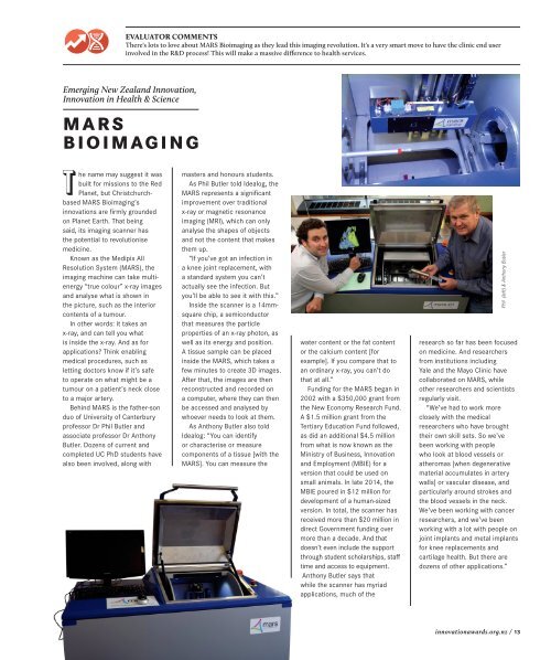
Known as the Medipix All<br />
Resolution System (MARS), the<br />
imaging machine can take multienergy<br />
“true colour” x-ray images<br />
and analyse what is shown in<br />
the picture, such as the interior<br />
contents of a tumour.<br />
In other words: it takes an<br />
x-ray, and can tell you what<br />
is inside the x-ray. And as for<br />
applications? Think enabling<br />
medical procedures, such as<br />
letting doctors know if it’s safe<br />
to operate on what might be a<br />
tumour on a patient’s neck close<br />
to a major artery.<br />
Behind MARS is the father-son<br />
duo of University of Canterbury<br />
professor Dr Phil Butler and<br />
associate professor Dr Anthony<br />
Butler. Dozens of current and<br />
completed UC PhD students have<br />
also been involved, along with<br />
masters and honours students.<br />
As Phil Butler told Idealog, the<br />
MARS represents a significant<br />
improvement over traditional<br />
x-ray or magnetic resonance<br />
imaging (MRI), which can only<br />
analyse the shapes of objects<br />
and not the content that makes<br />
them up.<br />
“If you’ve got an infection in<br />
a knee joint replacement, with<br />
a standard system you can’t<br />
actually see the infection. But<br />
you’ll be able to see it with this.”<br />
Inside the scanner is a 14mmsquare<br />
chip, a semiconductor<br />
that measures the particle<br />
properties of an x-ray photon, as<br />
well as its energy and position.<br />
A tissue sample can be placed<br />
inside the MARS, which takes a<br />
few minutes to create 3D images.<br />
After that, the images are then<br />
reconstructed and recorded on<br />
a computer, where they can then<br />
be accessed and analysed by<br />
whoever needs to look at them.<br />
As Anthony Butler also told<br />
Idealog: “You can identify<br />
or characterise or measure<br />
components of a tissue [with the<br />
MARS]. You can measure the<br />
water content or the fat content<br />
or the calcium content [for<br />
example]. If you compare that to<br />
an ordinary x-ray, you can’t do<br />
that at all.”<br />
Funding for the MARS began in<br />
2002 with a $350,000 grant from<br />
the New Economy Research Fund.<br />
A $1.5 million grant from the<br />
Tertiary Education Fund followed,<br />
as did an additional $4.5 million<br />
from what is now known as the<br />
Ministry of Business, <strong>Innovation</strong><br />
and Employment (MBIE) for a<br />
version that could be used on<br />
small animals. In late 2014, the<br />
MBIE poured in $12 million for<br />
development of a human-sized<br />
version. In total, the scanner has<br />
received more than $20 million in<br />
direct Government funding over<br />
more than a decade. And that<br />
doesn’t even include the support<br />
through student scholarships, staff<br />
time and access to equipment.<br />
Anthony Butler says that<br />
while the scanner has myriad<br />
applications, much of the<br />
Phil (left) & Anthony Butler<br />
research so far has been focused<br />
on medicine. And researchers<br />
from institutions including<br />
Yale and the Mayo Clinic have<br />
collaborated on MARS, while<br />
other researchers and scientists<br />
regularly visit.<br />
“We’ve had to work more<br />
closely with the medical<br />
researchers who have brought<br />
their own skill sets. So we’ve<br />
been working with people<br />
who look at blood vessels or<br />
atheromas [when degenerative<br />
material accumulates in artery<br />
walls] or vascular disease, and<br />
particularly around strokes and<br />
the blood vessels in the neck.<br />
We’ve been working with cancer<br />
researchers, and we’ve been<br />
working with a lot with people on<br />
joint implants and metal implants<br />
for knee replacements and<br />
cartilage health. But there are<br />
dozens of other applications.”<br />
innovationawards.org.nz / 13