cancer research at peter mac research at the forefront of discovery
cancer research at peter mac research at the forefront of discovery
cancer research at peter mac research at the forefront of discovery
You also want an ePaper? Increase the reach of your titles
YUMPU automatically turns print PDFs into web optimized ePapers that Google loves.
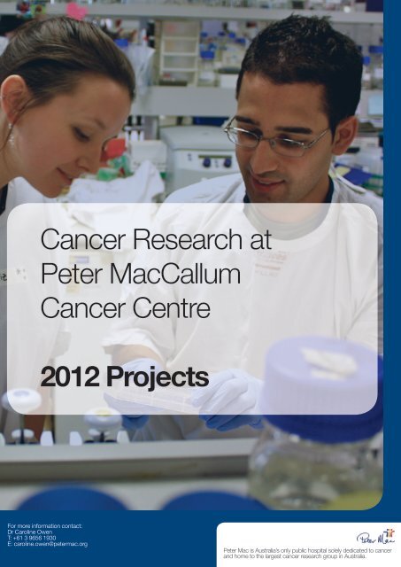
Cancer Research <strong>at</strong><br />
Peter MacCallum<br />
Cancer Centre<br />
2012 Projects<br />
For more inform<strong>at</strong>ion contact:<br />
Dr Caroline Owen<br />
T: +61 3 9656 1930<br />
E: caroline.owen@<strong>peter</strong><strong>mac</strong>.org<br />
Peter Mac is Australia’s only public hospital solely dedic<strong>at</strong>ed to <strong>cancer</strong><br />
and home to <strong>the</strong> largest <strong>cancer</strong> <strong>research</strong> group in Australia.
CANCER RESEARCH AT PETER MAC<br />
RESEARCH AT THE FOREFRONT OF DISCOVERY<br />
RESEARCH OVERVIEW<br />
For 60 years, Peter Mac has been providing high quality tre<strong>at</strong>ment and multidisciplinary care for <strong>cancer</strong><br />
p<strong>at</strong>ients and <strong>the</strong>ir families. It houses Australia’s largest and most progressive <strong>cancer</strong> <strong>research</strong> group, one <strong>of</strong><br />
only a handful <strong>of</strong> sites outside <strong>the</strong> United St<strong>at</strong>es where scientists and clinicians work side-by-side.<br />
Peter Mac’s unique integr<strong>at</strong>ion <strong>of</strong> science and clinical care enables <strong>the</strong> development and applic<strong>at</strong>ion <strong>of</strong><br />
innov<strong>at</strong>ive new methods <strong>of</strong> <strong>cancer</strong> diagnosis, tre<strong>at</strong>ment and educ<strong>at</strong>ion. This has enabled Peter Mac to build an<br />
intern<strong>at</strong>ional reput<strong>at</strong>ion for its clinical and <strong>research</strong> work.<br />
As Australia’s foremost <strong>cancer</strong> centre Peter Mac’s mission is to provide quality tre<strong>at</strong>ment and support to<br />
p<strong>at</strong>ients and <strong>the</strong>ir families, and broadly influence <strong>cancer</strong> care in <strong>the</strong> community through multidisciplinary<br />
p<strong>at</strong>ient care services, <strong>research</strong> and educ<strong>at</strong>ion.<br />
Cancer is a complex set <strong>of</strong> diseases, and modern <strong>cancer</strong> <strong>research</strong> institutes like <strong>the</strong> Peter Mac <strong>the</strong>refore<br />
conduct <strong>research</strong> th<strong>at</strong> covers a diversity <strong>of</strong> topics th<strong>at</strong> range from labor<strong>at</strong>ory-based studies into <strong>the</strong><br />
fundamental mechanisms <strong>of</strong> cell growth, transl<strong>at</strong>ional studies th<strong>at</strong> seek more accur<strong>at</strong>e <strong>cancer</strong> diagnosis,<br />
clinical trials with novel tre<strong>at</strong>ments, and <strong>research</strong> aimed to improve supportive care.<br />
The proximity and strong collabor<strong>at</strong>ive links <strong>of</strong> clinicians and scientists provides unique opportunities for medical<br />
advances to be moved from <strong>the</strong> ‘bench to <strong>the</strong> bedside' and for clinically orient<strong>at</strong>ed questions to guide our<br />
<strong>research</strong> agenda. As such, our <strong>research</strong> programs are having a pr<strong>of</strong>ound impact on <strong>the</strong> understanding <strong>of</strong><br />
<strong>cancer</strong> biology and are leading to more effective and individualised p<strong>at</strong>ient care.<br />
WHY PETER MAC?<br />
Collabor<strong>at</strong>ive interaction with n<strong>at</strong>ional and intern<strong>at</strong>ional peers is a lynchpin <strong>of</strong> any vibrant program. Peter Mac<br />
is continually seeking to work with <strong>the</strong> best worldwide and <strong>the</strong> world’s best are increasingly seeking out Peter<br />
Mac <strong>research</strong>ers to interact with.<br />
Why work or study <strong>at</strong> Peter Mac? In speaking to current and past <strong>research</strong>ers and students, it is immedi<strong>at</strong>ely<br />
evident th<strong>at</strong> <strong>the</strong> two factors most strongly influencing <strong>the</strong>ir decision to join and stay <strong>at</strong> Peter Mac are firstly, <strong>the</strong><br />
opportunity to be mentored by a strong and collegi<strong>at</strong>e group <strong>of</strong> senior <strong>research</strong>ers and secondly, <strong>the</strong> superb<br />
<strong>research</strong> infrastructure th<strong>at</strong> enables <strong>the</strong>m to perform virtually any type <strong>of</strong> experiment <strong>the</strong>y require <strong>at</strong> affordable<br />
cost. This is a strong vindic<strong>at</strong>ion <strong>of</strong> our str<strong>at</strong>egy <strong>of</strong> identifying, seeding and supporting <strong>the</strong> growth <strong>of</strong> an<br />
enabling environment, both in terms <strong>of</strong> talented senior personnel and first-class <strong>research</strong> infrastructure.<br />
RESEARCH STRUCTURE<br />
CANCER RESEARCH DIVISION<br />
The Cancer Research Division <strong>at</strong> Peter Mac is home to over 420 labor<strong>at</strong>ory-based scientists and support staff,<br />
including approxim<strong>at</strong>ely 80 higher degree (mainly PhD) and Honours students. Supported by nine core<br />
technology pl<strong>at</strong>forms, our 26 <strong>research</strong> labor<strong>at</strong>ories are organized into six programs <strong>of</strong> labor<strong>at</strong>ory-based<br />
programs and transl<strong>at</strong>ional <strong>research</strong>:<br />
• Cancer Cell Biology • Growth Control & Differenti<strong>at</strong>ion<br />
• Cancer Genetics & Genomics • Cancer Therapeutics<br />
• Cancer Immunology Research • Tumour Angiogenesis<br />
Research is supported by our core facilities and pl<strong>at</strong>form technologies. These core infrastructure groups are<br />
<strong>the</strong> backbone <strong>of</strong> <strong>the</strong> division and ensure th<strong>at</strong> <strong>the</strong> <strong>research</strong>ers are outfitted with <strong>the</strong> equipment and expertise<br />
needed to facilit<strong>at</strong>e <strong>the</strong>ir <strong>research</strong>. An important role <strong>of</strong> <strong>the</strong> Core Groups is to also identify, import, and develop<br />
new technologies.<br />
CLINICAL RESEARCH<br />
1
Peter Mac is proud <strong>of</strong> its long history <strong>of</strong> involvement in clinical <strong>research</strong>. The structure <strong>of</strong> clinical services <strong>at</strong><br />
Peter Mac fosters an environment in which clinicians from various specialties can work toge<strong>the</strong>r with allied<br />
health and supportive care staff on clinical <strong>research</strong> projects with a disease-specific focus. Research in <strong>the</strong><br />
clinical services are structured into <strong>the</strong> following areas:<br />
Breast Assoc. Pr<strong>of</strong>. Boon Chua boon.chua@<strong>peter</strong><strong>mac</strong>.org<br />
Gastrointestinal: Dr Michael Michael michael.michael@<strong>peter</strong><strong>mac</strong>.org<br />
Gynae-oncology: Dr Kailash Narayan kailash.narayan@<strong>peter</strong><strong>mac</strong>.org<br />
Haem<strong>at</strong>ology: Assoc Pr<strong>of</strong> John Seymour john.seymour@<strong>peter</strong><strong>mac</strong>.org<br />
Head and Neck: Assoc Pr<strong>of</strong> June Corry june.corry@<strong>peter</strong><strong>mac</strong>.org<br />
Lung: Assoc Pr<strong>of</strong> David Ball david.ball@<strong>peter</strong><strong>mac</strong>.org<br />
Melanoma and Skin Assoc. Pr<strong>of</strong> David Speakman david.speakman@<strong>peter</strong><strong>mac</strong>.org<br />
Neuro-oncology Dr Damian Tange damian.tange@<strong>peter</strong><strong>mac</strong>.org<br />
Paedi<strong>at</strong>ric, Adolescent, Young Adult and L<strong>at</strong>e Effects: Dr Greg Wheeler greg.wheeler@<strong>peter</strong><strong>mac</strong>.org<br />
Sarcoma: Pr<strong>of</strong> Peter Choong <strong>peter</strong>.choong@<strong>peter</strong><strong>mac</strong>.org<br />
Uro-oncology Dr Farshad Foroudi farshad.foroudi@<strong>peter</strong><strong>mac</strong>.org<br />
PLATFORM TECHNOLOGIES<br />
Peter Mac has pl<strong>at</strong>form technologies th<strong>at</strong> underpin an enabling environment and allow Peter Mac <strong>research</strong>ers<br />
to be intern<strong>at</strong>ionally competitive in an increasingly technology-driven environment. Peter Mac’s core<br />
technologies and expertise are also made available to external <strong>research</strong>ers on a collabor<strong>at</strong>ive or cost-recovery<br />
basis, <strong>the</strong>reby increasing <strong>research</strong> output in <strong>the</strong> wider bioscience community. Key technologies <strong>at</strong> Peter Mac<br />
include:<br />
Biost<strong>at</strong>istics <strong>at</strong> Peter Mac is <strong>the</strong> leading biost<strong>at</strong>istical centre focusing on <strong>cancer</strong> clinical trials in Australia.<br />
The centre provides st<strong>at</strong>istical expertise for n<strong>at</strong>ional <strong>cancer</strong> trials groups including <strong>the</strong> Trans Tasman<br />
Radi<strong>at</strong>ion Oncology Group (TROG) and <strong>the</strong> Australasian Leukaemia and Lymphoma Study Group (ALLG).<br />
Clinical <strong>research</strong> nurse core. Peter Mac currently has a team <strong>of</strong> <strong>research</strong> nurses to support a sophistic<strong>at</strong>ed<br />
clinical and transl<strong>at</strong>ional <strong>research</strong> program. These nurses provide necessary skills to coordin<strong>at</strong>e phase I firstin-man<br />
clinical trials involving complex procedures such as tumor biopsies for evalu<strong>at</strong>ion <strong>of</strong> molecular targets,<br />
serial PET scans and complex phar<strong>mac</strong>okinetic sampling.<br />
Flow Cytometry and Cell Sorting. We <strong>of</strong>fer multi-parameter (five colour) flow cytometric analysis for<br />
identifying rare popul<strong>at</strong>ions <strong>of</strong> cells within complex mixtures such as human bone marrow, and two fully<br />
supported fluorescent activ<strong>at</strong>ed cell sorting (FACS) instruments for isol<strong>at</strong>ing cells such as blood progenitor<br />
cells under sterile conditions.<br />
Genomics, Microarray Technology and Predictive Medicine. Peter Mac has been <strong>the</strong> leading site in<br />
Australia in <strong>the</strong> applic<strong>at</strong>ion <strong>of</strong> gene microarray technology for predicting outcomes <strong>of</strong> human <strong>cancer</strong> or an<br />
individual’s likely response to a given <strong>the</strong>rapy.<br />
Microscopy Imaging and Research Core Facility is a world-class facility encompassing all aspects <strong>of</strong><br />
microscopy such as immun<strong>of</strong>luorescence, laser capture, confocal and transmission electron microscopy.<br />
Molecular P<strong>at</strong>hology is a central pl<strong>at</strong>form to successful transl<strong>at</strong>ional <strong>research</strong> by providing robust diagnostic<br />
molecular analyses <strong>of</strong> tumours. Molecular P<strong>at</strong>hology <strong>at</strong> Peter Mac provides diagnostic testing for familial<br />
breast and colorectal <strong>cancer</strong>, and is a n<strong>at</strong>ional reference centre for testing for specific mut<strong>at</strong>ions in <strong>cancer</strong><br />
samples.<br />
Molecular Imaging. The centre for molecular imaging is a world leader in <strong>the</strong> clinical use <strong>of</strong> PET scanning in<br />
<strong>cancer</strong>. The facility includes three chemists, contains a cyclotron, two small animal PET scanners for<br />
transl<strong>at</strong>ional <strong>research</strong> and autom<strong>at</strong>ed production facilities for a number <strong>of</strong> novel tracers. These tracers provide<br />
<strong>the</strong> capacity to image diverse biological processes including hypoxia, lipid syn<strong>the</strong>sis, cell prolifer<strong>at</strong>ion and<br />
amino acid transport.<br />
Tissue/Tumour Bank. Peter Mac has been a leader in <strong>the</strong> development <strong>of</strong> sophistic<strong>at</strong>ed biospecimen and<br />
clinically annot<strong>at</strong>ed <strong>cancer</strong> samples collection. We are <strong>the</strong> host institute for <strong>the</strong> Australian Biospecimen Bank<br />
(ABN-Oncology), a federally funded project to enable n<strong>at</strong>ional <strong>cancer</strong> sample collection and facilit<strong>at</strong>ed access<br />
to tissue resources.<br />
Transgenic and SPF Facility. We currently breed and maintain approxim<strong>at</strong>ely 20,000 mice, representing<br />
over 130 different strains <strong>of</strong> transgenic and gene-targeted mice th<strong>at</strong> are immune-deficient or <strong>cancer</strong>. Peter<br />
Mac’s Animal Ethics Committee (AEC) has an important role in overseeing <strong>the</strong> ethical conduct <strong>of</strong> any work<br />
involving <strong>the</strong> use <strong>of</strong> animals for scientific purposes, conforming to <strong>the</strong> NHMRC Australian Code <strong>of</strong> Practice for<br />
<strong>the</strong> Care and Use <strong>of</strong> Animals for Scientific Purposes.<br />
2
RESEARCH EDUCATION PROGRAM<br />
Peter Mac brings toge<strong>the</strong>r Educ<strong>at</strong>ion and Research in a dynamic and life changing way. We strive to develop<br />
a world-class educ<strong>at</strong>ional experience for students <strong>at</strong> a leading Australian <strong>cancer</strong> <strong>research</strong> institution.<br />
The majority <strong>of</strong> students completing projects <strong>at</strong> Peter Mac are enrolled through <strong>the</strong> University <strong>of</strong> Melbourne<br />
and o<strong>the</strong>r Victorian universities. However, we welcome inquiries from students from all Universities throughout<br />
Australia and overseas. We boast a diverse student popul<strong>at</strong>ion from all over <strong>the</strong> world.<br />
Our postgradu<strong>at</strong>e PhD and Honours contribute significantly to <strong>the</strong> success <strong>of</strong> Peter Mac, and our Cancer<br />
Research Division is home to over 80 honours and post-gradu<strong>at</strong>e students. The <strong>at</strong>traction <strong>of</strong> high-quality<br />
students to undertake Doctor <strong>of</strong> Philosophy (PhD), Doctor <strong>of</strong> Medical Science (DMedSci), medical doctor<strong>at</strong>es,<br />
and Honours programs in our labor<strong>at</strong>ories continues as a priority. Our comprehensive student program<br />
includes mentor programs, dedic<strong>at</strong>ed student scientific review committees, onsite workshops and seminars, an<br />
annual retre<strong>at</strong>, and opportunities to contribute to our community and outreach programs.<br />
The following pages highlight some <strong>of</strong> <strong>the</strong> projects available for future students in 2012.<br />
If you are interested in a particular project, use <strong>the</strong> contact details to follow up with <strong>the</strong> listed supervisors to<br />
learn more about <strong>the</strong> project.<br />
For more inform<strong>at</strong>ion about <strong>research</strong> opportunities in <strong>the</strong> Cancer Research Division contact:<br />
Dr Caroline Owen, Educ<strong>at</strong>ion & Communic<strong>at</strong>ion Coordin<strong>at</strong>or (Research)<br />
Peter MacCallum Cancer Centre, St. Andrew’s Place, East Melbourne, Victoria, Australia 3002<br />
Tel: +61 3 9656 1930 Email: caroline.owen@<strong>peter</strong><strong>mac</strong>.org<br />
or visit:<br />
http://www.<strong>peter</strong><strong>mac</strong>.org/<strong>research</strong>/Educ<strong>at</strong>ionCareers<br />
PROJECT DESCRIPTIONS BY PROGRAM<br />
CANCER GENETICS PROGRAM page 4<br />
Cancer Genetics & Genomics page 4<br />
Sarcoma Genomics & Genetics page 5<br />
Surgical Oncology page 6<br />
VBCRC Genetics page 7<br />
Molecular P<strong>at</strong>hology page 8<br />
CANCER IMMUNOLOGY PROGRAM page 9<br />
Cancer Cell De<strong>at</strong>h page 9<br />
Cellular Immunity page 9<br />
Immune Signalling page 10<br />
Immuno<strong>the</strong>rapy page 11<br />
Haem<strong>at</strong>ology Immunology Transl<strong>at</strong>ional<br />
Research Labor<strong>at</strong>ory (HITRL) page 12<br />
CANCER THERAPEUTICS PROGRAM page 13<br />
Gene Regul<strong>at</strong>ion page 13<br />
Melanoma Research page 14<br />
Victorian Centre for Functional<br />
Genomics page 14<br />
Molecular Imaging & Transl<strong>at</strong>ional Medicine<br />
Program page 14<br />
CANCER CELL BIOLOGY PROGRAM page 15<br />
Cell Cycle & Development page 15<br />
Cell Cycle and Cancer Genetics page 16<br />
Cell Growth & Prolifer<strong>at</strong>ion page 17<br />
Epi<strong>the</strong>lial Stem Cell Biology page 18<br />
Metastasis Research page 18<br />
Molecular Radi<strong>at</strong>ion Biology page 19<br />
Tumour Suppression page 20<br />
Tumour Microenvironment page 21<br />
TUMOUR ANGIOGENESIS PROGRAM page 23<br />
ONCOGENIC SIGNALLING & GROWTH<br />
CONTROL PROGRAM page 24<br />
CLINICAL RESEARCH page 31<br />
FURTHER INFORMATION page 31<br />
3
CANCER GENOMICS PROGRAM<br />
www.<strong>peter</strong><strong>mac</strong>.org/Research/CancerGenomicsProgram<br />
The Cancer Genomics program seeks to use sophistic<strong>at</strong>ed high throughput genomic technologies to improve our<br />
understanding <strong>of</strong> <strong>the</strong> biology <strong>of</strong> <strong>cancer</strong> and to progress <strong>the</strong> clinical management <strong>of</strong> <strong>cancer</strong> p<strong>at</strong>ients through <strong>the</strong><br />
development <strong>of</strong> individualized approaches to tre<strong>at</strong>ment. Research in <strong>the</strong> program focuses primarily on breast, upper<br />
gastrointestinal and ovarian <strong>cancer</strong>s and sarcoma, and involves some <strong>of</strong> <strong>the</strong> largest familial and popul<strong>at</strong>ion-based <strong>cancer</strong><br />
cohorts in <strong>the</strong> world. These studies address questions <strong>of</strong> general importance to solid <strong>cancer</strong>s, including inherited<br />
susceptibility to <strong>cancer</strong> and genome-wide changes in gene expression, as well as more specific questions such as<br />
prediction <strong>of</strong> response to <strong>the</strong>rapy and <strong>the</strong> use <strong>of</strong> gene expression pr<strong>of</strong>iling for accur<strong>at</strong>e <strong>cancer</strong> diagnosis.<br />
CANCER GENETICS & GENOMICS<br />
MOLECULAR ANALYSIS OF PLATINUM<br />
RESISTANCE IN OVARIAN CANCER<br />
Supervisors: Pr<strong>of</strong>. David Bowtell, Dr Dariush<br />
Etemadmoghadam and Dr Prue Cowin<br />
Ovarian <strong>cancer</strong> is <strong>the</strong> 5th most common <strong>cancer</strong> in<br />
women, and most lethal gynaecologic malignancy.<br />
Despite aggressive surgery and pl<strong>at</strong>inum-based<br />
chemo<strong>the</strong>rapy, <strong>the</strong> majority <strong>of</strong> women experience<br />
recurrence and ~70% will succumb to <strong>the</strong> disease.<br />
Resistance to chemo<strong>the</strong>rapy, or pl<strong>at</strong>inum resistance, is<br />
<strong>the</strong> major barrier to long-term remissions, however <strong>the</strong><br />
underlying molecular mechanisms are poorly<br />
understood. We are part <strong>of</strong> <strong>the</strong> Australian Ovarian<br />
Cancer Study (AOCS), one <strong>of</strong> <strong>the</strong> largest ovarian <strong>cancer</strong><br />
cohort studies in <strong>the</strong> world. We are also one <strong>of</strong> <strong>the</strong> two<br />
Australian projects funded through a $27 million NHMRC<br />
grant to particip<strong>at</strong>e in <strong>the</strong> Intern<strong>at</strong>ional Cancer Genomics<br />
Consortium (ICGC).<br />
We recently performed a combined gene expression and<br />
DNA copy number change (CNC) analysis <strong>of</strong> serous<br />
ovarian <strong>cancer</strong> in a well-defined cohort <strong>of</strong> women who<br />
failed primary <strong>the</strong>rapy. We identified 19q12 amplific<strong>at</strong>ion<br />
as <strong>the</strong> most dominant amplicon associ<strong>at</strong>ed with primary<br />
tre<strong>at</strong>ment failure. The 19q12 amplific<strong>at</strong>ion is a high-level<br />
focal amplific<strong>at</strong>ion th<strong>at</strong> consistently targets a cluster <strong>of</strong><br />
only several genes; including <strong>the</strong> cell cycle gene<br />
CCNE1; and URI, recently been associ<strong>at</strong>ed with<br />
activ<strong>at</strong>ion <strong>of</strong> <strong>the</strong> mTOR/S6K p<strong>at</strong>hway and control <strong>of</strong><br />
apoptosis. It is not yet clear how <strong>the</strong>se genes or o<strong>the</strong>r<br />
cooper<strong>at</strong>ing mut<strong>at</strong>ions may contribute to primary<br />
chemo<strong>the</strong>rapy resistance.<br />
Molecular and functional explor<strong>at</strong>ion into mechanisms <strong>of</strong><br />
pl<strong>at</strong>inum-resistance in ovarian <strong>cancer</strong> will form <strong>the</strong> basis<br />
<strong>of</strong> <strong>the</strong> project. The student will learn key molecular<br />
biological techniques and will be exposed to large-scale<br />
human genetic studies th<strong>at</strong> are making use <strong>of</strong> <strong>the</strong><br />
emerging technologies, including microarrays and next<br />
gener<strong>at</strong>ion sequencing. This honours project will provide<br />
<strong>the</strong> student with <strong>the</strong> opportunity to contribute insights<br />
into one <strong>of</strong> <strong>the</strong> most clinically significant questions in<br />
ovarian <strong>cancer</strong>, pl<strong>at</strong>inum resistance.<br />
For more inform<strong>at</strong>ion about this project contact:<br />
Pr<strong>of</strong>. David Bowtell, Tel: +61 3 9656 1356, E-mail:<br />
david.bowtell@<strong>peter</strong><strong>mac</strong>.org<br />
MOLECULAR ANALYSIS OF OVARIAN CLEAR CELL<br />
CARCINOMAS<br />
Supervisors: Pr<strong>of</strong>. David Bowtell, Dr Dariush<br />
Etemadmoghadam, Dr Prue Cowin<br />
Ovarian clear cell adenocarcinoma (OCCA) is a clinically<br />
significant subtype <strong>of</strong> ovarian <strong>cancer</strong>, accounting for<br />
~10% <strong>of</strong> invasive ovarian <strong>cancer</strong>s. OCCA have a poor<br />
response r<strong>at</strong>e to standard <strong>the</strong>rapy (only 11-15%),<br />
indic<strong>at</strong>ing <strong>the</strong> need for novel <strong>the</strong>rapies. The occurrence<br />
<strong>of</strong> OCCA is associ<strong>at</strong>ed with co-existent endometriosis<br />
and may arise from endometriotic cysts, however very<br />
little is known <strong>of</strong> OCCA biology. To understand its<br />
molecular drivers, our labor<strong>at</strong>ory recently performed <strong>the</strong><br />
most extensive molecular analysis <strong>of</strong> OCCA to d<strong>at</strong>e. We<br />
found very consistent d<strong>at</strong>a associ<strong>at</strong>ed with de-regul<strong>at</strong>ed<br />
cytokine signalling. A central observ<strong>at</strong>ion was <strong>the</strong><br />
induction <strong>of</strong> hypoxia response genes including<br />
IL6/pSTAT3/HIF as measured by microarray,<br />
biochemical studies in OCCA cell lines and<br />
immunohistochemical staining <strong>of</strong> human tumour<br />
samples. IL6 has tumour-promoting actions on both<br />
malignant and stromal cells in a range <strong>of</strong> experimental<br />
<strong>cancer</strong> models, is a downstream effector <strong>of</strong> oncogenic<br />
ras, and has been implic<strong>at</strong>ed in several human <strong>cancer</strong>s.<br />
This project will investig<strong>at</strong>e <strong>the</strong> functional significance <strong>of</strong><br />
IL6 activ<strong>at</strong>ion in ovarian <strong>cancer</strong> cell lines. Expression <strong>of</strong><br />
HIF2a/EPAS1 is <strong>the</strong> most highly correl<strong>at</strong>ed gene with IL6<br />
in OCCA tumour samples. Interestingly, <strong>the</strong> hypoxic<br />
response in renal clear cell carcinoma cells is dependent<br />
on HIF2a. This observ<strong>at</strong>ion suggests a role for HIF2a in<br />
OCCA. Through molecular and functional techniques,<br />
<strong>the</strong> student will explore a model where strong upregul<strong>at</strong>ion<br />
<strong>of</strong> IL6 expression leads to increased HIF<br />
expression, promoting a proangiogenic response and<br />
facilit<strong>at</strong>ion adaption <strong>of</strong> <strong>cancer</strong> cells to hypoxia. This<br />
project will provide <strong>the</strong> <strong>the</strong> opportunity to contribute<br />
insights into one <strong>of</strong> <strong>the</strong> most clinically significant<br />
questions in ovarian <strong>cancer</strong>, pl<strong>at</strong>inum resistance.<br />
For more inform<strong>at</strong>ion about this project contact:<br />
Pr<strong>of</strong>essor David Bowtell, Tel: +61 3 9656 1356, E-mail:<br />
david.bowtell@<strong>peter</strong><strong>mac</strong>.org<br />
UNDERSTANDING DRIVERS OF A NOVEL<br />
MOLECULAR SUBTYPE OF HIGH-GRADE SEROUS<br />
OVARIAN CANCER<br />
Supervisors: Pr<strong>of</strong> David Bowtell, Dr Dariush<br />
Etemadmoghadam, Dr Prue Cowin<br />
Ovarian <strong>cancer</strong> is <strong>the</strong> 5-6th most common cause <strong>of</strong><br />
<strong>cancer</strong> de<strong>at</strong>h in women in Western countries, with ~800<br />
de<strong>at</strong>hs per year in Australia, with high-grade serous<br />
ovarian <strong>cancer</strong>s accounting for <strong>the</strong> majority <strong>of</strong> de<strong>at</strong>hs<br />
(>60%). Recently, molecular subtyping <strong>of</strong> ovarian <strong>cancer</strong><br />
has revealed four molecular c<strong>at</strong>egories <strong>of</strong> HG-SOC.<br />
4
Each molecular subtype, design<strong>at</strong>ed C1, C2, C4 and C5<br />
by Tothill et al, presents with a distinct expression<br />
p<strong>at</strong>tern and differing clinical outcomes. We are part <strong>of</strong><br />
<strong>the</strong> Australian Ovarian Cancer Study (AOCS), one <strong>of</strong> <strong>the</strong><br />
largest ovarian <strong>cancer</strong> cohort studies in <strong>the</strong> world. We<br />
are also one <strong>of</strong> <strong>the</strong> two Australian projects funded<br />
through a $27 million NHMRC grant to particip<strong>at</strong>e in <strong>the</strong><br />
Intern<strong>at</strong>ional Cancer Genomics Consortium (ICGC).<br />
We have recently shown th<strong>at</strong> <strong>the</strong> C5 subtype is<br />
associ<strong>at</strong>ed with amplific<strong>at</strong>ion and over-expression <strong>of</strong><br />
MYCN, over-expression <strong>of</strong> LIN28B, repression <strong>of</strong> Let-7<br />
family members, and over-expression <strong>of</strong> HMGA2. This<br />
work for <strong>the</strong> first time defines an oncogenic p<strong>at</strong>hway<br />
specific to a molecular subtype <strong>of</strong> serous ovarian<br />
<strong>cancer</strong>s, and opens a new door to p<strong>at</strong>ient tailored<br />
molecular <strong>the</strong>rapies.<br />
This project involves fur<strong>the</strong>r definition <strong>of</strong> this oncogenic<br />
p<strong>at</strong>hway through specific over-expression and<br />
knockdown <strong>of</strong> MYCN in ovarian <strong>cancer</strong> cell lines in vitro.<br />
The student will learn key molecular biological and tissue<br />
culture techniques. The Bowtell lab has a very strong<br />
reput<strong>at</strong>ion in <strong>cancer</strong> genetics and genomics, and in<br />
fundamental studies in <strong>cancer</strong> cell biology.<br />
For more inform<strong>at</strong>ion about this project contact:<br />
Pr<strong>of</strong>. David Bowtell, Tel: +61 3 9656 1356, E-mail:<br />
david.bowtell@<strong>peter</strong><strong>mac</strong>.org<br />
VALIDATION OF CANDIDATE GENES INVOLVED IN<br />
THE PROGRESSION OF GASTRIC CANCER<br />
Supervisors: Assoc Pr<strong>of</strong> Alex Boussioutas, Dr. Rita<br />
Busuttil<br />
Gastric <strong>cancer</strong> (GC) is <strong>the</strong> fourth most common <strong>cancer</strong><br />
globally and in many western countries is usually only<br />
diagnosed <strong>at</strong> advanced stage giving p<strong>at</strong>ients a 5-year<br />
survival r<strong>at</strong>e <strong>of</strong> less than 20%. GC has distinct<br />
premalignant stages th<strong>at</strong> have significant propensity to<br />
progress. The premalignant cascade consists <strong>of</strong> easily<br />
identifiable histological stages from chronic <strong>at</strong>rophic<br />
gastritis (ChG), intestinal metaplasia (IM) and dysplasia.<br />
The progression through <strong>the</strong>se stages, particularly IM,<br />
takes years, <strong>of</strong>fering a large window <strong>of</strong> opportunity to<br />
intervene. Not all p<strong>at</strong>ients with IM will progress and<br />
selection <strong>of</strong> p<strong>at</strong>ients for high-risk surveillance would<br />
reduce <strong>the</strong> burden <strong>of</strong> unnecessary screening, p<strong>at</strong>ient<br />
anxiety and improve outcomes due to early detection <strong>of</strong><br />
disease.<br />
Rel<strong>at</strong>ively little is known about <strong>the</strong> key genetic events<br />
leading to IM. Our labor<strong>at</strong>ory is currently in <strong>the</strong> process<br />
<strong>of</strong> completing <strong>the</strong> first comprehensive analysis <strong>of</strong> IM in<br />
<strong>the</strong> world and seeks to identify candid<strong>at</strong>e genes involved<br />
in <strong>the</strong> progression <strong>of</strong> IM to GC th<strong>at</strong> can be used to<br />
reliably predict <strong>the</strong> progression to GC in humans by<br />
using a genomics based approach. Identific<strong>at</strong>ion <strong>of</strong> such<br />
genes <strong>of</strong>fers an opportunity to study <strong>the</strong> molecular<br />
mechanisms involved and pinpoint targets for prevention<br />
and <strong>the</strong>rapy. The aim <strong>of</strong> this project is valid<strong>at</strong>e <strong>the</strong>se<br />
candid<strong>at</strong>e genes using an independent d<strong>at</strong>a set and <strong>the</strong>n<br />
characterizing <strong>the</strong>se genes using functional assays and<br />
animal models.<br />
The project will use broad range techniques including<br />
bioinform<strong>at</strong>ics, cell culture, animal models and molecular<br />
biology.<br />
For more inform<strong>at</strong>ion about this project contact:<br />
Assoc. Pr<strong>of</strong>. Alex Boussioutas, Tel: +61 3 9656 1287, Email:<br />
alex.boussioutas@<strong>peter</strong><strong>mac</strong>.org<br />
Dr. Rita Busuttil, Tel: +61 3 9656 1287, E-mail:<br />
rita.busuttil@<strong>peter</strong><strong>mac</strong>.org<br />
ROLE OF THE TUMOUR MICROENVIRONMENT IN<br />
GASTRIC CANCER<br />
Supervisors: Assoc. Pr<strong>of</strong>. Alex Boussioutas, Dr. Rita<br />
Busuttil<br />
Gastric <strong>cancer</strong> (GC) is <strong>the</strong> fourth most common <strong>cancer</strong><br />
globally and 7th in incidence in Australia. It has a poor<br />
survival r<strong>at</strong>e which can be <strong>at</strong>tributed to <strong>the</strong> advanced<br />
stage <strong>at</strong> diagnosis in most p<strong>at</strong>ients. The molecular and<br />
cellular mechanisms underlying <strong>the</strong> development <strong>of</strong> GC<br />
are not well described.<br />
Traditionally <strong>cancer</strong> <strong>research</strong> involved studying <strong>the</strong><br />
<strong>cancer</strong> cell itself. More recently, <strong>the</strong>re has been growing<br />
interest in studying <strong>the</strong> normal cells and molecules which<br />
surround <strong>the</strong> <strong>cancer</strong> cell. This tumour microenvironment<br />
consists <strong>of</strong> a variety <strong>of</strong> stromal cell types including cells<br />
such as fibroblasts. It is believed th<strong>at</strong> <strong>the</strong> dynamic<br />
communic<strong>at</strong>ion between tumour cells and <strong>the</strong><br />
surrounding cell types may play a major role in <strong>cancer</strong><br />
initi<strong>at</strong>ion, progression and establishment <strong>of</strong> metast<strong>at</strong>ic<br />
disease. The aim <strong>of</strong> this project is to investig<strong>at</strong>e tumourstromal<br />
interactions in gastric <strong>cancer</strong> utilizing established<br />
and primary cell lines. Once <strong>the</strong> molecular p<strong>at</strong>hways by<br />
which a tumour cell progresses has been elucid<strong>at</strong>ed it is<br />
possible th<strong>at</strong> <strong>the</strong>se processes could be exploited in <strong>the</strong><br />
development <strong>of</strong> novel <strong>the</strong>rapeutics.<br />
This project will use a broad range <strong>of</strong> techniques such as<br />
live cell microscopy, cell culture techniques and siRNA<br />
to interrog<strong>at</strong>e <strong>the</strong> function <strong>of</strong> gene products th<strong>at</strong><br />
influence tumour-stroma communic<strong>at</strong>ion.<br />
Our previous genomic experiments has provided us with<br />
a number <strong>of</strong> exciting candid<strong>at</strong>e genes th<strong>at</strong> may be<br />
involved in this interaction. This is novel <strong>research</strong> th<strong>at</strong><br />
may have a major benefit to our understanding <strong>of</strong> <strong>cancer</strong><br />
and improve p<strong>at</strong>ient outcomes.<br />
For more inform<strong>at</strong>ion about this project contact:<br />
Assoc. Pr<strong>of</strong>. Alex Boussioutas, Tel: +61 3 9656 1287, Email:<br />
alex.boussioutas@<strong>peter</strong><strong>mac</strong>.org<br />
Dr. Rita Busuttil, Tel: +61 3 9656 1287, E-mail:<br />
rita.busuttil@<strong>peter</strong><strong>mac</strong>.org<br />
SARCOMA GENOMICS & GENETICS<br />
ROLE OF IMMUNOMODULATORS IN THE<br />
DEVELOPMENT & PROGRESSION OF<br />
OSTEOSARCOMA IN VIVO.<br />
Supervisors: Assoc. Pr<strong>of</strong>. David Thomas, Dr. Maya<br />
Kansara<br />
The Sarcoma Genetics and Genomics labor<strong>at</strong>ory studies<br />
tumours <strong>of</strong> s<strong>of</strong>t tissue and bone. Osteosarcoma is <strong>the</strong><br />
most common <strong>cancer</strong> <strong>of</strong> bone. These tumours are highly<br />
metast<strong>at</strong>ic and <strong>of</strong>ten metastasise to lung via <strong>the</strong><br />
hem<strong>at</strong>ogenous route. Tre<strong>at</strong>ment involves aggressive<br />
surgery with intensive adjuvant chemo<strong>the</strong>rapy. Although<br />
<strong>the</strong>se measures have improved prognosis, a third <strong>of</strong><br />
those diagnosed will die from this disease.<br />
Understanding how osteosarcoma arises and persists<br />
will enable <strong>the</strong> development <strong>of</strong> targeted <strong>the</strong>rapies. The<br />
skeleton and <strong>the</strong> immune system share a number <strong>of</strong><br />
cytokines and transcription factors and <strong>the</strong>refore may<br />
mutually influence each o<strong>the</strong>r; <strong>the</strong> study <strong>of</strong> <strong>the</strong>se cells<br />
and <strong>the</strong>ir interactions has been termed<br />
5
osteoimmunology. In this project we will investig<strong>at</strong>e <strong>the</strong><br />
interaction between <strong>the</strong> immune system and bone<br />
<strong>cancer</strong> in an in vivo mouse model <strong>of</strong> osteosarcoma. The<br />
project will use broad range techniques including mouse<br />
models <strong>of</strong> <strong>cancer</strong>, histology, cell culture, flow cytometry,<br />
and molecular pr<strong>of</strong>iling.<br />
For more inform<strong>at</strong>ion about this project contact:<br />
Assoc. Pr<strong>of</strong>. David Thomas. Tel +61 3 9656 1238 Email<br />
david.thomas@<strong>peter</strong><strong>mac</strong>.org<br />
Dr. Maya Kansara Tel +61 3 9656 1618 Email<br />
maya.kansara@<strong>peter</strong><strong>mac</strong>.org<br />
SURGICAL ONCOLOGY<br />
HOW DO PIK3CA MUTATIONS CAUSE CANCER?<br />
Supervisor: Assoc. Pr<strong>of</strong>. Wayne Phillips<br />
The phosphoinositide 3-kinase (PI3K)/Akt signalling<br />
p<strong>at</strong>hway controls a range <strong>of</strong> fundamental cellular<br />
processes which, when de-regul<strong>at</strong>ed, are considered<br />
hallmarks <strong>of</strong> <strong>cancer</strong>. While it is now well established th<strong>at</strong><br />
som<strong>at</strong>ic mut<strong>at</strong>ions in PIK3CA, <strong>the</strong> gene coding for <strong>the</strong><br />
p110α c<strong>at</strong>alytic subunit <strong>of</strong> PI3K, are one <strong>of</strong> <strong>the</strong> most<br />
common, and thus potentially one <strong>of</strong> <strong>the</strong> most important,<br />
genetic abnormalities in many human tumours.<br />
However, it remains unclear how <strong>the</strong>se mut<strong>at</strong>ions drive<br />
tumourigenesis.<br />
We have recently gener<strong>at</strong>ed a novel mutant mouse with<br />
which to study <strong>the</strong> role <strong>of</strong> PIK3CA mut<strong>at</strong>ion in vivo and in<br />
vitro. This mouse has been designed with a Cre<br />
recombinase (Cre)-inducible knock-in <strong>of</strong> <strong>the</strong> most<br />
common tumour-associ<strong>at</strong>ed PI3K mut<strong>at</strong>ion,<br />
PIK3CAH1047R. Our str<strong>at</strong>egy <strong>of</strong> making <strong>the</strong> knock-in<br />
inducible with Cre allows us to target <strong>the</strong> expression <strong>of</strong><br />
<strong>the</strong> mutant to specific tissues using mice expressing Cre<br />
under <strong>the</strong> control <strong>of</strong> appropri<strong>at</strong>e tissue-specific<br />
promoters. We can also knock-in <strong>the</strong> mut<strong>at</strong>ion into cells<br />
growing in culture allowing us to examine <strong>the</strong> effects <strong>of</strong><br />
PIK3CAH1047R mut<strong>at</strong>ion in vitro under defined<br />
conditions.<br />
Two potential projects are being <strong>of</strong>fered.<br />
(1) PIK3CAH1047R in oesophageal <strong>cancer</strong>.<br />
Oesophageal epi<strong>the</strong>lial cells will be isol<strong>at</strong>ed from<br />
our PIK3CAH1047R mouse and use our novel 3D in<br />
vivo culture systems to examine <strong>the</strong> effect <strong>of</strong><br />
PIK3CAH1047R expression in <strong>the</strong> growth and<br />
differenti<strong>at</strong>ion <strong>of</strong> <strong>the</strong> oesophageal epi<strong>the</strong>lium and<br />
<strong>the</strong> development <strong>of</strong> <strong>cancer</strong>.<br />
(2) PIK3CAH1047R in breast <strong>cancer</strong>. Mammary<br />
epi<strong>the</strong>lial cells will be isol<strong>at</strong>ed from our<br />
PIK3CAH1047R mouse and use <strong>the</strong>se to examine<br />
<strong>the</strong> signaling p<strong>at</strong>hways by which PIK3CAH1047R<br />
induces tumourigenesis and regul<strong>at</strong>es <strong>the</strong> growth<br />
and differenti<strong>at</strong>ion <strong>of</strong> mammary epi<strong>the</strong>lial cells.<br />
For more inform<strong>at</strong>ion about this project contact:<br />
Assoc. Pr<strong>of</strong>. Wayne Phillips, Tel: +61 3 9656 1842;<br />
Email: wayne.phillips@<strong>peter</strong><strong>mac</strong>.org<br />
UNDERSTANDING BARRETT’S OESOPHAGUS AND<br />
OESOPHAGEAL ADENOCARCINOMA.<br />
Supervisors: Dr. Nicholas Clemons and Assoc. Pr<strong>of</strong>.<br />
Wayne Phillips<br />
Over <strong>the</strong> past thirty years <strong>the</strong>re has been a dram<strong>at</strong>ic<br />
increase in <strong>the</strong> incidence and prevalence <strong>of</strong><br />
oesophageal adenocarcinoma, a <strong>cancer</strong> with particularly<br />
high mortality. The reason for <strong>the</strong> increase is not clear<br />
but is thought to reflect an increase in <strong>the</strong> occurrence <strong>of</strong><br />
its recognised precursor lesion, Barrett’s oesophagus.<br />
Barrett’s oesophagus is a metaplastic abnormality in<br />
which <strong>the</strong> normal str<strong>at</strong>ified squamous epi<strong>the</strong>lium <strong>of</strong> <strong>the</strong><br />
oesophagus is replaced by an intestinal-type columnar<br />
epi<strong>the</strong>lium. The risk <strong>of</strong> adenocarcinoma in p<strong>at</strong>ients with<br />
Barrett’s oesophagus is approxim<strong>at</strong>ely 30-125-fold<br />
gre<strong>at</strong>er than th<strong>at</strong> in <strong>the</strong> general popul<strong>at</strong>ion. The origin <strong>of</strong><br />
Barrett’s oesophagus is a m<strong>at</strong>ter <strong>of</strong> conjecture. There is<br />
compelling etiological evidence th<strong>at</strong> gastro-oesophageal<br />
reflux disease is a major contributing factor but <strong>the</strong><br />
actual molecular and cellular mechanism(s) underlying<br />
<strong>the</strong> phenotypic change are not clear. Fur<strong>the</strong>rmore, it is<br />
unclear wh<strong>at</strong> <strong>the</strong> key molecular drivers <strong>of</strong> progression<br />
from Barrett’s to <strong>cancer</strong> are, which has contributed to <strong>the</strong><br />
clinical problem <strong>of</strong> indentifying those p<strong>at</strong>ients with<br />
Barrett’s oesophagus th<strong>at</strong> are most <strong>at</strong> risk <strong>of</strong> progression<br />
to <strong>cancer</strong>.<br />
Our group has developed novel in vivo and in vitro<br />
models th<strong>at</strong> allow <strong>the</strong> 3-D reconstitution <strong>of</strong> <strong>the</strong><br />
oesophageal epi<strong>the</strong>lium from mouse or human tissue<br />
and cell lines. Projects are available for Honours or PhD<br />
students to use <strong>the</strong>se models to investig<strong>at</strong>e <strong>the</strong><br />
molecular and cellular mechanisms underlying <strong>the</strong><br />
development <strong>of</strong> Barrett’s oesophagus and/or <strong>the</strong><br />
progression <strong>of</strong> Barrett’s oesophagus to adenocarcinoma.<br />
Possible projects include:<br />
• Determining <strong>the</strong> signalling p<strong>at</strong>hways involved in <strong>the</strong><br />
transition <strong>of</strong> <strong>the</strong> normal squamous oesophageal<br />
epi<strong>the</strong>lium to Barrett’s intestinal-like epi<strong>the</strong>lium.<br />
Possible p<strong>at</strong>hways/genes to study include: Sonic<br />
Hedgehog signalling p<strong>at</strong>hway, SOX9, GATA and<br />
HNF transcription factors and microRNAs miR203<br />
and miR205<br />
• Investig<strong>at</strong>ing <strong>the</strong> effect <strong>of</strong> <strong>the</strong> stromal<br />
microenvironment in Barrett’s carcinogenesis<br />
• Investig<strong>at</strong>ing Aurora A kinase inhibition in<br />
combin<strong>at</strong>ion with chemo<strong>the</strong>rapy as a novel tre<strong>at</strong>ment<br />
for oesophageal adenocarcinoma<br />
• Elucid<strong>at</strong>ing a role for miniSox9, a novel splice variant<br />
<strong>of</strong> Sox9, in Barrett’s carcinogenesis<br />
Understanding <strong>the</strong> biology underlying this condition will<br />
ultim<strong>at</strong>ely help us to design effective str<strong>at</strong>egies for <strong>the</strong><br />
management and tre<strong>at</strong>ment <strong>of</strong> Barrett’s oesophagus and<br />
to predict, and/or prevent, progression to oesophageal<br />
adenocarcinoma.<br />
References:<br />
1. • Phillips WA, et al. J Gastenterol Hep<strong>at</strong>ol 2011 26(4): 639-48<br />
2. • Wang DH, et al. Gastroenterology 2010 138(5): 1820-22.<br />
For more inform<strong>at</strong>ion about this project contact:<br />
Dr Nicholas Clemons, Tel: +61 3 9656 1849; Email:<br />
nicholas.clemons@<strong>peter</strong><strong>mac</strong>.org<br />
6
VBCRC CANCER GENETICS<br />
The Cancer Genetics labor<strong>at</strong>ory uses an integr<strong>at</strong>ive<br />
genomics approach to investig<strong>at</strong>e genes involved in<br />
breast and ovarian <strong>cancer</strong>, whereby d<strong>at</strong>a from several<br />
genome-wide pl<strong>at</strong>forms are combined to more rapidly<br />
define critical <strong>cancer</strong>-causing genes.<br />
GENOMIC ANALYSIS OF EARLY BREAST<br />
NEOPLASMS<br />
Supervisor: Assoc. Pr<strong>of</strong>. Ian Campbell,<br />
We have previously undertaken genomic analysis <strong>of</strong><br />
DCIS, <strong>the</strong> immedi<strong>at</strong>e precursor to invasive breast<br />
carcinoma, and found th<strong>at</strong> in many cases this tumour<br />
already contains a plethora <strong>of</strong> genomic events highly<br />
similar to IDC. In order to identify <strong>the</strong> earliest genomic<br />
events in <strong>the</strong> development <strong>of</strong> breast <strong>cancer</strong>, this project<br />
will analyse less advanced breast neoplasms such as<br />
<strong>at</strong>ypical ductal hyperplasia (ADH). These early lesions<br />
have not been previously analysed <strong>at</strong> high resolution<br />
and are likely to contain few, but highly relevant,<br />
genomic events.<br />
Techniques used in <strong>the</strong> project will include cutting edge<br />
technologies such as whole-exome next gener<strong>at</strong>ion<br />
sequencing as well as microdissection <strong>of</strong> tumour<br />
m<strong>at</strong>erial, DNA/RNA extraction, and expression<br />
microarrays. There will be a strong bioinform<strong>at</strong>ics<br />
component and potentially functional assays <strong>of</strong><br />
candid<strong>at</strong>e genes in cell culture.<br />
For more inform<strong>at</strong>ion about this project contact:<br />
Assoc. Pr<strong>of</strong>. Ian Campbell,<br />
Email: ian.campbell@<strong>peter</strong><strong>mac</strong>.org<br />
IDENTIFICATION OF HIGHLY PENETRANT GENES IN<br />
FAMILIAL BREAST AND OTHER CANCERS USING<br />
NEXT-GENERATION SEQUENCING<br />
Supervisor: Assoc. Pr<strong>of</strong>. Ian Campbell, Dr Ella<br />
Thompson, Dr Alison Trainer<br />
The ability to identify disease-causing mut<strong>at</strong>ions in highrisk<br />
<strong>cancer</strong> families has broad implic<strong>at</strong>ions for those<br />
affected in terms <strong>of</strong> risk assessment and management<br />
as well as tre<strong>at</strong>ment. A major initi<strong>at</strong>ive over <strong>the</strong> last year<br />
has been <strong>the</strong> applic<strong>at</strong>ion <strong>of</strong> next gener<strong>at</strong>ion sequencing<br />
(NGS) to identify <strong>cancer</strong> predisposition genes. We are<br />
performing whole exome sequence analysis <strong>of</strong> germline<br />
DNA from multiple affected rel<strong>at</strong>ives from over 75 high<br />
risk non-BRCA1/non-BRCA2 breast <strong>cancer</strong> families with<br />
<strong>the</strong> aim <strong>of</strong> identifying segreg<strong>at</strong>ing, rare, non-synonymous<br />
variants th<strong>at</strong> are likely to include novel predisposing<br />
mut<strong>at</strong>ions. In addition, we also aim to analyse families<br />
with o<strong>the</strong>r <strong>cancer</strong> types including male breast <strong>cancer</strong>,<br />
colorectal <strong>cancer</strong> and papillary thyroid <strong>cancer</strong> to identify<br />
<strong>the</strong> predisposing genes.<br />
This project will perform and analyse NGS d<strong>at</strong>a to<br />
identify candid<strong>at</strong>e gene variants identified and valid<strong>at</strong>e<br />
<strong>the</strong>se variants in <strong>the</strong> family in which <strong>the</strong> variant was<br />
found including segreg<strong>at</strong>ion analysis. After valid<strong>at</strong>ion,<br />
<strong>the</strong> gene will be fur<strong>the</strong>r analysed for mut<strong>at</strong>ions in o<strong>the</strong>r<br />
families and individuals with <strong>the</strong> same <strong>cancer</strong> type. For<br />
breast <strong>cancer</strong> this l<strong>at</strong>ter valid<strong>at</strong>ion may be undertaken<br />
using a boutique exon capture and NGS <strong>of</strong> 200<br />
additional breast <strong>cancer</strong> families. Techniques used will<br />
include DNA sequencing (NGS and Sanger), PCR, high<br />
resolution melting and potentially assays <strong>of</strong> gene<br />
transcription or function.<br />
For more inform<strong>at</strong>ion about this project contact:<br />
Assoc. Pr<strong>of</strong>. Ian Campbell,<br />
Email: ian.campbell@<strong>peter</strong><strong>mac</strong>.org<br />
MUCINOUS OVARIAN CARCINOMA IS A DISTINCT<br />
OVARIAN SUBTYPE REQUIRING ALTERNATIVE<br />
CHEMOTHERAPEUTIC REGIMES<br />
Supervisors: Assoc. Pr<strong>of</strong>. Ian Campbell, Dr. Kylie<br />
Gorringe<br />
Mucinous ovarian carcinoma (MOC) differs in<br />
appearance and behavior from <strong>the</strong> o<strong>the</strong>r common<br />
epi<strong>the</strong>lial ovarian <strong>cancer</strong> subtypes. MOC is frequently<br />
confused with metastases from organs such as <strong>the</strong><br />
appendix, but it is not known if this resemblance<br />
extends to similarities in genetic alter<strong>at</strong>ions. Advanced<br />
MOC does not respond well to conventional ovarian<br />
<strong>cancer</strong> chemo<strong>the</strong>rapies, indic<strong>at</strong>ing a need for more<br />
subtype-specific <strong>the</strong>rapies. We hypo<strong>the</strong>sis th<strong>at</strong> genomic<br />
aberr<strong>at</strong>ions in MOC will be similar to those in mucinous<br />
<strong>cancer</strong>s from o<strong>the</strong>r organs. Consequently, MOC may be<br />
better tre<strong>at</strong>ed with chemo<strong>the</strong>rapeutics th<strong>at</strong> show<br />
success with o<strong>the</strong>r mucinous tumours.<br />
This project will obtain genomic d<strong>at</strong>a from primary MOC<br />
and compare this with d<strong>at</strong>a from metastases to <strong>the</strong><br />
ovary from extra-ovarian sites (initially presenting as<br />
ovarian), appendiceal tumours, diffuse gastric tumours,<br />
mucinous colorectal tumours and pancre<strong>at</strong>ic tumours.<br />
Techniques used will include copy number and<br />
expression arrays and next-gener<strong>at</strong>ion sequencing. Cell<br />
lines represent<strong>at</strong>ive <strong>of</strong> MOC will be used to compare<br />
tre<strong>at</strong>ments with typical ovarian chemo<strong>the</strong>rapies such as<br />
cispl<strong>at</strong>in with <strong>the</strong>rapies more commonly used in o<strong>the</strong>r<br />
<strong>cancer</strong> types such as colorectal <strong>cancer</strong>s.<br />
For more inform<strong>at</strong>ion about this project contact:<br />
Assoc. Pr<strong>of</strong>. Ian Campbell, Email:<br />
ian.campbell@<strong>peter</strong><strong>mac</strong>.org<br />
Dr. Kylie Gorringe, Email: kylie.gorringe@<strong>peter</strong><strong>mac</strong>.org<br />
FUNCTIONAL AND GENETIC CHARACTERISATION<br />
OF OVARIAN ONCOGENES<br />
Supervisors: Assoc. Pr<strong>of</strong>. Ian Campbell, Dr. Kylie<br />
Gorringe<br />
Ovarian <strong>cancer</strong> is a disease characterised by complex<br />
genomic rearrangements including high-level copy<br />
number amplific<strong>at</strong>ions but <strong>the</strong> majority <strong>of</strong> <strong>the</strong> genes in<br />
<strong>the</strong>se regions th<strong>at</strong> drive ovarian <strong>cancer</strong> remain<br />
unidentified. C<strong>at</strong>aloguing <strong>the</strong>se target genes will provide<br />
useful insights into disease etiology and may provide an<br />
opportunity to develop novel diagnostic and <strong>the</strong>rapeutic<br />
interventions. We have recently undertaken a highthroughput<br />
siRNA knockdown screen <strong>of</strong> 300 candid<strong>at</strong>e<br />
genes loc<strong>at</strong>ed in recurrent regions <strong>of</strong> high-level gene<br />
amplific<strong>at</strong>ion and have identified a number <strong>of</strong> promising<br />
candid<strong>at</strong>e oncogenes. This project will select a subset <strong>of</strong><br />
<strong>the</strong>se genes for fur<strong>the</strong>r characteris<strong>at</strong>ion, including<br />
valid<strong>at</strong>ion <strong>of</strong> <strong>the</strong> effect <strong>of</strong> knockdown, screening tumours<br />
for mut<strong>at</strong>ions using custom exon capture and NGS,<br />
stable shRNA gene knockdown in cell culture for various<br />
in vitro and in vivo assays, <strong>the</strong> effects <strong>of</strong> small molecule<br />
inhibitors and immunohistochemistry.<br />
For more inform<strong>at</strong>ion about this project contact:<br />
Assoc. Pr<strong>of</strong>. Ian Campbell, Email:<br />
ian.campbell@<strong>peter</strong><strong>mac</strong>.org<br />
Dr. Kylie Gorringe, Email: kylie.gorringe@<strong>peter</strong><strong>mac</strong>.org<br />
7
MOLECULAR PATHOLOGY<br />
IT’S IN THE BLOOD: THE EFFECT OF<br />
CONSTITUTIONAL METHYLATION ON DISEASE<br />
PREDISPOSITION.<br />
Supervisors: Assoc. Pr<strong>of</strong>. Alexander Dobrovic, Dr.<br />
Michael McManus<br />
Constitutional methyl<strong>at</strong>ion refers to <strong>the</strong> presence <strong>of</strong> <strong>the</strong><br />
aberrant methyl<strong>at</strong>ion <strong>of</strong> a given gene through multiple<br />
tissues <strong>of</strong> <strong>the</strong> body. In most cases, it arises from <strong>the</strong><br />
combin<strong>at</strong>ion <strong>of</strong> <strong>the</strong> presence <strong>of</strong> a predisposing genetic<br />
variant in cis and a predisposing genetic background <strong>of</strong><br />
modifiers in trans. The methyl<strong>at</strong>ion seems to arise<br />
stochastically <strong>of</strong>ten giving rise to mosaic p<strong>at</strong>terns.<br />
Constitutional methyl<strong>at</strong>ion <strong>of</strong> familial <strong>cancer</strong> genes has<br />
been recently described as a new mechanism <strong>of</strong> <strong>cancer</strong><br />
predisposition [1]. It predisposes to a similar spectrum<br />
<strong>of</strong> <strong>cancer</strong>s as germline mut<strong>at</strong>ions in <strong>the</strong> same gene in<br />
those cases where <strong>the</strong> constitutional methyl<strong>at</strong>ion is<br />
acting as <strong>the</strong> driver <strong>of</strong> tumorigenesis. Moreover, it may<br />
drive a similar cluster <strong>of</strong> p<strong>at</strong>hological fe<strong>at</strong>ures within<br />
each <strong>of</strong> those tumours. This may in turn allow <strong>the</strong><br />
deduction <strong>of</strong> whe<strong>the</strong>r methyl<strong>at</strong>ion in a particular gene is<br />
acting as a driver or a passenger. In <strong>the</strong> case <strong>of</strong><br />
BRCA1, we have recently shown th<strong>at</strong> detectable<br />
methyl<strong>at</strong>ion <strong>of</strong> <strong>the</strong> BRCA1 gene in <strong>the</strong> peripheral blood<br />
is strongly associ<strong>at</strong>ed with <strong>the</strong> same characteristic<br />
<strong>cancer</strong> p<strong>at</strong>hology as BRCA1 germline mut<strong>at</strong>ion [2].<br />
The role <strong>of</strong> individual alleles <strong>of</strong> sequence variants in<br />
determining methyl<strong>at</strong>ion has become an area <strong>of</strong> active<br />
<strong>research</strong>. We have shown th<strong>at</strong> constitutional<br />
methyl<strong>at</strong>ion in <strong>the</strong> peripheral blood <strong>of</strong> <strong>the</strong> MGMT gene<br />
is highly dependent on <strong>the</strong> presence <strong>of</strong> <strong>the</strong> T allele <strong>of</strong><br />
<strong>the</strong> rs16906252 promoter SNP [3]. Trans effects are<br />
also likely to be important in determining methyl<strong>at</strong>ion as<br />
<strong>the</strong> T allele does not always result in detectable<br />
methyl<strong>at</strong>ion and overall levels <strong>of</strong> mosaicism are quite<br />
variable.<br />
The project seeks to examine <strong>the</strong> hypo<strong>the</strong>sis th<strong>at</strong> a<br />
significant proportion <strong>of</strong> early onset <strong>cancer</strong> can be<br />
explained by constitutional methyl<strong>at</strong>ion. We also<br />
propose th<strong>at</strong> constitutional methyl<strong>at</strong>ion may also<br />
underlie <strong>the</strong> development <strong>of</strong> o<strong>the</strong>r diseases <strong>of</strong> middle<br />
and old age. Details <strong>of</strong> <strong>the</strong> project will be worked out<br />
with <strong>the</strong> student <strong>at</strong> <strong>the</strong> time <strong>of</strong> commencement.<br />
References<br />
1. Dobrovic A, Kristensen LS. Int J Biochem Cell Biol. 41: pp.<br />
34-39, 2009.<br />
2. Wong EM et al.. Cancer Prev Res. 4:23-33. 2011.<br />
3. Candiloro IL, Dobrovic A. Cancer Prev Res, 2: pp. 862-7,<br />
2009<br />
For more inform<strong>at</strong>ion about this project contact:<br />
Assoc. Pr<strong>of</strong>. Alexander Dobrovic, Tel: +61 3 9656 1807,<br />
Email: alexander.dobrovic@<strong>peter</strong><strong>mac</strong>.org<br />
IDENTIFICATION OF BIOMARKERS IN PATIENTS<br />
WITH DUCTAL CARCINOMA IN SITU.<br />
The advent <strong>of</strong> mammographic breast <strong>cancer</strong> screening<br />
has resulted in a significant increase in <strong>the</strong> number <strong>of</strong><br />
breast <strong>cancer</strong>s such th<strong>at</strong> DCIS accounts for ~ 20-25% <strong>of</strong><br />
all breast <strong>cancer</strong>s. The identific<strong>at</strong>ion <strong>of</strong> appropri<strong>at</strong>e<br />
tre<strong>at</strong>ment options is particularly urgent for DCIS where<br />
<strong>the</strong>rapy is primarily aimed <strong>at</strong> reducing <strong>the</strong> risk <strong>of</strong><br />
recurrence and progression to invasive carcinoma.<br />
Tre<strong>at</strong>ing with radio<strong>the</strong>rapy and hormonal <strong>the</strong>rapy<br />
reduces <strong>the</strong> risk <strong>of</strong> recurrence (both for DCIS and<br />
invasive disease) by ~50% but for most p<strong>at</strong>ients this<br />
would involve substantial over tre<strong>at</strong>ment and use <strong>of</strong><br />
valuable resources. With <strong>the</strong> increasing costs <strong>of</strong> <strong>cancer</strong><br />
<strong>the</strong>rapeutics and targeted drugs, it will become essential<br />
to accur<strong>at</strong>ely identify p<strong>at</strong>ients th<strong>at</strong> will benefit from<br />
particular tre<strong>at</strong>ment regimens. Although conventional<br />
p<strong>at</strong>hological measures and some candid<strong>at</strong>e biomarkers<br />
have been reported to be useful in predicting relapse<br />
<strong>the</strong>ir sensitivity and specificity are low. Thus it is critical<br />
th<strong>at</strong> detailed evidence on <strong>the</strong> biology and <strong>the</strong> selection <strong>of</strong><br />
tre<strong>at</strong>ment are g<strong>at</strong>hered to support p<strong>at</strong>ients with DCIS.<br />
We propose to study a well-characterized cohort <strong>of</strong><br />
cases <strong>of</strong> human DCIS to identify biomarkers th<strong>at</strong> can be<br />
used to determine whe<strong>the</strong>r a p<strong>at</strong>ient is likely to recur or<br />
progress to invasive carcinoma. We propose to extract<br />
DNA and miRNAs and examine for methyl<strong>at</strong>ion and<br />
miRNA differences using methodologies th<strong>at</strong> are up and<br />
running within <strong>the</strong> labor<strong>at</strong>ory. Candid<strong>at</strong>es will be<br />
confirmed on an independent valid<strong>at</strong>ion cohort.<br />
Candid<strong>at</strong>es will be exposed to st<strong>at</strong>e <strong>of</strong> <strong>the</strong> art molecular<br />
p<strong>at</strong>hological methods, human p<strong>at</strong>hology and interact with<br />
diagnostic and <strong>research</strong> sections <strong>of</strong> <strong>the</strong> Institute. The<br />
goal is to develop assays th<strong>at</strong> can be used in <strong>the</strong><br />
diagnostic labor<strong>at</strong>ory for p<strong>at</strong>ient care.<br />
For more inform<strong>at</strong>ion about this project contact:<br />
Pr<strong>of</strong>. Stephen Fox, Tel: +61 3 9656 1515 Email:<br />
stephen.fox@<strong>peter</strong><strong>mac</strong>.org<br />
Assoc. Pr<strong>of</strong>. Alexander Dobrovic, Tel: +61 3 9656 1807,<br />
Email: alexander.dobrovic@<strong>peter</strong><strong>mac</strong>.org<br />
IDENTIFYING THE ROLE OF LYMPHATIC VESSEL<br />
REMODELLING IN HUMAN CANCER<br />
Supervisors: Pr<strong>of</strong>. Stephen Fox, Assoc. Pr<strong>of</strong>. Marc<br />
Achen, Assoc. Pr<strong>of</strong>.. Steven Stacker<br />
Angiogenesis, <strong>the</strong> gener<strong>at</strong>ion <strong>of</strong> blood vessels from <strong>the</strong><br />
existing blood supply, is essential for tumour growth and<br />
metastases. Indeed <strong>cancer</strong>s cannot grow beyond 2 mm<br />
in diameter without establishing a blood supply. Recent<br />
progress in <strong>the</strong> molecular characteris<strong>at</strong>ion <strong>of</strong> blood<br />
vessels has led to <strong>the</strong> development <strong>of</strong> a new class <strong>of</strong><br />
anti-<strong>cancer</strong> drugs targeting “angiogenesis”. As an<br />
extension <strong>of</strong> <strong>the</strong>se studies our Program <strong>of</strong> <strong>research</strong> is<br />
developing ways to explore <strong>the</strong> role <strong>of</strong> lymph<strong>at</strong>ic vessels<br />
and <strong>the</strong> process <strong>of</strong> lymphangiogenesis in tumor growth<br />
and metastasis. In collabor<strong>at</strong>ion with <strong>the</strong> Victorian<br />
Centre for Functional Genomics we have undertaken a<br />
genome-wide siRNA screen to uncover molecules th<strong>at</strong><br />
are functionally important for <strong>the</strong> migr<strong>at</strong>ion <strong>of</strong> human<br />
lymph<strong>at</strong>ic endo<strong>the</strong>lial cells and <strong>the</strong>ir capacity to remodel<br />
and form new vessels. We have identified a group <strong>of</strong><br />
genes which represents about 0.25% <strong>of</strong> <strong>the</strong> human<br />
genome which is critically important for lymph<strong>at</strong>ic<br />
endo<strong>the</strong>lial cell function in primary human cells.<br />
Our <strong>research</strong> program seeks to determine <strong>the</strong> role <strong>of</strong><br />
<strong>the</strong>se genes in <strong>the</strong> growth and progression <strong>of</strong> human<br />
<strong>cancer</strong> using a system<strong>at</strong>ic analysis <strong>of</strong> human tumor<br />
samples. This would include p<strong>at</strong>ient tissue sets which<br />
contain lymph<strong>at</strong>ic vessels within primary tumors <strong>of</strong><br />
different organs, sentinel lymph nodes and p<strong>at</strong>ient tissue<br />
before and after specific tre<strong>at</strong>ment regimes including<br />
chemo<strong>the</strong>rapy, immuno<strong>the</strong>rapy and radio<strong>the</strong>rapy. The<br />
project will incorpor<strong>at</strong>e techniques <strong>of</strong> modern molecular<br />
and cellular biology with molecular p<strong>at</strong>hology and<br />
bioinform<strong>at</strong>ics. The p<strong>at</strong>tern and level <strong>of</strong> expression <strong>of</strong><br />
such factors in human <strong>cancer</strong>s and <strong>the</strong>ir regul<strong>at</strong>ion gives<br />
<strong>the</strong> project is a unique transl<strong>at</strong>ional opportunity<br />
combining expertise from a basic <strong>research</strong> <strong>discovery</strong><br />
program to a fully functional molecular p<strong>at</strong>hology<br />
8
esearch unit. The expected outcomes <strong>of</strong> <strong>the</strong> project are<br />
<strong>the</strong> development <strong>of</strong> diagnostic tools for human disease<br />
involving <strong>the</strong> lymph<strong>at</strong>ic network.<br />
For more inform<strong>at</strong>ion about this project contact:<br />
Pr<strong>of</strong>. Stephen Fox, Tel: +61 3 9656-5263;<br />
stephen.fox@<strong>peter</strong><strong>mac</strong>.org;<br />
Assoc. Pr<strong>of</strong>. Marc Achen, Tel: +61 3 9656-<br />
5264marc.achen@<strong>peter</strong><strong>mac</strong>.org;;<br />
Assoc. Pr<strong>of</strong>. Steven Stacker, Tel: +61 3 9656-5263<br />
steven.stacker@<strong>peter</strong><strong>mac</strong>.org<br />
See also Angiogenesis Program, page 22<br />
CANCER IMMUNOLOGY RESEARCH PROGRAM<br />
www.<strong>peter</strong><strong>mac</strong>.org/Research/CancerImmunologyProgram<br />
The Cancer Immunology Program is identifying ways in which <strong>the</strong> immune system can be harnessed to prevent and<br />
control <strong>cancer</strong>. We are interested in <strong>the</strong> very early stages <strong>of</strong> how immune cells can pick up and respond to <strong>the</strong> presence<br />
<strong>of</strong> <strong>cancer</strong> cells. We have demonstr<strong>at</strong>ed th<strong>at</strong> specific toxins made by “killer T cells” can prevent <strong>the</strong> onset <strong>of</strong> certain<br />
<strong>cancer</strong>s (immune surveillance), and are developing genetic technologies to modify and expand <strong>the</strong> activity <strong>of</strong> <strong>the</strong>se cells<br />
to tre<strong>at</strong> established malignancies. In addition, we are defining <strong>the</strong> molecular means by which new classes <strong>of</strong> anti-<strong>cancer</strong><br />
drugs kill <strong>cancer</strong> cells, so th<strong>at</strong> r<strong>at</strong>ional choices can be made on <strong>the</strong> most appropri<strong>at</strong>e <strong>cancer</strong> chemo<strong>the</strong>rapy for a p<strong>at</strong>ient.<br />
CANCER CELL DEATH<br />
REGULATION AND FUNCTION OF PERFORIN, A<br />
KEY EFFECTOR MOLECULE OF CYTOTOXIC<br />
LYMPHOCYTES<br />
Supervisors: Dr. Ilia Voskoboinik, Pr<strong>of</strong>. Joe Trapani<br />
Perforin is a pore-forming toxin expressed in cytotoxic<br />
lymphocytes, a subtype <strong>of</strong> immune cells, which<br />
recognise and destroy virus-infected or <strong>cancer</strong>ous cells.<br />
Functional perforin is essential for <strong>the</strong> killing activity <strong>of</strong><br />
cytotoxic lymphocytes and, more generally, for<br />
maintaining immune homoeostasis. We have<br />
demonstr<strong>at</strong>ed th<strong>at</strong> <strong>the</strong> loss <strong>of</strong> PRF function due to<br />
detrimental bi-allelic mut<strong>at</strong>ions in <strong>the</strong> perforin gene leads<br />
to a c<strong>at</strong>astrophic immunoregul<strong>at</strong>ory disorder, Familial<br />
Haemophagocytic Lymphohistiocytosis (FHL) [1]. At <strong>the</strong><br />
same time, partial perforin deficiency appeared to be a<br />
strong predisposition factor for haem<strong>at</strong>ological<br />
malignancies in early adolescence [2].<br />
Our biochemical studies have revealed <strong>the</strong> structural<br />
basis for perforin membrane binding [3] and<br />
oligomeris<strong>at</strong>ion [4], which were fur<strong>the</strong>r supported by <strong>the</strong><br />
structural studies <strong>of</strong> perforin monomer and <strong>the</strong> entire<br />
pore [5]. We have also identified a unique intracellular<br />
transport mechanism th<strong>at</strong> protects cytotoxic lymphocytes<br />
as <strong>the</strong>y shuttle <strong>the</strong> toxic perforin protein through <strong>the</strong><br />
secretory p<strong>at</strong>hway to <strong>the</strong> cell surface [6].<br />
While <strong>the</strong>se studies have led to some <strong>of</strong> <strong>the</strong> major<br />
advances in <strong>the</strong> field, <strong>the</strong>y also raised new important<br />
questions, which we are now in position to address.<br />
A prospective student will be a part <strong>of</strong> a successful<br />
multidisciplinary <strong>research</strong> team <strong>of</strong> immunologists,<br />
biochemists, cell biologists, geneticists and clinical<br />
scientists.<br />
We <strong>of</strong>fer <strong>the</strong> following projects:<br />
• Structure-function analysis <strong>of</strong> perforin,<br />
• Genetic regul<strong>at</strong>ion <strong>of</strong> perforin in <strong>cancer</strong> p<strong>at</strong>ients,<br />
• Molecular basis <strong>of</strong> granule exocytosis in cytotoxic<br />
lymphocytes.<br />
References<br />
1. Voskoboinik et al. (2004) J. Exp. Med., 200, 811-16.<br />
2. Chia et al. (2009) Proc. N<strong>at</strong>l. Acad. Sci. USA, 106, 9809-14.<br />
3. Voskoboinik et al. (2004) J. Biol. Chem., 280, 8426-34.<br />
4. Baran et al. (2009) Immunity, 30, 684-95.<br />
5. Law et al. (2010) N<strong>at</strong>ure, 468, 447-51.<br />
6. Brennan et al. (2011) Immunity, 34, 879-92.<br />
For more inform<strong>at</strong>ion about <strong>the</strong>se projects contact:<br />
Dr. Ilia Voskoboinik, Tel: +61 3 9656 1657, Email:<br />
ilia.voskoboinik@<strong>peter</strong><strong>mac</strong>.org<br />
Pr<strong>of</strong>. Joe Trapani, Tel: +61 3 9656 1326, Email:<br />
joe.trapani@<strong>peter</strong><strong>mac</strong>.org<br />
CELLULAR IMMUNITY<br />
RECOGNITION OF H2-M3 BY LY49 AND<br />
SUBSEQUENT REGULATION OF NK CELL<br />
RESPONSES.<br />
N<strong>at</strong>ural Killer (NK) cells clearly contribute to immune<br />
responses to <strong>cancer</strong> and viruses. Unlike adaptive<br />
immune lymphocytes such as B and T cells, <strong>the</strong> receptor<br />
repertoire <strong>of</strong> NK cells is independent <strong>of</strong> rearrangement<br />
and requires ano<strong>the</strong>r form <strong>of</strong> regul<strong>at</strong>ion to medi<strong>at</strong>e<br />
specificity. Each NK cell expresses a range <strong>of</strong><br />
stimul<strong>at</strong>ory or inhibitory receptors, which allows <strong>the</strong>m to<br />
target cells with increased or decreased ligand<br />
expression. The regul<strong>at</strong>ion <strong>of</strong> NK cell responses is<br />
balanced by interactions th<strong>at</strong> transmit signals medi<strong>at</strong>ing<br />
activ<strong>at</strong>ion or inhibition. Under normal conditions NK cells<br />
are repressed through <strong>the</strong> recognition <strong>of</strong> MHC class I by<br />
inhibitory Ly49 molecules. Among this family, Ly49A is<br />
enigm<strong>at</strong>ic in th<strong>at</strong> it is expressed in mice where <strong>the</strong> MHC<br />
is not recognised. We have described a novel p<strong>at</strong>hway<br />
<strong>of</strong> NK cell regul<strong>at</strong>ion through Ly49A recognition <strong>of</strong> <strong>the</strong><br />
non-classical MHC class I molecule H2-M3. Our results<br />
demonstr<strong>at</strong>e th<strong>at</strong> <strong>the</strong> absence <strong>of</strong> H2-M3 prevents<br />
licensing in C57BL/6J mice, which results in NK cell<br />
hypo responsiveness and manifests as increases in<br />
tumour burden. In contrast, blockade <strong>of</strong> H2-M3 in mice<br />
with a recognised Ly49A ligand results in increased NK<br />
cell activ<strong>at</strong>ion and a reduction in tumour burden.<br />
We are seeking an honours student to pursue a role for<br />
o<strong>the</strong>r Ly49 molecules in <strong>the</strong> recognition <strong>of</strong> H2-M3. This<br />
project will entail cloning and expression <strong>of</strong> plasmids,<br />
animal handling, tissue culture <strong>of</strong> primary cells and cell<br />
lines as well as Flow Cytometry.<br />
For more inform<strong>at</strong>ion about this project contact:<br />
Dr Daniel Andrews, Tel: +61 3 9656 1752, E-mail:<br />
Daniel.andrews@<strong>peter</strong><strong>mac</strong>.org<br />
9
GRANZYMES AS MEDIATORS OF CYTOKINE<br />
RELEASE.<br />
Supervisors : Dr. Dan Andrews, Dr. Nikola Baschuk and<br />
Pr<strong>of</strong>. Mark Smyth<br />
Lymphocyte perforin and serine protease granzymes are<br />
well-recognized extrinsic medi<strong>at</strong>ors <strong>of</strong> apoptosis. We<br />
have demonstr<strong>at</strong>ed th<strong>at</strong> cytotoxic lymphocyte granule<br />
components pr<strong>of</strong>oundly augment <strong>the</strong> myeloid cell<br />
inflamm<strong>at</strong>ory cytokine cascade in response to TLR4<br />
lig<strong>at</strong>ion. A lack <strong>of</strong> granzyme M (GrzM) results in reduced<br />
serum IL-1a, IL-1b, TNF, and IFN-g levels and<br />
significantly reduced susceptibility to lethal<br />
endotoxicosis. These altered responses were also<br />
observed in granzyme A-deficient but not granzyme Bdeficient<br />
mice. A role for APC–NK cell cross-talk in <strong>the</strong><br />
inflamm<strong>at</strong>ory cascade was highlighted, as GrzM was<br />
exclusively expressed by NK cells and resistance to LPS<br />
was also observed on a RAG-1/GrzM-double deficient<br />
background. Collectively, <strong>the</strong> d<strong>at</strong>a suggest th<strong>at</strong> NK cell<br />
GrzM augments <strong>the</strong> inflamm<strong>at</strong>ory cascade downstream<br />
<strong>of</strong> LPS-TLR4 signaling, which ultim<strong>at</strong>ely results in lethal<br />
endotoxicosis.<br />
We are seeking an honours student to pursue <strong>the</strong><br />
mechanisms by which granzyme M induces cytokine<br />
release. This project will entail biochemical<br />
experiment<strong>at</strong>ion (Western Blot), cloning and expression<br />
<strong>of</strong> plasmids, animal handling, tissue culture <strong>of</strong> primary<br />
cells and cell lines as well as Flow Cytometry.<br />
For more inform<strong>at</strong>ion about this project contact:<br />
Dr Daniel Andrews, Tel: +61 3 9656 1752, E-mail:<br />
Daniel.andrews@<strong>peter</strong><strong>mac</strong>.org<br />
Dr Nikola Baschuk, Tel: +61 3 9656 1752, E-mail:<br />
nikola.bsdchuk@<strong>peter</strong><strong>mac</strong>.org<br />
THE IDENTITY, ROLE AND FUNCTION OF IMMUNE<br />
CELLS AT SITES OF METASTASIS IN BREAST<br />
CANCER.<br />
Supervisors: Dr. Andreas Moeller & Pr<strong>of</strong>. Mark Smyth<br />
Immune cells are key medi<strong>at</strong>ors <strong>of</strong> anti-tumour and protumour<br />
functions <strong>at</strong> both <strong>the</strong> primary and metast<strong>at</strong>ic<br />
sites <strong>of</strong> breast <strong>cancer</strong>. While <strong>the</strong> role <strong>of</strong> monocyte<br />
derived cells <strong>at</strong> <strong>the</strong> primary site <strong>of</strong> breast <strong>cancer</strong> is<br />
slowly being understood, <strong>the</strong>re is very little known about<br />
<strong>the</strong> role and composition <strong>of</strong> immune cells <strong>at</strong> metast<strong>at</strong>ic<br />
sites. We have gener<strong>at</strong>ed a unique breast <strong>cancer</strong><br />
model, utilizing fluorescently marked immune and<br />
tumours cells, to assess <strong>the</strong> immune cell infiltr<strong>at</strong>e <strong>at</strong><br />
metast<strong>at</strong>ic sites in breast <strong>cancer</strong>. We found th<strong>at</strong> immune<br />
cell infiltr<strong>at</strong>ion into metast<strong>at</strong>ic sites is directed and<br />
orchestr<strong>at</strong>ed by <strong>the</strong> primary tumours, and results in a<br />
permissive environment <strong>at</strong> secondary sites for<br />
metast<strong>at</strong>ic outgrowth.<br />
In this project, bone marrow chimeras and orthotopic<br />
breast <strong>cancer</strong> mouse models will be used to determine<br />
<strong>the</strong> composition and function <strong>of</strong> <strong>the</strong> immune cell infiltr<strong>at</strong>e<br />
<strong>at</strong> <strong>the</strong> metast<strong>at</strong>ic site. Using FACS and<br />
Immunohistology, <strong>the</strong> immune cell lineages will be<br />
investig<strong>at</strong>ed in gre<strong>at</strong> detail. Isol<strong>at</strong>ed immune cells, which<br />
are induced by tumours to popul<strong>at</strong>e <strong>the</strong> metast<strong>at</strong>ic site,<br />
will be assessed for <strong>the</strong>ir cytokine production.<br />
Fur<strong>the</strong>rmore, <strong>the</strong> frequency <strong>of</strong> metastasis will be<br />
assessed in experiments where <strong>the</strong> immune cell<br />
lineages which popul<strong>at</strong>e <strong>the</strong> metast<strong>at</strong>ic site have been<br />
abl<strong>at</strong>ed. These experiments will provide an insight into<br />
<strong>the</strong> metast<strong>at</strong>ic progression <strong>of</strong> breast <strong>cancer</strong> and will be<br />
<strong>the</strong> first <strong>of</strong> <strong>the</strong>ir kind. Ultim<strong>at</strong>ely <strong>the</strong> goal is to understand<br />
<strong>the</strong> identity, role and function <strong>of</strong> immune cells <strong>at</strong> <strong>the</strong><br />
metast<strong>at</strong>ic site and explore <strong>the</strong> potential to use this<br />
inform<strong>at</strong>ion to reduce metast<strong>at</strong>ic tumour burden in<br />
breast <strong>cancer</strong> p<strong>at</strong>ients. Students will have access to<br />
unique reagents and mice, and will acquire skills in<br />
mouse tumour model experiment<strong>at</strong>ion, immune cell<br />
isol<strong>at</strong>ion, multi-colour flow cytometry, IHC, and o<strong>the</strong>r<br />
basic cellular immunology techniques.<br />
For more inform<strong>at</strong>ion about this project contact:<br />
Dr. Andreas Moeller, Tel: +61 3 9656 1287, E-mail:<br />
andreas.moeller@<strong>peter</strong><strong>mac</strong>.org<br />
Pr<strong>of</strong>. Mark Smyth,Tel: +61 3 9656 3728, E-mail:<br />
mark.smyth@<strong>peter</strong><strong>mac</strong>.org<br />
ESCAPE BY BREAST CANCER CELLS<br />
Supervisors: Dr Belinda Parker, Dr Daniel Andrews<br />
Over 80% <strong>of</strong> p<strong>at</strong>ients who die from <strong>the</strong>ir breast <strong>cancer</strong><br />
succumb due to <strong>the</strong> development <strong>of</strong> metast<strong>at</strong>ic disease.<br />
The mechanisms <strong>of</strong> breast <strong>cancer</strong> spread to bone are<br />
largely unknown. Our recent studies using a unique<br />
model <strong>of</strong> breast <strong>cancer</strong> have revealed th<strong>at</strong> <strong>cancer</strong> cells<br />
growing in bone suppress an immune defence p<strong>at</strong>hway<br />
called <strong>the</strong> Type I interferon p<strong>at</strong>hway, and th<strong>at</strong> restor<strong>at</strong>ion<br />
<strong>of</strong> this p<strong>at</strong>hway blocks <strong>cancer</strong> spread.<br />
Our studies provide evidence th<strong>at</strong> breast <strong>cancer</strong> cells<br />
have <strong>the</strong> ability to modul<strong>at</strong>e <strong>the</strong> immune response to<br />
avoid being recognised and elimin<strong>at</strong>ed. This allows<br />
breast tumour cells to survive in bone and grow into<br />
lethal tumours th<strong>at</strong> are currently untre<strong>at</strong>able. We are<br />
looking to extend <strong>the</strong>se studies. This project aims to<br />
identify <strong>the</strong> immune responses th<strong>at</strong> are activ<strong>at</strong>ed in<br />
response to this p<strong>at</strong>hway and if restor<strong>at</strong>ion <strong>of</strong> such<br />
responses is critical in blocking <strong>the</strong> spread <strong>of</strong> breast<br />
<strong>cancer</strong> to bone.<br />
Through both n<strong>at</strong>ional and intern<strong>at</strong>ional collabor<strong>at</strong>ions,<br />
we have access to rare models <strong>of</strong> breast <strong>cancer</strong> and <strong>the</strong><br />
tools required to study <strong>the</strong> role <strong>of</strong> Type I IFNs <strong>at</strong> an<br />
intern<strong>at</strong>ionally competitive level. A broad range <strong>of</strong><br />
techniques will be utilised for this PhD project, including<br />
histological analysis, flow cytometry, confocal and light<br />
microscopy, real time PCR, molecular cloning, cell<br />
culture, animal models <strong>of</strong> breast <strong>cancer</strong>, in vitro and in<br />
vivo metastasis assays.<br />
For more inform<strong>at</strong>ion about this project contact:<br />
Dr Belinda Parker, Tel: +61 3 9656 1285, Email:<br />
belinda.parker@<strong>peter</strong><strong>mac</strong>.org<br />
Dr Daniel Andrews, Tel: +61 3 9656 1752, Email:<br />
daniel.andrews@<strong>peter</strong><strong>mac</strong>.org<br />
IMMUNE SIGNALLING<br />
THE ROLE OF SIGNALING & POLARITY PROTEINS IN<br />
ASYMMETRIC CELL DIVISION OF T LYMPHOCYTES<br />
Supervisors: Dr Jane Oliaro and Dr Sarah Russell<br />
Our labor<strong>at</strong>ory studies <strong>the</strong> role <strong>of</strong> signaling and polarity<br />
proteins in T lymphocyte biology. Polarity - or <strong>the</strong><br />
compartmentalis<strong>at</strong>ion <strong>of</strong> proteins within a cell - is critical<br />
for T lymphocyte functions such as migr<strong>at</strong>ion,<br />
immunological synapse form<strong>at</strong>ion and cytotoxic activity<br />
during an immune response. More recently, we<br />
demonstr<strong>at</strong>ed th<strong>at</strong> asymmetric cell division <strong>of</strong> T<br />
lymphocytes in response to antigen present<strong>at</strong>ion may be<br />
used to gener<strong>at</strong>e effector and memory T lymphocytes<br />
(Chang et al. Science, 2007) - a process regul<strong>at</strong>ed by<br />
polarity proteins in o<strong>the</strong>r cell types. This observ<strong>at</strong>ion<br />
10
provides a potential mechanism for gener<strong>at</strong>ing <strong>the</strong><br />
diversity <strong>of</strong> T lymphocytes required for an effective<br />
immune response, and suggests th<strong>at</strong> a conserved<br />
mechanism based on asymmetric cell division also<br />
exists in immune cells.<br />
This project will investig<strong>at</strong>e <strong>the</strong> role <strong>of</strong> polarity proteins<br />
and <strong>the</strong> cell surface receptor, CD46, in <strong>the</strong> regul<strong>at</strong>ion <strong>of</strong><br />
asymmetric cell division <strong>of</strong> T lymphocytes. CD46 is a<br />
receptor for a number <strong>of</strong> p<strong>at</strong>hogens, including measles<br />
virus, and signaling through CD46 can affect T<br />
lymphocyte polarity, function and f<strong>at</strong>e in human T and<br />
NK cells (Oliaro et al. PNAS, 2006). CD46 has<br />
previously been shown to interact with members <strong>of</strong> <strong>the</strong><br />
polarity network, and p<strong>at</strong>hogens th<strong>at</strong> bind to CD46 may<br />
utilise this to alter T lymphocyte responses. The project<br />
will focus on how polarity proteins control asymmetric<br />
cell division, whe<strong>the</strong>r signaling through CD46 affects this<br />
process, and wh<strong>at</strong> <strong>the</strong> consequences are for T<br />
lymphocyte f<strong>at</strong>e and function.<br />
The project will involve some animal experiment<strong>at</strong>ion,<br />
immunological techniques such as tissue culture and<br />
flow cytometry, and both fixed and live imaging <strong>of</strong> cells<br />
using our st<strong>at</strong>e <strong>of</strong> <strong>the</strong> art microscopy facilities.<br />
For more inform<strong>at</strong>ion about this project contact:<br />
Dr. Jane Oliaro, Tel: +61 3 9656 1657, Email:<br />
jane.oliaro@<strong>peter</strong><strong>mac</strong>.org<br />
THE ROLE OF POLARITY PROTEINS AND<br />
ASYMMETRIC CELL DIVISION IN LYMPHOCYTE<br />
FATE DECISIONS AND CANCER<br />
Supervisor: Dr Sarah Russell<br />
Cellular diversity during development is <strong>of</strong>ten gener<strong>at</strong>ed<br />
through <strong>the</strong> segreg<strong>at</strong>ion <strong>of</strong> cell f<strong>at</strong>e determinants into<br />
one daughter cell upon cell division, a process termed<br />
asymmetric cell division (ACD). ACD is classically<br />
considered to occur in <strong>the</strong> cells <strong>of</strong> solid tissues, and is<br />
controlled by a group <strong>of</strong> proteins including <strong>the</strong> Scribble<br />
and Par3 complex, many <strong>of</strong> which have been identified<br />
as tumour suppressor genes in Drosophila. Recent<br />
studies from a number <strong>of</strong> groups have led to a working<br />
model by which <strong>the</strong>se genes exert tumour suppressive<br />
effects by coordin<strong>at</strong>ing ACD <strong>of</strong> epi<strong>the</strong>lial cells. By<br />
orchestr<strong>at</strong>ing cell f<strong>at</strong>es such as self renewal potential,<br />
control <strong>of</strong> ACD can play important roles in <strong>the</strong> initi<strong>at</strong>ion<br />
and prognosis <strong>of</strong> epi<strong>the</strong>lial <strong>cancer</strong>s.<br />
Our labor<strong>at</strong>ory has recently obtained exciting new d<strong>at</strong>a<br />
to indic<strong>at</strong>e th<strong>at</strong> a similar process controls <strong>the</strong> f<strong>at</strong>e<br />
decisions <strong>of</strong> lymphocytes. We have now developed a<br />
number <strong>of</strong> mouse models with which to test <strong>the</strong> role <strong>of</strong><br />
polarity and asymmetric cell division in hem<strong>at</strong>opoiesis,<br />
immunity and leukemogenesis, and have gener<strong>at</strong>ed<br />
exciting preliminary d<strong>at</strong>a to indic<strong>at</strong>e <strong>the</strong> involvement <strong>of</strong><br />
polarity in <strong>the</strong>se processes. PhD projects are available to<br />
use <strong>the</strong>se mouse models to investig<strong>at</strong>e <strong>the</strong> effects <strong>of</strong><br />
deletion <strong>of</strong> individual polarity proteins on <strong>the</strong><br />
development <strong>of</strong> hem<strong>at</strong>opoietic cells, to determine<br />
whe<strong>the</strong>r <strong>the</strong> polarity proteins influence <strong>the</strong> development<br />
or progression <strong>of</strong> leukaemia, and to establish how<br />
phenotypes in <strong>the</strong>se mice rel<strong>at</strong>e to lymphocyte polarity.<br />
The project involves a wide range <strong>of</strong> techniques,<br />
including animal experiment<strong>at</strong>ion, multi-parameter flow<br />
cytometry and st<strong>at</strong>e-<strong>of</strong>-<strong>the</strong>-art microscopy.<br />
For more inform<strong>at</strong>ion about this project contact:<br />
Dr. Sarah Russell, Tel: +61 3 9656 3727, Email:<br />
sarah.russell@<strong>peter</strong><strong>mac</strong>.org<br />
DEVELOPMENT OF A MICROFLUIDIC BIOREACTOR<br />
USING NOVEL DYNAMIC VALVING FOR CELL<br />
MANIPULATION<br />
Supervisor: Dr Sarah Russell<br />
The elucid<strong>at</strong>ion <strong>of</strong> biological signalling p<strong>at</strong>hways for<br />
diverse processes such as <strong>cancer</strong> and immunity has<br />
recently been revolutionized by access to highthroughput<br />
functional genomic screens. Such screens<br />
have cre<strong>at</strong>ed a currently unmet need for micr<strong>of</strong>luidics<br />
devices th<strong>at</strong> will enable very high throughput<br />
(parallelism) and fast analysis time. The aim <strong>of</strong> this<br />
project is to fabric<strong>at</strong>e micr<strong>of</strong>luidic devices with which to<br />
control and monitor <strong>the</strong> response <strong>of</strong> cells to genomic and<br />
phar<strong>mac</strong>ological intervention. Novel mechanisms for<br />
trapping and releasing cells based on dynamic valving<br />
using pneum<strong>at</strong>ically oper<strong>at</strong>ed valves will be developed.<br />
A range <strong>of</strong> micr<strong>of</strong>abric<strong>at</strong>ion techniques will be used to<br />
design, fabric<strong>at</strong>e and integr<strong>at</strong>e <strong>the</strong> microstructures and<br />
valves, including femtosecond pulse laser etching, CO2<br />
laser cutting, hot embossing and s<strong>of</strong>t lithography. The<br />
student will primarily work <strong>at</strong> <strong>the</strong> Centre for Micro-<br />
Photonics, but will also interact with biologists <strong>at</strong> Peter<br />
Mac to assess <strong>the</strong> applicability <strong>of</strong> <strong>the</strong> micr<strong>of</strong>abric<strong>at</strong>ed<br />
devices.<br />
For more inform<strong>at</strong>ion about this project contact:<br />
Dr. Sarah Russell, Tel: +61 3 9656 3727, Email:<br />
sarah.russell@<strong>peter</strong><strong>mac</strong>.org<br />
IMMUNOTHERAPY<br />
USING CAANCER TO FIGHT CANCER<br />
Supervisors: Assoc Pr<strong>of</strong> Michael Kershaw, Dr Phil Darcy<br />
Research has shown th<strong>at</strong> <strong>cancer</strong> can develop resistance<br />
to chemo<strong>the</strong>rapy and radio<strong>the</strong>rapy, and its pervasive<br />
n<strong>at</strong>ure can make a mockery <strong>of</strong> our best <strong>at</strong>tempts <strong>at</strong><br />
surgical removal. Cancer can also evade our most<br />
powerful immune <strong>the</strong>rapies through a number <strong>of</strong><br />
mechanisms th<strong>at</strong> include stealth and potent suppression<br />
<strong>of</strong> immunity.<br />
Given <strong>the</strong> resilience and vers<strong>at</strong>ility <strong>of</strong> <strong>cancer</strong>, in this<br />
project we wish to test <strong>the</strong> concept th<strong>at</strong> <strong>the</strong> best way to<br />
fight <strong>cancer</strong> might be with ano<strong>the</strong>r <strong>cancer</strong>. We propose<br />
to produce a kind <strong>of</strong> leukaemia th<strong>at</strong> we can manipul<strong>at</strong>e<br />
to possess many <strong>cancer</strong>-fighting abilities, while retaining<br />
absolute control over its growth and malignant<br />
properties. Leukocytes endowed with <strong>the</strong> abilities to<br />
localize to tumours, execute cytotoxicity and overcome<br />
immune inhibition, while retaining <strong>the</strong> <strong>cancer</strong>-associ<strong>at</strong>ed<br />
<strong>at</strong>tributes <strong>of</strong> persistence and resistance to de<strong>at</strong>h, might<br />
be<strong>at</strong> <strong>cancer</strong> cells <strong>at</strong> <strong>the</strong>ir own game.<br />
Currently, T cells can be genetically modified to<br />
recognize and respond to <strong>cancer</strong> cells in a limited way,<br />
and adoptive transfer <strong>of</strong> <strong>the</strong>se T cells can inhibit small<br />
tumours in mice. Larger widespread disease is refractory<br />
to this form <strong>of</strong> immuno<strong>the</strong>rapy. Factors th<strong>at</strong> contribute to<br />
<strong>the</strong> failure <strong>of</strong> adoptive immuno<strong>the</strong>rapy include a poor<br />
ability <strong>of</strong> T cells to localize to tumours, toge<strong>the</strong>r with<br />
inhibition <strong>of</strong> T cell activity <strong>at</strong> <strong>the</strong> tumour site and a failure<br />
to persist in large numbers. Genes exist th<strong>at</strong> can be<br />
used to remedy <strong>the</strong>se problems, but our ability to get<br />
enough genes into mouse primary T cells to s<strong>at</strong>isfy all<br />
<strong>the</strong>se requirements is limited by <strong>the</strong> low ability <strong>of</strong> mouse<br />
T cells to grow in vitro. Therefore, immortalizing mouse T<br />
cells would give us <strong>the</strong> opportunity to insert many genes<br />
and test <strong>the</strong>ir ability to defe<strong>at</strong> <strong>cancer</strong>.<br />
11
Immortaliz<strong>at</strong>ion will be achieved using oncogenes in a<br />
tetracycline-inducible genetic vector system. This project<br />
will use molecular biology and a range <strong>of</strong> immunological<br />
assays to determine <strong>the</strong> anti-tumour activity <strong>of</strong> genemodified<br />
immune cells in mouse models <strong>of</strong> <strong>cancer</strong>.<br />
For more inform<strong>at</strong>ion about this project contact:<br />
Assoc. Pr<strong>of</strong>. Michael Kershaw, Tel: +61 3 9656 1177;<br />
Email: michael.kershaw@<strong>peter</strong><strong>mac</strong>.org<br />
PROVIDING ‘HELP’ FOR EFFECTIVE CANCER<br />
IMMUNOTHERAPY<br />
Supervisors Dr. Phillip Darcy, Assoc Pr<strong>of</strong> Michael Kershaw<br />
Adoptive immuno<strong>the</strong>rapy involving transfer <strong>of</strong> tumourspecific<br />
T cells into p<strong>at</strong>ients has shown remarkable antitumour<br />
effects and has led to remission <strong>of</strong> disease in a<br />
proportion <strong>of</strong> p<strong>at</strong>ients with metast<strong>at</strong>ic melanoma. To<br />
broaden this approach against o<strong>the</strong>r malignancies,<br />
genetic modific<strong>at</strong>ion <strong>of</strong> T cells with single-chain (scFv)<br />
chimeric receptors th<strong>at</strong> specifically target tumour<br />
associ<strong>at</strong>ed antigen has emerged as a promising<br />
approach1,2. To d<strong>at</strong>e, evidence in p<strong>at</strong>ients and in<br />
immunocompromised mouse models have shown a<br />
correl<strong>at</strong>ion between effective anti-tumour responses and<br />
increased transfer <strong>of</strong> CD4+ T cell helper cells. However,<br />
<strong>the</strong> question <strong>of</strong> whe<strong>the</strong>r increased transfer <strong>of</strong> genemodified<br />
CD4+ T cells may cause associ<strong>at</strong>ed p<strong>at</strong>hology<br />
has not been properly assessed. The proposed study<br />
will employ novel tools involving transgenic mice which<br />
express target antigen on both normal tissue and<br />
tumour cells. These models more closely reflect <strong>the</strong><br />
p<strong>at</strong>ient setting. Studies will be undertaken to genetically<br />
modify enriched CD8+ and CD4+ T cell popul<strong>at</strong>ions with<br />
anti-tumour scFv receptors and evalu<strong>at</strong>e both <strong>the</strong> antitumour<br />
efficacy and potential p<strong>at</strong>hology to normal tissue<br />
following adoptive transfer <strong>of</strong> CD8+ and CD4+ T cells <strong>at</strong><br />
varying r<strong>at</strong>ios. This project’s results will have direct<br />
implic<strong>at</strong>ions for enhancing this type <strong>of</strong> <strong>the</strong>rapy for<br />
<strong>cancer</strong> p<strong>at</strong>ients.<br />
The project will involve a number <strong>of</strong> molecular and<br />
biochemical methods including flow cytometry, ELISA,<br />
cytokine and prolifer<strong>at</strong>ion assays. The student will also<br />
become competent in tissue culture (retroviral<br />
transduction <strong>of</strong> T cells and tumour cells) and handling <strong>of</strong><br />
mice. We are looking for a highly motiv<strong>at</strong>ed student<br />
who is interested in developing effective tre<strong>at</strong>ments for<br />
<strong>cancer</strong>.<br />
For more inform<strong>at</strong>ion about this project contact:<br />
Dr. Phillip Darcy, Tel: +61 3 9656 1177; Email:<br />
phil.darcy@<strong>peter</strong><strong>mac</strong>.org<br />
Assoc. Pr<strong>of</strong>. Michael Kershaw, Tel: +61 3 9656 1177;<br />
Email: michael.kershaw@<strong>peter</strong><strong>mac</strong>.org<br />
References<br />
1. Morgan RA, et al. Science 2006;314:126-9.<br />
2. Kershaw MH, et al. N<strong>at</strong> Rev Immunol 2005;5:928-40.<br />
HAEMATOLOGY IMMUNOLOGY<br />
TRANSLATIONAL RESEARCH<br />
ESTABLISHING A HUMANIZED MOUSE MODEL OF<br />
CANCER<br />
Supervisors: Dr Andy Hsu, Paul Neeson<br />
Research into <strong>the</strong> causes and tre<strong>at</strong>ment <strong>of</strong> <strong>cancer</strong><br />
ideally requires pre-clinical animal models th<strong>at</strong> closely<br />
recapitul<strong>at</strong>e human disease. Our labor<strong>at</strong>ory specializes<br />
in blood <strong>cancer</strong>s with a focus on multiple myeloma (MM)<br />
and chronic lymphocytic leukemia (CLL), which are<br />
malignancies <strong>of</strong> <strong>the</strong> bone marrow.<br />
There are no accur<strong>at</strong>e models <strong>of</strong> human MM and CLL in<br />
mice as most models use mouse <strong>cancer</strong> cells. For <strong>the</strong><br />
few models th<strong>at</strong> use human MM or CLL cells, <strong>the</strong>se are<br />
transplanted into immune-deficient mice th<strong>at</strong> lack an<br />
immune system to prevent graft rejection. However this<br />
is not ideal as <strong>the</strong>se models do not have an active<br />
immune system. Recently, a world wide groundbreaking<br />
study described a ‘humanized’ mouse model whereby<br />
human stem cells are engrafted into immune-deficient<br />
mice to establish a functional human immune system.<br />
Our lab aims to take this fur<strong>the</strong>r by establishing<br />
humanized mouse models using stem cells from MM or<br />
CLL p<strong>at</strong>ients. Once <strong>the</strong>se mice are adults, we aim to<br />
transplant in MM or CLL cells (from <strong>the</strong> same p<strong>at</strong>ients<br />
where <strong>the</strong> stem cells were derived), thus cre<strong>at</strong>ing a<br />
world’s first p<strong>at</strong>ient-derived humanized mouse model.<br />
These models harboring both a functional human<br />
immune system and <strong>cancer</strong> will become exceptionally<br />
powerful tools to study <strong>cancer</strong> biology and <strong>the</strong>rapy. The<br />
aims <strong>of</strong> this exciting Honours project are two-fold: 1) to<br />
optimize <strong>the</strong> humanized mouse model, and 2) to<br />
establish <strong>the</strong> first humanized MM and/or CLL model.<br />
Traggiai et al. Development <strong>of</strong> a human adaptive immune<br />
system in cord blood-cell transplanted mice. Science, 304:104<br />
(2004).<br />
For more inform<strong>at</strong>ion about <strong>the</strong>se project contact:<br />
Dr. Andy Hsu, Tel: +61 3 9656 3657, Email:<br />
andy.hsu@<strong>peter</strong><strong>mac</strong>.org,<br />
Dr. Paul Neeson, Tel: +61 3 9656 3657, Email:<br />
paul.neeson@<strong>peter</strong><strong>mac</strong>.org<br />
12
CANCER THERAPEUTICS RESEARCH PROGRAM<br />
www.<strong>peter</strong><strong>mac</strong>.org/Research/CancerTherapeuticsProgram<br />
The Cancer Therapeutics program, toge<strong>the</strong>r with <strong>the</strong> Molecular Imaging and Transl<strong>at</strong>ional Medicine Program, integr<strong>at</strong>es<br />
various basic <strong>research</strong> activities, pl<strong>at</strong>form technologies, and pre-clinical model systems available within <strong>the</strong> Peter Mac to<br />
discover, develop, characterise and refine novel <strong>cancer</strong> <strong>the</strong>rapeutics for clinical use. Basic <strong>research</strong> within <strong>the</strong> program is<br />
focused on increased understanding <strong>of</strong> <strong>the</strong> biological basis <strong>of</strong> disease p<strong>at</strong>terns and tre<strong>at</strong>ment outcomes observed in <strong>the</strong><br />
clinic, preclinical testing <strong>of</strong> novel <strong>the</strong>rapeutics, development <strong>of</strong> imaging methods and biomarker assays to follow tre<strong>at</strong>ment<br />
efficacy and investig<strong>at</strong>ion <strong>of</strong> cellular p<strong>at</strong>hways involved in response to anti<strong>cancer</strong> <strong>the</strong>rapies.<br />
GENE REGULATION<br />
NEW THERAPIES FOR JAK2-DRIVEN<br />
MALIGNANCIES<br />
Supervisors: Dr Michaela Waibel, Dr Vanessa Solomon,<br />
Pr<strong>of</strong> Ricky Johnstone<br />
Activ<strong>at</strong>ing mut<strong>at</strong>ions in <strong>the</strong> JAK2 tyrosine kinase have<br />
been associ<strong>at</strong>ed with hem<strong>at</strong>ological malignancies<br />
including acute leukaemias and myeloprolifer<strong>at</strong>ive<br />
neoplasms such as polycy<strong>the</strong>mia vera and essential<br />
thrombocy<strong>the</strong>mia. Several clinical trials are currently<br />
underway to test <strong>the</strong> efficacy <strong>of</strong> specific inhibitors <strong>of</strong><br />
JAK2 in <strong>the</strong> tre<strong>at</strong>ment <strong>of</strong> <strong>the</strong>se diseases; thus far <strong>the</strong>y<br />
have shown only moder<strong>at</strong>e success, and <strong>the</strong>re remains<br />
a need for improved <strong>the</strong>rapies for JAK2-driven diseases.<br />
We have been working to define <strong>the</strong> molecular p<strong>at</strong>hways<br />
th<strong>at</strong> are activ<strong>at</strong>ed by constitutively active JAK2 mutants,<br />
and to use this knowledge to r<strong>at</strong>ionally design new<br />
molecularly targetted <strong>the</strong>rapies to tre<strong>at</strong> JAK2-driven<br />
malignancies. This project will determine <strong>the</strong> role <strong>of</strong> <strong>the</strong><br />
PI3 Kinase/AKT p<strong>at</strong>hway in JAK2-driven leukaemias and<br />
its interactions with o<strong>the</strong>r signalling p<strong>at</strong>hways. We will<br />
build on our experience with murine models <strong>of</strong> JAK2driven<br />
acute lymphocytic leukaemia and molecularly<br />
targetted agents, using in vitro, in vivo and ex vivo<br />
analyses to determine <strong>the</strong> efficacy <strong>of</strong> inhibitors <strong>of</strong> <strong>the</strong> PI3<br />
Kinase/AKT p<strong>at</strong>hway in tre<strong>at</strong>ing disease. A PhD project<br />
extend this work into o<strong>the</strong>r JAK2-driven diseases<br />
including xenografted human samples.<br />
For more inform<strong>at</strong>ion about <strong>the</strong>se projects contact:<br />
Assoc. Pr<strong>of</strong>. Ricky Johnstone, Tel: +61 3 9656 3727;<br />
Email: ricky.johnstone@<strong>peter</strong><strong>mac</strong>.org<br />
DEVELOPMENT OF GENETICALLY ENGINEERED<br />
MOUSE MODELS OF AML TO STUDY<br />
TUMOURIGENESIS AND RESPONSE TO NOVEL<br />
THERAPEUTICS.<br />
Supervisor: Pr<strong>of</strong>. Ricky Johnstone<br />
The genetic heterogeneity <strong>of</strong> <strong>cancer</strong> influences <strong>the</strong><br />
trajectory <strong>of</strong> tumour progression and may underlie<br />
clinical vari<strong>at</strong>ion in <strong>the</strong>rapy response. To model such<br />
heterogeneity, we aim to produce genetically and<br />
p<strong>at</strong>hologically accur<strong>at</strong>e mouse models <strong>of</strong> common forms<br />
<strong>of</strong> human acute myeloid leukaemia (AML) expressing<br />
oncogenic fusion proteins involving <strong>the</strong> mixed lineage<br />
leukaemia gene (MLL). MLL is fused with one <strong>of</strong> over<br />
60 distinct partner genes through chromosomal<br />
transloc<strong>at</strong>ions in various human acute leukaemias,<br />
resulting in <strong>the</strong> form<strong>at</strong>ion <strong>of</strong> multiple MLL fusion proteins<br />
(MLL-FPs). MLL-FPs are capable <strong>of</strong> leukemic<br />
transform<strong>at</strong>ion and dysregul<strong>at</strong>ion <strong>of</strong> multiple genes,<br />
<strong>of</strong>ten through <strong>the</strong> aberrant recruitment <strong>of</strong> epigenetic<br />
modifying enzymes such as histone deacetylases and<br />
methyltransferases. Using retoviral gene transduction <strong>of</strong><br />
hemopoietic stem cells to express diverse MLL-FPs we<br />
will produce mice th<strong>at</strong> develop AML driven by different<br />
ongogenic fusion proteins. These mice will be utilized to<br />
study disease onset and progression and determine <strong>the</strong><br />
oncogenic potential <strong>of</strong> different MLL-FPs. Moreover,<br />
<strong>the</strong>se mouse models will be used to test <strong>the</strong> efficacy <strong>of</strong><br />
epigenetic modifying agents such as histone dacetylase<br />
inhibitors and histone methyltransferase inhibitors as<br />
well as conventional chemo<strong>the</strong>rapeutic drugs.<br />
These studies will assess <strong>the</strong> importance <strong>of</strong> genetic<br />
inform<strong>at</strong>ion in guiding <strong>the</strong> tre<strong>at</strong>ment <strong>of</strong> human AML and<br />
determine if genetically engineered mouse models <strong>of</strong><br />
human <strong>cancer</strong> can accur<strong>at</strong>ely predict <strong>the</strong>rapy response<br />
in p<strong>at</strong>ients.<br />
For more inform<strong>at</strong>ion about this project contact:<br />
Assoc. Pr<strong>of</strong>. Ricky Johnstone, Tel: +61 3 9656 3727;<br />
Email: ricky.johnstone@<strong>peter</strong><strong>mac</strong>.org<br />
DEVELOPING NEW THERAPEUTIC STRATEGIES TO<br />
TREAT MULTIPLE MYELOMA.<br />
Supervisor: Pr<strong>of</strong>. Ricky Johnstone<br />
Multiple myeloma (MM) is an incurable disease and<br />
<strong>the</strong>re is clearly an unmet medical need for new<br />
<strong>the</strong>rapeutic options. The primary aims <strong>of</strong> this project are<br />
to utilize a novel mouse model <strong>of</strong> MM to assess <strong>the</strong><br />
<strong>the</strong>rapeutic effects <strong>of</strong> combining novel tumour cellselective<br />
apoptosis-inducing small molecules and<br />
biological agents. We will additionally dissect <strong>the</strong><br />
molecular and biological events th<strong>at</strong> underpin <strong>the</strong> single<br />
agent and combin<strong>at</strong>ion <strong>the</strong>rapy effects. The agents<br />
under investig<strong>at</strong>ion are <strong>the</strong> histone deacetylase inhibitor<br />
(HDACi) vorinost<strong>at</strong>, <strong>the</strong> proteosome inhibitor bortezomib<br />
and activ<strong>at</strong>ors <strong>of</strong> <strong>the</strong> TRAIL de<strong>at</strong>h receptor p<strong>at</strong>hway. As<br />
single agents, <strong>the</strong>se are ei<strong>the</strong>r currently in use for <strong>the</strong><br />
tre<strong>at</strong>ment <strong>of</strong> MM or are in clinical development. We and<br />
o<strong>the</strong>rs have recently demonstr<strong>at</strong>ed th<strong>at</strong> a combin<strong>at</strong>ion <strong>of</strong><br />
vorinost<strong>at</strong> and <strong>the</strong> agonistic anti-TRAIL receptor<br />
monoclonal antibody (mAb) MD5-1, or bortezomib and<br />
MD5-1 can cure mice bearing <strong>cancer</strong>s th<strong>at</strong> are resistant<br />
to ei<strong>the</strong>r agent used as a mono<strong>the</strong>rapy. Moreover, a<br />
combin<strong>at</strong>ion <strong>of</strong> bortezomib and HDACi has been shown<br />
to synergistically kill a variety <strong>of</strong> tumour cell lines.<br />
We hypo<strong>the</strong>sise th<strong>at</strong> novel anti-<strong>cancer</strong> agents such as<br />
vorinost<strong>at</strong>, bortezomib and activ<strong>at</strong>ors <strong>of</strong> <strong>the</strong> TRAIL<br />
p<strong>at</strong>hway used alone or more likely in combin<strong>at</strong>ion will<br />
provide <strong>the</strong>rapeutic benefit for p<strong>at</strong>ients with MM. In<br />
addition, we propose th<strong>at</strong> a detailed understanding <strong>of</strong><br />
<strong>the</strong> molecular mechanisms <strong>of</strong> action <strong>of</strong> novel anti-<strong>cancer</strong><br />
agents will lead to <strong>the</strong> development <strong>of</strong> r<strong>at</strong>ional<br />
combin<strong>at</strong>ion str<strong>at</strong>egies th<strong>at</strong> will provide significantly<br />
gre<strong>at</strong>er <strong>the</strong>rapeutic benefit than single agent <strong>the</strong>rapy.<br />
For more inform<strong>at</strong>ion about <strong>the</strong>se projects contact:<br />
Assoc. Pr<strong>of</strong>. Ricky Johnstone, Tel: +61 3 9656 3727;<br />
Email: ricky.johnstone@<strong>peter</strong><strong>mac</strong>.org<br />
13
MELANOMA RESEARCH<br />
Melanoma is a common <strong>cancer</strong> and a source <strong>of</strong><br />
significant morbidity and mortality in our community,<br />
particularly among 20-40 year-olds where it is <strong>the</strong> most<br />
common cause <strong>of</strong> <strong>cancer</strong> de<strong>at</strong>h. As a result, melanoma<br />
causes <strong>the</strong> second/third most years <strong>of</strong> lost productive life<br />
<strong>of</strong> all <strong>cancer</strong>s in males/females. Disturbingly, <strong>the</strong><br />
incidence <strong>of</strong> melanoma is increasing in Australia and<br />
de<strong>at</strong>hs <strong>at</strong>tributable to <strong>the</strong> disease are projected to<br />
increase accordingly. Compounding <strong>the</strong> increasing<br />
disease and economic burden imposed by melanoma in<br />
our community is <strong>the</strong> lack <strong>of</strong> effective <strong>the</strong>rapies for<br />
p<strong>at</strong>ients with advanced disease.<br />
Our <strong>research</strong> program seeks to address <strong>the</strong> problem <strong>of</strong><br />
melanoma using two approaches. First, through<br />
improving understanding <strong>of</strong> normal melanocyte<br />
development, we aim to identify mechanisms <strong>of</strong><br />
melanomagenesis and <strong>the</strong>reby develop str<strong>at</strong>egies for<br />
improved disease prevention. Second, through use <strong>of</strong> a<br />
highly efficient model <strong>of</strong> human melanoma progression<br />
th<strong>at</strong> replic<strong>at</strong>es closely <strong>the</strong> biology <strong>of</strong> this disease in<br />
p<strong>at</strong>ients, we aim to identify mechanisms <strong>of</strong> melanoma<br />
propag<strong>at</strong>ion and metastasis. Our close links with <strong>the</strong><br />
clinical <strong>research</strong> activities <strong>of</strong> <strong>the</strong> Peter Mac Melanoma<br />
Unit enable rapid clinical transl<strong>at</strong>ion <strong>of</strong> our lab<br />
discoveries in order to help p<strong>at</strong>ients.<br />
CHARACTERIZATION OF NORMAL MELANOCYTE<br />
DEVELOPMENT<br />
Supervisors: Dr. Mark Shackleton, Assoc. Pr<strong>of</strong>. Grant<br />
McArthur<br />
This project will adapt classical stem cell biology<br />
techniques developed in o<strong>the</strong>r solid organ systems<br />
(N<strong>at</strong>ure 439:84) to <strong>the</strong> study <strong>of</strong> normal melanocyte<br />
development. Using sophistic<strong>at</strong>ed mouse models,<br />
melanocytes <strong>at</strong> different stages <strong>of</strong> development will be<br />
conditionally tagged, isol<strong>at</strong>ed and functionally<br />
characterized. The oncogenic effects <strong>of</strong> various genetic<br />
and environmental stimuli on melanocyte lineage<br />
subpopul<strong>at</strong>ions will <strong>the</strong>n be studied, and str<strong>at</strong>egies<br />
explored to prevent oncogenesis in different contexts.<br />
Complementary studies <strong>of</strong> human melanocyte<br />
development will also be performed. The techniques<br />
used will include working with mouse models, cell<br />
culture, flow cytometry and cell sorting, microarraybased<br />
gene expression studies, and quantit<strong>at</strong>ive PCR.<br />
For more inform<strong>at</strong>ion about this project contact:<br />
Dr. Mark Shackleton, Tel: +61 3 9656 5235; Email:<br />
mark.shackleton@<strong>peter</strong><strong>mac</strong>.org<br />
IDENTIFICATION OF DETERMINANTS OF<br />
MELANOMA PROGRESSION<br />
Supervisors: Dr. Mark Shackleton and Assoc. Pr<strong>of</strong>.<br />
Grant McArthur<br />
This project will use a novel human melanoma<br />
tumourigenesis assay (N<strong>at</strong>ure 456:593) to study how<br />
melanomas progress once <strong>the</strong>y have formed. This assay<br />
<strong>of</strong>fers a unique opportunity to study human <strong>cancer</strong><br />
biology, genetics and epigenetics <strong>at</strong> <strong>the</strong> clonal level. By<br />
comparing <strong>the</strong> malignant and molecular properties <strong>of</strong><br />
sister clonal tumours, this approach enables direct<br />
correl<strong>at</strong>ion <strong>of</strong> tumour phenotype and<br />
genotype/epigenotype in a way th<strong>at</strong> is likely to reveal<br />
those molecular aberr<strong>at</strong>ions th<strong>at</strong> are functionally relevant<br />
to malignant progression and potentially target-able by<br />
modern <strong>the</strong>rapeutic approaches. Multiple projects within<br />
this framework are planned, involving a wide range <strong>of</strong><br />
techniques: working with fresh human tumour<br />
specimens and analyzing clinical d<strong>at</strong>a, mouse handing<br />
and surgery, immunostaining and flow cytometry, and<br />
molecular studies such as SNP genotyping, DNA<br />
methyl<strong>at</strong>ion analysis, NextGen sequencing and<br />
functional genomics.<br />
For more inform<strong>at</strong>ion about this project contact:<br />
Dr. Mark Shackleton, Tel: +61 3 9656 5235; Email:<br />
mark.shackleton@<strong>peter</strong><strong>mac</strong>.org<br />
THE ROLE OF POLARITY REGULATORS IN<br />
MELANOMA<br />
Supervisors: Dr P<strong>at</strong>rick Humbert, Dr Mark Shackleton<br />
See Cell Cycle & Cancer Genetics in Cell Biology<br />
Program page 16<br />
VICTORIAN CENTRE FOR<br />
FUNCTIONAL GENOMICS<br />
USING FUNCTIONAL GENOMICS APPROACHES TO<br />
IDENTIFY GENES THAT REGULATE BREAST<br />
CARCINOMA INVASION AND METASTASIS<br />
Supervisors: Dr. Kaylene Simpson, Assoc. Pr<strong>of</strong>. Robin<br />
Anderson<br />
The Functional Genomics Facility <strong>at</strong> <strong>the</strong> Peter Mac<br />
provides <strong>the</strong> infrastructure, resources and expertise to<br />
perform genome-scale RNA interference screens. We<br />
<strong>of</strong>fer both shRNA and siRNA str<strong>at</strong>egies to knockdown<br />
gene expression to <strong>research</strong>ers Australia-wide. Such<br />
access places us in <strong>the</strong> unique and exciting position <strong>of</strong><br />
enabling and discussing cutting edge gene <strong>discovery</strong><br />
projects from diverse scientific backgrounds on a daily<br />
basis. The Facility also has a <strong>research</strong> interest in <strong>the</strong><br />
mechanisms regul<strong>at</strong>ing breast carcinoma invasion and<br />
metastasis and this project would be directly involved<br />
with Assoc Pr<strong>of</strong> Robin Anderson's metastasis labor<strong>at</strong>ory.<br />
We are interested in speaking with people who share an<br />
interest in fundamental gene <strong>discovery</strong>, particularly in<br />
rel<strong>at</strong>ion to breast <strong>cancer</strong>. We have well established cellbased<br />
and animal tumour models in which genes<br />
promoting invasion and metastasis can be tested<br />
functionally and <strong>the</strong>ir mechanism <strong>of</strong> action explored.<br />
Projects rel<strong>at</strong>ed to identifying novel gene targets th<strong>at</strong><br />
may regul<strong>at</strong>e <strong>the</strong> cytoskeletal changes associ<strong>at</strong>ed with<br />
acquisition <strong>of</strong> cell motility, invasion and ultim<strong>at</strong>ely<br />
metastasis are on <strong>of</strong>fer.<br />
For more inform<strong>at</strong>ion about this project contact:<br />
Dr Kaylene Simpson. Tel: +61 3 9656 1790; Email:<br />
kaylene.simpson@petre<strong>mac</strong>.org<br />
MOLECULAR IMAGING &<br />
TRANSLATIONAL MEDICINE PROGRAM<br />
The Centre for Molecular Imaging coordin<strong>at</strong>es<br />
transl<strong>at</strong>ional <strong>research</strong> in human subjects, building on our<br />
expertise in both clinical and pre-clinical <strong>cancer</strong> imaging<br />
using PET.<br />
The major focus <strong>of</strong> our program is <strong>the</strong> development <strong>of</strong><br />
novel radiophar<strong>mac</strong>eutical tracers for PET and in using<br />
metabolic imaging to facilit<strong>at</strong>e drug development. Major<br />
initi<strong>at</strong>ives underpinning <strong>the</strong>se roles include being a core<br />
partner in a cooper<strong>at</strong>ive <strong>research</strong> consortium (CRC) for<br />
Biomedical Imaging Development and being a preferred<br />
14
clinical imaging site for n<strong>at</strong>ional and intern<strong>at</strong>ional drug<br />
development trials. The l<strong>at</strong>ter role has gener<strong>at</strong>ed<br />
substantial industry support for phase I/II trials <strong>of</strong> new<br />
drugs th<strong>at</strong> utilise advanced imaging techniques to<br />
monitor <strong>the</strong>rapeutic response. Radionuclide <strong>the</strong>rapy,<br />
including novel radioligands and combin<strong>at</strong>ion tre<strong>at</strong>ment<br />
with radiosensitising chemo<strong>the</strong>rapy, is an important<br />
aspect <strong>of</strong> <strong>the</strong> clinical program.<br />
18 F -FPHCys AS A RELIABLE IMAGING BIOMARKER<br />
TO MONITOR EARLY EFFICACY OF PI3K/mTOR<br />
TARGETED CANCER THERAPIES BY POSITRON<br />
EMISSION TOMOGRAPHY (PET)<br />
Supervisors: Dr. Delphine Denoyer, Dr. Carleen<br />
Cullinane, Pr<strong>of</strong>. Rod Hicks<br />
Abnormal cell signalling contributes to <strong>the</strong> progression <strong>of</strong><br />
many <strong>cancer</strong>s. The development <strong>of</strong> anti<strong>cancer</strong> drugs<br />
th<strong>at</strong> specifically targets <strong>the</strong> PI3K/mTOR signalling has<br />
dram<strong>at</strong>ically improved <strong>the</strong> way <strong>cancer</strong> p<strong>at</strong>ients are<br />
tre<strong>at</strong>ed. However, PI3K/mTOR targeted <strong>the</strong>rapy is not<br />
always successful, even when <strong>the</strong> tumours harbour <strong>the</strong><br />
malfunctioning proteins. Consequently, non-responding<br />
p<strong>at</strong>ients are <strong>of</strong>ten tre<strong>at</strong>ed unnecessarily when <strong>the</strong>y<br />
would o<strong>the</strong>rwise respond to altern<strong>at</strong>ive <strong>the</strong>rapies.<br />
Currently, multiple biopsies are <strong>the</strong> only way drug<br />
efficacy can be determined <strong>at</strong> <strong>the</strong> molecular level. This is<br />
<strong>of</strong>ten impractical for some p<strong>at</strong>ients due to <strong>the</strong><br />
inaccessibility <strong>of</strong> <strong>the</strong> tumour. Positron Emission<br />
Tomography (PET) allows serial non-invasive imaging <strong>of</strong><br />
<strong>the</strong> tumour and has <strong>the</strong> potential to identify early<br />
molecular changes induced by tre<strong>at</strong>ment. However,<br />
<strong>the</strong>re are currently no reliable imaging biomarkers<br />
available to monitor PI3K/mTOR targeting tre<strong>at</strong>ment by<br />
PET. Recently, we identified a new amino acid analogue<br />
<strong>of</strong> methionine, 18F-FPHCys, with excellent potential for<br />
imaging solid tumours. Tumours are 18F-FPHCys-avid<br />
due to <strong>the</strong>ir high expression <strong>of</strong> LAT1 amino acid<br />
transporter compared to normal tissues. We have<br />
observed th<strong>at</strong> blocking PI3K/mTOR p<strong>at</strong>hway significantly<br />
decreased tumour cell accumul<strong>at</strong>ion <strong>of</strong> 18F-FPHCys<br />
indic<strong>at</strong>ing a strong rel<strong>at</strong>ionship between PI3K/mTOR<br />
signalling and LAT1-dependent 18F-FPHCys uptake.<br />
The overall objective <strong>of</strong> this <strong>research</strong> project is to<br />
investig<strong>at</strong>e <strong>the</strong> potential <strong>of</strong> 18F-FPHCys-PET as a<br />
reliable imaging tool for <strong>the</strong> detection <strong>of</strong> early molecular<br />
responses to PI3K/mTOR tre<strong>at</strong>ment in <strong>cancer</strong>. This<br />
project is suitable for Honours students and will make<br />
use <strong>of</strong> various techniques including cell culture, protein<br />
and molecular techniques (protein immunoblotting,<br />
immunohistochemistry, real time quantit<strong>at</strong>ive PCR, use<br />
<strong>of</strong> siRNA technology) as well as in vivo PET imaging.<br />
For more inform<strong>at</strong>ion about this project contact:<br />
Dr. Delphine Denoyer, Tel: +61 3 9656 1274, Email:<br />
Delphine.Denoyer@<strong>peter</strong><strong>mac</strong>.org<br />
Dr. Carleen Cullinane, Tel: +61 3 9656 1275, Email:<br />
carleen.cullinane@<strong>peter</strong><strong>mac</strong>.org<br />
Pr<strong>of</strong>. Rod Hicks, Tel: +61 3 9656 1854, Email:<br />
Rod.Hicks@<strong>peter</strong><strong>mac</strong>.org<br />
CAMBRIDGE INSTITUTE FOR MEDICAL<br />
RESEARCH – PETER MACCALLUM<br />
CANCER CENTRE COLLABORATION<br />
CHROMATIN MODIFYING ENZYMES IN THE<br />
HAEMATOLOGICAL MALIGNANCIES<br />
Supervisors: Dr. Mark Dawson (Cambridge), Pr<strong>of</strong> Ricky<br />
Johnstone, Assoc. Pr<strong>of</strong>. Grant McArthur<br />
Loc<strong>at</strong>ion: Cambridge Institute For Medical Research,<br />
University <strong>of</strong> Cambridge, UK and Peter MacCallum<br />
Cancer Centre, University <strong>of</strong> Melbourne, Australia<br />
A PhD student position is available <strong>at</strong> <strong>the</strong> Cambridge<br />
Institute For Medical Research under <strong>the</strong> supervision <strong>of</strong><br />
Dr. Mark Dawson. The position is available for 3 years<br />
and <strong>the</strong> successful candid<strong>at</strong>e will spend <strong>the</strong> initial phase<br />
<strong>of</strong> <strong>the</strong>ir PhD <strong>at</strong> Cambridge, UK and will complete <strong>the</strong>ir<br />
PhD in Melbourne, Australia.<br />
The aim <strong>of</strong> our <strong>research</strong> is to define <strong>the</strong> molecular<br />
mechanisms by which chrom<strong>at</strong>in-modifying enzymes<br />
contribute to <strong>the</strong> development and/or maintenance <strong>of</strong><br />
haem<strong>at</strong>ological malignancies. We have a particular<br />
interest in understanding how <strong>the</strong> dysregul<strong>at</strong>ion <strong>of</strong> <strong>the</strong>se<br />
enzymes contributes to <strong>the</strong> process <strong>of</strong> self-renewal in<br />
leukaemia stem cells. Chrom<strong>at</strong>in is a combin<strong>at</strong>ion <strong>of</strong><br />
DNA and proteins called histones. The chemical<br />
modific<strong>at</strong>ion <strong>of</strong> both histones and DNA by highly<br />
conserved enzymes plays a critical role in <strong>the</strong> regul<strong>at</strong>ion<br />
<strong>of</strong> all DNA based processes such as transcription, DNA<br />
repair and replic<strong>at</strong>ion. Consequently, when <strong>the</strong>se<br />
enzymes are mut<strong>at</strong>ed <strong>the</strong> results are <strong>of</strong>ten devast<strong>at</strong>ing<br />
and lead to <strong>the</strong> induction and/or maintenance <strong>of</strong> various<br />
<strong>cancer</strong>s including leukaemia. Understanding <strong>the</strong><br />
molecular mechanisms by which <strong>the</strong>se enzymes<br />
contribute to <strong>cancer</strong> will provide a unique opportunity to<br />
specifically target <strong>the</strong>se enzymes with novel epigenetic<br />
<strong>the</strong>rapies.<br />
The <strong>research</strong> will initially be conducted <strong>at</strong> <strong>the</strong> University<br />
<strong>of</strong> Cambridge, one <strong>of</strong> <strong>the</strong> world’s most prestigious<br />
academic institutions with a renowned reput<strong>at</strong>ion for<br />
excellence in scientific <strong>research</strong> and achievement. This<br />
unique position will afford <strong>the</strong> successful candid<strong>at</strong>e <strong>the</strong><br />
opportunity to be involved with <strong>research</strong> groups th<strong>at</strong><br />
have pioneered <strong>the</strong> fields <strong>of</strong> <strong>cancer</strong> stem cells and<br />
chrom<strong>at</strong>in biology.<br />
Recent public<strong>at</strong>ions rel<strong>at</strong>ing to this subject area:<br />
Dawson MA et al., N<strong>at</strong>ure 2011;<br />
Dawson MA et al., N<strong>at</strong>ure 2009;<br />
Griffiths DS et al., N<strong>at</strong>ure Cell Biology 2011;<br />
Dawson MA et al., Molecular Cell 2010<br />
Huntly BJ el al., Cancer Cell 2004.<br />
For more inform<strong>at</strong>ion about this project contact:<br />
Assoc. Pr<strong>of</strong>. Ricky Johnstone, Tel: +61 3 9656 3727;<br />
Email: ricky.johnstone@<strong>peter</strong><strong>mac</strong>.org<br />
Dr Mark Dawson, Email: mafd2@cam.ac.uk<br />
15
CELL BIOLOGY RESEARCH PROGRAM<br />
www.<strong>peter</strong><strong>mac</strong>.org/Research/CancerCellBiologyProgram<br />
Diverse areas <strong>of</strong> cellular and molecular biology important in <strong>cancer</strong> are being explored within this program. We are looking<br />
<strong>at</strong> regul<strong>at</strong>ion <strong>of</strong> <strong>the</strong> cell cycle, <strong>the</strong> signalling p<strong>at</strong>hways within tumour cells and host cells adjacent to tumours th<strong>at</strong> drive<br />
tumour growth and metastasis and <strong>the</strong> importance <strong>of</strong> <strong>the</strong> aberrant regul<strong>at</strong>ion <strong>of</strong> apoptosis in tumour cells. Since<br />
radio<strong>the</strong>rapy is a major tre<strong>at</strong>ment modality for <strong>cancer</strong>, we are investig<strong>at</strong>ing how radi<strong>at</strong>ion kills cells and ways to improve <strong>the</strong><br />
applic<strong>at</strong>ion <strong>of</strong> this <strong>the</strong>rapy to p<strong>at</strong>ients.<br />
CELL CYCLE & DEVELOPMENT<br />
HOW CELL POLARITY REGULATORS AFFECT<br />
SIGNALLING PATHWAYS<br />
Supervisor: Dr Linda Parsons, Assoc. Pr<strong>of</strong>. Helena<br />
Richardson, Dr P<strong>at</strong>rick Humbert<br />
The cell cycle and development lab uses sophistic<strong>at</strong>ed<br />
genetic and cell biological analysis <strong>of</strong> <strong>the</strong> animal model<br />
system, <strong>the</strong> vinegar fly Drosophila, to address<br />
fundamental questions in <strong>cancer</strong> biology. The cell cycle<br />
and development lab consists <strong>of</strong> a dynamic group <strong>of</strong><br />
people, with <strong>research</strong>ers <strong>at</strong> all stages <strong>of</strong> <strong>the</strong>ir career. Dr<br />
Linda Parsons is a senior postdoctoral <strong>research</strong>er with<br />
extensive experience in working with Drosophila and<br />
supervising students. The project will suit those who<br />
enjoy working on genetically tractable model organisms<br />
and are interested in a holistic understanding <strong>of</strong> <strong>cancer</strong>.<br />
The project will encompass a wide range <strong>of</strong> cell biology<br />
(microscopy), biochemistry, genetics and molecular<br />
biology techniques.<br />
This project focuses on how signaling p<strong>at</strong>hways are<br />
affected by <strong>the</strong> cell polarity (shape) regul<strong>at</strong>ors, Lgl,<br />
aPKC or Crb, in medi<strong>at</strong>ing <strong>the</strong>ir effects on cell<br />
prolifer<strong>at</strong>ion, survival and tumourigenesis. The project<br />
will involve genetic, biochemical and cell biological<br />
approaches to determine which signaling p<strong>at</strong>hways are<br />
altered and <strong>the</strong>ir functional importance upon depletion <strong>of</strong><br />
Lgl or activ<strong>at</strong>ion <strong>of</strong> aPKC or Crb in Drosophila. The<br />
<strong>research</strong> will also be extended to analyze <strong>the</strong> role <strong>of</strong> <strong>the</strong><br />
mammalian homologs <strong>of</strong> <strong>the</strong>se genes in <strong>the</strong> regul<strong>at</strong>ion <strong>of</strong><br />
signaling p<strong>at</strong>hways in mammalian cells in collabor<strong>at</strong>ion<br />
with Dr P<strong>at</strong>rick Humbert.<br />
For more inform<strong>at</strong>ion about this project contact:<br />
Assoc. Pr<strong>of</strong>. Helena Richardson, Tel: +61 3 9656 1466,<br />
Email: helena.richardson@<strong>peter</strong><strong>mac</strong>.org<br />
COOPERATIVE TUMOURIGENESIS: ANAYSIS OF<br />
NOVEL TUMOUR SUPPRESSORS IN RAS<br />
ONCOGENE DRIVEN EPITHELIAL TUMOURS<br />
Supervisor: Assoc. Pr<strong>of</strong>. Helena Richardson, Dr P<strong>at</strong>rick<br />
Humbert<br />
The cell cycle and development lab uses sophistic<strong>at</strong>ed<br />
genetic and cell biological analysis <strong>of</strong> <strong>the</strong> animal model<br />
system, <strong>the</strong> vinegar fly Drosophila, to address<br />
fundamental questions in <strong>cancer</strong> biology. The cell cycle<br />
and development lab consists <strong>of</strong> a dynamic group <strong>of</strong><br />
people, with <strong>research</strong>ers <strong>at</strong> all stages <strong>of</strong> <strong>the</strong>ir<br />
career. The project will suit those who enjoy working on<br />
genetically tractable model organisms and are interested<br />
in a holistic understanding <strong>of</strong> <strong>cancer</strong>. The project will<br />
encompass a wide range <strong>of</strong> cell biology (microscopy),<br />
biochemistry, genetics and molecular biology<br />
techniques.<br />
We have carried out a genetic screen in flies for novel<br />
tumour suppressors th<strong>at</strong> cooper<strong>at</strong>e with <strong>the</strong> Ras<br />
oncogene to promote invasive overgrowth <strong>of</strong> neural-<br />
epi<strong>the</strong>lial tissue. This project will use sophistic<strong>at</strong>ed<br />
Drosophila genetics, and molecular, cell biology and<br />
biochemical approaches to determine how one <strong>of</strong> <strong>the</strong>se<br />
novel tumour suppressors affects <strong>the</strong> hallmarks <strong>of</strong><br />
<strong>cancer</strong> in vivo in Drosophila. The project will extend into<br />
<strong>the</strong> analysis <strong>of</strong> <strong>the</strong> mammalian homolog <strong>of</strong> this gene in<br />
tumourigenesis in mammalian epi<strong>the</strong>lial cells, in<br />
collabor<strong>at</strong>ion with Dr P<strong>at</strong>rick Humbert.<br />
For more inform<strong>at</strong>ion about this project contact:<br />
Assoc. Pr<strong>of</strong>. Helena Richardson, Tel: +61 3 9656 1466,<br />
Email: helena.richardson@<strong>peter</strong><strong>mac</strong>.org<br />
ANALYSIS OF DROSOPHILA MODELS OF BRAIN<br />
CANCER<br />
Supervisor: Dr Kirsten Allan, Assoc. Pr<strong>of</strong>. Helena<br />
Richardson<br />
The cell cycle and development lab uses sophistic<strong>at</strong>ed<br />
genetic and cell biological analysis <strong>of</strong> <strong>the</strong> animal model<br />
system, <strong>the</strong> vinegar fly Drosophila, to address<br />
fundamental questions in <strong>cancer</strong> biology. The cell cycle<br />
and development lab consists <strong>of</strong> a dynamic group <strong>of</strong><br />
people, with <strong>research</strong>ers <strong>at</strong> all stages <strong>of</strong> <strong>the</strong>ir career. Dr<br />
Kirsten Allan is a postdoctoral <strong>research</strong>er with extensive<br />
experience in Drosophila. The projects will suit those<br />
who enjoy working on genetically tractable model<br />
organisms and are interested in a holistic understanding<br />
<strong>of</strong> <strong>cancer</strong>. The project will encompass a wide range <strong>of</strong><br />
cell biology (microscopy), biochemistry, genetics and<br />
molecular biology techniques.<br />
We are using sophistic<strong>at</strong>ed genetic and cell biological<br />
approaches to model human brain <strong>cancer</strong>s in<br />
Drosophila. In particular we are interested in<br />
understanding <strong>the</strong> importance <strong>of</strong> regul<strong>at</strong>ors <strong>of</strong> cellular<br />
architecture, including <strong>the</strong> actin cytoskeleton, in <strong>the</strong><br />
tumourigenic properties <strong>of</strong> <strong>the</strong>se <strong>cancer</strong>s. The project<br />
will involve <strong>the</strong> use <strong>of</strong> transgenic RNAi lines and small<br />
molecule inhibitors to knockdown <strong>the</strong>se regul<strong>at</strong>ors to<br />
assess <strong>the</strong>ir importance in brain tumour models.<br />
For more inform<strong>at</strong>ion about this project contact:<br />
Assoc. Pr<strong>of</strong>. Helena Richardson, Tel: +61 3 9656 1466,<br />
Email: helena.richardson@<strong>peter</strong><strong>mac</strong>.org<br />
CELL CYCLE & CANCER GENETICS<br />
FUNCTIONAL CHARACTERIZATION OF THE<br />
SCRIBBLE POLARITY NETWORK IN PROSTATE<br />
CANCER METASTASIS<br />
Supervisors: Dr P<strong>at</strong>rick Humbert and Dr Helen Pearson<br />
Cell polarity refers to <strong>the</strong> asymmetry <strong>of</strong> cells within a<br />
tissue and is a fundamental property <strong>of</strong> all mammalian<br />
cells. Loss <strong>of</strong> cell polarity (<strong>the</strong> orient<strong>at</strong>ion <strong>of</strong> cells within a<br />
tissue) is one <strong>of</strong> <strong>the</strong> hallmarks <strong>of</strong> epi<strong>the</strong>lial <strong>cancer</strong>, and is<br />
correl<strong>at</strong>ed with more aggressive and invasive <strong>cancer</strong>s.<br />
16
However how loss <strong>of</strong> cell polarity occurs and how it<br />
contributes <strong>at</strong> <strong>the</strong> molecular level to tumour form<strong>at</strong>ion<br />
remains poorly understood.<br />
Metast<strong>at</strong>ic spread <strong>of</strong> tumours is <strong>the</strong> major cause <strong>of</strong><br />
<strong>cancer</strong> rel<strong>at</strong>ed morbidity in p<strong>at</strong>ients. We have recently<br />
shown th<strong>at</strong> <strong>the</strong> tumour suppressive functions <strong>of</strong> <strong>the</strong> cell<br />
polarity gene Scribble are highly conserved in<br />
mammalian epi<strong>the</strong>lial tissues. Indeed, Our analysis <strong>of</strong><br />
mice genetically engineered to mut<strong>at</strong>e Scribble in<br />
prost<strong>at</strong>e tissue have indic<strong>at</strong>ed th<strong>at</strong> Scribble disruption<br />
can promote loss <strong>of</strong> cell polarity, hyperplasia and in<br />
combin<strong>at</strong>ion with oncogene activ<strong>at</strong>ion prost<strong>at</strong>e <strong>cancer</strong> in<br />
<strong>the</strong> mouse. Disruption <strong>of</strong> Scribble also correl<strong>at</strong>es with<br />
poor outcome in prost<strong>at</strong>e <strong>cancer</strong> p<strong>at</strong>ients. Whe<strong>the</strong>r<br />
disruption <strong>of</strong> human Scribble can contribute to tumour<br />
metastasis however is not known.<br />
This project aims to examine using in vitro and in vivo<br />
transplant<strong>at</strong>ion models <strong>the</strong> role Scribble and associ<strong>at</strong>ed<br />
proteins may play in prost<strong>at</strong>e <strong>cancer</strong> metastasis. A<br />
better understanding <strong>of</strong> this p<strong>at</strong>hway and how loss <strong>of</strong><br />
tissue architecture can occur and impact on <strong>cancer</strong><br />
progression will lead to <strong>the</strong> <strong>discovery</strong> <strong>of</strong> novel prognostic<br />
factors, novel chemo<strong>the</strong>rapeutic targets and<br />
fundamental insights in to epi<strong>the</strong>lial tumour biology and<br />
<strong>cancer</strong> progression.<br />
For more inform<strong>at</strong>ion about this project contact:<br />
Dr. P<strong>at</strong>rick Humbert, Tel: +61 3 9656 3526, Email:<br />
p<strong>at</strong>rick.humbert@<strong>peter</strong><strong>mac</strong>.org<br />
STRUCTURAL AND BIOCHEMICAL<br />
CHARACTERIZATION OF POLARITY COMPLEXES<br />
IN CANCER<br />
Supervisors: Dr P<strong>at</strong>rick Humbert and Dr N<strong>at</strong>han Godde<br />
Every cell in our body has an intrinsic orient<strong>at</strong>ion (or<br />
polarity) th<strong>at</strong> is controlled by a universal set <strong>of</strong> genes<br />
known as polarity genes. Loss <strong>of</strong> this orient<strong>at</strong>ion is a<br />
defining early fe<strong>at</strong>ure in <strong>cancer</strong>s, and has been linked to<br />
<strong>cancer</strong> spread or metastasis. Our team has previously<br />
identified <strong>the</strong> gene Scribble as a new human polarity<br />
gene th<strong>at</strong> controls cell orient<strong>at</strong>ion and tumour growth.<br />
Scribble works in concert with <strong>the</strong> two o<strong>the</strong>r proteins<br />
called Discs Large (Dlg) and Lethal giant larvae (Lgl) to<br />
define <strong>the</strong> polarity <strong>of</strong> a cell. Using mouse models and<br />
samples from tumour p<strong>at</strong>ients we have shown th<strong>at</strong><br />
Scribble acts as a suppressor <strong>of</strong> tumours. In particular,<br />
we have shown th<strong>at</strong> lowering levels <strong>of</strong> Scribble in normal<br />
cells increases <strong>the</strong> risk <strong>of</strong> <strong>cancer</strong> by disorganizing <strong>the</strong><br />
tissue and by increasing <strong>the</strong> speed <strong>at</strong> which cells grow<br />
within <strong>the</strong> tissue.<br />
We now need to establish how Scribble and its partners<br />
contribute to tumour form<strong>at</strong>ion and metastasis and<br />
clarify <strong>the</strong>ir molecular mechanism <strong>of</strong> action, to enable<br />
targeting <strong>of</strong> <strong>the</strong>se proteins for <strong>the</strong>rapeutic purposes. This<br />
projects aims to gain deeper insight into <strong>the</strong> n<strong>at</strong>ure <strong>of</strong><br />
<strong>the</strong> physical interactions th<strong>at</strong> allow Scribble and its<br />
partners to perform its function using combin<strong>at</strong>ion <strong>of</strong><br />
biochemical, yeast-two-hybrid, proteomics and functional<br />
assays. Structural and biochemical inform<strong>at</strong>ion will be<br />
valid<strong>at</strong>ed for <strong>the</strong>ir functional relevance in our well<br />
established mouse and cellular models, and <strong>the</strong>refore<br />
rapidly transl<strong>at</strong>ed into biological inform<strong>at</strong>ion directly<br />
relevant to human <strong>cancer</strong> p<strong>at</strong>ients and <strong>the</strong>ir outcome.<br />
We believe th<strong>at</strong> studies investig<strong>at</strong>ing <strong>the</strong> mechanism <strong>of</strong><br />
how this complex is formed may lead to <strong>the</strong> <strong>discovery</strong> <strong>of</strong><br />
new prognosis factors and new chemo<strong>the</strong>rapeutic<br />
targets, as well as a better understanding <strong>of</strong> <strong>cancer</strong><br />
biology and <strong>cancer</strong> progression.<br />
For more inform<strong>at</strong>ion about this project contact:<br />
Dr. P<strong>at</strong>rick Humbert, Tel: +61 3 9656 3526, Email:<br />
p<strong>at</strong>rick.humbert@<strong>peter</strong><strong>mac</strong>.org<br />
THE ROLE OF POLARITY REGULATORS IN<br />
MELANOMA<br />
Supervisors: Dr P<strong>at</strong>rick Humbert, Dr Mark Shackleton<br />
Cell polarity refers to <strong>the</strong> asymmetry <strong>of</strong> cells within a<br />
tissue and is a fundamental property <strong>of</strong> all mammalian<br />
cells. Loss <strong>of</strong> cell polarity (<strong>the</strong> orient<strong>at</strong>ion <strong>of</strong> cells within a<br />
tissue) is one <strong>of</strong> <strong>the</strong> hallmarks <strong>of</strong> solid tumours, and is<br />
correl<strong>at</strong>ed with more aggressive and invasive <strong>cancer</strong>s.<br />
This project aims to understand how cell polarity<br />
regul<strong>at</strong>ors can control Melanoma progression. In order<br />
to do this, you will characterize novel conditional mouse<br />
models engineered to allow specific loss <strong>of</strong> function <strong>of</strong><br />
polarity regul<strong>at</strong>ors in melanocytes. In addition, mice<br />
genetically sensitized to melanoma will be utilized to test<br />
<strong>the</strong> role <strong>of</strong> polarity regul<strong>at</strong>ors in <strong>the</strong> initi<strong>at</strong>ion and<br />
progression <strong>of</strong> melanoma. These studies will utilize a<br />
combin<strong>at</strong>ion <strong>of</strong> in vitro and in vivo analysis and will<br />
provide essential inform<strong>at</strong>ion as to how genes th<strong>at</strong><br />
regul<strong>at</strong>e tissue architecture can impact on melanoma.<br />
These studies may <strong>the</strong>refore lead to <strong>the</strong> <strong>discovery</strong> <strong>of</strong><br />
novel prognostic factors, novel chemo<strong>the</strong>rapeutic targets<br />
and fundamental insights in to melanocyte biology and<br />
melanoma progression.<br />
For more inform<strong>at</strong>ion about this project contact:<br />
Dr. P<strong>at</strong>rick Humbert, Tel: +61 3 9656 3526, Email:<br />
p<strong>at</strong>rick.humbert@<strong>peter</strong><strong>mac</strong>.org<br />
Dr. Mark Shackleton, Tel: +61 3 9656 5235; Email:<br />
mark.shackleton@<strong>peter</strong><strong>mac</strong>.org<br />
CELL GROWTH & PROLIFERATION<br />
THE HIPPO PATHWAY, REGENERATION AND<br />
CANCER<br />
Supervisor: Dr. Kieran Harvey<br />
In <strong>the</strong> cell growth and prolifer<strong>at</strong>ion labor<strong>at</strong>ory we are<br />
interested in how tissue growth is controlled during<br />
development and regener<strong>at</strong>ion. We are also interested<br />
in how deregul<strong>at</strong>ion <strong>of</strong> signalling p<strong>at</strong>hways th<strong>at</strong> control<br />
tissue growth contributes to <strong>the</strong> genesis <strong>of</strong> human<br />
<strong>cancer</strong>. We utilise <strong>the</strong> model organism, drosophila<br />
melanogaster (vinegar fly), and mammalian cell culture<br />
to discover and investig<strong>at</strong>e genes involved in tissue<br />
growth and <strong>cancer</strong>. Our approach is to identify genes<br />
involved in <strong>cancer</strong>ous-like growth in flies and <strong>the</strong>n use<br />
human and mouse models to determine whe<strong>the</strong>r <strong>the</strong><br />
human counterparts <strong>of</strong> <strong>the</strong>se fly <strong>cancer</strong> genes have a<br />
role in human <strong>cancer</strong>. Approxim<strong>at</strong>ely 70% <strong>of</strong> human<br />
disease genes are conserved in flies, making it an<br />
excellent model for <strong>the</strong>se studies.<br />
One newly identified signaling p<strong>at</strong>hway th<strong>at</strong> our<br />
labor<strong>at</strong>ory helped to discover and actively studies is <strong>the</strong><br />
salvador-warts-hippo (hippo) p<strong>at</strong>hway, which controls<br />
organ size during development (reviewed in references<br />
3 and 9, below). This p<strong>at</strong>hway controls organ size by<br />
restricting cells from growing and dividing excessively,<br />
properties central to <strong>the</strong> form<strong>at</strong>ion <strong>of</strong> <strong>cancer</strong>. The hippo<br />
p<strong>at</strong>hway is conserved in humans and several studies<br />
from our labor<strong>at</strong>ory and o<strong>the</strong>rs have implic<strong>at</strong>ed this<br />
p<strong>at</strong>hway in <strong>the</strong> genesis <strong>of</strong> human <strong>cancer</strong> (eg. 2). By<br />
studying various aspects <strong>of</strong> this p<strong>at</strong>hway we aim to<br />
understand how organ size is correctly specified during<br />
17
development, and how deregul<strong>at</strong>ion <strong>of</strong> this p<strong>at</strong>hway<br />
contributes to human <strong>cancer</strong>. Broad <strong>research</strong> aims:<br />
• to understand how <strong>the</strong> hippo p<strong>at</strong>hway controls <strong>the</strong> size<br />
<strong>of</strong> developing, and regener<strong>at</strong>ing, organs.<br />
• to understand how hippo p<strong>at</strong>hway deregul<strong>at</strong>ion<br />
contributes to <strong>the</strong> genesis <strong>of</strong> human <strong>cancer</strong>s.<br />
We currently have several projects based around <strong>the</strong>se<br />
aims which we would like to discuss with students.<br />
For more inform<strong>at</strong>ion about <strong>the</strong>se projects contact:<br />
Dr Kieran Harvey, Tel: +61 3 9656 1291, Email:<br />
kieran.harvey@<strong>peter</strong><strong>mac</strong>.org<br />
References from our labor<strong>at</strong>ory relevant to <strong>the</strong> project:<br />
1. F. Grusche et al. (2011). Dev Biol. 3550, 255-266.<br />
2. X. Zhang et al. (2011). Oncogene. 30, 2810-2822.<br />
3 F. Grusche et al. (2010). Curr Biol. 20, r574-582.<br />
4. C. Milton et al. (2010). Development. 137, 735-43.<br />
5. X. Zhang et al. (2009). Cancer Res. 69, 6033-6041.<br />
6. K.F. Harvey et al. (2008). J Cell Biol. 25, 691-696.<br />
7. F.C. Bennett and K.F. Harvey (2006). Curr Biol. 16, 2101-<br />
2110.<br />
8. K.F. Harvey et al. (2003). Cell 114, 457-467.<br />
9. N. Tapon et al. (2002). Cell 110, 467-478.<br />
EPITHELIAL STEM CELL BIOLOGY<br />
EPIDERMAL STEM CELLS AND THEIR<br />
MICROENVIRONMENT: INVESTIGATION OF THE<br />
MOLECULAR REGULATORS OF NORMAL SKIN<br />
REPLACEMENT, WOUND HEALING AND CANCER IN<br />
HUMAN SKIN<br />
Supervisors: Dr. Pritinder Kaur, and Dr. Holger Schlüter,<br />
Human skin epidermis is a constantly renewing tissue,<br />
where <strong>the</strong> combined action <strong>of</strong> rel<strong>at</strong>ively quiescent<br />
ker<strong>at</strong>inocyte stem cells and <strong>the</strong>ir committed progeny<br />
undergo tightly regul<strong>at</strong>ed prolifer<strong>at</strong>ion to replace normal<br />
skin tissue. A hallmark <strong>of</strong> stem cells is <strong>the</strong> capacity to<br />
regener<strong>at</strong>e <strong>the</strong> tissue <strong>of</strong> origin for a prolonged period <strong>of</strong><br />
time and to constantly self-renew. Our lab recently<br />
demonstr<strong>at</strong>ed in long-term tissue reconstitution assays<br />
th<strong>at</strong> <strong>the</strong> gre<strong>at</strong>est capability <strong>of</strong> tissue reconstitution<br />
resides within <strong>the</strong> candid<strong>at</strong>e ker<strong>at</strong>inocyte stem cells.<br />
However, it is also clear th<strong>at</strong> stem cell properties such<br />
as self-renewal and tissue replacement can be<br />
enhanced by cellular and molecular regul<strong>at</strong>ors found in<br />
<strong>the</strong> microenvironment, although this remains poorly<br />
characterized. A dermal cell type i.e. pericytes, isol<strong>at</strong>ed<br />
using specific antibodies and flow cytometry can<br />
enhance <strong>the</strong> prolifer<strong>at</strong>ive capacity <strong>of</strong> human ker<strong>at</strong>inocyte<br />
stem cells and <strong>the</strong>ir committed progeny independent <strong>of</strong><br />
angiogenesis. In epi<strong>the</strong>lial <strong>cancer</strong>s, pericyte involvement<br />
can predict <strong>cancer</strong> progression and disease-free survival<br />
in p<strong>at</strong>ients despite tre<strong>at</strong>ment with current <strong>cancer</strong><br />
<strong>the</strong>rapies.<br />
Two projects are available for PhD students with <strong>the</strong><br />
following aims:<br />
1. Investig<strong>at</strong>e <strong>the</strong> role <strong>of</strong> specific candid<strong>at</strong>e molecules<br />
syn<strong>the</strong>sised and secreted by pericytes identified in<br />
our labor<strong>at</strong>ory, th<strong>at</strong> will improve <strong>the</strong> growth <strong>of</strong> skin<br />
cells.<br />
2. Investig<strong>at</strong>e <strong>the</strong> role <strong>of</strong> <strong>the</strong> same molecules secreted<br />
by pericytes th<strong>at</strong> promote tumour growth in models <strong>of</strong><br />
skin, ovarian and breast <strong>cancer</strong>.<br />
Both projects use techniques routinely employed in our<br />
labor<strong>at</strong>ory such as tissue culture <strong>of</strong> primary cells, cell<br />
sorting using a fluorescence-activ<strong>at</strong>ed cell sorter,<br />
standard molecular and cell biological methods like<br />
cloning or SDS-PAGE, as well as microscopy (light and<br />
electron) and mouse models <strong>of</strong> tissue regener<strong>at</strong>ion,<br />
<strong>cancer</strong> and wound healing.<br />
The results <strong>of</strong> <strong>the</strong>se studies will have <strong>the</strong> following<br />
impact on human skin biology:<br />
1. Increase understanding <strong>of</strong> how stem cells and <strong>the</strong>ir<br />
environment manage to routinely replace skin cells<br />
2. Understand how stem cells escape from regul<strong>at</strong>ory<br />
mechanisms th<strong>at</strong> prevent <strong>cancer</strong> development<br />
3. Understand <strong>the</strong> role <strong>of</strong> stem cells and <strong>the</strong>ir<br />
environment in promoting wound healing<br />
4. Improve current methods for expanding skin cells for<br />
transplantion onto p<strong>at</strong>ients with large skin deficits<br />
e.g. burns p<strong>at</strong>ients.<br />
5. Improve diagnosis <strong>of</strong> p<strong>at</strong>ients with aggressive<br />
epi<strong>the</strong>lial <strong>cancer</strong>s e.g. ovarian & breast<br />
For more inform<strong>at</strong>ion about this project contact:<br />
Dr. Pritinder Kaur, Tel: +61 3 9656 3714; Email:<br />
pritinder.kaur@<strong>peter</strong><strong>mac</strong>.org<br />
Dr. Holger Schlüter, Tel: +61 3 9656 3713; Email:<br />
holger.schlueter@<strong>peter</strong><strong>mac</strong>.org<br />
METASTASIS RESEARCH<br />
REGULATION OF BREAST CANCER METASTASIS<br />
BY microRNA GENES<br />
Supervisor: Dr. Cameron Johnstone, Assoc. Pr<strong>of</strong>. Robin<br />
Anderson<br />
The project will explore <strong>the</strong> role <strong>of</strong> microRNA genes in<br />
breast <strong>cancer</strong> metastasis using both in vitro and in vivo<br />
approaches. MicroRNA genes currently under<br />
examin<strong>at</strong>ion in <strong>the</strong> labor<strong>at</strong>ory include those with potential<br />
roles in potenti<strong>at</strong>ing (miR-21), or inhibiting (miR-146,<br />
miR-15/16, and mir-200 families) metastasis. In vitro<br />
techniques to be used include prolifer<strong>at</strong>ion, adhesion,<br />
migr<strong>at</strong>ion, invasion, and apoptosis/anoikis assays. The<br />
labor<strong>at</strong>ory utilises both <strong>the</strong> '4T1' syngeneic mouse model<br />
<strong>of</strong> breast <strong>cancer</strong> progression as well as human breast<br />
<strong>cancer</strong> cell line xenografts in immunocompromised mice<br />
to study metastasis in vivo. These approaches are<br />
underpinned by real-time bioluminescent (luciferase)<br />
and bi<strong>of</strong>luorescence (tdTom<strong>at</strong>o) imaging.<br />
For more inform<strong>at</strong>ion about this project contact:<br />
Dr. Cameron Johnstone, Tel: +61 3 9656 1771, Email:<br />
cameron.johnstone@<strong>peter</strong><strong>mac</strong>.org<br />
THE ROLE OF MYOEPITHELIAL PROTEINS IN<br />
BLOCKING BREAST CANCER INVASION<br />
Supervisors: Dr Belinda Parker, Dr Andreas Moeller<br />
Breast <strong>cancer</strong> begins in <strong>the</strong> epi<strong>the</strong>lial cells, a single layer<br />
<strong>of</strong> cells th<strong>at</strong> surround <strong>the</strong> lumen <strong>of</strong> breast ducts. Ductal<br />
carcinoma in situ (DCIS) is considered a benign form <strong>of</strong><br />
breast <strong>cancer</strong>, where tumour cells have started to divide<br />
but have not spread beyond <strong>the</strong> confines <strong>of</strong> <strong>the</strong> ducts<br />
nor have <strong>the</strong>y recruited new blood vessels. Invasion<br />
beyond <strong>the</strong> ducts and through <strong>the</strong> breast tissue (invasive<br />
ductal carcinoma; IDC) is associ<strong>at</strong>ed with an increased<br />
risk <strong>of</strong> metastasis. For progression to invasive<br />
carcinoma, tumour cells need to pass <strong>the</strong><br />
myoepi<strong>the</strong>lium, a layer <strong>of</strong> cells surrounding <strong>the</strong> ducts,<br />
and invade <strong>the</strong> surrounding protein m<strong>at</strong>rix. There is<br />
18
increasing evidence th<strong>at</strong> <strong>the</strong> myoepi<strong>the</strong>lium functions to<br />
suppress progression from DCIS to IDC, both by<br />
providing a physical barrier and by expressing specific<br />
proteins th<strong>at</strong> are inhibitory to tumour cell invasion.<br />
Our studies indic<strong>at</strong>e th<strong>at</strong> <strong>the</strong> cysteine c<strong>at</strong>hepsin<br />
proteases have important roles in tumour cell invasion<br />
and th<strong>at</strong> inhibitors <strong>of</strong> <strong>the</strong>se proteases, including <strong>the</strong><br />
physiological inhibitor stefin A, prevent <strong>cancer</strong><br />
progression by blocking invasion. In breast tissue stefin<br />
A is abundant in <strong>the</strong> myoepi<strong>the</strong>lial layer surrounding<br />
normal ducts, possibly inhibiting degrad<strong>at</strong>ion <strong>of</strong> <strong>the</strong><br />
m<strong>at</strong>rix surrounding <strong>the</strong> ductal system and preventing<br />
invasive carcinoma. The objectives <strong>of</strong> this project are to<br />
test <strong>the</strong> contribution <strong>of</strong> stefin A to myoepi<strong>the</strong>lial cellinduced<br />
suppression <strong>of</strong> invasion and determine if <strong>the</strong><br />
expression <strong>of</strong> this inhibitor in benign <strong>cancer</strong>s can predict<br />
risk <strong>of</strong> <strong>cancer</strong> recurrence in p<strong>at</strong>ients.<br />
This project will utilize in vitro and in vivo models <strong>of</strong><br />
tumourigenesis to test if stefin A in <strong>the</strong> myoepi<strong>the</strong>lial<br />
cells is critical for suppressing <strong>the</strong> progression <strong>of</strong> DCIS<br />
to IDC. A broad range <strong>of</strong> techniques will be used<br />
including 3D tissue culture, confocal and light<br />
microscopy, molecular cloning, shRNA interference,<br />
immunohistochemistry and immun<strong>of</strong>luoresence and in<br />
vitro invasion assays<br />
For more inform<strong>at</strong>ion about this project contact:<br />
Dr Belinda Parker, Tel: +61 3 9656 1285, Email:<br />
belinda.parker@<strong>peter</strong><strong>mac</strong>.org<br />
Dr Andreas Moeller, Tel: +61 3 9656 1287, E-mail:<br />
andreas.moeller@<strong>peter</strong><strong>mac</strong>.org<br />
MECHANISMS AND THERAPY OF BREAST CANCER<br />
METASTASIS TO BRAIN<br />
Supervisor: Dr. Normand Pouliot<br />
Breast <strong>cancer</strong> affects 1 in 10 women in Australia and is<br />
f<strong>at</strong>al once it has spread to and compromised <strong>the</strong> function<br />
<strong>of</strong> distant organs such as <strong>the</strong> brain. However, <strong>the</strong><br />
molecular mechanisms regul<strong>at</strong>ing metastasis to brain<br />
remain largely unknown, due in part to <strong>the</strong> lack <strong>of</strong><br />
clinically relevant animal models in which to study<br />
disease progression. Moreover, despite improved<br />
detection methods and <strong>the</strong> introduction <strong>of</strong> novel targeted<br />
<strong>the</strong>rapies against systemic disease extending <strong>the</strong> life <strong>of</strong><br />
p<strong>at</strong>ients, <strong>the</strong> lack <strong>of</strong> effectiveness <strong>of</strong> <strong>the</strong>se tre<strong>at</strong>ments<br />
against brain metastasis has led to an increase in <strong>the</strong><br />
incidence <strong>of</strong> advanced breast <strong>cancer</strong> p<strong>at</strong>ients<br />
developing aggressive brain metastases.<br />
We have now developed and characterised a unique<br />
and clinically relevant mouse model <strong>of</strong> spontaneous<br />
breast <strong>cancer</strong> metastasis to brain. The model is ideally<br />
suited to identify and investig<strong>at</strong>e <strong>the</strong> function <strong>of</strong> brain<br />
metastasis genes in vivo and to test novel <strong>the</strong>rapies<br />
against this devast<strong>at</strong>ing disease. Thus, <strong>the</strong> overall<br />
objective <strong>of</strong> this <strong>research</strong> project will be to characterise<br />
<strong>the</strong> function <strong>of</strong> selected genes in metastasis to brain and<br />
to test <strong>the</strong> efficacy <strong>of</strong> various inhibitors alone or in<br />
combin<strong>at</strong>ion with radi<strong>at</strong>ion against brain-metast<strong>at</strong>ic<br />
breast tumour cells both in vitro and in vivo. The project<br />
is suitable for both Honours and PhD students and will<br />
make use <strong>of</strong> a variety <strong>of</strong> techniques ranging from basic<br />
cell culture and in vitro functional assays (prolifer<strong>at</strong>ion,<br />
migr<strong>at</strong>ion, invasion, survival) to molecular techniques<br />
and in vivo <strong>the</strong>rapies in animals.<br />
For more inform<strong>at</strong>ion about this project contact:<br />
Dr. Normand Pouliot, Tel: +61 3 9656 1285; Email:<br />
normand.pouliot@<strong>peter</strong><strong>mac</strong>.org<br />
TARGETING TUMOUR-STROMA INTERACTIONS TO<br />
TREAT BREAST CANCER METASTASIS TO BONE.<br />
Supervisor: Dr. Normand Pouliot<br />
Our labor<strong>at</strong>ory focuses on <strong>the</strong> identific<strong>at</strong>ion and<br />
characteris<strong>at</strong>ion <strong>of</strong> genes involved in <strong>the</strong> spread<br />
(metastasis) <strong>of</strong> breast tumours to specific organs. By far,<br />
<strong>the</strong> most commonly affected organ in advanced breast<br />
<strong>cancer</strong> p<strong>at</strong>ients is bone, leading to severe and<br />
debilit<strong>at</strong>ing skeletal complic<strong>at</strong>ions. While recent work<br />
has led to <strong>the</strong> development <strong>of</strong> more effective <strong>the</strong>rapies<br />
against bone metastases, a particular subtype <strong>of</strong><br />
aggressive breast tumours called triple neg<strong>at</strong>ive (TN)<br />
remains largely resistant to tre<strong>at</strong>ment. Therefore,<br />
targeting factors present in <strong>the</strong> bone environment th<strong>at</strong><br />
support <strong>the</strong> growth <strong>of</strong> TN tumours r<strong>at</strong>her than directly<br />
targeting tumour cells has been proposed as an<br />
altern<strong>at</strong>ive/complementary to prevent or delay <strong>the</strong><br />
development <strong>of</strong> TN bone metastases in advanced breast<br />
<strong>cancer</strong> p<strong>at</strong>ients. To test this, <strong>the</strong> project will make use <strong>of</strong><br />
a unique mouse model <strong>of</strong> TN breast <strong>cancer</strong> th<strong>at</strong><br />
aggressively metastasises to bone to investig<strong>at</strong>e <strong>the</strong><br />
effectiveness <strong>of</strong> <strong>the</strong>rapies targeting various stromal<br />
factors thought to contribute to bone metastasis<br />
including <strong>the</strong> extracellular m<strong>at</strong>rix protein laminin-511 and<br />
two soluble factors secreted by bone cells, transforming<br />
growth factor-β and Gas6. The project is suitable for<br />
both Honours and PhD students and will make use <strong>of</strong> a<br />
variety <strong>of</strong> techniques ranging from basic cell culture, in<br />
vitro functional assays (prolifer<strong>at</strong>ion, migr<strong>at</strong>ion, invasion,<br />
fluorescence imaging, survival), immunohistochemistry,<br />
molecular techniques and <strong>the</strong> use <strong>of</strong> inhibitors for in vivo<br />
<strong>the</strong>rapy in animals.<br />
For more inform<strong>at</strong>ion about this project contact:<br />
Dr. Normand Pouliot, Tel: +61 3 9656 1285; Email:<br />
normand.pouliot@<strong>peter</strong><strong>mac</strong>.org<br />
INVESTIGATING THE MECHANISMS OF IMMUNE<br />
ESCAPE BY BREAST CANCER CELLS<br />
Supervisors: Dr Belinda Parker, Dr Daniel Andrews<br />
See Cellular Immunity Labor<strong>at</strong>ory in <strong>the</strong> Cancer<br />
Immunology Program.<br />
MOLECULAR RADIATION BIOLOGY<br />
PROTECTION OF RADIATION-INDUCED DAMAGE<br />
BY METHYLPROAMINE AND ANALOGUES<br />
Supervisors: Dr. Pavel Lobachevsky, Dr Alesia<br />
Ivashkevich & Assoc. Pr<strong>of</strong>. Roger Martin<br />
Normal tissue damage associ<strong>at</strong>ed with radio<strong>the</strong>rapy has<br />
motiv<strong>at</strong>ed Peter Mac’s development <strong>of</strong> a new class <strong>of</strong><br />
DNA-binding radioprotecting drugs th<strong>at</strong> could be applied<br />
topically to normal tissues <strong>at</strong> risk. Methylproamine, <strong>the</strong><br />
lead compound, reduces radi<strong>at</strong>ion induced cell kill <strong>at</strong> low<br />
concentr<strong>at</strong>ions, apparently by reducing radi<strong>at</strong>ion-induced<br />
DNA damage. Our program <strong>of</strong> lead optimiz<strong>at</strong>ion,<br />
collabor<strong>at</strong>ing with Assoc Pr<strong>of</strong> Jon<strong>at</strong>han White (University<br />
<strong>of</strong> Melbourne, School <strong>of</strong> Chemistry), has identified new,<br />
improved analogues, and pro<strong>of</strong>-<strong>of</strong>-principle <strong>of</strong> topical<br />
radioprotection has been established in mouse oral<br />
mucosa.<br />
Our <strong>research</strong> has raised more fundamental questions<br />
which are potential <strong>research</strong> projects. Many <strong>of</strong> <strong>the</strong>se<br />
questions concern <strong>the</strong> mechanism <strong>of</strong> radioprotection,<br />
and in this context, our collection <strong>of</strong> methylproamine<br />
analogues with a range <strong>of</strong> radioprotective efficacies are<br />
19
an important resource. The mechanistic<br />
questions/issues include:<br />
• Protection <strong>of</strong> radi<strong>at</strong>ion-induced DNA damage by<br />
methylproamine. There is evidence in support <strong>of</strong> <strong>the</strong><br />
hypo<strong>the</strong>sis th<strong>at</strong> radioprotection is due to repair <strong>of</strong><br />
initial radi<strong>at</strong>ion-induced DNA damage. Subsiduary<br />
questions concern <strong>the</strong> spectrum <strong>of</strong> chemical lesions<br />
involved, and <strong>the</strong> extent to which each <strong>of</strong> <strong>the</strong>se is<br />
“repaired’ or suppressed by DNA-bound<br />
radioprotector. This project will involve studies <strong>of</strong><br />
DNA damage and protection in both purified DNA<br />
using <strong>the</strong> plasmid model and DNA in cultured cells.<br />
• Investig<strong>at</strong>ions <strong>of</strong> combin<strong>at</strong>ions <strong>of</strong> radioprotectors.<br />
Our recent studies indic<strong>at</strong>e th<strong>at</strong> combin<strong>at</strong>ions <strong>of</strong><br />
radioprotectors can provide a gre<strong>at</strong>er extent <strong>of</strong><br />
radioprotection th<strong>at</strong>n single agents. This project will<br />
explore this potential, first in cell culture systems,<br />
and <strong>the</strong>n in mouse models.<br />
For more inform<strong>at</strong>ion about this project contact:<br />
Assoc. Pr<strong>of</strong>. Roger Martin, Tel: +61 3 9656 1290; Email:<br />
roger.martin@<strong>peter</strong><strong>mac</strong>.org<br />
Dr. Pavel Lobachevsky, Tel: +61 3 9656 1357; Email:<br />
pavel.lobachevsky@<strong>peter</strong><strong>mac</strong>.org<br />
THE POTENTIAL OF DNA LIGANDS LABELLED<br />
WITH AUGER-EMITTERS FOR<br />
ENDORADIOTHERAPY AND IMAGING OF<br />
TUMOURS.<br />
Supervisors: Dr. Pavel Lobachevsky, Assoc. Pr<strong>of</strong>. Roger<br />
Martin<br />
Auger-emitters are a special class <strong>of</strong> radioisotopes<br />
characterized by highly focused radiochemical damage.<br />
The general requirement to position an Auger-emitter<br />
very close to DNA to fully exploit <strong>the</strong> cytotoxic potential<br />
<strong>of</strong> Auger emission is well-established, but a more recent<br />
development is <strong>the</strong> integr<strong>at</strong>ion <strong>of</strong> this requirement into a<br />
general tumour targeting str<strong>at</strong>egy. The focus <strong>of</strong> this<br />
str<strong>at</strong>egy is a conjug<strong>at</strong>e <strong>of</strong> <strong>the</strong> radioactive DNA ligand<br />
linked to a tumour targeting protein specific for an<br />
appropri<strong>at</strong>e cell surface receptor. The combin<strong>at</strong>ion <strong>of</strong><br />
both positron and Auger electron emission in <strong>the</strong> decay<br />
scheme <strong>of</strong> 124 I presents very special opportunities. The<br />
positron-emission fe<strong>at</strong>ure <strong>of</strong> 124 I enables this “sink” effect<br />
to be exploited in <strong>the</strong> context <strong>of</strong> PET imaging generally,<br />
as well as in conjunction with Auger <strong>the</strong>rapy. Adoption<br />
<strong>of</strong> <strong>the</strong> general str<strong>at</strong>egy for imaging-only applic<strong>at</strong>ions<br />
requires re-design <strong>of</strong> <strong>the</strong> DNA ligands so th<strong>at</strong> <strong>the</strong> iodine<br />
<strong>at</strong>om is positioned away from <strong>the</strong> DNA helix so as to<br />
minimise DNA damage (as distinct from maximising<br />
DNA damage for combined <strong>the</strong>rapy/PET imaging<br />
objective).<br />
Potential are in two distinct arenas:<br />
• Design and evalu<strong>at</strong>ion <strong>of</strong> new labelled DNA ligands.<br />
In collabor<strong>at</strong>ion with Assoc Pr<strong>of</strong> Jon<strong>at</strong>han White<br />
(University <strong>of</strong> Melbourne, School <strong>of</strong> Chemistry), new<br />
iodin<strong>at</strong>ed DNA ligands are being designed and<br />
syn<strong>the</strong>sised. These ligands have <strong>the</strong> iodine <strong>at</strong>om<br />
positioned <strong>at</strong> varying distances from <strong>the</strong> axis <strong>of</strong> <strong>the</strong><br />
DNA helix. The overall aim <strong>of</strong> <strong>the</strong> program is to<br />
establish <strong>the</strong> rel<strong>at</strong>ionship between distance <strong>of</strong> <strong>the</strong><br />
iodine <strong>at</strong>om from DNA and <strong>the</strong> extent <strong>of</strong> DNA<br />
breakage, particularly DNA double-strand breaks<br />
(DNAdsbs). This project will focus on a new ligand<br />
designed to exhibit minimal DNA damage.<br />
• Explor<strong>at</strong>ion <strong>of</strong> various new targeting protein/receptor<br />
systems for tumour imaging/<strong>the</strong>rapy. In collabor<strong>at</strong>ion<br />
with <strong>the</strong> CRC for BioImaging Development, headed<br />
by Pr<strong>of</strong>essor Rod Hicks, <strong>the</strong> current emphasis is on<br />
<strong>the</strong> som<strong>at</strong>ost<strong>at</strong>in receptor system and octreotide<br />
deriv<strong>at</strong>ives as targeting peptides. Som<strong>at</strong>ost<strong>at</strong>in<br />
receptors are overexpressed in a range <strong>of</strong> tumours.<br />
This project aims to conjug<strong>at</strong>e <strong>the</strong> DNA ligand<br />
labelled with an Auger electron emitting isotope to<br />
octreotide and investig<strong>at</strong>e biological properties <strong>of</strong> <strong>the</strong><br />
conjug<strong>at</strong>e such as affinity to somastot<strong>at</strong>in receptors,<br />
intracellular distribution, radiotoxicity, biodistribution<br />
in an animal model.<br />
For more inform<strong>at</strong>ion about this project contact:<br />
Assoc. Pr<strong>of</strong>. Roger Martin, Tel: +61 3 9656 1357; Email:<br />
roger.martin@<strong>peter</strong><strong>mac</strong>.org<br />
Dr. Pavel Lobachevsky, Tel: +61 3 9656 1357; Email:<br />
pavel.lobachevsky@<strong>peter</strong><strong>mac</strong>.org<br />
EVALUATION OF RADIOSENSITIVITY OF<br />
RADIOTHERAPY PATIENTS.<br />
Supervisors: Dr. Pavel Lobachevsky, Dr Alesia<br />
Ivashkevitch, Assoc. Pr<strong>of</strong>. Roger Martin<br />
In <strong>the</strong> context <strong>of</strong> <strong>the</strong> radioprotector program described<br />
above, a rel<strong>at</strong>ively new assay <strong>of</strong> radi<strong>at</strong>ion-induced DNA<br />
damage has been established in <strong>the</strong> lab. The assay is<br />
based on a very early event in <strong>the</strong> response <strong>of</strong> cells to<br />
radi<strong>at</strong>ion-induced DNA damage, namely<br />
phosphoryl<strong>at</strong>ion <strong>of</strong> histone H2AX to form γ-H2AX.<br />
Fluorescent foci are detectable in cells by<br />
immun<strong>of</strong>luorescence using labelled γ-H2AX-antibodies,<br />
within minutes after irradi<strong>at</strong>ion. Preliminary studies have<br />
established a radi<strong>at</strong>ion dose-response for isol<strong>at</strong>ed<br />
human lymphocytes irradi<strong>at</strong>ed ex vivo, and this will be<br />
evalu<strong>at</strong>ed as <strong>the</strong> basis for an assay <strong>of</strong> radiosensitivity <strong>of</strong><br />
radio<strong>the</strong>rapy p<strong>at</strong>ients. The assay is facilit<strong>at</strong>ed by<br />
s<strong>of</strong>tware developed by Dr Lobachevsky for autom<strong>at</strong>ic<br />
counting <strong>of</strong> γ-H2AX foci. If <strong>the</strong> assay is valid<strong>at</strong>ed, it has<br />
obvious applic<strong>at</strong>ion for design <strong>of</strong> individualised<br />
radio<strong>the</strong>rapy.<br />
This project involves collabor<strong>at</strong>ors <strong>at</strong> Peter Mac (Pr<strong>of</strong>.<br />
Stephen Fox and Assoc. Pr<strong>of</strong>.Trevor Leung) and NIH in<br />
USA (Drs William Bonner and Olga Sedelnikova).<br />
For more inform<strong>at</strong>ion about this project contact:<br />
Assoc. Pr<strong>of</strong>. Roger Martin, Tel: +61 3 9656 1357; Email:<br />
roger.martin@<strong>peter</strong><strong>mac</strong>.org<br />
Dr. Pavel Lobachevsky, Tel: +61 3 9656 1357; Email:<br />
pavel.lobachevsky@<strong>peter</strong><strong>mac</strong>.org<br />
DEVELOPMENT OF A NOVEL SCREENING TEST TO<br />
ASSESS INDIVIDUAL RADIOSENSITIVITY OF<br />
RADIOTHERAPY PATIENTS<br />
Supervisor: Dr. Olga Martin<br />
A few percent <strong>of</strong> p<strong>at</strong>ients experience abnormally severe<br />
side-effects <strong>of</strong> radio<strong>the</strong>rapy, due to inn<strong>at</strong>e (genetically<br />
determined) radiosensitivity (RS). If such p<strong>at</strong>ients could<br />
be identified, this would enable radi<strong>at</strong>ion oncologists to<br />
prescribe customized radio<strong>the</strong>rapy schedules, with<br />
increased prospects <strong>of</strong> cure.<br />
The importance <strong>of</strong> this clinical problem has prompted<br />
numerous <strong>at</strong>tempts to develop effective assays by using<br />
blood and o<strong>the</strong>r normal tissues from radio<strong>the</strong>rapy<br />
p<strong>at</strong>ients. However until recently, <strong>the</strong>se have been<br />
unsuccessful, largely due to limit<strong>at</strong>ions <strong>of</strong> methodology,<br />
such as sensitivity, reproducibility, reliability and a long<br />
time delay (2-3 weeks) to obtain <strong>the</strong> results. Also,<br />
20
previous assays required th<strong>at</strong> <strong>the</strong> cells were<br />
“transformed” so <strong>the</strong>y could prolifer<strong>at</strong>e indefinitely, and<br />
this procedure itself changes <strong>the</strong>ir RS.<br />
An important fe<strong>at</strong>ure <strong>of</strong> this project is <strong>the</strong> rapid “readout”<br />
<strong>of</strong> <strong>the</strong> γ-H2AX assay, which enables use <strong>of</strong><br />
untransformed primary cells. The assay is based on a<br />
very early event in <strong>the</strong> response <strong>of</strong> cells to radi<strong>at</strong>ioninduced<br />
DNA damage, namely phosphoryl<strong>at</strong>ion <strong>of</strong><br />
histone H2AX to form γ-H2AX. Fluorescent foci are<br />
detectable in cells by immun<strong>of</strong>luorescence using<br />
labelled anti-γ-H2AX antibodies, within minutes after<br />
irradi<strong>at</strong>ion. We are using primary lymphocytes and<br />
plucked hair follicles, which are subsequently irradi<strong>at</strong>ed<br />
and <strong>the</strong>n assayed. In <strong>the</strong> context <strong>of</strong> valid<strong>at</strong>ion, <strong>the</strong>re is<br />
<strong>the</strong> access to a retrospective collection <strong>of</strong> ex-RT p<strong>at</strong>ients<br />
<strong>at</strong> PeterMac, who experienced abnormally severe sideeffects,<br />
toge<strong>the</strong>r with m<strong>at</strong>ched controls. The d<strong>at</strong>abase <strong>of</strong><br />
ex-RT p<strong>at</strong>ients was assembled by Dr Trevor Leong, a<br />
radi<strong>at</strong>ion oncologist. The first stage <strong>of</strong> <strong>the</strong> project<br />
involves testing <strong>the</strong> ex-RT p<strong>at</strong>ients, and comparison <strong>of</strong><br />
<strong>the</strong> assay results with <strong>the</strong> extent <strong>of</strong> clinically reported<br />
radi<strong>at</strong>ion-induced side effects.<br />
This project involves collabor<strong>at</strong>ors <strong>at</strong> Peter Mac (Pr<strong>of</strong>.<br />
Stephen Fox and Assoc. Pr<strong>of</strong>.Trevor Leung)<br />
For more inform<strong>at</strong>ion about this project contact:<br />
Dr Olga Martin (Department <strong>of</strong> Radi<strong>at</strong>ion Oncology), Tel:<br />
+61 3 9656 1357; Email: olga.martin@<strong>peter</strong><strong>mac</strong>.org<br />
TUMOUR SUPPRESSION<br />
The major goal <strong>of</strong> our <strong>research</strong> is to restore tumour<br />
suppression to trigger selective elimin<strong>at</strong>ion <strong>of</strong> <strong>cancer</strong><br />
cells. To achieve th<strong>at</strong> we need to identify <strong>the</strong> key<br />
regul<strong>at</strong>ory nodes in tumour suppression.<br />
Ultim<strong>at</strong>ely, we aim to apply and transl<strong>at</strong>e <strong>the</strong> knowledge<br />
we are deriving from our basic <strong>research</strong> to <strong>the</strong> <strong>cancer</strong><br />
clinic. The work in our lab focuses on two key tumour<br />
suppressors, p53 and PML (promyelocytic leukemia<br />
protein). P53 is considered to be <strong>the</strong> most important<br />
agent <strong>of</strong> <strong>the</strong> body for fighting <strong>cancer</strong>. It plays a key role<br />
in <strong>the</strong> cellular response to stress conditions, such as<br />
damage to DNA. Upon activ<strong>at</strong>ion, p53 acts to elimin<strong>at</strong>e<br />
cells <strong>of</strong> <strong>cancer</strong>ous potential by halting <strong>the</strong>ir growth. In<br />
70% <strong>of</strong> all human <strong>cancer</strong>s, it loses its anti<strong>cancer</strong><br />
properties through direct mut<strong>at</strong>ions. The mutant forms <strong>of</strong><br />
p53 acts to promote <strong>cancer</strong> and metastasis. Likewise,<br />
PML is commonly targeted during <strong>cancer</strong> development<br />
and its loss contributes to aggressive and invasive<br />
<strong>cancer</strong>s. To better tre<strong>at</strong> <strong>cancer</strong> we need to understand<br />
how <strong>the</strong>se key tumour suppressors are controlled in<br />
normal and <strong>cancer</strong> cells. Our lab discovered how <strong>the</strong><br />
protein stability <strong>of</strong> p53 (Haupt et al., 1997 N<strong>at</strong>ure) and<br />
PML (Luoria-Hayon et al., 2009, CDD) is regul<strong>at</strong>ed. We<br />
are using this inform<strong>at</strong>ion in order to manipul<strong>at</strong>e <strong>the</strong><br />
expression and function <strong>of</strong> <strong>the</strong>se tumour suppressors in<br />
order to trigger selective killing <strong>of</strong> <strong>cancer</strong> cells. We are<br />
seeking Honours and PhD students to undertake<br />
projects on this area <strong>of</strong> <strong>research</strong>. Three examples are<br />
outlined below, but o<strong>the</strong>r projects are available.<br />
EXPLORING THE REGULATION OF MUTANT p53<br />
Supervisors: Assoc Pr<strong>of</strong>. Ygal Haupt, Dr. Sue Haupt<br />
The p53 tumour suppressor is tightly controlled by an<br />
extensive network <strong>of</strong> positive and neg<strong>at</strong>ive regul<strong>at</strong>ors.<br />
Work from our lab and o<strong>the</strong>rs revealed th<strong>at</strong> <strong>the</strong> tumour<br />
suppressor PML is a key positive regul<strong>at</strong>or <strong>of</strong> p53.<br />
Since mutant p53 is <strong>the</strong> most common genetic event in<br />
human <strong>cancer</strong> we have investig<strong>at</strong>ed how it is regul<strong>at</strong>ed<br />
in <strong>cancer</strong> cells. Surprisingly, we found th<strong>at</strong> PML also<br />
activ<strong>at</strong>es <strong>the</strong> function <strong>of</strong> mutant forms <strong>of</strong> p53.<br />
Specifically, we showed th<strong>at</strong> PML is essential for <strong>the</strong><br />
oncogenic activities <strong>of</strong> mutant p53, suggesting th<strong>at</strong><br />
tumour suppression by PML depends on <strong>the</strong> st<strong>at</strong>us <strong>of</strong><br />
p53 (Haupt et al., 2009 Cancer Res). These findings<br />
demonstr<strong>at</strong>ed th<strong>at</strong> PML regul<strong>at</strong>es both mutant and wild<br />
type p53. This imposes a potential risk to <strong>cancer</strong><br />
p<strong>at</strong>ients th<strong>at</strong> express mutant p53.<br />
The aim <strong>of</strong> <strong>the</strong> first project is to explore <strong>the</strong> interplay<br />
between mutant p53 and key regul<strong>at</strong>ors <strong>of</strong> wild type<br />
p53. The project will involve work with <strong>cancer</strong> cell lines<br />
and testing our findings in transgenic mouse models. In<br />
addition <strong>the</strong> project will expose students to a variety <strong>of</strong><br />
molecular, cellular and biochemical techniques.<br />
The aim <strong>of</strong> <strong>the</strong> second project is to use <strong>the</strong> results<br />
derived from a whole genome shRNA screen for novel<br />
regul<strong>at</strong>ors <strong>of</strong> mutant p53 stability. This project will<br />
involve valid<strong>at</strong>ion <strong>of</strong> <strong>the</strong> best hits from <strong>the</strong> screen and<br />
characteriz<strong>at</strong>ion <strong>of</strong> <strong>the</strong>ir role in <strong>the</strong> regul<strong>at</strong>ion <strong>of</strong> mutant<br />
p53. The project will involve ubiquitin<strong>at</strong>ion assays, cell<br />
culture and mouse models.<br />
REGULATION OF THE ONCOGENIC PML-RARa IN<br />
ACUTE PROMYELOCYTIC LUEKEMIA (APL).<br />
Supervisor: Assoc. Pr<strong>of</strong>. Ygal Haupt<br />
We have recently discovered th<strong>at</strong> <strong>the</strong> E6AP E3 ligase<br />
regul<strong>at</strong>es <strong>the</strong> tumour suppressor PML (Luoria-Hayon et<br />
al., 2009 CDD). When PML is transloc<strong>at</strong>ed to <strong>the</strong><br />
retinoic acid receptor (RARa) a fusion protein PML-<br />
RARa is gener<strong>at</strong>ed which inhibits myeloid<br />
differenti<strong>at</strong>ion. This fusion protein is <strong>the</strong> prototype <strong>of</strong><br />
APL and it exists in <strong>the</strong> majority <strong>of</strong> p<strong>at</strong>ients. Tre<strong>at</strong>ments<br />
<strong>of</strong> APL p<strong>at</strong>ients with all trans retinoic acid (ATRA) or<br />
arsenic trioxide promotes <strong>the</strong> degrad<strong>at</strong>ion <strong>of</strong> <strong>the</strong> fusion<br />
protein, and results in terminal differenti<strong>at</strong>ion <strong>of</strong> APL<br />
cells and is responsible for <strong>the</strong> high r<strong>at</strong>e <strong>of</strong> successful<br />
cure <strong>of</strong> <strong>the</strong>se p<strong>at</strong>ients. However, <strong>the</strong> mechanisms by<br />
which <strong>the</strong>se drugs promote <strong>the</strong> degrad<strong>at</strong>ion <strong>of</strong> <strong>the</strong><br />
fusion protein are poorly understood. The aim <strong>of</strong> this<br />
project is to explore <strong>the</strong> role <strong>of</strong> E6AP in <strong>the</strong> regul<strong>at</strong>ion <strong>of</strong><br />
<strong>the</strong> PML-RARa and to define <strong>the</strong> role <strong>of</strong> E6AP in <strong>the</strong><br />
response <strong>of</strong> APL cells to <strong>the</strong>se drugs. This will be<br />
achieved by measuring <strong>the</strong> st<strong>at</strong>us <strong>of</strong> PML-RARa in APL<br />
cells expressing or lacking E6AP. The project will<br />
involve a variety <strong>of</strong> molecular, cellular and biochemical<br />
techniques.<br />
THE INVOLVEMENT OF THE UBIQUITIN<br />
PROTEASOME SYSTEM IN CANCER<br />
DEVELOPMENT.<br />
Supervisor: Assoc. Pr<strong>of</strong>. Ygal Haupt, Dr. Kamil<br />
Wolyniec, Dr. Sue Haupt<br />
We have recently discovered th<strong>at</strong> E6AP is <strong>the</strong> major<br />
regul<strong>at</strong>or <strong>of</strong> PML stability by acting as its E3 ubiquitin<br />
ligase (Luoria-Hayon et al., 2009 CDD). PML is<br />
frequently downregul<strong>at</strong>ed or lost in many different<br />
<strong>cancer</strong> types however <strong>the</strong> mechanism is unknown. We<br />
hypo<strong>the</strong>size th<strong>at</strong> this PML loss is due to upregul<strong>at</strong>ion <strong>of</strong><br />
E6AP. Our recent results strongly support this<br />
hypo<strong>the</strong>sis in prost<strong>at</strong>e <strong>cancer</strong> and in B lymphoma. The<br />
aim <strong>of</strong> this project is to explore <strong>the</strong> type <strong>of</strong> <strong>cancer</strong>s in<br />
which <strong>the</strong> E6AP/PML tumour suppression p<strong>at</strong>hway is<br />
deregul<strong>at</strong>ed, and to test whe<strong>the</strong>r restor<strong>at</strong>ion <strong>of</strong> PML by<br />
interference with E6AP is an effective mechanism to<br />
inhibit <strong>cancer</strong> cell growth. This would provide a<br />
r<strong>at</strong>ionale for novel anti-<strong>cancer</strong> tre<strong>at</strong>ment. The project<br />
21
will involve measuring <strong>the</strong> st<strong>at</strong>us <strong>of</strong> E6AP and PML in<br />
human <strong>cancer</strong> samples. Fur<strong>the</strong>r, <strong>the</strong> hypo<strong>the</strong>sis will be<br />
tested in <strong>cancer</strong> cell lines and in relevant mouse<br />
models for <strong>cancer</strong>.<br />
For more inform<strong>at</strong>ion about <strong>the</strong>se projects contact:<br />
Assoc. Pr<strong>of</strong>. Ygal Haupt Tel: +61 3 9656 5871<br />
E-Mail: ygal.haupt@<strong>peter</strong><strong>mac</strong>.org<br />
MANIPULATING METAL IONS AS A NOVEL<br />
APPROACH TO CANCER TREATMENT<br />
Elev<strong>at</strong>ed copper in both malignant tissue and serum is<br />
emerging as a hallmark <strong>of</strong> <strong>cancer</strong>, having been<br />
established in a range <strong>of</strong> <strong>cancer</strong> types including breast,<br />
ovarian, cervical, lung, sto<strong>mac</strong>h, prost<strong>at</strong>e and leukemia.<br />
We are developing a <strong>the</strong>rapeutic approach aimed <strong>at</strong><br />
exploiting elev<strong>at</strong>ed copper in <strong>cancer</strong> cells, which<br />
involves <strong>the</strong> use <strong>of</strong> copper specific ionophores. An<br />
ionophore by definition transports specific metal(s) into<br />
cells <strong>of</strong>ten allowing <strong>the</strong>m to become bioavailable. We<br />
have recently discovered th<strong>at</strong> prost<strong>at</strong>e <strong>cancer</strong> cells have<br />
a remarkable elev<strong>at</strong>ion in intracellular copper and are<br />
consequently sensitive to copper-ionophore tre<strong>at</strong>ment.<br />
We have demonstr<strong>at</strong>ed th<strong>at</strong> <strong>the</strong> copper-ionophore<br />
clioquinol can selectively target and destroy <strong>cancer</strong>ous<br />
prost<strong>at</strong>e cells without harming normal prost<strong>at</strong>e cells (see<br />
http://www.ncbi.nlm.nih.gov/pubmed/21426304). As<br />
copper is critically involved in multiple facets <strong>of</strong> <strong>cancer</strong><br />
development and progression and th<strong>at</strong> copper<br />
accumul<strong>at</strong>ion is emerging as a hallmark <strong>of</strong> <strong>cancer</strong>, an<br />
exciting opportunity exists to develop a <strong>the</strong>rapeutic<br />
str<strong>at</strong>egy th<strong>at</strong> might be applicable to a variety <strong>of</strong> <strong>cancer</strong><br />
types.<br />
Our project brings toge<strong>the</strong>r <strong>the</strong> fields <strong>of</strong> <strong>cancer</strong> <strong>research</strong><br />
and copper biology, to interrog<strong>at</strong>e and develop coppertargeted<br />
tre<strong>at</strong>ments for prost<strong>at</strong>e <strong>cancer</strong>. Specifically, we<br />
aim to (1) determine <strong>at</strong> wh<strong>at</strong> stage in prost<strong>at</strong>e <strong>cancer</strong><br />
development intracellular copper accumul<strong>at</strong>es, (2) when<br />
during prost<strong>at</strong>e <strong>cancer</strong> development cells become<br />
sensitive to copper-ionophore tre<strong>at</strong>ment, (3) how<br />
prost<strong>at</strong>e <strong>cancer</strong> development affects cellular copper<br />
homeostasis and (4) to evalu<strong>at</strong>e <strong>the</strong> <strong>the</strong>rapeutic efficacy<br />
<strong>of</strong> copper-ionophores in <strong>the</strong> tre<strong>at</strong>ment <strong>of</strong> human prost<strong>at</strong>e<br />
<strong>cancer</strong> using preclinical mouse models. We are looking<br />
for motiv<strong>at</strong>ed and enthusiastic students (both Honours<br />
and PhD) to help achieve some <strong>of</strong> <strong>the</strong>se aims. Students<br />
will carry out <strong>research</strong> in a broad field <strong>of</strong> disciplines<br />
ranging from, molecular biology, biochemistry, cell<br />
biology, animal models and metallomics.<br />
For more inform<strong>at</strong>ion about this project contact:<br />
Dr. Michael C<strong>at</strong>er, Tel +61 3 9656 5889, Email:<br />
michael.c<strong>at</strong>er@<strong>peter</strong><strong>mac</strong>.org or<br />
Assoc. Pr<strong>of</strong>. Ygal Haupt, Tel +61 3 9656 5871, Email:<br />
ygal.haupt@<strong>peter</strong><strong>mac</strong>.org.<br />
TUMOUR MICROENVIRONMENT<br />
INVESTIGATING THE TUMOUR/STROMA<br />
MICROENVIRONMENT IN BREAST CANCER<br />
Supervisors: Dr. Andreas Moeller<br />
Breast <strong>cancer</strong> is <strong>the</strong> most common <strong>cancer</strong> in women,<br />
with approxim<strong>at</strong>ely one in ten women developing this<br />
disease during <strong>the</strong>ir lifetime. The metast<strong>at</strong>ic spread <strong>of</strong><br />
<strong>the</strong> tumour to distant organs is <strong>the</strong> most common cause<br />
<strong>of</strong> morbidity in breast <strong>cancer</strong> p<strong>at</strong>ients, and tre<strong>at</strong>ment<br />
options for metast<strong>at</strong>ic disease are limited. Our <strong>research</strong><br />
focuses on understanding <strong>the</strong> progression <strong>of</strong> breast<br />
<strong>cancer</strong>s, especially <strong>the</strong> interaction <strong>of</strong> tumour cells with<br />
surrounding cells th<strong>at</strong> make up <strong>the</strong> tumour<br />
microenvironment. These cells include infiltr<strong>at</strong>ing<br />
immune and mesenchymal cell lineages, th<strong>at</strong> play a role<br />
in hypoxia and neo-angiogenesis (development <strong>of</strong> new<br />
blood vessels) and <strong>the</strong> process <strong>of</strong> metastasis to distant<br />
organs. We are developing new approaches to transl<strong>at</strong>e<br />
this knowledge into novel tre<strong>at</strong>ment options for breast<br />
<strong>cancer</strong> p<strong>at</strong>ients. To achieve <strong>the</strong>se aims, we have<br />
gener<strong>at</strong>ed novel knockout and transgenic breast <strong>cancer</strong><br />
mouse models, which we are using in conjunction with<br />
st<strong>at</strong>e-<strong>of</strong>-<strong>the</strong>-art techniques including imaging, FACS,<br />
microarray, and drug development.<br />
This project will investig<strong>at</strong>e <strong>the</strong> interaction <strong>of</strong> tumour<br />
cells with <strong>the</strong>ir surrounding stroma, and how this<br />
interaction enables <strong>the</strong> tumour to grow and eventually<br />
metastasise. Understanding <strong>the</strong> rel<strong>at</strong>ionship <strong>of</strong> tumour<br />
cells with <strong>the</strong> surrounding stroma in <strong>the</strong> tumour<br />
microenvironment is crucial in order to <strong>the</strong>rapeutically<br />
target tumour progression. Apart from tumour cells, <strong>the</strong><br />
tumour microenvironment is comprised <strong>of</strong> several<br />
different cells types, including endo<strong>the</strong>lial cells (involved<br />
in new blood vessel form<strong>at</strong>ion), fibroblasts (involved in<br />
wound healing and fibrotic responses) and immune cells<br />
(involved in immune surveillance). Using isol<strong>at</strong>ed cell<br />
lines and co-culture experiments, cytokine and growth<br />
factor secretion will be assessed by <strong>the</strong> student and<br />
observ<strong>at</strong>ions verified by qPCR, ELISA and IHC on both<br />
individual cell lines and tumour samples. This approach<br />
will shed light on <strong>the</strong> communic<strong>at</strong>ion between tumour<br />
cells and <strong>the</strong>ir surrounding, non-tumour cell lineages.<br />
For more inform<strong>at</strong>ion about this project contact:<br />
Dr. Andreas Moeller, Tel: +61 3 9656 1287, E-mail:<br />
andreas.moeller@<strong>peter</strong><strong>mac</strong>.org<br />
THE IDENTITY, ROLE & FUNCTION OF IMMUNE<br />
CELLS AT SITES OF METASTASIS IN BREAST<br />
CANCER.<br />
Supervisors: Dr. Andreas Moeller & Pr<strong>of</strong>. Mark Smyth<br />
See Cellular Immunity in Cancer Immunology<br />
Program<br />
22
TUMOUR ANGIOGENESIS PROGRAM<br />
www.<strong>peter</strong><strong>mac</strong>.org/Research/TumourAngiogenesisProgram<br />
A tumour establishes a blood supply by secreting proteins th<strong>at</strong> <strong>at</strong>tract <strong>the</strong> growth <strong>of</strong> blood vessels (angiogenesis) from<br />
surrounding tissue. This allows <strong>the</strong> tumour to grow more rapidly and to spread to distant sites in <strong>the</strong> body via <strong>the</strong> blood<br />
vascul<strong>at</strong>ure. This spread <strong>of</strong> tumour cells is known as metastasis and is <strong>the</strong> most lethal aspect <strong>of</strong> <strong>cancer</strong>. We and o<strong>the</strong>rs<br />
have shown th<strong>at</strong> tumours also <strong>at</strong>tract <strong>the</strong> growth <strong>of</strong> lymph<strong>at</strong>ic vessels (lymphangiogenesis) which facilit<strong>at</strong>es metast<strong>at</strong>ic<br />
spread via <strong>the</strong> lymph<strong>at</strong>ics.<br />
We seek to identify and characterize <strong>the</strong> signalling p<strong>at</strong>hways which control tumour angiogenesis and lymphangiogenesis,<br />
with a view to targeting <strong>the</strong>m in <strong>the</strong> clinic to restrict <strong>the</strong> growth and spread <strong>of</strong> <strong>cancer</strong>. These p<strong>at</strong>hways include those<br />
triggered by <strong>the</strong> vascular endo<strong>the</strong>lial growth factors (VEGFs) and VEGF receptors on blood vessels and lymph<strong>at</strong>ics. This<br />
approach to <strong>cancer</strong> <strong>the</strong>rapeutics has previously given rise to <strong>the</strong>rapeutic agents, including a neutralizing antibody targeting<br />
VEGF-A, known as Avastin (or bevacizumab).<br />
Ano<strong>the</strong>r focus <strong>of</strong> <strong>the</strong> labor<strong>at</strong>ory is an altern<strong>at</strong>ive molecular signaling p<strong>at</strong>hway involving <strong>the</strong> Wnt ligands and <strong>the</strong> Ryk receptor<br />
th<strong>at</strong> is thought to be important in <strong>cancer</strong> biology. This intriguing p<strong>at</strong>hway is essential for embryonic development and may<br />
play a role in <strong>the</strong> prolifer<strong>at</strong>ion <strong>of</strong> <strong>cancer</strong> cells and tumour metastasis. A deeper understanding <strong>of</strong> this p<strong>at</strong>hway will facilit<strong>at</strong>e<br />
<strong>at</strong>tempts to inhibit Wnt/Ryk signaling in <strong>the</strong> setting <strong>of</strong> <strong>cancer</strong>. Our studies <strong>of</strong> signaling for tumour growth and metastasis<br />
involve technologies rel<strong>at</strong>ing to molecular biology, cell biology, protein chemistry, developmental biology and genetic models<br />
<strong>of</strong> disease.<br />
DEFINING SIGNALLING PATHWAYS THAT<br />
CONTROL THE RESPONSE OF ENDOTHELIUM TO<br />
CANCER THERAPY<br />
Supervisor: Assoc. Pr<strong>of</strong>. Steven Stacker<br />
The form<strong>at</strong>ion and alter<strong>at</strong>ion <strong>of</strong> blood and lymph<strong>at</strong>ic<br />
vessels are important for <strong>the</strong> growth and spread <strong>of</strong><br />
spread <strong>of</strong> <strong>cancer</strong>. Targeting <strong>the</strong> process <strong>of</strong> angiogenesis<br />
by blocking <strong>the</strong> action <strong>of</strong> vascular growth factors with<br />
agents such as Avastin has already shown promise for<br />
<strong>cancer</strong> <strong>the</strong>rapy. To understand <strong>the</strong> response <strong>of</strong><br />
endo<strong>the</strong>lial cells to <strong>cancer</strong> <strong>the</strong>rapy, including biologicals,<br />
radio<strong>the</strong>rapy and chemo<strong>the</strong>rapy we are establishing<br />
screening systems using human endo<strong>the</strong>lial cells <strong>of</strong><br />
blood vascular and lymph<strong>at</strong>ic origin. These screens will<br />
involve <strong>the</strong> selection <strong>of</strong> endo<strong>the</strong>lial cells in various anti<strong>cancer</strong><br />
<strong>the</strong>rapeutic regimes, followed by rescue using a<br />
library <strong>of</strong> shRNA. Such screens would give insight into<br />
<strong>the</strong> signalling p<strong>at</strong>hways required for escape from general<br />
anti-<strong>cancer</strong> <strong>the</strong>rapeutics and current anti-angiogenesis<br />
approaches.<br />
This project will investig<strong>at</strong>e a specific endo<strong>the</strong>lial subset<br />
in combin<strong>at</strong>ion with a suitable anti-<strong>cancer</strong> tre<strong>at</strong>ment<br />
protocol, and rescue using a library <strong>of</strong> shRNA from <strong>the</strong><br />
Victorian Centre for Functional Genomics. The student<br />
will identify and characterise specific genes involved in<br />
<strong>the</strong> rescue and <strong>the</strong>se would be fur<strong>the</strong>r characterised<br />
using in vitro and in vivo assays <strong>of</strong> angiogenesis and/or<br />
lymphangiogenesis th<strong>at</strong> are established in <strong>the</strong><br />
labor<strong>at</strong>ory. This inform<strong>at</strong>ion wo;; form <strong>the</strong> basis for<br />
developing an understanding <strong>of</strong> <strong>the</strong> complex signalling<br />
networks present in endo<strong>the</strong>lial cells and how <strong>the</strong>se may<br />
be targeted in <strong>cancer</strong>.<br />
During this project <strong>the</strong> student will develop skills<br />
including tissue culture <strong>of</strong> primary cells, in vitro cell and<br />
molecular biology assays, use <strong>of</strong> si and shRNA to<br />
enable gene knockdown and bioinform<strong>at</strong>ics analysis.<br />
The project is designed to isol<strong>at</strong>e key molecules th<strong>at</strong><br />
control <strong>the</strong> endo<strong>the</strong>lium <strong>of</strong> blood and lymph<strong>at</strong>ic vessels<br />
in <strong>the</strong> context <strong>of</strong> anti-<strong>cancer</strong> <strong>the</strong>rapy.<br />
For more inform<strong>at</strong>ion about this project contact:<br />
Assoc. Pr<strong>of</strong>. Steven Stacker, Tel: +61 3 9656 5263,<br />
Email: steven.stacker@<strong>peter</strong><strong>mac</strong>.org<br />
ANALYSIS OF MOLECULES REGULATING THE<br />
GROWTH OF BLOOD VESSELS AND LYMPHATICS<br />
IN CANCER<br />
The growth <strong>of</strong> blood vessels (angiogenesis) and<br />
lymph<strong>at</strong>ic vessels (lymphangiogenesis) is central to <strong>the</strong><br />
growth and spread <strong>of</strong> <strong>cancer</strong>. In <strong>the</strong> Tumour<br />
Angiogenesis Program, Peter MacCallum Cancer<br />
Centre, we have identified protein growth factors, cell<br />
surface receptors and signalling p<strong>at</strong>hways th<strong>at</strong> control<br />
<strong>the</strong>se important processes, including members <strong>of</strong> <strong>the</strong><br />
vascular endo<strong>the</strong>lial growth factor (VEGF) family. The<br />
project will explore <strong>the</strong> regul<strong>at</strong>ion <strong>of</strong> <strong>the</strong>se molecules in<br />
<strong>cancer</strong> and normal development, with a focus on in vivo<br />
studies using mouse genetic models <strong>of</strong> disease<br />
developed in our Program. Our Program is a leader in<br />
such in vivo models <strong>of</strong> angiogenesis and<br />
lymphangiogenesis, and we have established novel<br />
models <strong>of</strong> growth factor activ<strong>at</strong>ion and function important<br />
for this project. The project will have a view to<br />
<strong>the</strong>rapeutics designed to restrict tumour angiogenesis<br />
and lymphangiogenesis, and <strong>the</strong>reby inhibit <strong>the</strong> spread<br />
<strong>of</strong> <strong>cancer</strong>. It will have elements <strong>of</strong> transl<strong>at</strong>ional<br />
<strong>research</strong>, and will involve <strong>the</strong> l<strong>at</strong>est techniques <strong>of</strong> cell<br />
biology, molecular biology, microscopy, molecular<br />
p<strong>at</strong>hology, genetics, bioinform<strong>at</strong>ics and biotechnology.<br />
References::<br />
Achen et al., Cancer Cell 7:121-127, 2005<br />
Francois et al., N<strong>at</strong>ure 456:643-648, 2008<br />
Harris et al., FASEB J. 25:2615-2625, 2011<br />
McColl et al., FASEB J. 21:1088-1098, 2007<br />
McColl et al., J. Exp. Med. 198:863-868, 2003<br />
For more inform<strong>at</strong>ion about this project contact:<br />
Assoc. Pr<strong>of</strong>. Marc Achen; Tel: 61-3-9656-5264; Email:<br />
Marc.achen@<strong>peter</strong><strong>mac</strong>.org<br />
TARGETING GROWTH FACTOR ACTIVATION IN<br />
ANIMAL MODELS OF CANCER<br />
Supervisor: Assoc. Pr<strong>of</strong>. Marc Achen<br />
Angiogenesis, growth <strong>of</strong> lymph<strong>at</strong>ic vessels<br />
(lymphangiogenesis), immune suppression and<br />
recruitment <strong>of</strong> tumour stroma are important fe<strong>at</strong>ures <strong>of</strong><br />
<strong>cancer</strong> biology th<strong>at</strong> are in part driven by protein growth<br />
factors. Many <strong>of</strong> <strong>the</strong>se growth factors are secreted from<br />
tumour cells in inactive forms which require activ<strong>at</strong>ion by<br />
23
proteases. For example, we have shown th<strong>at</strong> proteolytic<br />
activ<strong>at</strong>ion <strong>of</strong> members <strong>of</strong> <strong>the</strong> vascular endo<strong>the</strong>lial growth<br />
factor (VEGF) family <strong>of</strong> proteins is an important step in<br />
promoting tumour angiogenesis and lymphangiogenesis<br />
in <strong>cancer</strong> which in turn facilit<strong>at</strong>es tumour growth and<br />
spread. We have shown th<strong>at</strong> <strong>the</strong> proteases involved are<br />
members <strong>of</strong> <strong>the</strong> proprotein convertase (PC) family <strong>of</strong><br />
enzymes which also activ<strong>at</strong>e a range <strong>of</strong> o<strong>the</strong>r growth<br />
factors with roles in <strong>cancer</strong> biology. These findings<br />
suggest th<strong>at</strong> targeting members <strong>of</strong> <strong>the</strong> PC family may be<br />
useful for restricting <strong>the</strong> growth and spread <strong>of</strong> <strong>cancer</strong>.<br />
This project will explore <strong>the</strong> effect <strong>of</strong> targeting individual<br />
members <strong>of</strong> <strong>the</strong> PC family on tumour growth and spread<br />
in animal models. PCs in tumour cells or tumour stroma<br />
will be targeted by siRNAs, small molecule inhibitors or<br />
o<strong>the</strong>r technologies and effects on primary tumour growth<br />
and metast<strong>at</strong>ic spread will be monitored.<br />
During this project <strong>the</strong> student will develop skills in cell<br />
biology, tissue culture and animal models <strong>of</strong> disease. It<br />
is envisaged th<strong>at</strong> <strong>the</strong> student will ascertain if targeting<br />
certain families <strong>of</strong> proteases which activ<strong>at</strong>e growth<br />
factors in <strong>cancer</strong> is a beneficial str<strong>at</strong>egy for restricting<br />
<strong>the</strong> growth and spread <strong>of</strong> <strong>cancer</strong>.<br />
For more inform<strong>at</strong>ion about this project contact:<br />
Assoc. Pr<strong>of</strong>. Marc Achen; Tel: 61-3-9656-5264; Email:<br />
Marc.achen@<strong>peter</strong><strong>mac</strong>.org<br />
ONCOLGENIC SIGNALLING AND GROWTH CONTROL AND PROGRAM<br />
www.<strong>peter</strong><strong>mac</strong>.org/Research/OncogenicSignallingGrowthControlProgram<br />
The Oncogenic Signalling and Growth Control Program consists <strong>of</strong> three groups (Pearson, Hannan and McArthur) with<br />
extensive and complementary expertise in areas ranging from proteomics and protein chemistry, through signal<br />
transduction and cell biology to <strong>the</strong> regul<strong>at</strong>ion <strong>of</strong> gene transcription and protein transl<strong>at</strong>ion. A major focus <strong>of</strong> our work is in<br />
understanding <strong>the</strong> mechanisms <strong>of</strong> regul<strong>at</strong>ion <strong>of</strong> ribosome biogenesis, and protein transl<strong>at</strong>ion, and how <strong>the</strong>se processes<br />
impact on differenti<strong>at</strong>ion and are corrupted in tumour cells. A number <strong>of</strong> current projects employ st<strong>at</strong>e-<strong>of</strong>-<strong>the</strong>-art<br />
biochemistry, molecular and cell biology to characterize <strong>the</strong> basis <strong>of</strong> <strong>the</strong> regul<strong>at</strong>ion <strong>of</strong> <strong>the</strong>se fundamental processes.<br />
REGULATION OF CHROMATIN REMODELING OF<br />
THE rRNA GENES IN MOUSE EMBRYONIC STEM<br />
CELLS<br />
Supervisors: Assoc. Pr<strong>of</strong>. Ross Hannan, Dr Nadine Hein<br />
In som<strong>at</strong>ic mammalian cells over 50% <strong>of</strong> <strong>the</strong> ~200 copies<br />
<strong>of</strong> <strong>the</strong> rRNA genes are silenced though CpG methyl<strong>at</strong>ion.<br />
Given th<strong>at</strong> rRNA gene transcription is limiting for growth<br />
silencing <strong>of</strong> half <strong>the</strong> rRNA gene capacity seems counter<br />
intuitive. Interestingly <strong>the</strong>re is an increasing body <strong>of</strong><br />
evidence to suggest th<strong>at</strong> rDNA silencing is critical not<br />
only for suppressing unwanted recombin<strong>at</strong>ion within <strong>the</strong><br />
rDNA repe<strong>at</strong> and general nucleolar organiz<strong>at</strong>ion but also<br />
for controlling, to some extent, general heterochrom<strong>at</strong>in<br />
<strong>of</strong> <strong>the</strong> nucleus. For example in budding yeast <strong>the</strong> NAD+dependent<br />
histone deacetylase Sir2 (Silent inform<strong>at</strong>ion<br />
regul<strong>at</strong>or 2) is required to repress recombin<strong>at</strong>ion in <strong>the</strong><br />
rDNA locus and promote yeast longevity. Similarly,<br />
Drosophila mutants th<strong>at</strong> are defective in <strong>the</strong> histone H3<br />
Lys 9 (H3K9) methyltransferase Su(var)3-9 and o<strong>the</strong>r<br />
factors involved in heterochrom<strong>at</strong>in form<strong>at</strong>ion exhibit<br />
increased nucleolar fragment<strong>at</strong>ion and <strong>the</strong> gener<strong>at</strong>ion <strong>of</strong><br />
extrachromosomal rDNA circles. In addition to<br />
suppression <strong>of</strong> recombin<strong>at</strong>ion, it appears th<strong>at</strong> <strong>the</strong><br />
perinucleolar heterochrom<strong>at</strong>in might serve as a distinct<br />
nuclear space with a primary function in maintaining<br />
repressive chrom<strong>at</strong>in st<strong>at</strong>es. For example <strong>the</strong> inactive X<br />
chromosome (Xi) must continuously visit <strong>the</strong><br />
perinucleolar compartment during S phase to maintain its<br />
epigenetic st<strong>at</strong>us. Similarly it has been well documented<br />
th<strong>at</strong> a fraction <strong>of</strong> human centromeres cluster around<br />
nucleoli in vivo. Thus <strong>the</strong> reason for <strong>the</strong> silencing <strong>of</strong> over<br />
50% <strong>of</strong> <strong>the</strong> rRNA genes repe<strong>at</strong>s in mammalian cells is<br />
not clear. In our view it seems unlikely it is to simply to<br />
limit rRNA syn<strong>the</strong>sis r<strong>at</strong>es and growth. This is because<br />
experimental reactiv<strong>at</strong>ion <strong>of</strong> silent rRNA genes following<br />
deletion <strong>of</strong> <strong>the</strong> methyltransferese DMNT1 severely<br />
disrupts nucleolar structure and function and has been<br />
associ<strong>at</strong>ed with no change or even decreased rRNA<br />
gene transcription (T. Moss pers commun).<br />
(i) This project will use mouse embryonic stems (ES)<br />
cells to explore <strong>the</strong> biological function <strong>of</strong> rRNA gene<br />
silencing. Unlike som<strong>at</strong>ic cells mouse ES cells are highly<br />
tolerant to large scale CpG demethyl<strong>at</strong>ion and thus<br />
provide a mechanism to explore its consequences on<br />
rDNA transcription and nucleolar/nucleoplasmic structure<br />
and function. The effects <strong>of</strong> methyl<strong>at</strong>ion independent<br />
manipul<strong>at</strong>ion <strong>of</strong> <strong>the</strong> proportion <strong>of</strong> active rRNA genes, rchrom<strong>at</strong>in<br />
remodeling, cell morphology and rDNA<br />
stability will be examined in ES cells through<br />
manipul<strong>at</strong>ing levels <strong>of</strong> <strong>the</strong> Pol I transcription factor UBF.<br />
This will be compared to <strong>the</strong> effects <strong>of</strong> altering<br />
methyl<strong>at</strong>ion dependent silencing though manipul<strong>at</strong>ion <strong>of</strong><br />
DNMT1 levels.<br />
(ii) The Pol I-specific transcription initi<strong>at</strong>ion factor Rrn3<br />
and <strong>the</strong> cytoarchitectural Pol I transcription factor UBF<br />
are thought to play a critical role in integr<strong>at</strong>ing growth<br />
factor, nutrient and stress signals with rDNA<br />
transcription. In som<strong>at</strong>ic cells deletion <strong>of</strong> UBF and Rrn3<br />
is lethal. Surprisingly while UBF null mice die before<br />
implant<strong>at</strong>ion, Rrn3 null mice survive until E9.5. These<br />
d<strong>at</strong>a suggest th<strong>at</strong> <strong>at</strong> very early stage in development<br />
rDNA transcription may be independent <strong>of</strong> Rrn3 activity.<br />
This project would test this hypo<strong>the</strong>sis using inducible<br />
knockout <strong>of</strong> Rrn3.<br />
For more inform<strong>at</strong>ion about this project contact:<br />
Assoc. Pr<strong>of</strong>. Ross Hannan, Tel: +61 3 9656 1747, Email:<br />
Ross.Hannan@<strong>peter</strong><strong>mac</strong>.org<br />
Dr Nadine Hein, Tel: +61 3 9656 1806, Email:<br />
nadine.hein@<strong>peter</strong><strong>mac</strong>.org<br />
HIJACKING GROWTH FACTOR RECEPTOR<br />
SIGNALING IN CANCER<br />
Supervisors: Dr. Amee George and Assoc. Pr<strong>of</strong>. Ross<br />
Hannan<br />
Aberrant receptor tyrosine kinase (RTK) signalling leads<br />
to <strong>the</strong> activ<strong>at</strong>ion <strong>of</strong> a plethora <strong>of</strong> downstream signalling<br />
p<strong>at</strong>hways (such as <strong>the</strong> MAPK and PI3K p<strong>at</strong>hways)<br />
involved in growth, prolifer<strong>at</strong>ion, migr<strong>at</strong>ion, invasion and<br />
angiogenesis, all <strong>of</strong> which are important factors in <strong>the</strong><br />
development and progression <strong>of</strong> <strong>cancer</strong>. One<br />
mechanism proposed to contribute to <strong>the</strong> dysregul<strong>at</strong>ion<br />
<strong>of</strong> this signalling is a phenomenon called G proteincoupled<br />
receptor (GPCR) RTK transactiv<strong>at</strong>ion, where<br />
GPCR signalling mechanisms ‘hijack’ well-known cellular<br />
24
signalling <strong>mac</strong>hinery to drive various cellular outcomes<br />
distal <strong>of</strong> <strong>the</strong> RTK. In particular, our lab has an interest in<br />
<strong>the</strong> transactiv<strong>at</strong>ion <strong>of</strong> <strong>the</strong> epidermal growth factor<br />
receptor (EGFR) by signalling through <strong>the</strong> well-known<br />
renin-angiotensin system type I receptor (AT1R), a<br />
GPCR. This is known to occur in selected cell types, and<br />
while <strong>the</strong> exact mechanism remains unclear, current<br />
<strong>the</strong>ories suggest Gq/11-dependent metalloproteases,<br />
such as members <strong>of</strong> <strong>the</strong> ADAM family <strong>of</strong> proteins (e.g.<br />
ADAM 10, 12 and 17) are activ<strong>at</strong>ed after stimul<strong>at</strong>ion <strong>of</strong><br />
<strong>the</strong> receptor with angiotensin II (AngII), causing <strong>the</strong><br />
shedding <strong>of</strong> EGF ligands, which activ<strong>at</strong>e <strong>the</strong> EGFR and<br />
promote cellular growth. We have developed a stable<br />
cellular model <strong>of</strong> AngII-medi<strong>at</strong>ed EGFR transactiv<strong>at</strong>ion,<br />
and work is currently underway to perform a high-content<br />
genome-wide functional genomics screen (using RNAi<br />
technologies) through <strong>the</strong> Victorian Centre for Functional<br />
Genomics (VCFG) to determine <strong>the</strong> molecular<br />
mechanisms underlying EGFR transactiv<strong>at</strong>ion.<br />
To investig<strong>at</strong>e this mechanism in <strong>cancer</strong> in more detail,<br />
we will screen a number <strong>of</strong> human <strong>cancer</strong> cell lines for<br />
expression <strong>of</strong> <strong>the</strong> AT1R using qRT-PCR, and following<br />
on from this, test <strong>the</strong>ir propensity to transactiv<strong>at</strong>e RTKs<br />
(such as EGFR) by stimul<strong>at</strong>ing <strong>the</strong> cells with AngII and<br />
look <strong>at</strong> activ<strong>at</strong>ion <strong>of</strong> signalling molecules by Western<br />
blotting analysis. We will also introduce <strong>the</strong> receptor into<br />
<strong>cancer</strong> cells using retroviral delivery, use FACS to enrich<br />
<strong>the</strong> popul<strong>at</strong>ion <strong>of</strong> cells containing <strong>the</strong> ectopically<br />
expressed receptor, and subsequently determine<br />
whe<strong>the</strong>r overexpression <strong>of</strong> <strong>the</strong> AT1R increases <strong>the</strong><br />
likelihood <strong>of</strong> cells to transactiv<strong>at</strong>e RTKs. We will clone<br />
<strong>the</strong> constitutively active mutant AT1R (N111G) receptor<br />
into a retroviral vector and stably introduce this receptor<br />
into untransformed, partially transformed or fully<br />
transformed human cell lines to determine <strong>the</strong> effect <strong>of</strong><br />
<strong>the</strong> activ<strong>at</strong>ed receptor on cellular growth and<br />
prolifer<strong>at</strong>ion, migr<strong>at</strong>ion and invasion in an array <strong>of</strong><br />
different assays.<br />
For more inform<strong>at</strong>ion about <strong>the</strong>se projects contact:<br />
Assoc. Pr<strong>of</strong>. Ross Hannan, Tel: +61 3 9656 1747, Email:<br />
ross.hannan@<strong>peter</strong><strong>mac</strong>.org<br />
Dr. Amee George, Tel: +61 3 9656 3758, Email:<br />
amee.george@<strong>peter</strong><strong>mac</strong>.org<br />
A COMPLETELY NEW STRATEGY FOR THE<br />
TREATMENT OF CANCER BASED ON DISRUPTION<br />
OF RNA POLYMERASE I<br />
Supervisors: Assoc. Pr<strong>of</strong>. Ross Hannan, Dr Gretchen<br />
Poortinga, Dr Nadine Hein, Dr Megan Byw<strong>at</strong>er<br />
Increased transcription <strong>of</strong> ribosomal RNA (rRNA) genes<br />
(rDNA) by Pol I is a common fe<strong>at</strong>ure <strong>of</strong> human <strong>cancer</strong><br />
but it’s contribution to <strong>the</strong> malignant phenotype until now<br />
has remained unclear. To explore this we have<br />
collabor<strong>at</strong>ed with Cylene Phar<strong>mac</strong>euticals to develop<br />
and test <strong>the</strong> world’s first small molecule inhibitors <strong>of</strong> Pol I<br />
as <strong>the</strong>rapeutics for <strong>cancer</strong>. We have shown th<strong>at</strong> one<br />
such inhibitor, CX-5461, induces a period <strong>of</strong> complete<br />
disease remission in mice transplanted with Em-Myc<br />
lymphomas (a model which recapitul<strong>at</strong>es many <strong>of</strong> <strong>the</strong><br />
fe<strong>at</strong>ures <strong>of</strong> human Burkitt’s lymphoma), while<br />
maintaining a normal B-cell popul<strong>at</strong>ion. Tumour specific<br />
cell de<strong>at</strong>h in response to CX-5461 was rapid, occurring<br />
well before ribosome levels decreased, due to p53dependent<br />
induction <strong>of</strong> apoptosis as a consequence <strong>of</strong><br />
activ<strong>at</strong>ion <strong>of</strong> a recently described nucleolar surveillance<br />
p<strong>at</strong>hway . We also observed similar responses in mouse<br />
models <strong>of</strong> acute myeloid leukaemia (eg., AML1/ETO9a;<br />
MLL/ENL). Encouraged by this d<strong>at</strong>a, we carried out a<br />
preliminary survey <strong>of</strong> which types <strong>of</strong> human <strong>cancer</strong>s best<br />
respond to Pol I inhibition. Our d<strong>at</strong>a demonstr<strong>at</strong>ed th<strong>at</strong><br />
CX-5461 significantly increased <strong>the</strong> anti-prolifer<strong>at</strong>ive<br />
capacity in human haem<strong>at</strong>ological tumour cell lines<br />
compared to solid tumours dependent on <strong>the</strong> mut<strong>at</strong>ional<br />
st<strong>at</strong>us <strong>of</strong> p53. Thus we hypo<strong>the</strong>sise th<strong>at</strong> haem<strong>at</strong>ological<br />
malignancies have a unique nucleolar biology th<strong>at</strong> render<br />
<strong>the</strong>m especially susceptible to activ<strong>at</strong>ion <strong>of</strong> p53 and<br />
apoptotic cell de<strong>at</strong>h following perturb<strong>at</strong>ions <strong>of</strong> ribosome<br />
biogenesis. Thus we propose th<strong>at</strong> CX-5461 and o<strong>the</strong>r<br />
str<strong>at</strong>egies th<strong>at</strong> target Pol I transcription represent a novel<br />
class <strong>of</strong> drugs to tre<strong>at</strong> a broad range <strong>of</strong> human <strong>cancer</strong>s<br />
<strong>of</strong> haem<strong>at</strong>ological origin.<br />
There are multiple projects available suitable for Hons<br />
and PhD projects th<strong>at</strong> will extend <strong>the</strong> intriguing d<strong>at</strong>a<br />
outlined above to address <strong>the</strong> following specific aims; (i)<br />
to determine whe<strong>the</strong>r <strong>the</strong> efficacy <strong>of</strong> CX-5461 in<br />
transgenic models <strong>of</strong> lymphoma and leukaemia are<br />
recapitul<strong>at</strong>ed in human hem<strong>at</strong>ological <strong>cancer</strong>s and to<br />
determine which subtypes <strong>of</strong> haem<strong>at</strong>ological <strong>cancer</strong>s<br />
best respond; (ii) to identify <strong>the</strong> molecular mechanism(s)<br />
by which inhibition <strong>of</strong> Pol I transcription by CX-5461<br />
selectively kills malignant B cells; (iii) to determine how<br />
<strong>the</strong> <strong>the</strong>rapeutic efficacy <strong>of</strong> CX-5461 compares to existing<br />
<strong>the</strong>rapeutic approaches and whe<strong>the</strong>r it has efficacy in<br />
malignancies th<strong>at</strong> have failed conventional <strong>the</strong>rapy; (iv)<br />
to determine <strong>the</strong> mechanisms underlying relapse to<br />
inhibition <strong>of</strong> Pol I transcription; (v) to identify predictive<br />
biomarkers <strong>of</strong> efficacy <strong>of</strong> CX-5461 in human<br />
haem<strong>at</strong>ological <strong>cancer</strong>s; (vi) to determine whe<strong>the</strong>r rDNA<br />
class switching <strong>of</strong> rRNA genes underlies <strong>the</strong> sensitivity <strong>of</strong><br />
haem<strong>at</strong>ological malignancies to inhibition <strong>of</strong> Pol I and<br />
whe<strong>the</strong>r class switching can serve as a predic<strong>at</strong>ive<br />
marker for sensitivity to CX-5461. Methodologies: These<br />
studies will employ a broad range <strong>research</strong> techniques<br />
including but not limited to transgenic animal models <strong>of</strong><br />
malignancy xenograft studies; molecular biology;<br />
chrom<strong>at</strong>in biology; next gen sequencing; biochemistry;<br />
protein chemistry; extensive cell culture approaches;<br />
cellular signaling studies; immun<strong>of</strong>luroescence and in<br />
situ hybridis<strong>at</strong>ion.<br />
For more inform<strong>at</strong>ion about this project contact:<br />
Assoc. Pr<strong>of</strong>. Ross Hannan, Tel: +61 3 9656 1747, Email:<br />
Ross.Hannan@<strong>peter</strong><strong>mac</strong>.org<br />
r-CHROMATIN REMODELING DURNG MALIGNANT<br />
TRANSFORMATION<br />
Supervisors: Dr Elaine Sanij, Dr Gretchen Poortinga<br />
The major focus <strong>of</strong> our work is in understanding <strong>the</strong><br />
mechanisms <strong>of</strong> regul<strong>at</strong>ion <strong>of</strong> ribosome biogenesis, and<br />
protein transl<strong>at</strong>ion and to determine how <strong>the</strong>se<br />
processes impact on differenti<strong>at</strong>ion and are corrupted in<br />
tumour cells. This is a critical area <strong>of</strong> <strong>cancer</strong> <strong>research</strong> as<br />
increasing evidence suggests th<strong>at</strong> deregul<strong>at</strong>ion <strong>of</strong> mRNA<br />
transl<strong>at</strong>ion ei<strong>the</strong>r <strong>at</strong> <strong>the</strong> level <strong>of</strong> ribosome biogenesis or<br />
function is <strong>at</strong> least permissive, and may be directly<br />
contributory to <strong>the</strong> etiology and progression <strong>of</strong> <strong>cancer</strong>.<br />
However <strong>the</strong> molecular processes th<strong>at</strong> regul<strong>at</strong>e ribosome<br />
biogenesis and transl<strong>at</strong>ion are complex and <strong>the</strong>ir<br />
rel<strong>at</strong>ionship to <strong>cancer</strong> p<strong>at</strong>hology is only just beginning to<br />
be understood. Fundamental questions th<strong>at</strong> our <strong>research</strong><br />
is <strong>at</strong>tempting to address include: (1) does aberrant<br />
regul<strong>at</strong>ion <strong>of</strong> components <strong>of</strong> <strong>the</strong> ribosome biogenesis<br />
<strong>mac</strong>hinery directly contribute to <strong>the</strong> transformed<br />
phenotype and is this due to <strong>the</strong> deregul<strong>at</strong>ion <strong>of</strong> total or<br />
specific mRNA transl<strong>at</strong>ion?; (2) which components <strong>of</strong> <strong>the</strong><br />
ribosome biogenesis <strong>mac</strong>hinery are deregul<strong>at</strong>ed in<br />
<strong>cancer</strong> cells and wh<strong>at</strong> are <strong>the</strong> molecular mechanism(s)<br />
25
esponsible for <strong>the</strong>ir deregul<strong>at</strong>ion?; and (3) do any <strong>of</strong> <strong>the</strong><br />
identified factors represent suitable targets for <strong>cancer</strong><br />
<strong>the</strong>rapy? Our long-term goal is to develop novel<br />
<strong>the</strong>rapeutic approaches th<strong>at</strong> target fundamental aspects<br />
<strong>of</strong> <strong>cancer</strong> growth control and to transl<strong>at</strong>e <strong>the</strong>se to <strong>the</strong><br />
clinic.<br />
A typical human cell contains ~200 copies <strong>of</strong> <strong>the</strong> rRNA<br />
genes arranged in tandemly repe<strong>at</strong>ed arrays loc<strong>at</strong>ed in<br />
nucleolar organizing regions (NORs). Remarkably,<br />
despite rRNA gene transcription being limiting for growth,<br />
over 50% <strong>of</strong> <strong>the</strong> rRNA genes are believed to be<br />
transcriptionally silent <strong>at</strong> any one time. The epigenetic<br />
mechanisms controlling <strong>the</strong> activity st<strong>at</strong>us <strong>of</strong> individual<br />
ribosomal genes and <strong>the</strong> reasons why a majority are<br />
silenced in higher eukaryotes remain unresolved<br />
questions. The prevailing model is th<strong>at</strong> <strong>the</strong> rel<strong>at</strong>ive<br />
amounts <strong>of</strong> active and inactive ribosomal genes are<br />
stably maintained and are not regul<strong>at</strong>ed in higher<br />
eukaryotic cells. Contrary to this we have found th<strong>at</strong> <strong>the</strong><br />
pool <strong>of</strong> active ribosomal genes is not st<strong>at</strong>ic as previously<br />
thought but decreases during terminal differenti<strong>at</strong>ion <strong>of</strong><br />
granulocytes, most likely due to decreased expression <strong>of</strong><br />
a Pol I transcription factor UBF. Moreover our<br />
preliminary studies with a model <strong>of</strong> B-cell lymphoma<br />
demonstr<strong>at</strong>ed th<strong>at</strong> when a B-cell progresses towards<br />
malignancy <strong>the</strong>re is a dram<strong>at</strong>ic reactiv<strong>at</strong>ion <strong>of</strong> previously<br />
pseudo-silent ribosomal genes th<strong>at</strong> correl<strong>at</strong>es with<br />
increased UBF expression and UBF loading on <strong>the</strong> rRNA<br />
genes. This project will examine <strong>the</strong> hypo<strong>the</strong>sis th<strong>at</strong><br />
alter<strong>at</strong>ions in <strong>the</strong> UBF expression and number <strong>of</strong> active<br />
rRNA genes is an important first step in <strong>the</strong> overactiv<strong>at</strong>ion<br />
<strong>of</strong> ribosome biogenesis th<strong>at</strong> is associ<strong>at</strong>ed with<br />
cell transform<strong>at</strong>ion. The study will focus on <strong>the</strong> Em-Myc<br />
model <strong>of</strong> lymphoma and use genetic and epigenetic<br />
approaches to modul<strong>at</strong>e UBF levels in <strong>the</strong>se malignant<br />
cells to examine <strong>the</strong> consequences <strong>of</strong> manipul<strong>at</strong>ing<br />
active rDNA for <strong>the</strong> development <strong>of</strong> lymphoma. Fur<strong>the</strong>r<br />
we will examine in detail <strong>the</strong> chrom<strong>at</strong>in <strong>of</strong> <strong>the</strong> rDNA<br />
repe<strong>at</strong>s during transition from premalignacy to<br />
malignancy in order to determine <strong>the</strong> epigenetic marks<br />
and mechanisms responsible for <strong>the</strong> conversion <strong>of</strong><br />
pseudosilenced rDNA to active rDNA.<br />
For more inform<strong>at</strong>ion about this project contact:<br />
Dr Elaine Sanij, Tel: +61 3 9656 3758, Email:<br />
elaine.sanij@<strong>peter</strong><strong>mac</strong>.org<br />
Dr Gretchen Poortinga, Tel: +61 3 9656 1279, Email:<br />
gretchen.poortinga@<strong>peter</strong><strong>mac</strong>.org<br />
ROLE OF AP-1 TRANSCRIPTION FACTORS DURING<br />
CANCER PROGRESSION<br />
Supervisors: Dr Amardeep Dhillon, Assoc. Pr<strong>of</strong>. Ross<br />
Hannan<br />
The spread <strong>of</strong> tumours by invasion and metastasis leads<br />
to <strong>the</strong> destruction <strong>of</strong> vital body functions is <strong>the</strong> major<br />
cause <strong>of</strong> mortality in many types <strong>of</strong> <strong>cancer</strong>s. These<br />
complex processes require tumour cells to modify gene<br />
expression in order to adapt to new environments and<br />
overcome physiological barriers maintaining tissue<br />
homeostasis. The orchestr<strong>at</strong>ion <strong>of</strong> <strong>the</strong>se events is<br />
medi<strong>at</strong>ed by transcription factors embedded within<br />
specific oncogene or tumour suppressor networks<br />
oper<strong>at</strong>ing in <strong>the</strong> cell.<br />
Activ<strong>at</strong>or Protein-1 (AP-1) transcription factors consist <strong>of</strong><br />
specific, context-dependent protein complexes formed<br />
predominantly by members <strong>of</strong> <strong>the</strong> Fos (c-Fos, FosB, Fra-<br />
1, Fra-1), Jun (c-Jun, JunB, JunD), ATF and MAF<br />
families. They regul<strong>at</strong>e gene expression in response to<br />
numerous physiological (e.g. growth factors, cytokines)<br />
and p<strong>at</strong>hological (e.g. oncogenes, stresses) stimuli. They<br />
also represent a key point <strong>of</strong> convergence for many<br />
common <strong>cancer</strong>-associ<strong>at</strong>ed signaling networks.<br />
This project aims to identify direct targets and gene<br />
expression programs regul<strong>at</strong>ed by AP-1 in metast<strong>at</strong>ic<br />
colorectal <strong>cancer</strong>s. The work will involve a combin<strong>at</strong>ion<br />
<strong>of</strong> genomic analyses, bioinform<strong>at</strong>ics, cell biological<br />
approaches and analysis <strong>of</strong> p<strong>at</strong>ient tissue specimens.<br />
This <strong>research</strong> is expected to provide new insights into<br />
how <strong>the</strong> expression <strong>of</strong> genes required for <strong>the</strong> spread <strong>of</strong><br />
CRC is orchestr<strong>at</strong>ed, uncover gene expression<br />
sign<strong>at</strong>ures predictive <strong>of</strong> CRC progression, and identify<br />
new <strong>the</strong>rapeutic targets to impede <strong>the</strong> progression <strong>of</strong><br />
aggressive forms <strong>of</strong> <strong>the</strong> disease.<br />
For more inform<strong>at</strong>ion about this project contact:<br />
Dr. Amardeep Dhillon, Tel: +61 3 9656 1279, Email:<br />
Amardeep.Dhillon@<strong>peter</strong><strong>mac</strong>.org<br />
Assoc. Pr<strong>of</strong>. Ross Hannan, Tel: +61 3 9656 1747, Email:<br />
Ross.Hannan@<strong>peter</strong><strong>mac</strong>.org<br />
REGULATION OF TRANSCRIPTION BY AP-1<br />
COMPLEXES IN CANCER CELLS<br />
Supervisors: Dr Amardeep Dhillon, Assoc. Pr<strong>of</strong>. Ross<br />
Hannan<br />
The progression <strong>of</strong> <strong>cancer</strong>s involves dynamic changes in<br />
gene expression, which is orchestr<strong>at</strong>ed by transcription<br />
factors embedded within specific oncogene or tumour<br />
suppressor networks. Neutralising <strong>the</strong> activities <strong>of</strong> <strong>the</strong>se<br />
transcription factors may <strong>the</strong>refore represent an<br />
<strong>at</strong>tractive str<strong>at</strong>egy to impede <strong>cancer</strong> progression. Our<br />
long-term goal is to develop this concept and test its<br />
clinical potential.<br />
The Activ<strong>at</strong>or Protein-1 (AP-1) transcription factor<br />
complex is a downstream target <strong>of</strong> many common<br />
<strong>cancer</strong>-associ<strong>at</strong>ed signaling networks. It consists <strong>of</strong> a<br />
context-specific dimeric core formed predominantly by<br />
members <strong>of</strong> <strong>the</strong> Fos (c-Fos, FosB, Fra-1, Fra-1), Jun (c-<br />
Jun, JunB, JunD), ATF and MAF families. These dimers<br />
bind to DNA and regul<strong>at</strong>e gene expression in response<br />
to numerous physiological (e.g. growth factors,<br />
cytokines) and p<strong>at</strong>hological (e.g. oncogenes, stresses)<br />
stimuli.<br />
We have recently used proteomics to examine <strong>the</strong> n<strong>at</strong>ure<br />
<strong>of</strong> AP-1 complexes required for <strong>the</strong> expression <strong>of</strong> key<br />
genes involved in tumour progression. This project aims<br />
to unravel <strong>the</strong> mechanism by which <strong>the</strong> AP-1 complexes<br />
regul<strong>at</strong>e transcription <strong>of</strong> <strong>the</strong>se genes. The work will<br />
involve functional characteris<strong>at</strong>ion <strong>of</strong> candid<strong>at</strong>e AP-1associ<strong>at</strong>ed<br />
chrom<strong>at</strong>in remodeling factors and<br />
components <strong>of</strong> <strong>the</strong> RNA polymerase II complex th<strong>at</strong> we<br />
have recently identified. In addition, we will develop new<br />
proteomic approaches to characterise n<strong>at</strong>ive<br />
transcription factor complexes, which we will use to<br />
determine <strong>the</strong> molecular composition <strong>of</strong> different AP-1<br />
dimeric complexes in tumour cells.<br />
For more inform<strong>at</strong>ion about this project contact:<br />
Dr. Amardeep Dhillon, Tel: +61 3 9656 1279, Email:<br />
Amardeep.Dhillon@<strong>peter</strong><strong>mac</strong>.org<br />
Assoc. Pr<strong>of</strong>. Ross Hannan, Tel: +61 3 9656 1747, Email:<br />
Ross.Hannan@<strong>peter</strong><strong>mac</strong>.org<br />
26
GENOME WIDE LOSS RNA INTERFERENCE<br />
SCREENS TO IDENTIFY GENES MODULATE rDNA<br />
TRANSCRIPTION AND CELL SIZE<br />
Supervisors: Assoc. Pr<strong>of</strong>. Ross Hannan, Assoc., Pr<strong>of</strong>.<br />
Rick Pearson, Dr K<strong>at</strong>e Hannan.<br />
The problem was most succinctly put in a recent review<br />
“Size is a fundamental <strong>at</strong>tribute impacting cellular design,<br />
fitness, and function. Size homeostasis requires a<br />
doubling <strong>of</strong> cell mass with each division. In yeast,<br />
division is delayed until a critical size has been achieved.<br />
In metazoans, cell cycles can be actively coupled to<br />
growth, but in certain cell types extracellular signals may<br />
independently induce growth and division. Despite a long<br />
history <strong>of</strong> study, <strong>the</strong> fascin<strong>at</strong>ing mechanisms th<strong>at</strong> control<br />
cell size have resisted molecular genetic insight.”<br />
We plan to use RNAi to functionally screen <strong>the</strong> entire<br />
human genome to identify genes th<strong>at</strong> couple growth to<br />
prolifer<strong>at</strong>ion and which rDNA transcription and regul<strong>at</strong>e<br />
cell size. We have recently established <strong>the</strong> Victorian<br />
Centre for Functional Genomics (VCFG), a facility<br />
accessible to all Australian medical <strong>research</strong>ers th<strong>at</strong><br />
uses two complimentary approaches to perform<br />
functional genome wide loss <strong>of</strong> function screens. Firstly it<br />
utilizes high-throughput short hairpin microRNAi (shRNAmir)<br />
libraries delivered by pools <strong>of</strong> lentivirus for largescale<br />
functional genomic screens. This technology is <strong>the</strong><br />
first <strong>of</strong> its kind in Australia and <strong>of</strong>fers <strong>the</strong> unique<br />
opportunity to perform genome-wide loss <strong>of</strong> function<br />
screens. However <strong>the</strong> system does have limit<strong>at</strong>ions th<strong>at</strong><br />
constrain <strong>the</strong> scope <strong>of</strong> screens th<strong>at</strong> can be performed.<br />
As such, we also use Dhar<strong>mac</strong>on siRNA libraries<br />
allowing knockdown <strong>of</strong> a single gene in a single well <strong>of</strong> a<br />
tissue culture pl<strong>at</strong>e serve as complementary pl<strong>at</strong>forms to<br />
th<strong>at</strong> already available within <strong>the</strong> VCFG. Such a<br />
complementary system incorpor<strong>at</strong>ing robotics and<br />
integr<strong>at</strong>ed high content, fluorescence-based image<br />
analysis to allow high throughput screening is warranted<br />
if we are to develop a truly world class facility th<strong>at</strong><br />
provides Australian <strong>research</strong>ers with unparalleled access<br />
to cutting edge whole genome knockdown technologies.<br />
We plan to use <strong>the</strong> Dhar<strong>mac</strong>on RNAi library to<br />
functionally screen <strong>the</strong> entire human genome to identify<br />
genes th<strong>at</strong> couple growth to prolifer<strong>at</strong>ion and which<br />
regul<strong>at</strong>e rDNA transcription and cell size. Two types <strong>of</strong><br />
screens will be employed:<br />
(i) Screens for regul<strong>at</strong>ors <strong>of</strong> Pol I transcription based<br />
on a 45S reporter; This screen is based on <strong>the</strong><br />
observ<strong>at</strong>ion th<strong>at</strong> despite a rel<strong>at</strong>ively good understanding<br />
<strong>of</strong> a few core components <strong>of</strong> <strong>the</strong> Pol I transcription<br />
appar<strong>at</strong>us, for <strong>the</strong> large part <strong>the</strong> signalling p<strong>at</strong>hways and<br />
downstream protein complexes th<strong>at</strong> regul<strong>at</strong>e mammalian<br />
rDNA transcription are poorly defined. To identify<br />
regul<strong>at</strong>ors <strong>of</strong> rDNA transcription, stable cell lines coexpressing<br />
<strong>the</strong> Cherry red expression vector (pmCherry-<br />
1, Clontech) and Pol1shRNAi-Red knockdown construct<br />
will be selected by FACS for clones th<strong>at</strong> exhibit half<br />
maximal red florescence (HEK-PolI-shRNAI-Red Med )<br />
compared to control cells expressing <strong>the</strong> cherry red<br />
vector alone. Such clones when screened with <strong>the</strong><br />
shRNA mir library will allow for <strong>the</strong> identific<strong>at</strong>ion <strong>of</strong><br />
repressors (increased red fluorescence) or activ<strong>at</strong>ors<br />
(reduced red fluorescence) <strong>of</strong> Pol I transcription.<br />
(ii) Screens th<strong>at</strong> re-couple growth to prolifer<strong>at</strong>ion:<br />
This screen is based on <strong>the</strong> following observ<strong>at</strong>ions;<br />
some growth factors / genes are able to drive increased<br />
prolifer<strong>at</strong>ion, th<strong>at</strong> is to say <strong>the</strong>y drive growth which leads<br />
to cells th<strong>at</strong> cycle faster <strong>at</strong> <strong>the</strong> same size as <strong>the</strong> parental<br />
cells. Ano<strong>the</strong>r set <strong>of</strong> growth factors / genes drive<br />
increased growth which doesn’t lead to increased<br />
prolifer<strong>at</strong>ion but r<strong>at</strong>her larger cells th<strong>at</strong> divide ei<strong>the</strong>r <strong>at</strong> <strong>the</strong><br />
same r<strong>at</strong>e or a slower r<strong>at</strong>e than <strong>the</strong> parental cells. Why<br />
for this l<strong>at</strong>er group <strong>the</strong> increased growth doesn’t lead to<br />
increased prolifer<strong>at</strong>ion is not clear but it suggests th<strong>at</strong><br />
additional signals are needed. We have found th<strong>at</strong> overexpression<br />
<strong>of</strong> AKT increases cell growth in <strong>the</strong><br />
immortalised human fibroblasts (BJ cells) without driving<br />
prolifer<strong>at</strong>ion, resulting in a large cell phenotype. In<br />
contrast, overexpression <strong>of</strong> AKT in transformed BJs<br />
leads to increased prolifer<strong>at</strong>ion <strong>at</strong> a normal size. We<br />
hypo<strong>the</strong>sise th<strong>at</strong> in tumour cells, additional mut<strong>at</strong>ions are<br />
required to convert increased PI3K/AKTdependentsignalling<br />
into a prolifer<strong>at</strong>ive advantage. To<br />
address this hypo<strong>the</strong>sis we will use a genome-wide<br />
screen using shRNA mirs and monitor cell size to identify<br />
genes required to recouple <strong>the</strong> AKT large size phenotype<br />
to a prolifer<strong>at</strong>ive advantage.<br />
(iii) Screens to identify signals th<strong>at</strong> couple Pol I<br />
transcription to <strong>the</strong> cell cycle and senescence. We<br />
are currently collabor<strong>at</strong>ing with Cylene Phar<strong>mac</strong>euticals<br />
to test a range <strong>of</strong> novel small molecule Pol I inhibitors<br />
with <strong>the</strong> ultim<strong>at</strong>e aim <strong>of</strong> using <strong>the</strong>se drugs as clinical<br />
interventions to regress <strong>cancer</strong>. We have demonstr<strong>at</strong>ed<br />
th<strong>at</strong> CX-5461 (SL-1 inhibitor) induces cell cycle arrest <strong>at</strong><br />
G1 and G2 and potentially senescence in BJ fibroblasts.<br />
In this study we will use <strong>the</strong> Dhar<strong>mac</strong>on siRNA libraries<br />
to functionally screen <strong>the</strong> human genome for genes th<strong>at</strong><br />
confer resistance to CX-5461 induced cell cycle arrest<br />
and senescence. These studies will provide important<br />
insights into <strong>the</strong> signaling p<strong>at</strong>hways th<strong>at</strong> eman<strong>at</strong>ed form<br />
<strong>the</strong> nucleolus upon inactiv<strong>at</strong>ion <strong>of</strong> Pol I transcription.<br />
They will also identify genes th<strong>at</strong> confer resistance to<br />
CX-5461 which will be clinically relevant if Pol I inhibitors<br />
are to be used for <strong>the</strong>rapeutic intervention in <strong>cancer</strong>.<br />
For more inform<strong>at</strong>ion about this project contact:<br />
Dr K<strong>at</strong>e Hannan, Tel: +61 3 9656 1279, Email:<br />
k<strong>at</strong>e.hannan@<strong>peter</strong><strong>mac</strong>.org<br />
USING STRUCTURAL BIOLOGY APPROACHES TO<br />
UNDERSTAND THE MOLECULAR MECHANISMS OF<br />
NEW GENERATION RNA POLYMERASE I<br />
INHIBITORS TO TREAT CANCER.<br />
Supervisors: Assoc. Pr<strong>of</strong>. Ross Hannan, Dr Bill<br />
McKinstry<br />
Transcription <strong>of</strong> <strong>the</strong> ribosomal RNA (rRNA) genes<br />
(rDNA) by <strong>the</strong> dedic<strong>at</strong>ed RNA Polymerase I (Pol I) gives<br />
rise to <strong>the</strong> 18S and 28S rRNAs, which along with <strong>the</strong> 5S<br />
rRNA form <strong>the</strong> nucleic acid backbone <strong>of</strong> ribosomes. The<br />
abundance and activity <strong>of</strong> ribosomes dict<strong>at</strong>es <strong>the</strong> protein<br />
syn<strong>the</strong>tic capacity <strong>of</strong> a cell and thus ribosome biogenesis<br />
constitutes a fundamental r<strong>at</strong>e-limiting step for growth<br />
and prolifer<strong>at</strong>ion. Perhaps not surprisingly dysregul<strong>at</strong>ion<br />
<strong>of</strong> ribosome biogenesis is a consistent fe<strong>at</strong>ure <strong>of</strong> human<br />
<strong>cancer</strong>. However, its contribution to <strong>the</strong> malignant<br />
phenotype until now has remained unclear. To directly<br />
test whe<strong>the</strong>r rRNA syn<strong>the</strong>sis might represent a novel<br />
target for <strong>cancer</strong> <strong>the</strong>rapy we have collabor<strong>at</strong>ed with<br />
Cylene Phar<strong>mac</strong>euticals to develop and assess <strong>the</strong><br />
world¹s first small molecule inhibitors <strong>of</strong> rDNA<br />
transcription by Pol I. One such inhibitor CX-5461<br />
(Drygin et al Cancer Res 2010) , is orally available and<br />
inhibits Pol I-driven transcription in <strong>the</strong> low nanomolar<br />
range by preventing <strong>the</strong> associ<strong>at</strong>ion <strong>of</strong> <strong>the</strong> Pol I-specific<br />
transcription initi<strong>at</strong>ion selectivity factor SL-1 with <strong>the</strong><br />
rDNA promoter, exhibiting gre<strong>at</strong>er than 200-fold<br />
selectivity rel<strong>at</strong>ive to <strong>the</strong> inhibition <strong>of</strong> Pol II-driven<br />
transcription.<br />
27
In order to better understand <strong>the</strong> mechanism <strong>of</strong> action <strong>of</strong><br />
CX-5461 and to gener<strong>at</strong>e 2nd gener<strong>at</strong>ion inhibitors <strong>of</strong> Pol<br />
I transcription, we propose to determine <strong>the</strong> crystal<br />
structure <strong>of</strong> SL-1 alone and in complex with CX-5461.<br />
To do this we will use high throughput cloning<br />
approaches to produce multiple expression constructs <strong>of</strong><br />
SL-1 and combine this with parallel expression and<br />
purific<strong>at</strong>ion studies to maximise <strong>the</strong> likelihood <strong>of</strong><br />
obtaining soluble SL-1 protein for crystallis<strong>at</strong>ion trials.<br />
Crystallis<strong>at</strong>ion trials will be performed <strong>at</strong> CSIRO’s<br />
Collabor<strong>at</strong>ive Crystallis<strong>at</strong>ion Centre, which provides<br />
st<strong>at</strong>e-<strong>of</strong>-<strong>the</strong>-art <strong>mac</strong>romolecular crystallis<strong>at</strong>ion facilities<br />
using high throughput robotics and nano-drop technology<br />
to screen 100’s <strong>of</strong> different crystallis<strong>at</strong>ion conditions<br />
using minimal amounts <strong>of</strong> protein. X-ray diffraction<br />
studies on <strong>the</strong> crystals will be undertaken <strong>at</strong> <strong>the</strong><br />
Australian synchrotron, and diffraction d<strong>at</strong>a processed by<br />
standard crystallographic programs. The structure <strong>of</strong><br />
CX-5461 bound to SL-1 will provide us <strong>the</strong> blueprint to<br />
develop 2 nd gener<strong>at</strong>ion inhibitors <strong>of</strong> Pol1 transcription<br />
using libraries <strong>of</strong> drug fragments and surface plasmon<br />
resonance, crystallography and NMR technologies.<br />
This will be a collabor<strong>at</strong>ive project between Ross<br />
Hannan <strong>at</strong> <strong>the</strong> Peter Mac, and Bill McKinstry <strong>at</strong> <strong>the</strong><br />
CSIRO M<strong>at</strong>erials Science and Engineering, Parkville<br />
Melbourne.<br />
For more inform<strong>at</strong>ion about this project contact:<br />
Assoc. Pr<strong>of</strong>. Ross Hannan , Tel: +61 3 9656 1747,<br />
Email: Ross.Hannan@<strong>peter</strong><strong>mac</strong>.org<br />
Dr. Bill McKinstry, Tel: +61 3 9662 7283, Email:<br />
bill.mckinstry@csiro.au<br />
EPIGENETIC REGULATION OF POL I<br />
TRANSCRIPTION BY UBF.<br />
Supervisors: Assoc. Pr<strong>of</strong>. Ross Hannan, and Dr. Elaine<br />
Sanij<br />
The RNA Polymerase-1 specific nucleolar transcription<br />
factor UBF belongs to <strong>the</strong> sequence non-specific class <strong>of</strong><br />
HMG1-box proteins. Through DNA binding <strong>of</strong> its HMG<br />
boxes, UBF is thought to induce a nucleosome -like<br />
structure termed <strong>the</strong> “enhancesome” which is thought to<br />
be essential for <strong>the</strong> assembly <strong>of</strong> <strong>the</strong> preiniti<strong>at</strong>ion complex<br />
(PIC) <strong>at</strong> <strong>the</strong> promoters <strong>of</strong> ribosomal genes (rDNA). The<br />
important role ascribed to UBF in regul<strong>at</strong>ion <strong>of</strong> rDNA<br />
transcription is reflected by observ<strong>at</strong>ions th<strong>at</strong> key<br />
growth/prolifer<strong>at</strong>ion regul<strong>at</strong>ory p<strong>at</strong>hways PI3K/mTOR/S6,<br />
Ras/Raf/ERK, c-MYC and cell cycle control, converge on<br />
this factor. We and o<strong>the</strong>rs have shown th<strong>at</strong> UBF DNA<br />
binding in vivo is not restricted to <strong>the</strong> promoter but is also<br />
found on multiple sites across <strong>the</strong> entire transcribed<br />
rDNA repe<strong>at</strong> suggesting roles for UBF in addition to, or<br />
o<strong>the</strong>r than, PIC form<strong>at</strong>ion. One interpret<strong>at</strong>ion <strong>of</strong> this d<strong>at</strong>a<br />
is th<strong>at</strong> UBF binding to high affinity sites across <strong>the</strong> active<br />
chrom<strong>at</strong>in structure <strong>of</strong> rDNA, ie UBF modul<strong>at</strong>es rchrom<strong>at</strong>in.<br />
We have begun studies to examine this<br />
possibility.<br />
We have used RNAi to deplete UBF in prolifer<strong>at</strong>ing cells<br />
and have examined <strong>the</strong> consequence on rDNA<br />
transcription, <strong>the</strong> structure <strong>of</strong> <strong>the</strong> nucleoli, and <strong>the</strong> site <strong>of</strong><br />
rDNA transcription. Transient transfection <strong>of</strong> NIH3T3<br />
cells with siRNA against UBF reduced endogenous<br />
levels <strong>of</strong> UBF by over 90%. Chrom<strong>at</strong>in<br />
immunoprecipit<strong>at</strong>ion (ChIP) revealed th<strong>at</strong> depletion <strong>of</strong><br />
UBF resulted in a significant decrease <strong>of</strong> UBF bound<br />
across <strong>the</strong> rDNA repe<strong>at</strong> and changes in <strong>the</strong> rel<strong>at</strong>ive<br />
levels <strong>of</strong> acetyl<strong>at</strong>ed histone associ<strong>at</strong>ed with <strong>the</strong> rDNA.<br />
Quantit<strong>at</strong>ive AgNOR staining using transmission electron<br />
microscopy demonstr<strong>at</strong>ed a >50% decrease in silver<br />
staining <strong>of</strong> <strong>the</strong> fibrillar centres (FC) <strong>of</strong> <strong>the</strong> nucleolus.<br />
Thus, our d<strong>at</strong>a suggest th<strong>at</strong> depletion <strong>of</strong> UBF leads to a<br />
decrease in <strong>the</strong> number <strong>of</strong> transcriptionally competent<br />
ribosomal genes. This conclusion was supported from<br />
<strong>the</strong> results <strong>of</strong> psoralen cross-linking experiments.<br />
Paradoxically <strong>the</strong> 70% reduction in transcribed genes<br />
leads to only a 20% decrease in rDNA transcription<br />
suggesting th<strong>at</strong> transcription on <strong>the</strong> remaining active<br />
genes is increased. There is a precedent for this in<br />
yeast. Interestingly knockdown <strong>of</strong> UBF using an inducible<br />
microRNA approach induced a G1-S cell cycle block<br />
before changes in rRNA levels or transl<strong>at</strong>ion were<br />
observed supporting recent studies suggesting th<strong>at</strong><br />
rDNA transcription is a check point for G1-S progression.<br />
Results from microarray experiments examining <strong>the</strong><br />
effect <strong>of</strong> acute UBF depletion on <strong>the</strong> cellular<br />
transcriptome suggest th<strong>at</strong> UBF has roles outside Pol I<br />
transcription.<br />
(i) Epigenetic Mechanism <strong>of</strong> UBF action: To define<br />
<strong>the</strong> effect <strong>of</strong> decreased UBF expression on <strong>the</strong> rchrom<strong>at</strong>in<br />
st<strong>at</strong>us and Pol I transcription. A particularly<br />
important question to come from our studies is whe<strong>the</strong>r<br />
active rDNA repe<strong>at</strong>s are nucleosomal or not. D<strong>at</strong>a from<br />
yeast suggest <strong>the</strong>y are not. Mammalian studies are<br />
unclear. Our d<strong>at</strong>a suggest <strong>the</strong>y are nucleosomal, or <strong>at</strong><br />
least associ<strong>at</strong>ed with histones, perhaps in a lexisomal<br />
st<strong>at</strong>e. Experiments in collabor<strong>at</strong>ion with Sui Huang<br />
(Northwestern Medical School, USA) and Lawrence<br />
Rothblum (Oklahoma Medical School, USA) will explore<br />
this question fur<strong>the</strong>r using a version <strong>of</strong> UBF targeted via<br />
a lac repressor fusion protein to a heterochrom<strong>at</strong>ic,<br />
amplified chromosome region containing lac oper<strong>at</strong>or<br />
repe<strong>at</strong>s. We have also developed a UBF rescue assay<br />
whereby we can rescue UBF RNAi medi<strong>at</strong>ed knockdown<br />
with RNAi resistant versions <strong>of</strong> UBF. This will allow us to<br />
undertake a detailed structure function analysis <strong>of</strong> UBF<br />
to differenti<strong>at</strong>e <strong>the</strong> domains in this factor required for<br />
gene activ<strong>at</strong>ion and chrom<strong>at</strong>in remodeling etc. Ano<strong>the</strong>r<br />
interesting rel<strong>at</strong>ed project underway is to examine <strong>the</strong><br />
Pol I independent roles <strong>of</strong> UBF by microarray and ChIP-<br />
Sequencing. An additional long-term experiment will be<br />
crystallize UBF1 and UBF2 on <strong>the</strong> rDNA to obtain<br />
structural d<strong>at</strong>a to explain <strong>the</strong>ir differing abilities to<br />
regul<strong>at</strong>e chrom<strong>at</strong>in.<br />
Cell culture studies combined with inducible viral<br />
medi<strong>at</strong>ed UBF RNAi and over expression, structure<br />
function analysis, epigenetic modific<strong>at</strong>ions <strong>at</strong> <strong>the</strong> rDNA<br />
locus both biochemically (ChIP, ChIP-chop, C3 assays,<br />
methyl<strong>at</strong>ion), and cell biologically (confocal IF, FISH,<br />
Fibre-FISH etc).<br />
(ii) Epigenetic regul<strong>at</strong>ion <strong>of</strong> Pol I transcription during<br />
differenti<strong>at</strong>ion and development: To determine<br />
whe<strong>the</strong>r changes in UBF expression are a<br />
physiologically relevant mechanism by which Pol I<br />
transcription is regul<strong>at</strong>ed in mammalian cells and<br />
whe<strong>the</strong>r <strong>the</strong> decreased expression <strong>of</strong> UBF is a<br />
prerequisite for loss <strong>of</strong> rDNA transcription during<br />
differenti<strong>at</strong>ion and quiescence. Specifically, we have<br />
demonstr<strong>at</strong>ed th<strong>at</strong> UBF expression drops dram<strong>at</strong>ically<br />
during differenti<strong>at</strong>ion <strong>of</strong> skeletal muscle, cardiac muscle,<br />
and more recently in granulocytes which correl<strong>at</strong>es with<br />
decreased rDNA transcription. We will examine if <strong>the</strong><br />
loss <strong>of</strong> UBF during differenti<strong>at</strong>ion also leads to a<br />
decreased number <strong>of</strong> active genes and altered rchrom<strong>at</strong>in.<br />
We will determine whe<strong>the</strong>r enforced<br />
expression <strong>of</strong> UBF can delay or block <strong>the</strong> down<br />
regul<strong>at</strong>ion <strong>of</strong> rDNA transcription and differenti<strong>at</strong>ion. We<br />
will examine whe<strong>the</strong>r enforced expression <strong>of</strong> UBF in<br />
28
differenti<strong>at</strong>ed cells (ei<strong>the</strong>r cardiomycytes or differenti<strong>at</strong>ed<br />
granulocytes) can lead to alter<strong>at</strong>ions in r-chrom<strong>at</strong>in and<br />
<strong>the</strong> number <strong>of</strong> transcriptionally competent genes. We<br />
have developed conditional KO <strong>of</strong> UBF with our<br />
collabor<strong>at</strong>or Tom Moss (Quebec, Canada) and we are<br />
making inducible transgenic RNAi approaches. This will<br />
allow us to look <strong>at</strong> <strong>the</strong> consequences <strong>of</strong> manipul<strong>at</strong>ing<br />
UBF levels and number <strong>of</strong> active genes during<br />
development, in genetic mouse models <strong>of</strong> <strong>cancer</strong> and in<br />
embryonic stem cells (<strong>the</strong> l<strong>at</strong>er being <strong>of</strong> considerable<br />
interest as <strong>the</strong>re is considerable anecdotal evidence th<strong>at</strong><br />
Pol I transcription is wired differently in stem cells).<br />
For more inform<strong>at</strong>ion about this project contact:<br />
Assoc. Pr<strong>of</strong>. Ross Hannan, Tel: +61 3 9656 1747, Email:<br />
Ross.Hannan@<strong>peter</strong><strong>mac</strong>.org<br />
Dr Elaine Sanij, Tel: +61 3 9656 3758, Email:<br />
elaine.sanij@<strong>peter</strong><strong>mac</strong>.org<br />
THE UPSTREAM BINDING FACTOR UBF<br />
REGULATES GENOME STABILITY<br />
Supervisors: Assoc. Pr<strong>of</strong>. Ross Hannan, Dr. Elaine Sanij<br />
RNA Polymerase I (Pol I) transcribes <strong>the</strong> 200 copies <strong>of</strong><br />
ribosomal DNA (rDNA) to produce <strong>the</strong> 45S precursor <strong>of</strong><br />
<strong>the</strong> 18S, 5.8S and 28S ribosomal RNAs (rRNAs), which<br />
toge<strong>the</strong>r with <strong>the</strong> 5S rRNA form <strong>the</strong> RNA backbone <strong>of</strong><br />
<strong>the</strong> ribosome. Initi<strong>at</strong>ion <strong>of</strong> rDNA transcription requires <strong>the</strong><br />
binding <strong>of</strong> <strong>the</strong> Selectivity Factor (SL1) and <strong>the</strong> Upstream<br />
Binding Factor (UBF), which toge<strong>the</strong>r form <strong>the</strong><br />
preiniti<strong>at</strong>ion complex <strong>at</strong> <strong>the</strong> rDNA promoter, facilit<strong>at</strong>ing<br />
<strong>the</strong> recruitment <strong>of</strong> Pol I.<br />
In addition to transcription initi<strong>at</strong>ion, UBF plays an<br />
equally important role in regul<strong>at</strong>ing promoter escape,<br />
transcription elong<strong>at</strong>ion and maintenance <strong>of</strong> <strong>the</strong> open<br />
chrom<strong>at</strong>in structure required for transcription.<br />
Interestingly, depletion <strong>of</strong> UBF does not affect <strong>the</strong> r<strong>at</strong>e <strong>of</strong><br />
rDNA transcription – a 70-80% reduction in UBF equ<strong>at</strong>es<br />
to a modest 15% reduction in rRNA syn<strong>the</strong>sis (Sanij, et<br />
al. 2008). Despite <strong>the</strong> rel<strong>at</strong>ively unchanged<br />
transcriptional ouput, cells eventually enter cell cycle<br />
arrest and exhibit chromosomal aberr<strong>at</strong>ions, nucleolar<br />
disorganiz<strong>at</strong>ion and senescence, suggesting th<strong>at</strong> UBF<br />
has additional non-nucleolar functions within <strong>the</strong> cell th<strong>at</strong>,<br />
when perturbed, lead to defects in cell cycle progression.<br />
Chrom<strong>at</strong>in Immunoprecipit<strong>at</strong>ion (ChIP) sequencing was<br />
performed to identify novel UBF target genes and <strong>the</strong><br />
results demonstr<strong>at</strong>ed UBF enrichment <strong>at</strong> 2212 genomic <br />
regions within 10kb <strong>of</strong> known Polymerase II (Pol II)<br />
transcription start sites (TSS). To determine if UBF might<br />
directly regul<strong>at</strong>e transcription <strong>of</strong> <strong>the</strong> identified genes, we<br />
performed expression array analysis in control and UBF<br />
depleted cells. Gene ontology analysis was performed to<br />
identify <strong>the</strong> molecular functions <strong>of</strong> genes whose<br />
expression significantly changed following UBF<br />
knockdown and were also bound by UBF within 200 bp<br />
<strong>of</strong> <strong>the</strong>ir TSS. The analysis demonstr<strong>at</strong>ed significant<br />
overrepresent<strong>at</strong>ion <strong>of</strong> genes belonging nucleosome<br />
organiz<strong>at</strong>ion and DNA packaging, chrom<strong>at</strong>in assembly<br />
and regul<strong>at</strong>ion <strong>of</strong> transcription, chrom<strong>at</strong>in condens<strong>at</strong>ion<br />
and G2-M cell cycle progression, and DNA repair. In<br />
addition, we have detected increased relocalis<strong>at</strong>ion <strong>of</strong><br />
UBF from <strong>the</strong> nucleoli into <strong>the</strong> nucleoplasm following<br />
inhibition <strong>of</strong> Pol I activity, which correl<strong>at</strong>es with increased<br />
UBF enrichment <strong>at</strong> a subset <strong>of</strong> <strong>the</strong>se Pol II genes. We<br />
<strong>the</strong>refore hypo<strong>the</strong>size th<strong>at</strong> UBF plays a global role in <strong>the</strong><br />
regul<strong>at</strong>ion <strong>of</strong> chrom<strong>at</strong>in through positive transcriptional<br />
regul<strong>at</strong>ion <strong>of</strong> histones, histone assembly factors and<br />
DNA repair genes. This project will address <strong>the</strong> following<br />
specific aims:<br />
1: to define <strong>the</strong> molecular mechanism by which UBF<br />
modul<strong>at</strong>es Pol II target gene expression and determine<br />
how UBF is selectively recruited to specific Pol II<br />
transcribed loci.<br />
2: to explore <strong>the</strong> biological relevance <strong>of</strong> UBF's role in Pol<br />
II gene transcription by addressing <strong>the</strong> hypo<strong>the</strong>sis th<strong>at</strong><br />
UBF is a dual Pol I and Pol II transcription factor which<br />
functions to allow cells to sense changes in Pol I<br />
transcription and to adapt appropri<strong>at</strong>ely through inducing<br />
global modific<strong>at</strong>ions in chrom<strong>at</strong>in.<br />
For more inform<strong>at</strong>ion about this project contact:<br />
Assoc. Pr<strong>of</strong>. Ross Hannan, Tel: +61 3 9656 1747, Email:<br />
Ross.Hannan@<strong>peter</strong><strong>mac</strong>.org<br />
Dr Elaine Sanij, Tel: +61 3 9656 3758, Email:<br />
elaine.sanij@<strong>peter</strong><strong>mac</strong>.org<br />
COOPERATION BETWEEN PI3K/AKT/mTOR AND<br />
MYC REGULATES RIBOSOME BIOGENEIS AND<br />
GROWTH DURING CANCER<br />
Supervisors: Assoc Pr<strong>of</strong>. Rick Pearson, Dr K<strong>at</strong>e Hannan,<br />
Assoc Pr<strong>of</strong>. Ross Hannan<br />
The molecular mechanism(s) th<strong>at</strong> activ<strong>at</strong>e Pol I<br />
transcription are still a m<strong>at</strong>ter for deb<strong>at</strong>e. Both direct<br />
activ<strong>at</strong>ion <strong>of</strong> Pol I transcription by growth factor<br />
dependent kinases (eg., ERK, PI3K and JNK)<br />
interaction with oncogenes and tumor suppressors (eg.,<br />
p53, Rb, Myc), and regul<strong>at</strong>ion by cell cycle kinases<br />
(CDK2/4/6) have been implic<strong>at</strong>ed in <strong>the</strong> process .<br />
Moreover, both initi<strong>at</strong>ion and elong<strong>at</strong>ion steps have been<br />
proposed to be r<strong>at</strong>e limiting.<br />
We have shown th<strong>at</strong> cells blocked in <strong>the</strong> cell cycle<br />
through over expression <strong>of</strong> p27 fail to down regul<strong>at</strong>ed<br />
rDNA transcription demonstr<strong>at</strong>ing, <strong>at</strong> least in this model<br />
system, th<strong>at</strong> growth factor dependent signaling is<br />
dispensable for high r<strong>at</strong>es <strong>of</strong> Pol I transcription. This is in<br />
direct odds with <strong>the</strong> prevailing models for Pol I activ<strong>at</strong>ion.<br />
One possibility is th<strong>at</strong> direct growth factor activ<strong>at</strong>ion <strong>of</strong><br />
Pol I transcription factors is necessary to stimul<strong>at</strong>e rDNA<br />
transcription in Go cells but as cell progress past start or<br />
during <strong>the</strong> ongoing cell cycle dependent kinases may<br />
play <strong>the</strong> prominent role in regul<strong>at</strong>ing rDNA transcription.<br />
Moreover we found th<strong>at</strong> acute, 3 hr removal <strong>of</strong> serum<br />
from exponentially growing NIH3T3 cells inhibited 45S<br />
syn<strong>the</strong>sis by 70% without changing Pol I loading<br />
suggesting elong<strong>at</strong>ion and/or processing had been<br />
preferentially inhibited. Sustained removal <strong>of</strong> serum for<br />
gre<strong>at</strong>er than 24hrs, was required to also reduce Pol I<br />
loading and thus initi<strong>at</strong>ion. Serum stimul<strong>at</strong>ion <strong>of</strong> serum<br />
starved cells, led to a slow recovery <strong>of</strong> rDNA<br />
transcription which did not reach maximal levels until 6-<br />
12hrs following stimul<strong>at</strong>ion, which correl<strong>at</strong>ed<br />
quantit<strong>at</strong>ively and temporally with increased Pol I<br />
loading. Consistent with this, over expression <strong>of</strong><br />
recombinant Rrn3 was able to increase Pol I loading and<br />
45S syn<strong>the</strong>sis above serum stimul<strong>at</strong>ed levels for up to<br />
3hrs following serum restimul<strong>at</strong>ion but not <strong>at</strong> l<strong>at</strong>er time<br />
points. Toge<strong>the</strong>r <strong>the</strong>se d<strong>at</strong>a suggest th<strong>at</strong> as cells move<br />
out <strong>of</strong> G0 arrest into G1, initi<strong>at</strong>ion is limiting for 45S<br />
syn<strong>the</strong>sis. However elong<strong>at</strong>ion and or processing quickly<br />
become r<strong>at</strong>e limiting <strong>at</strong> l<strong>at</strong>er stages. These d<strong>at</strong>a<br />
demonstr<strong>at</strong>e in exponentially growing cells Pol I<br />
elong<strong>at</strong>ion and or rRNA processing are major targets for<br />
growth factor signalling to ribosome biogenesis.<br />
With <strong>the</strong> advent <strong>of</strong> inducible RNAi approaches to rapidly<br />
silence <strong>the</strong> above kinases, <strong>the</strong> availability <strong>of</strong> specific<br />
29
inhibitors for cyclin-dependent growth factor kinases, and<br />
MEFS null for many <strong>of</strong> <strong>the</strong> above genes, it is now<br />
possible to take a holistic approach to elucid<strong>at</strong>e <strong>the</strong><br />
mechanism <strong>of</strong> activ<strong>at</strong>ion and maintenance <strong>of</strong> Pol I<br />
transcription in mammalian cells. We will use a<br />
combin<strong>at</strong>ion <strong>of</strong> <strong>the</strong> above approaches to define <strong>the</strong><br />
factors th<strong>at</strong> control Pol I transcription elong<strong>at</strong>ion and<br />
rRNA processing in response to growth factor<br />
manipul<strong>at</strong>ion. As part <strong>of</strong> this project we will cre<strong>at</strong>e novel<br />
molecular beacons to measure by Pol I transcription in<br />
individual cells by florescence microscopy, which will<br />
allow for <strong>the</strong> first time to follow rDNA transcription during<br />
<strong>the</strong> ongoing cell cycle and how it responds to inhibitors <strong>of</strong><br />
various signaling p<strong>at</strong>hways.<br />
For more inform<strong>at</strong>ion about this project contact:<br />
Dr K<strong>at</strong>e Hannan, Tel: +61 3 9656 1279, Email:<br />
k<strong>at</strong>e.hannan@<strong>peter</strong><strong>mac</strong>.org<br />
Assoc. Pr<strong>of</strong>. Rick Pearson, Tel: +61 3 9656 1247, Email:<br />
Rick.Pearson@<strong>peter</strong><strong>mac</strong>.org<br />
MECHANISMS OF RESISTANCE TO THE B-RAF<br />
INHIBITOR DRUG VEMURAFENIB<br />
Supervisors: Dr Petranel Ferrao and A/Pr Grant<br />
McArthur<br />
Vemurafenib (previously known as PLX4032) is a<br />
specific inhibitor <strong>of</strong> <strong>the</strong> mutant B-Raf V600E protein recently<br />
approved by <strong>the</strong> FDA (USA) for <strong>the</strong> tre<strong>at</strong>ment <strong>of</strong><br />
metast<strong>at</strong>ic melanoma. Dram<strong>at</strong>ic responses to<br />
Vemurafenib were observed in p<strong>at</strong>ients with metast<strong>at</strong>ic<br />
melanoma expressing B-Raf V600E during <strong>the</strong> clinical trials<br />
conducted <strong>at</strong> Peter Mac and o<strong>the</strong>r centres around <strong>the</strong><br />
world. However, some p<strong>at</strong>ients acquire resistance to <strong>the</strong><br />
drug. The main mechanism <strong>of</strong> resistance has been<br />
reported to be via re-activ<strong>at</strong>ion <strong>of</strong> <strong>the</strong> MAPK signalling<br />
p<strong>at</strong>hway ei<strong>the</strong>r upstream or downstream <strong>of</strong> Raf 1 .<br />
We have assessed <strong>the</strong> ability <strong>of</strong> various proteins<br />
commonly expressed and/or mut<strong>at</strong>ed in melanoma, to<br />
confer resistance to Vemurafenib in B-Raf V600E<br />
melanoma cells. This project will investig<strong>at</strong>e <strong>the</strong> factors<br />
determining <strong>the</strong> ability <strong>of</strong> various genes to confer<br />
resistance in melanoma, assessing expression <strong>of</strong> <strong>the</strong><br />
identified proteins able to confer resistance in clinical<br />
samples from melanoma p<strong>at</strong>ients showing resistance.<br />
The efficacy <strong>of</strong> selected combin<strong>at</strong>ional <strong>the</strong>rapies with<br />
Vemurafenib to overcome resistance medi<strong>at</strong>ed by<br />
specific oncoproteins, will also be assessed. The results<br />
will determine <strong>the</strong> potential benefit <strong>of</strong> combin<strong>at</strong>ion<br />
<strong>the</strong>rapies in <strong>the</strong> tre<strong>at</strong>ment <strong>of</strong> metast<strong>at</strong>ic melanoma, in an<br />
<strong>at</strong>tempt to avert or overcome drug resistance.<br />
1 Nazarian et al N<strong>at</strong>ure 2010 doi:10.1038/n<strong>at</strong>ure09626;<br />
Johannessen et al N<strong>at</strong>ure 2010 doi:10.1038/n<strong>at</strong>ure09627;<br />
Wagle et al JCO 2011 DOI: 10.1200/JCO.2010.33.2312<br />
For more inform<strong>at</strong>ion about this project contact:<br />
Dr Petranel Ferrao. Tel: 03 9656 1806. Email:<br />
petranel.ferrao@<strong>peter</strong><strong>mac</strong>.org<br />
THERAPEUTIC TARGETING OF THE PI3K PATHWAY<br />
IN ENDOMETRIAL AND OVARIAN CANCER.<br />
The oncogenic PI3K/mTOR p<strong>at</strong>hway is commonly<br />
deregul<strong>at</strong>ed in human <strong>cancer</strong>s and novel <strong>the</strong>rapies<br />
targeting this p<strong>at</strong>hway is an exciting focus <strong>of</strong> many<br />
clinical trials. We are interested in using genetically<br />
engineered mouse models to understand how PI3K<br />
p<strong>at</strong>hway mut<strong>at</strong>ions contribute to tumourigenesis; and<br />
<strong>the</strong>n utilizing such tumour models in pre-clinical<br />
evalu<strong>at</strong>ion <strong>of</strong> novel <strong>the</strong>rapeutics.<br />
Endometrial and ovarian <strong>cancer</strong> commonly exhibit<br />
mut<strong>at</strong>ions in multiple PI3K p<strong>at</strong>hway genes, including <strong>the</strong><br />
oncogene PIK3CA and tumour suppressor PTEN,<br />
making <strong>the</strong>se tumour types potentially susceptible to<br />
PI3K p<strong>at</strong>hway inhibitors. As such, endometrial <strong>cancer</strong><br />
p<strong>at</strong>ients are currently being tre<strong>at</strong>ed in human clinical<br />
trials using mTOR inhibitors. Our lab is currently in<br />
various stages <strong>of</strong> development <strong>of</strong> both endometrial and<br />
ovarian <strong>cancer</strong> mouse models via tissue specific genetic<br />
manipul<strong>at</strong>ion <strong>of</strong> <strong>the</strong>se PI3K p<strong>at</strong>hway components.<br />
This project will 1) explore <strong>the</strong> role <strong>of</strong> PI3K p<strong>at</strong>hway<br />
mut<strong>at</strong>ions in <strong>the</strong> development <strong>of</strong> disease in one <strong>of</strong> <strong>the</strong>se<br />
gynecological <strong>cancer</strong> models and/or 2) evalu<strong>at</strong>e <strong>the</strong> in<br />
vivo consequences <strong>of</strong> PI3K p<strong>at</strong>hway inhibition using<br />
novel targeted <strong>the</strong>rapeutics in tumour-bearing mice.<br />
These in vivo experiments may be complemented with<br />
in vitro cell culture analysis <strong>of</strong> drug efficacy in human<br />
<strong>cancer</strong> cell lines – analyzing drug responses such as<br />
perturb<strong>at</strong>ion <strong>of</strong> p<strong>at</strong>hway signaling, cell cycle, cell de<strong>at</strong>h<br />
or senescence.<br />
The project will use analysis <strong>of</strong> mouse models <strong>of</strong> <strong>cancer</strong><br />
and <strong>the</strong> preclinical evalu<strong>at</strong>ion <strong>of</strong> targeted <strong>the</strong>rapies, and<br />
ex vivo analysis <strong>of</strong> mutant tissues and/or tumours,<br />
immunohistochemistry, western blotting and microscopy;<br />
and in vitro cell culture and drug response analysis.<br />
For more inform<strong>at</strong>ion about this project contact:<br />
Dr K<strong>at</strong>hryn Kinross. Tel: 03 9656 1806. Email:<br />
k<strong>at</strong>hryn.kinross@<strong>peter</strong><strong>mac</strong>.org<br />
CHK INHIBITOR THERAPEUTICS IN THE<br />
TREATMENT OF VARIOUS MALIGNANCIES<br />
Supervisors: Dr Petranel Ferrao and A/Pr Grant<br />
McArthur<br />
Several specific inhibitors <strong>of</strong> <strong>the</strong> cell cycle checkpoint<br />
kinase CHK1 are currently in clinical trials. These drugs<br />
were originally developed for use in combin<strong>at</strong>ion with<br />
DNA damaging agents such as conventional<br />
chemo<strong>the</strong>rapeutic agents or irradi<strong>at</strong>ion. Our recent<br />
studies have demonstr<strong>at</strong>ed th<strong>at</strong> CHK inhibitors are<br />
effective as single agent <strong>the</strong>rapeutics in MYC-driven<br />
lymphoma 1 . We are currently valid<strong>at</strong>ing specific<br />
predictors <strong>of</strong> sensitivity to CHK inhibitor drugs in various<br />
o<strong>the</strong>r malignancies. Recent studies suggest th<strong>at</strong> CHK1 is<br />
a potential target for <strong>the</strong>rapy in Acute Myeloid Leukaemia<br />
(AML) and paedi<strong>at</strong>ric neuroblastoma.<br />
There are two separ<strong>at</strong>e projects:<br />
1. To assess <strong>the</strong> efficacy <strong>of</strong> several CHK1 inhibitor<br />
drugs in leukaemia, using in vitro drug response assays<br />
on human AML cell lines and in vivo <strong>the</strong>rapy experiments<br />
in spontaneous mouse models <strong>of</strong> leukaemia.<br />
2. To assess <strong>the</strong> efficacy <strong>of</strong> specific CHK1 inhibitor<br />
drugs in vitro on Myc driven human and murine<br />
neuroblastoma cell lines, and in a MYC-driven murine<br />
model <strong>of</strong> neuroblastoma in vivo.<br />
The results <strong>of</strong> <strong>the</strong>se projects will determine <strong>the</strong> potential<br />
benefit <strong>of</strong> CHK inhibitors in <strong>the</strong> tre<strong>at</strong>ment <strong>of</strong> two<br />
malignancies th<strong>at</strong> have poor prognosis following<br />
conventional <strong>the</strong>rapies currently used.<br />
1 Ferrao et al Oncogene 2011 adv online pub<br />
doi:10.1038/onc.2011.358<br />
For more inform<strong>at</strong>ion about this project contact:<br />
Dr Petranel Ferrao, Tel: +61 3 9656 1806, Email:<br />
petranel.ferrao@<strong>peter</strong><strong>mac</strong>.org<br />
30
CLINICAL RESEARCH<br />
Clinical <strong>research</strong> <strong>at</strong> Peter Mac is structured to reflect <strong>the</strong> multidisciplinary teams th<strong>at</strong> work toge<strong>the</strong>r to care for<br />
our p<strong>at</strong>ients. Clinicians from various specialties work toge<strong>the</strong>r with allied health and supportive care staff on<br />
clinical <strong>research</strong> projects with a disease-specific focus. In addition to <strong>the</strong> tumour stream clinical groups<br />
outlined earlier in this document, <strong>research</strong> in <strong>the</strong> clinical services are structured into <strong>the</strong> areas outlined below.<br />
SURGICAL ONCOLOGY CLINICAL RESEARCH<br />
For clinically focused Surgical Oncology <strong>research</strong> opportunities, contact Executive Director <strong>of</strong> Cancer Surgery:<br />
Assoc. Pr<strong>of</strong>. Alexander (Sandy) Heriot. Email Alexander.heriot@<strong>peter</strong><strong>mac</strong>.org<br />
RADIATION ONCOLOGY RESEARCH<br />
For clinically focused Radi<strong>at</strong>ion Oncology <strong>research</strong> opportunities, contact Executive Director <strong>of</strong> Radi<strong>at</strong>ion Oncology:<br />
Pr<strong>of</strong>. Gillian Duchesne. Email gillian.duchesne@<strong>peter</strong><strong>mac</strong>.org<br />
SUPPORTIVE CARE RESEARCH<br />
For clinically focused Supportive Care <strong>research</strong> opportunities, contact:<br />
Assoc. Pr<strong>of</strong>. Penny Sch<strong>of</strong>ield. Email penelope.sch<strong>of</strong>ield@<strong>peter</strong><strong>mac</strong>.org<br />
HAEMATOLOGY RESEARCH<br />
For clinically focused Haem<strong>at</strong>ology <strong>research</strong> opportunities, contact:<br />
Pr<strong>of</strong>. John Seymour. Email john.seymour@<strong>peter</strong><strong>mac</strong>.org<br />
MEDICAL ONCOLOGY RESEARCH<br />
For clinically focused Medical Oncology <strong>research</strong> opportunities, contact:<br />
Assoc. Pr<strong>of</strong>. Danny Rischin. Email danny.rischin@<strong>peter</strong><strong>mac</strong>.org<br />
NURSING AND RESEARCH<br />
For clinically focused Medical Oncology <strong>research</strong> opportunities, contact:<br />
Assoc. Pr<strong>of</strong>. Danny Rischin. Email danny.rischin@<strong>peter</strong><strong>mac</strong>.org<br />
FAMILIAL CANCER CENTRE<br />
For clinically focused familial <strong>cancer</strong> <strong>research</strong> opportunities, contact:<br />
Dr Gillian Mitchell. Email gillian.mitchell@<strong>peter</strong><strong>mac</strong>.org<br />
CONTACT AND FURTHER INFORMATION<br />
VISIT US ON THE WEB FOR MORE INFORMATION ABOUT:<br />
Our <strong>research</strong>, our people, annual reports, <strong>the</strong> labor<strong>at</strong>ories and <strong>research</strong> facilities, our history, public<strong>at</strong>ions,<br />
where we are and maps <strong>of</strong> how to find Peter Mac.<br />
http://www.<strong>peter</strong><strong>mac</strong>.org/<strong>research</strong><br />
PETER MACCALLUM CANCER CENTRE<br />
RESEARCH DIVISION<br />
Smorgan Family Building<br />
St Andrews Place, East Melbourne<br />
VIC 3002<br />
Postal Address:<br />
Locked Bag 1 A’Beckett Street, VIC 8006<br />
Phone: +61 3 9656 1238<br />
Fax: +61 3 9656 1411<br />
EDUCATION AND COMMUNICATIONS<br />
COORDINATOR<br />
Dr Caroline Owen<br />
Email: caroline.owen@<strong>peter</strong><strong>mac</strong>.org<br />
Phone: +61 3 9656 1930<br />
http://www.<strong>peter</strong><strong>mac</strong>.org/<strong>research</strong>/Educ<strong>at</strong>ionCareers<br />
31


