Hemiplegic Shoulder Pain - Physical Therapy
Hemiplegic Shoulder Pain - Physical Therapy
Hemiplegic Shoulder Pain - Physical Therapy

Create successful ePaper yourself
Turn your PDF publications into a flip-book with our unique Google optimized e-Paper software.
<strong>Hemiplegic</strong> <strong>Shoulder</strong> <strong>Pain</strong><br />
Judy W Griffin<br />
PHYS THER. 1986; 66:1884-1893.<br />
The online version of this article, along with updated information and services, can<br />
be found online at: http://ptjournal.apta.org/content/66/12/1884<br />
Collections<br />
e-Letters<br />
This article, along with others on similar topics, appears<br />
in the following collection(s):<br />
Injuries and Conditions: <strong>Shoulder</strong><br />
<strong>Pain</strong><br />
Parkinson Disease and Parkinsonian Disorders<br />
Stroke (Geriatrics)<br />
Stroke (Neurology)<br />
To submit an e-Letter on this article, click here or click on<br />
"Submit a response" in the right-hand menu under<br />
"Responses" in the online version of this article.<br />
E-mail alerts Sign up here to receive free e-mail alerts<br />
Downloaded from<br />
http://ptjournal.apta.org/ by guest on December 15, 2012
<strong>Hemiplegic</strong> <strong>Shoulder</strong> <strong>Pain</strong><br />
JUDY W. GRIFFIN<br />
<strong>Shoulder</strong> pain and stiffness.are, unfortunately,<br />
frequent complications in<br />
hemiplegia. The following clinical picture<br />
of hemiplegic shoulder pain (HSP)<br />
has been described by several authorities.<br />
1-3 The patient frequently has severe<br />
paralysis; glenohumeral joint (GHJ)<br />
subluxation or edema of the wrist and<br />
hand also may exist. <strong>Pain</strong> may be localized<br />
to the shoulder or can radiate to<br />
include the elbow and hand. Localized<br />
tenderness over the biceps brachii and<br />
supraspinatus tendons frequently is<br />
present. Although pain may be present<br />
at rest, the patient complains of increased<br />
pain with attempted passive motion<br />
or with a dependent position of the<br />
arm. The most painful and limited<br />
shoulder movement is usually lateral<br />
(external) rotation, which is followed in<br />
severity by abduction. 4-6 <strong>Pain</strong> may intensify<br />
at night, interfering with sleep.<br />
Many authorities believe spasticity is<br />
characteristic of the HSP syndrome, 7-9<br />
although flaccidity also has been described<br />
as the associated state of tone. 1<br />
No significant relationship between sex<br />
or hemiplegic side appears to exist. 4,10<br />
Duration of hemiplegia appears to be<br />
significantly related to HSP, although<br />
HSP can develop in the early weeks after<br />
stroke. In a longitudinal study of 135<br />
patients, Brocklehurst et al noted that<br />
pain and stiffness were present in 16%<br />
of the patients two weeks after the stroke<br />
and had developed in an additional 27%<br />
Ms. Griffin is Associate Professor, Department of<br />
Rehabilitation Sciences, University of Tennessee,<br />
Memphis, 800 Madison Ave, Memphis, TN 38163<br />
(USA).<br />
This article reviews the literature relevant to the possible causes, prevention,<br />
and treatment of hemiplegic shoulder pain. <strong>Shoulder</strong> pain and stiffness impede<br />
the rehabilitation of patients with hemiplegia. The cause of this complication is<br />
unknown, but it may be related to the severity of neurological deficits, preexisting<br />
or posthemiplegic soft tissue injury, subluxation, brachial plexus injury, or shoulder-hand<br />
syndrome. <strong>Shoulder</strong> pain may be preventable if risk factors can be<br />
identified and appropriate prophylaxis applied. Resolution of the condition depends<br />
on diagnosis and effective treatment at the onset of the symptoms. More<br />
clinical research is needed to clarify the cause of hemiplegic shoulder pain and<br />
to document the efficacy of prophylactic and treatment methods.<br />
Key Words: Cerebrovascular disorders, Hemiplegia, <strong>Pain</strong>, <strong>Physical</strong> therapy,<br />
<strong>Shoulder</strong>.<br />
after one year. 11 Liao et al noted a significant<br />
correlation between HSP and<br />
the duration of hemiplegia before physical<br />
therapy was begun. 4 Studies of patients<br />
with HSP have reported an average<br />
onset time after the stroke to be two<br />
to three months. 4,10,12<br />
<strong>Hemiplegic</strong> shoulder pain impedes<br />
and prolongs rehabilitation. <strong>Pain</strong> and<br />
limited shoulder range of motion interfere<br />
with self-care activities, impede balance,<br />
and create difficulty with transfers<br />
and ambulation. Liao et al found that<br />
patients with HSP demonstrated significantly<br />
less motor recovery in the upper<br />
limb and achieved less ambulatory success<br />
than comparable patients without<br />
HSP. 4 <strong>Shoulder</strong> pain limits patient ability<br />
and desire to participate in social as<br />
well as physical activities, and it contributes<br />
to anxiety, frustration, and discouragement.<br />
According to Braun et al,<br />
the patient who has pain when he<br />
moves will remain immobile. If he<br />
also has pain at rest, he usually withdraws<br />
from any rehabilitation program.<br />
13<br />
DEFINITION OF THE PROBLEM<br />
Epidemiological data concerning<br />
HSP have been gathered from patients<br />
enrolled in outpatient or inpatient rehabilitation<br />
programs; these patients<br />
inevitably are the most severely physically<br />
disabled. 11 Thus, the incidence of<br />
HSP essentially is unknown in patients<br />
not participating in rehabilitation programs,<br />
in patients with mild physical<br />
disability, or in patients after discharge<br />
from rehabilitation. In the general population<br />
of hemiplegic patients enrolled<br />
in rehabilitation programs, incidences of<br />
HSP have been reported at 38% 14 and<br />
70%. 15 Incidences at the time of patient<br />
admission into rehabilitation programs<br />
have varied from 28% 4 to 67%. 5 Development<br />
of HSP during the rehabilitation<br />
phase has been reported, 4,5 and one<br />
group of investigators noted that 72%<br />
of their patients developed HSP during<br />
rehabilitation. 16 Variability in reported<br />
incidences may be a result of patient<br />
differences in duration of hemiplegia<br />
and severity of paralysis, in addition to<br />
differences among investigators in defining<br />
HSP.<br />
A major obstacle to determining the<br />
magnitude of the problem is a lack of<br />
an accepted set of diagnostic criteria.<br />
<strong>Hemiplegic</strong> shoulder pain remains a<br />
nebulous clinical entity, defined differently<br />
by each investigator. <strong>Pain</strong> is an<br />
elusive symptom, defying quantitative<br />
measurement even in patients with intact<br />
sensorimotor, cognitive, and communicative<br />
faculties. The presence of<br />
pain in hemiplegic patients has been<br />
defined variously as discomfort with<br />
pressure over the supraspinatus or biceps<br />
brachii tendons 14 or shoulder joint 1<br />
and complaint of pain at rest or with<br />
movement. 17 <strong>Pain</strong> has been graded from<br />
moderate to severe, based on its radiation<br />
or location. 17 Several investigators<br />
reporting on HSP have not defined pain<br />
at all. 11,16,18-21<br />
A reduced amplitude of passive shoulder<br />
ROM frequently is included as part<br />
of the definition of HSP. Improvement<br />
or worsening of the pain is considered<br />
to be reflected by an increase or decrease<br />
of passive ROM. 4-6,22 Differences exist,<br />
however, among investigators in their<br />
descriptions of ROM. Examples include<br />
1884 PHYSICAL THERAPY<br />
Downloaded from<br />
http://ptjournal.apta.org/ by guest on December 15, 2012
percentage of patients who could not<br />
tolerate passive flexion to 90 degrees, 14<br />
description of ROM as limited or not<br />
limited, 1 and present or absent limitations<br />
of passive ROM. 11 Some investigators<br />
used a rating scale, combining<br />
percentage of limited ROM with the<br />
amount of associated pain. 5,6 A few research<br />
studies have reported direct goniometric<br />
measurements of each shoulder<br />
motion. 4,22<br />
Diagnosis of HSP typically is accomplished<br />
clinically, based on a combination<br />
of signs and symptoms. A need<br />
exists, however, for commonly agreed<br />
on diagnostic criteria and methods for<br />
measuring pain and ROM. Otherwise,<br />
the findings of different investigators are<br />
not comparable concerning either the<br />
incidence of HSP or the efficacy of prophylaxis<br />
and treatment.<br />
The pathogenesis of HSP remains unclear.<br />
Etiologic agents most commonly<br />
mentioned in the literature are subluxation,<br />
rotator cuff tears, severe paralysis,<br />
brachial plexus injuries, exacerbation of<br />
preexisting pathological conditions, and<br />
improper exercise or handling of the<br />
paralyzed arm. In more severe cases, a<br />
shoulder-hand syndrome (SHS) may exist.<br />
Chronic frozen shoulder also has<br />
been reported as the associated state of<br />
patients with HSP. Comprehensive<br />
long-range studies to identify the cause<br />
of HSP have not been conducted yet.<br />
Many investigators have sought to determine<br />
the incidence of single factors<br />
such as subluxation or rotator cuff tears;<br />
some have studied only patients with<br />
HSP, and others have studied the incidence<br />
of single factors in a general hemiplegic<br />
population.<br />
Purpose of Paper<br />
A single cause of HSP probably does<br />
not exist; the pathogenesis no doubt is a<br />
complex series of interrelated factors.<br />
Prospective, controlled research is lacking<br />
concerning the causes of HSP and<br />
efficacy of treatment. Several studies,<br />
however, have examined the association<br />
of HSP with one or more of the above<br />
potential etiologic factors. One purpose<br />
of this article is to review those studies.<br />
The ultimate value of such a review<br />
would be 1) to identify early after the<br />
stroke the patients who might be at risk<br />
and 2) to describe prophylaxis. <strong>Hemiplegic</strong><br />
shoulder pain should be viewed<br />
as a largely preventable complication<br />
after stroke. Additional purposes of this<br />
article are to discuss the management of<br />
HSP and to summarize general approaches<br />
for further study of HSP.<br />
POSSIBLE CAUSES<br />
Neurological Deficits<br />
The reported positive association between<br />
HSP and loss of motor functions<br />
is impressive. Patients with slight paresis<br />
rarely complain of shoulder pain,<br />
whereas patients having severe paralysis<br />
frequently develop shoulder problems<br />
during rehabilitation. 3 Najenson et al<br />
noted 84% of hemiplegic patients with<br />
severe paralysis had moderate or severe<br />
shoulder pain. 17 In a prospective study<br />
of hemiplegic patients, Fugl-Meyer et al<br />
noted joint pain and limited ROM that<br />
developed in patients who initially had<br />
poor motor function. 18 In patients having<br />
had neurosurgery, the development<br />
of a stiff painful shoulder was found to<br />
be correlated significantly with both the<br />
extent of hemiparesis and impairment<br />
of consciousness. 10<br />
The trauma that may be associated<br />
with handling a paralyzed upper limb<br />
can aggravate preexisting conditions<br />
such as arthritis or tendinitis. 23 Paralysis<br />
imposes a condition of immobility that<br />
may contribute to the development of<br />
GHJ stiffness. Paralysis also may predispose<br />
the affected shoulder to subluxation<br />
or traction injury, which secondarily<br />
may cause pain.<br />
Abnormal tone—either flaccidity or<br />
spasticity—has been mentioned as an<br />
etiological factor in HSP. Because abnormal<br />
tone coexists with deficits in<br />
voluntary motor function, shoulder immobility<br />
and, therefore, danger of contractures<br />
result. Spasticity tends to be<br />
mentioned more frequently as being associated<br />
with HSP, perhaps because<br />
spasticity is the predominant condition<br />
over time in chronic hemiplegia. In a<br />
recent study of patients with cerebrovascular<br />
accidents (CVAs) with HSP, 88%<br />
were classified as spastic and 24% flaccid.<br />
16 Flaccidity and spasticity will be<br />
included in further sections as they<br />
relate to other possible causative agents.<br />
Other neurological deficits such as<br />
sensory loss, visual field deficits, communication<br />
disorders, loss of body image,<br />
and sensory-integrative disorders<br />
affect the prognosis for upper limb function.<br />
24 These deficits in combination<br />
with motor loss and abnormal muscle<br />
tone may increase the risk of HSP.<br />
Soft Tissue Injury<br />
Influence of preexisting pathological<br />
conditions. The painless excursions of<br />
shoulder flexion and abduction normally<br />
decrease with age. 25 One factor<br />
contributing to this decreased range is<br />
that of postural changes. The elderly<br />
individual develops increased dorsal kyphosis<br />
with resultant downward scapular<br />
rotation. 25 This position lowers the<br />
coracoacromial arch, causing impingement<br />
early in the range of GHJ flexion<br />
or abduction. 26 Also contributing to decreased<br />
shoulder ROM are the many<br />
degenerative changes in articular surfaces<br />
and soft tissue that occur with age.<br />
Such degenerative changes may or may<br />
not have been asymptomatic before the<br />
onset of the hemiplegia.<br />
Degenerative changes of the acromioclavicular<br />
joint, glenoid labrum, and articular<br />
cartilage of the glenoid fossa appear<br />
after the second decade of life, 27,28<br />
and hypertrophy of synovial tissue develops<br />
by age 30 years. 27 Between the<br />
fourth and fifth decades of life, and increasing<br />
with age, significant degenerative<br />
changes occur in the periarticular<br />
soft tissue: The biceps brachii tendon<br />
thickens and shreds; the rotator cuff,<br />
particularly the supraspinatus muscle,<br />
thins and frays; calcific deposits may<br />
appear in the rotator cuff tendons. 27 The<br />
most severe tears in the rotator cuff<br />
occur from ages 60 to 70 years. 27 With<br />
the wear and tear of normal use, degenerated<br />
areas of the rotator cuff and the<br />
biceps brachii tendon become edematous,<br />
and adjacent bursae thicken. The<br />
significant result is reduced space under<br />
the coracoacromial arch, with an increased<br />
likelihood of impingement and<br />
pain. Although extensive degenerative<br />
changes can be present without symptoms<br />
or an impairment of function, the<br />
additional stress of paralysis can activate<br />
previously asymptomatic abnormalities.<br />
2327<br />
Impingement during passive shoulder<br />
flexion and abduction. Impingement of<br />
soft tissue may occur during shoulder<br />
flexion or abduction if the humerus is<br />
compressed against the coracoacromial<br />
arch. Severe impingement can cause rotator<br />
cuff ischemia, 23 soft tissue injury,<br />
and pain. 3 Impingement usually is minimized<br />
by two mechanisms: upward rotation<br />
of the scapula, which elevates the<br />
coracoacromial arch, and humeral rotation,<br />
which alters the position of humeral<br />
tubercles in relation to the arch.<br />
Medial rotation must accompany flexion,<br />
and lateral rotation must accompany<br />
abduction to avoid impingement.<br />
2729<br />
In the flaccid paralytic state, the<br />
weight of the unsupported arm can<br />
lower the coracoacromial arch, increasing<br />
the danger of impingement during<br />
Volume 66 / Number 12, December 1986 1885<br />
Downloaded from<br />
http://ptjournal.apta.org/ by guest on December 15, 2012
passive elevation of the arm. Spasticity<br />
may increase the likelihood of impingement<br />
during GHJ abduction, because<br />
spastic rhomboid muscles fix the scapula<br />
in downward rotation, and spastic<br />
adductor-medial rotator muscles prevent<br />
lateral humeral rotation. 8,23 Spasticity,<br />
thus, might contribute to impingement<br />
during active and passive GHJ<br />
abduction. Several authorities in stroke<br />
rehabilitation have discussed the importance<br />
of reducing tone and adhering to<br />
biomechanical principles during passive<br />
elevation of the spastic shoulder. 30-32<br />
Improper passive exercise by health<br />
care professionals, family, or patients<br />
themselves may be a major cause of<br />
shoulder pain in hemiplegic patients.<br />
3,8,11,33 Failure to provide upward<br />
scapular rotation and lateral humeral<br />
rotation during passive abduction are<br />
the most frequently mentioned mechanisms<br />
of pain. Use of overhead pulleys,<br />
furthermore, has been cited as a cause<br />
of impingement pain 17,34 and even rotator<br />
cuff rupture. 17 No controlled studies,<br />
however, have been conducted in this<br />
area.<br />
Rotator cuff rupture. Partial tears of<br />
the rotator cuff are common after 50 or<br />
60 years of age. 27-28 Wear that is secondary<br />
to impingement has been cited as a<br />
major contributing factor in rotator cuff<br />
tears. 35 A fall or stress causing forcible<br />
abduction without lateral rotation can<br />
convert a partial tear to a complete rupture.<br />
36 Degenerative changes, however,<br />
can be so severe that even minor injuries<br />
in nonhemiplegic patients can precipitate<br />
a complete tear. 27 Patients with rotator<br />
cuff rupture have symptoms of<br />
severe pain, especially with passive flexion<br />
or abduction; tenderness with palpation<br />
at the rupture site; and atrophy<br />
and weakness of the lateral rotator and<br />
the deltoid muscles. 27,37<br />
Several arthrographic studies in hemiplegic<br />
patients have revealed a high incidence<br />
of rotator cuff tears. Ruptures<br />
were found in 33% of hemiplegic patients<br />
with painful, stiff shoulders; none<br />
of the patients had a premorbid history<br />
of pathological conditions of the shoulder.<br />
38 Examining only patients with severe<br />
paralysis, Najenson et al found a<br />
40% incidence of rotator cuff rupture<br />
on the affected side compared with a<br />
16% incidence on the nonaffected<br />
side. 17 Najenson et al further associated<br />
pain with rotator cuff rupture and subluxation;<br />
all of the 11 patients with severe<br />
pain had subluxation, and 10 had<br />
rupture of the rotator cuff. 17<br />
The cause of rotator cuff rupture in<br />
hemiplegia has been identified as impingement<br />
during passive abduction<br />
above 90 degrees 3 and as forced abduction<br />
without lateral rotation, especially<br />
with the use of overhead pulleys. 17 Rotator<br />
cuff rupture also may be facilitated<br />
by prior degenerative changes and by<br />
subluxation, 17 trauma during falls, 38 and<br />
repeated small traction injuries. 39<br />
Adhesive capsulitis. Conversely,<br />
other investigators have noted that adhesive<br />
capsular changes, not rotator cuff<br />
ruptures, are the predominant findings<br />
during arthrography of hemiplegic<br />
shoulders. Rizk et al performed arthrographic<br />
studies of 30 patients with stiff,<br />
painful shoulders and reported a 77%<br />
incidence of adhesive capsulitis; they<br />
saw no evidence of rotator cuff rupture.<br />
12 Hakuno et al, performing arthrography<br />
on both affected and unaffected<br />
shoulders in 77 randomly selected<br />
hemiplegic patients, found an equal incidence<br />
of rotator cuff rupture on affected<br />
and unaffected sides (22% and<br />
24%, respectively). 40 Hakuno et al further<br />
reported that adhesive changes were<br />
noted the most frequently during arthrography.<br />
Although adhesive changes<br />
were noted in both paralyzed and nonparalyzed<br />
shoulders, 55% of the paralyzed<br />
shoulders with adhesions had multiple<br />
adhesion sites compared with 4%<br />
of nonparalyzed shoulders. Adhesions<br />
in the GHJ and the subscapularis bursae<br />
were associated significantly with limited<br />
GHJ motion; adhesions in the bicipital<br />
tendon sleeve were associated significantly<br />
with subluxation but not with<br />
ROM restriction. 40<br />
The classical clinical picture for adhesive<br />
capsulitis 41 is strikingly similar to<br />
that described previously for HSP. The<br />
cause of adhesive capsulitis even in nonhemiplegic<br />
patients is unknown. 27 The<br />
most significantly related factors in development<br />
seem to be prolonged immobilization<br />
secondary to pain and age<br />
greater than 40 years. 27,28,41,42 <strong>Pain</strong> secondary<br />
to impingement may be a precipitating<br />
factor. 41 DePalma has noted<br />
that the ages of the greatest incidence of<br />
adhesive capsulitis and the most degenerative<br />
soft tissue changes are the same<br />
(40 to 60 years). 27 Suprascapular neuropathy<br />
also has been associated with<br />
frozen shoulder. 43<br />
In hemiplegic patients, the development<br />
of adhesive capsulitis has been<br />
related to paralysis, 10,40 unconsciousness,<br />
10 impingement pain, 3 and subluxation.<br />
20 The precise cause, however, remains<br />
obscure.<br />
Glenohumeral joint malalignment. Inferior<br />
subluxation of the GHJ appears<br />
more frequently in patients with hemiplegia<br />
than in other geriatric patients.<br />
Reviewing radiographs of 300 geriatric<br />
patients, Carpenter and Millard reported<br />
a 5% incidence of subluxation;<br />
80% of the subluxations were superior<br />
in the nonhemiplegic patients. 44 All 5 of<br />
the hemiplegic patients had inferior subluxation,<br />
and 4 of these had bilateral<br />
subluxation. The magnitude of inferior<br />
subluxation on the affected side, however,<br />
was much greater than on the unaffected<br />
side. Radiographic studies by<br />
other investigators have not demonstrated<br />
subluxation on the unaffected<br />
side. 245<br />
In hemiplegic patients, without regard<br />
to severity of paralysis or time since the<br />
onset of the hemiplegia, the incidence<br />
of inferior subluxation has been reported<br />
variously as 30%, 2,40 56%, 14 and<br />
64%. 20 In patients with severe paralysis,<br />
the reported incidences of subluxation<br />
usually are substantially higher: 66%, 2<br />
81%, 17 and 92%. 46<br />
Subluxation appears to develop during<br />
the several weeks immediately after<br />
the stroke, when flaccid paralysis prevents<br />
normal muscle response to loading.<br />
47 Using radiological examinations<br />
of stroke patients within 24 hours of<br />
their admission to the hospital, Smith et<br />
al found 60% of their patients with complete<br />
paralysis had GHJ malalignment.<br />
48<br />
Some evidence exists that subluxation<br />
may be irreversible after a certain period<br />
of time, regardless of return of active<br />
motion or spasticity in the deltoid and<br />
supraspinatus muscles. Longitudinal<br />
electromyographic studies demonstrated<br />
that subluxation developed during<br />
the flaccid period and did not occur<br />
after the supraspinatus muscle demonstrated<br />
EMG activity in response to<br />
loading. Having developed, however,<br />
the subluxation did not reverse even<br />
when supraspinatus muscle EMG activity<br />
became evident. 17 Some anecdotal<br />
reports indicate that, in many patients<br />
who have chronic subluxation at rest,<br />
temporary reduction of the subluxation<br />
can occur during ambulation 9 or during<br />
voluntary effort. 14<br />
Removal of the usual glenohumeral<br />
stabilizing mechanisms described by<br />
Basmajian and Bazant 49 permits inferior<br />
displacement of the humeral head in<br />
relation to the glenoid fossa: Specifically,<br />
superior tilting of the glenoid fossa<br />
is reversed secondarily to paralysis of<br />
the muscles that rotate the scapula up-<br />
1886 PHYSICAL THERAPY<br />
Downloaded from<br />
http://ptjournal.apta.org/ by guest on December 15, 2012
ward and secondarily to the weight of<br />
the unsupported arm; the normal passive<br />
restraint mechanism from the<br />
superior capsule and coracohumeral ligament<br />
is ineffective secondarily to scapular<br />
downward rotation; and the normal<br />
active restraint mechanism of the posterior<br />
deltoid and supraspinatus muscle<br />
contractions is inactive secondarily to<br />
the upper motor neuron paralysis. The<br />
gravitational pull on the arm when the<br />
patient is upright creates an unopposed<br />
distracting force with resulting inferior<br />
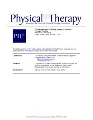
subluxation of the humeral head 23 (Figure).<br />
Although the cause of inferior subluxation<br />
usually is attributed to paralysis<br />
resulting from CVA, other possible<br />
causes are rotator cuff tear 17 and brachial<br />
plexus upper trunk injury. 23,50<br />
Spasticity also has been mentioned as<br />
contributing to subluxation by positioning<br />
the scapula in downward rotation.<br />
23,30 The real association between<br />
subluxation and spasticity, however, is<br />
unclear.<br />
How dangerous is subluxation? Several<br />
authorities in rehabilitation discount<br />
any importance of subluxation,<br />
30,32,51 although they offer little<br />
evidence to support their view. Others<br />
believe that subluxation may be a major<br />
cause of HSP because of the resulting<br />
overstretching of the GHJ soft tissue.<br />
1,20,46 Passive dependence of the unsupported<br />
arm, even in an uninvolved<br />
shoulder, elongates the rotator cuff and<br />
creates ischemia in the biceps brachii<br />
tendon and in the "critical zone" of the<br />
rotator cuff. 26,28 Hakuno et al found a<br />
significant correlation between subluxation<br />
and arthrographically demonstrated<br />
adhesive changes in the bicipital<br />
tendon sleeve. 40 Subluxation may cause<br />
traction injury to the upper trunk of the<br />
brachial plexus. 50 Ring et al reported<br />
EMG evidence of axillary nerve dysfunction<br />
in patients with GHJ subluxation<br />
and hypothesized that initial stages<br />
of subluxation may cause axillary nerve<br />
compression. 52<br />
The relationship of pain to subluxation<br />
is unclear. Some investigators have<br />
reported no correlation between incidences<br />
of subluxation and pain. 12,14 Najenson<br />
et al, however, found 93% of<br />
their hemiplegic patients with severe paralysis<br />
that was accompanied by moderate<br />
or severe pain had subluxation. 17<br />
Although subluxation may not be associated<br />
with pain in the early stage of<br />
hemiplegia, if subluxation continues<br />
into the chronic spastic stage, the association<br />
of pain and limited motion may<br />
be higher. 13 A recent study found sub<br />
luxation was associated frequently with<br />
HSP; 85% of the patients with spasticity<br />
had HSP, and subluxation was present<br />
in 67% of these patients. 16<br />
Brachial plexus and peripheral nerve<br />
injuries. Electromyographic evidence of<br />
lower motor neuron involvement has<br />
been reported in hemiplegic patients<br />
and is found twice as frequently in the<br />
upper as in the lower limb. 23 Brachial<br />
plexus injury may be a source of shoulder<br />
pain and a cause of subluxation.<br />
Chino reported 75% of his hemiplegic<br />
patients with subluxation demonstrated<br />
Figure. Basmajian's principle. A) The normal<br />
position of the shoulder prevents subluxation<br />
because the humeral head cannot<br />
displace laterally out of the upwardly tilted<br />
glenoid fossa (dotted arrow). B) Drooping of<br />
the shoulder in some hemiplegic patients permits<br />
subluxation of the humeral head out of<br />
the downwardly tilted glenoid fossa (dotted<br />
arrow). (Reprinted by permission of W. B.<br />
Saunders Co. 92 )<br />
neuropathic responses in the deltoid and<br />
supraspinatus muscles. 50 Kaplan et al<br />
found EMG changes consistent with injury<br />
of the upper trunk of the brachial<br />
plexus in five hemiplegic patients with<br />
flaccid paralysis and atrophy of the<br />
shoulder musculature. 53 Electromyographic<br />
evidence of pressure neuropathy<br />
of the ulnar nerve and brachial plexus<br />
neuropathies was reported by Moskowitz<br />
and Porter in eight hemiplegic patients.<br />
54 Other investigators have found<br />
no electrophysiological evidence of brachial<br />
plexus injuries in hemiplegic patients.<br />
12,19,46,47,55<br />
Signs of neuropathy are superimposed<br />
on the hemiplegic paresis. They include<br />
extensive segmental atrophy, an atypical<br />
pattern of return of motor function (ie,<br />
distal before proximal return), and electrophysiological<br />
signs. 54<br />
All investigators reporting brachial<br />
plexus injuries in hemiplegic patients<br />
identify the probable mechanism of injury<br />
as a combination of flaccid paralysis<br />
and traction injury. Traction stress is<br />
caused by either lack of support of the<br />
paralyzed flaccid shoulder 50,52-54 or pulls<br />
on the flaccid arm by personnel who<br />
move the patient. 54,56 Patients who are<br />
unconscious are at the greatest risk of<br />
having traction and pressure neuropathies.<br />
56<br />
The occurrence of a brachial plexus<br />
injury can increase rehabilitation time<br />
greatly. The superimposition of a lower<br />
motor neuron injury on the upper motor<br />
neuron lesion increases the likelihood<br />
for the development of painful<br />
contractures. 54 A brachial plexus injury<br />
constitutes a major setback for recovery<br />
of active motion; because of the slow<br />
rate of nerve regeneration, functional<br />
recovery of shoulder movement may be<br />
delayed by 8 to 12 months. 53<br />
<strong>Shoulder</strong>-hand syndrome. This reflex<br />
neurovascular disorder is characterized<br />
by pain and hyperesthesia, edema,<br />
trophic changes, and vasomotor instability.<br />
Signs and symptoms vary among<br />
patients, and onset may be dramatic. 57<br />
The temporal sequence of signs has been<br />
described by many authorities. 58-60 Early<br />
manifestations include pain and limited<br />
motion in the shoulder, wrist, and hand;<br />
the most restricted motions are shoulder<br />
lateral rotation and abduction, wrist<br />
dorsiflexion, and flexion of the metacarpophalangeal<br />
and proximal interphalangeal<br />
joints. Edema is evident in the wrist<br />
and finger joints, with increased skin<br />
temperature, rubor, and hyperhidrosis<br />
or hypohidrosis. As the condition progresses,<br />
pain decreases and stiffness predominates.<br />
The skin becomes shiny, cyanotic,<br />
and hyperhidrotic. Intrinsic<br />
muscle atrophy, nail changes, and thickening<br />
of subcutaneous tissue in the finger<br />
joints and palmar fascia develop.<br />
Osteoporosis is demonstrable radiographically.<br />
The syndrome may last<br />
from months to years. 61<br />
<strong>Shoulder</strong>-hand syndrome is known to<br />
develop after internal lesions such as<br />
CVA, myocardial infarction, cervical osteoarthritis,<br />
or spondylosis. The syndrome<br />
also develops often after trauma<br />
such as peripheral nerve injury, fracture,<br />
or spinal cord injury. 58,59,62 The precise<br />
Volume 66 / Number 12, December 1986 1887<br />
Downloaded from<br />
http://ptjournal.apta.org/ by guest on December 15, 2012
mechanisms triggering SHS are unknown.<br />
Tissue injury may serve as a<br />
focus of noxious stimuli, altering central<br />
control of the sympathetic nervous system.<br />
58 Immobilization of the limb because<br />
of injury, debility, pain, or paralysis<br />
may result in decreased sensory input<br />
from the limb to the central nervous<br />
system, causing an imbalance in the central<br />
neural control of the sympathetic<br />
nervous system. 58 Inactivity of muscles<br />
that perform pumping actions in the<br />
hand and the axilla (secondary to paralysis<br />
or immobilization) may cause altered<br />
circulation and edema. 23,27,63,64<br />
In hemiplegia, however, vasomotor<br />
changes may not be related necessarily<br />
to paralysis. 51,65 The cerebrovascular lesion<br />
may disturb central vasomotor regulation,<br />
resulting in arteriolar vasodilation<br />
of the upper limb. 65 Lesion of the<br />
premotor and anterior motor cortical<br />
regions may cause SHS after a stroke. 66<br />
The incidence of SHS among hemiplegic<br />
patients is unclear. Ring et al<br />
found no cases in 24 patients followed<br />
one year after a stroke. 52 In a retrospective<br />
study of 540 hemiplegic patients,<br />
Davis et al reported a 12.5% incidence<br />
of SHS. 57 Van Ouwenaller et al found<br />
the incidence of SHS in 215 hemiplegic<br />
patients was 28%. 16 Finch and Harvey,<br />
in a prospective study of 20 patients<br />
hospitalized after stroke, reported 50%<br />
developed signs of SHS. 67 Of hemiplegic<br />
patients with shoulder pain, the percentage<br />
of patients with signs of SHS<br />
has been cited as 36% 6 and 32%. 16<br />
Davis et al reported that most of their<br />
patients who developed SHS did so two<br />
to four months after the stroke, and they<br />
hypothesized that the incidence was the<br />
least in patients with mild involvement.<br />
57 Mossman also reported that the<br />
syndrome was more typical of patients<br />
with little or no voluntary motor ability.<br />
51 Two studies comparing hemiplegic<br />
patients with and without SHS, however,<br />
reported no difference between<br />
groups in active motor ability or muscle<br />
tone. 6,67 Van Ouwenaller et al found a<br />
higher incidence of SHS in patients with<br />
spasticity than in those without. 16<br />
<strong>Hemiplegic</strong> patients with SHS have<br />
been found to be significantly more confused<br />
and have greater sensory loss than<br />
other hemiplegic patients; they also have<br />
a higher incidence of subluxation than<br />
other hemiplegic patients. 67 Weiss and<br />
Ellis stated that hemiplegic patients with<br />
prior myocardial infarction may be at<br />
special risk for developing SHS. 65 Confirming<br />
this finding, Finch and Harvey<br />
reported that half of the patients devel<br />
oping the syndrome had a history of<br />
cardiac abnormalities. 67<br />
Thalamic pain. A burning, excruciating<br />
pain may involve the face, tongue,<br />
and thorax as well as the affected<br />
limbs. 68 Marked proprioceptive loss<br />
usually is present, 68 and the patient may<br />
neglect the paretic side except during a<br />
painful episode. 69 Peculiar sensations relating<br />
to the limb, feelings such as absence,<br />
twisting, or changes in size, may<br />
be present. The pain may be constant<br />
but is exacerbated by changes in emotional<br />
states, visual or auditory stimuli,<br />
temperature, or cutaneous stimuli. 68,69<br />
Vasomotor disturbances may be present<br />
on the affected side, characterized by<br />
cycles of skin coolness, pallor, and hyperhidrosis,<br />
alternating with warmth,<br />
rubor, and edema.<br />
A lesion of the lateral thalamus, the<br />
posterior limb of the internal capsule, or<br />
the parietal lobe is believed to be responsible.<br />
68,69 The incidence is unknown.<br />
Thalamic pain responds poorly to treatment.<br />
70<br />
PROPHYLAXIS AND<br />
TREATMENT<br />
Although cases of HSP may result<br />
directly from CNS lesions, many may<br />
be caused by faulty treatment after the<br />
stroke. 71 Careless handling of the paralyzed<br />
upper limb at any time after the<br />
onset of hemiplegia can precipitate<br />
shoulder injury and pain. For prophylaxis<br />
to be effective, however, it must<br />
begin immediately after the stroke. After<br />
the pain cycle has begun, resultant anxiety,<br />
overprotection, and disuse inevitably<br />
produce decreased ROM and contractures.<br />
23<br />
Treatment of the many varieties of<br />
HSP represents a complex and difficult<br />
problem. The ideal treatment is prevention;<br />
the next best management is to<br />
implement a treatment program immediately<br />
when the first painful symptoms<br />
appear.<br />
Positioning and Handling of the<br />
Dependent Patient<br />
Education of the patient and everyone<br />
associated with the care of the patient in<br />
the acute period after the stroke is critical<br />
to the success of a positioning and<br />
handling program. According to Mulley,<br />
pain in the shoulder is a complex subject<br />
that has not yet been approached<br />
systematically, but even in our present<br />
state of ignorance we can prevent<br />
much pain and limitation of movement<br />
in the shoulder by careful positioning<br />
and handling. 33<br />
Adequate support and protection of<br />
the flaccid arm are essential when the<br />
patient is lifted or moved in bed. 48,71,72<br />
All persons moving the dependent patient<br />
must be instructed concerning the<br />
hazards of pulling on the affected arm.<br />
Methods of upper limb handling when<br />
the patient is moved in bed or is assisted<br />
to a sitting position have been described.<br />
30 ' 32 ' 72 " 74<br />
Positioning for the flaccid limb in bed<br />
also is important, and protocols are discussed<br />
in several sources. 23,30,32,72,74,75<br />
The typical spasticity pattern of scapular<br />
retraction, neck flexion to the affected<br />
side, shoulder medial rotation and adduction,<br />
and elbow-wrist flexion should<br />
be avoided. Pillows can be used to support<br />
and elevate the limb to lessen dependent<br />
edema of the hand. 74,76,77 When<br />
the patient is in the supine position, the<br />
shoulder should be supported in a flexion-abduction-lateral<br />
rotation position.<br />
71,72 Bohannon et al have described<br />
a method of shoulder positioning in bed<br />
using a device made of foam and Velcro.<br />
78 Chino recommended overhead<br />
suspension of the arm in bed. 50 When<br />
the patient is sidelying, the affected arm<br />
should be flexed and rotated laterally<br />
with the shoulder girdle protracted. 71<br />
Limited sidelying on the affected side<br />
sometimes is recommended. 77 When the<br />
patient is sidelying on the unaffected<br />
side, the affected shoulder should be<br />
supported on pillows and not allowed to<br />
lie in horizontal adduction. A horizontally<br />
adducted position may exert traction<br />
of the suprascapular nerve and<br />
cause HSP. 43<br />
Facilitation of Active Motion<br />
Early facilitation of activity in muscle<br />
groups producing protraction and upward<br />
rotation of the scapula and flexionabduction<br />
of the shoulder is essential.<br />
Methods to inhibit spasticity contributing<br />
to scapular retraction and shoulder<br />
adduction and medial rotation likewise<br />
are essential if spasticity is present. A<br />
discussion of these methods is beyond<br />
the scope of this article; descriptions of<br />
facilitation-inhibition programs may be<br />
found in several sources. 23,30-32,74,75,79,80<br />
Unfortunately, little controlled research<br />
is available documenting the relative efficacy<br />
of these methods.<br />
Electrical stimulation and EMG biofeedback,<br />
alone or in combination, have<br />
emerged as promising therapeutic adjuncts<br />
in the facilitation of voluntary<br />
motion and the inhibition of spasticity.<br />
81-85 Most studies relating to the efficacy<br />
of these methods, however, have<br />
1888 PHYSICAL THERAPY<br />
Downloaded from<br />
http://ptjournal.apta.org/ by guest on December 15, 2012
een of distal extremity function, that<br />
is, facilitation of ankle dorsiflexion and<br />
wrist-finger extension. Few studies have<br />
been published relating to the effect of<br />
biofeedback on proximal muscle function.<br />
Lee et al found no difference in<br />
the deltoid muscle EMG activity between<br />
patients receiving biofeedback<br />
and patients in conventional physical<br />
therapy and placebo groups. 86 Inglis et<br />
al, however, found that hemiplegic patients<br />
receiving EMG biofeedback and<br />
physical therapy improved more in<br />
shoulder strength and ROM than patients<br />
receiving physical therapy alone. 87<br />
Patients with proprioceptive impairment<br />
may have limited success with biofeedback<br />
training, 88 although some evidence<br />
indicates sensory loss may not<br />
preclude improvement with biofeedback.<br />
89 Electrical stimulation and biofeedback<br />
may be effective in preventing<br />
or reducing GHJ subluxation, as will be<br />
discussed in the next section.<br />
Subluxation<br />
Prevention of subluxation requires<br />
the institution of prophylactic methods<br />
in the first few weeks of flaccid paralysis.<br />
46,50 After spasticity or active shoulder<br />
motion, or both, appear, subluxation<br />
seems unlikely to develop. 47<br />
Objective documentation of subluxation<br />
requires radiographs with the patient<br />
in the upright, not supine, position.<br />
17,20,46,48 Likewise, the efficacy of<br />
methods for preventing or reducing subluxation<br />
should be demonstrated radiographically.<br />
Shai et al hypothesized<br />
that earlier radiologic diagnosis of subluxation<br />
(ie, before grade I or II changes<br />
are demonstrated) might enable more<br />
effective prevention than if it is delayed.<br />
21 In their study, 12 of 14 patients<br />
showing early, positive radiologic signs<br />
of subluxation subsequently developed<br />
a painful shoulder, and 4 developed subluxation.<br />
Shai et al suggested that patients<br />
showing early radiologic signs of<br />
subluxation might be most likely to benefit<br />
from early orthotic intervention.<br />
When the patient is upright, the arm<br />
should be supported and positioned to<br />
prevent unopposed gravitational stress<br />
on the soft tissue of the GHJ. Various<br />
methods have been described for upper<br />
limb positioning in the wheelchair: pillows,<br />
a padded arm trough attached to<br />
the wheelchair arm or a lapboard, 30,72 or<br />
an overhead wheelchair sling. 15,23,72,90<br />
None of these methods has been proven<br />
to be more effective than others in preventing<br />
or reducing subluxation.<br />
Some support of the flaccid arm is<br />
necessary when the patient is ambulating.<br />
The patient with severe perceptual<br />
deficits and denial may need a sling for<br />
protecting the arm against trauma. 74 A<br />
multitude of sling types have been described.<br />
30,91-96 Elements to be considered<br />
in the choice of slings for particular<br />
patient problems have been summarized.<br />
95,96 External support can be discontinued<br />
when muscle tone around the<br />
GHJ is adequate to prevent subluxation.<br />
91 If needed, a sling should be used<br />
only during ambulation; the sling cannot<br />
be allowed to contribute to shoulder<br />
immobilization. An exercise program<br />
always should accompany the use of a<br />
sling. 24<br />
Controversy abounds concerning the<br />
use of slings; disadvantages to their use<br />
have been enumerated by many authorities.<br />
34,71,94,97 The major purpose of a<br />
sling is to provide an upward force to<br />
maintain the humeral head in the glenoid<br />
fossa. Unfortunately, little documentation<br />
exists in the form of radiological<br />
evidence to indicate which of the<br />
plethora of sling types produces the<br />
most effective reduction. In the only<br />
controlled, longitudinal study on sling<br />
efficacy, Hurd et al found one type of<br />
sling ineffective in preventing subluxation.<br />
19 Sodring 94 and Rajaram and<br />
Holta 98 reported radiographic evidence<br />
of GHJ reduction with two types of<br />
slings; however, no controlled research<br />
study was conducted to demonstrate the<br />
efficacy of subluxation in prophylaxis.<br />
In circumstances when the presence<br />
of subluxation has been determined to<br />
be the primary cause of HSP, use of a<br />
Varney brace has been reported to be<br />
successful; patients became asymptomatic<br />
in five to seven days. 99 After the<br />
subluxation is reduced and the shoulder<br />
is asymptomatic, an active and passive<br />
exercise program is important to improve<br />
and maintain ROM. 99<br />
Electrical stimulation of the supraspinatus<br />
and deltoid muscles can be used<br />
as an "electrical orthosis" for maintaining<br />
the alignment of the GHJ and has<br />
been recommended as an effective substitute<br />
for a sling. 83 Studies conducted at<br />
Rancho Los Amigos Rehabilitation<br />
Engineering Center reported that patients<br />
who were receiving electrical stimulation<br />
to the posterior deltoid and<br />
supraspinatus muscles demonstrated radiological<br />
evidence of reduced subluxation.<br />
100,101 Some evidence existed that<br />
the reduction was maintained after the<br />
stimulation was discontinued. 100 No<br />
prospective longitudinal studies could<br />
be located, however, documenting the<br />
efficacy of electrical stimulation for the<br />
treatment of subluxation. (See the article<br />
by L. Baker in this issue.)<br />
Electromyographic biofeedback has<br />
been reported to be successful in reducing<br />
subluxation, 82,85 although little prospective<br />
research seems to exist. In a<br />
controlled study on hemiplegic patients<br />
with subluxation, Shahani et al noted<br />
equal effectiveness of an EMG biofeedback<br />
program and a "conventional"<br />
physical therapy program in reducing<br />
subluxation; a correlation existed between<br />
the decrease in subluxation and<br />
the increase in strength and tone in the<br />
shoulder muscles. 102<br />
In a clinical report, Burstein recommended<br />
weight bearing on the affected<br />
arm and manual reduction of the subluxation<br />
during exercise as effective<br />
methods in relieving HSP. 103 Smith presented<br />
two case reports describing the<br />
reduction of subluxation and the increase<br />
of active abduction using an intensive<br />
facilitation program for the supraspinatus,<br />
deltoid, and serratus anterior<br />
muscles. 104<br />
Avoidance of Impingement<br />
During Passive Exercise<br />
Immobilization contributes to loss of<br />
motion, and passive ROM is an essential<br />
prophylactic measure to prevent joint<br />
stiffness and soft tissue contractures.<br />
The effectiveness of passive ROM, however,<br />
does not necessarily indicate that<br />
all types of shoulder exercise are beneficial<br />
or that a patient who begins to<br />
complain of pain during exercise requires<br />
more aggressive exercise therapy.<br />
11 Improperly administered passive<br />
exercise can cause impingement, resulting<br />
in soft tissue trauma, inflammation,<br />
and pain. 3,8,17,34,50 Increased exercise of<br />
the wrong type can contribute to the<br />
development of a stiff, painful shoulder.<br />
All persons performing passive exercise<br />
to the flaccid or spastic arm, from<br />
professional staff to family and the patient<br />
himself, must be taught the correct<br />
exercise techniques. Certainly, manual<br />
reduction of subluxation, if it exists,<br />
should be accomplished before passively<br />
elevating the arm. Medial or lateral rotation<br />
must accompany flexion or abduction,<br />
and scapular upward rotation<br />
must accompany either rotation to<br />
avoid soft tissue impingement. Spasticity<br />
must be reduced before elevating<br />
the arm more than 90 degrees. Overhead<br />
pulleys provide neither correct humeral<br />
nor correct scapular rotation during pas-<br />
Volume 66 / Number 12, December 1986 1889<br />
Downloaded from<br />
http://ptjournal.apta.org/ by guest on December 15, 2012
sive elevation and are a likely cause of<br />
impingement.<br />
An important question to be addressed<br />
is how much ROM is "enough."<br />
All medical professionals seem "programmed"<br />
to take each joint through its<br />
full ROM. As previously noted, complete<br />
ROM to a young physical therapist<br />
is not necessarily complete ROM to a<br />
geriatric stroke patient. Perhaps a more<br />
realistic goal is the maintenance of painfree<br />
functional ROM in the shoulder.<br />
The affected arm must be pain free to<br />
serve even in an assistive role. 38 Functional<br />
ROM has been defined as flexion<br />
to 100 degrees, abduction to 90 degrees,<br />
lateral rotation to 30 degrees, and medial<br />
rotation to 70 degrees. 90<br />
Another aspect to be considered is<br />
whether abduction should be included<br />
in passive exercise, in view of its tendency<br />
to cause soft tissue impingement.<br />
Some authorities recommend that abduction<br />
should be performed only in the<br />
scapular plane, that is, 30 degrees to 40<br />
degrees from the frontal plane, 3,34 because<br />
lateral rotation is not required to<br />
prevent impingement. 27 Curtailing the<br />
amplitude of passive abduction in exercise<br />
programs might preserve functional<br />
ROM and minimize impingement pain.<br />
If impingement during ROM is determined<br />
to be the etiological factor in<br />
pain, then the amplitude of passive motion<br />
should be kept within the pain-free<br />
range. 15 Caldwell et al reported that pain<br />
subsided in 43% of their patients with<br />
HSP when the amplitude of passive<br />
ROM was limited. 15<br />
Should the patient begin to complain<br />
of pain during passive ROM, a thorough<br />
evaluation to assess the cause of pain<br />
should be conducted before pain and<br />
stiffness become established.<br />
Diagnosis of Established <strong>Pain</strong>ful<br />
Conditions<br />
If a patient has an established painful,<br />
stiff shoulder, the first step in treatment<br />
is the identification of the cause. A carefully<br />
detailed medical history is important<br />
to determine the prestroke history<br />
of any shoulder pathological conditions<br />
and to identify any known trauma that<br />
occurred after the stroke, such as<br />
falls. 23,27 A correct diagnosis of the cause<br />
is essential if the patient is to be helped."<br />
A thorough diagnostic examination of<br />
the neck and shoulder should be conducted<br />
to identify cervical spine pathological<br />
factors, rotator cuff or biceps<br />
brachii tendinitis, or GHJ or acromioclavicular<br />
joint arthritis. 42,99 Confirmatory<br />
evidence of rotator cuff tears and<br />
adhesive capsulitis can be obtained with<br />
arthrography. Radiographs can rule out<br />
humeral fracture 3 and ectopic ossification.<br />
105 Electrophysiological studies can<br />
be used to identify brachial plexus injurv.<br />
<strong>Shoulder</strong>-hand syndrome may be<br />
confirmed by a positive response to stellate<br />
ganglion block. 58<br />
Conservative Management<br />
If subluxation is identified as the causative<br />
agent, several treatment methods<br />
exist. <strong>Pain</strong> and stiffness in a subluxated<br />
shoulder, however, are not necessarily a<br />
result of subluxation. 15,102 Krempen et al<br />
examined 37 patients with pain and subluxation<br />
and found 16 patients had<br />
other, orthopedic causes for the pain."<br />
Conservative orthopedic management<br />
of patients with diagnoses of tendinitis,<br />
bursitis, and rotator cuff tears is<br />
similar. The goals of treatment are pain<br />
reduction and improvement of ROM.<br />
Symptoms may be reduced with oral<br />
anti-inflammatory agents and analgesics<br />
and injections of lidocaine and steroids<br />
into trigger points, the subacromial bursae,<br />
or the joint space. 27,28,42,106 As pain<br />
decreases, active, passive, and assistive<br />
exercises are used to improve ROM.<br />
Applications of heat, cold, phonophoresis,<br />
and transcutaneous electrical<br />
nerve stimulation may be effective in<br />
relieving pain and encouraging motion.<br />
106 Applications of heat, however,<br />
have not been proven to be of particular<br />
benefit in the treatment of HSP. Liao et<br />
al found no significant difference in the<br />
amount of improvement in ROM between<br />
patients treated with exercise only<br />
versus those treated with a combination<br />
of heat and exercise therapy; exercise<br />
appeared to be the most effective treatment.<br />
4 Thus far, in studies of hemiplegic<br />
patients with unstated etiologic factors<br />
of pain, ultrasound 22 and TENS 6 have<br />
not been found to decrease pain or to<br />
contribute to increased range of shoulder<br />
motion.<br />
If an injury to the upper trunk has<br />
been diagnosed, the brachial plexus<br />
must be protected until regeneration can<br />
occur. 53 The shoulder should be splinted<br />
in 45 degrees of abduction, a treatment<br />
similar to that of postsurgical repair of<br />
the upper trunk. 107 Care must be taken<br />
to prevent further traction injury until<br />
regeneration occurs. The prognosis for<br />
recovery of function varies widely. 23<br />
Although evidence exists that adhesive<br />
capsulitis in nonhemiplegic patients<br />
is a self-limited disease resolving in one<br />
to three years, (see the article by C. T.<br />
Wadsworth in this issue), the same does<br />
not appear to be true of hemiplegic adhesive<br />
capsulitis. 12,42 A vigorous program<br />
of ice or heat applications and<br />
active and passive exercises can be combined<br />
with anti-inflammatory and analgesic<br />
medications, oral corticosteroids,<br />
and muscle relaxants. 41 Suprascapular<br />
nerve blocks, 108 corticosteroid injections<br />
into trigger points, and stellate ganglion<br />
blocks have been reported to be beneficial.<br />
23<br />
Antispasmodic medications may supplement<br />
inhibition and relaxation techniques.<br />
16 Motor-point blocks of the<br />
trapezius 16 and pectoralis major 109,110<br />
muscles may aid in controlling spasticity.<br />
In the management of SHS, early recognition<br />
is the key to a favorable prognosis;<br />
prompt treatment is essential to<br />
prevent permanent disability. 58,60,111 An<br />
aggressive exercise program is an essential<br />
component of treatment. 27,59 Active<br />
muscle contraction and joint movements<br />
are important, as are light weightbearing<br />
activities. Exercise must be performed<br />
within the pain-free range and<br />
frequently for short periods during the<br />
day. Modalities such as heat or ice treatments<br />
or TENS may be used to increase<br />
sensory input and reduce pain. 28,71,112<br />
Although TENS has been reported as<br />
helpful in the management of reflex<br />
sympathetic dystrophies, no research results<br />
appear to be reported on the effectiveness<br />
of TENS in hemiplegic SHS.<br />
Aggressive and prompt management<br />
of hand edema is important, because<br />
prolonged edema will precipitate perhaps<br />
irreversible hand stiffness. 27 Dependent<br />
positions of the hand should be<br />
avoided; an elevated position is preferable.<br />
Intermittent pneumatic compression<br />
may be of value in reducing hand<br />
edeipa, 23,90,113 and an elastic glove is<br />
helpful also in controlling edema. 71<br />
Systemic administration of corticosteroids<br />
for two to three weeks is recommended<br />
by several authorities for the<br />
management of SHS. 27,59,71 Davis et al<br />
reported a complete resolution of symptoms<br />
in 68 hemiplegic patients using<br />
oral steroids in combination with an<br />
intensive rehabilitation program. 57 A<br />
sympathetic block followed by intensive<br />
rehabilitation is cited as the most effective<br />
treatrnent by Some researchers.<br />
58,61,111 Sympathectomy is suggested<br />
in refractory cases. 58,59,111<br />
The emotional state of patients with<br />
severe HSP has been described as tense,<br />
hyperresponsive to pain, overemotional,<br />
apathetic, and emotionally dependent. 23<br />
Whether such emotional states contrib-<br />
1890 PHYSICAL THERAPY<br />
Downloaded from<br />
http://ptjournal.apta.org/ by guest on December 15, 2012
ute to the development of HSP or<br />
emerge as a result of the pain is unknown.<br />
111 In any event, appropriate psychological<br />
management is important for<br />
successful treatment. 23<br />
Surgical Management<br />
Surgical release of soft tissue may be<br />
of value if conservative methods have<br />
failed and if shoulder motion has become<br />
severely limited and painful. 112<br />
Recent improvements in rehabilitation<br />
techniques have decreased the need for<br />
surgical intervention. 75 The most frequently<br />
described surgical management<br />
involves resection of the subscapularis<br />
and pectoralis major tendons 15,114 and<br />
release of the capsule 115 ; insertions of<br />
the latissimus dorsi and teres major<br />
muscles also may be necessary. 116,117 Surgical<br />
release does not result in improved<br />
ROM without an intensive postoperative<br />
program of exercise and splinting to<br />
maintain and increase abduction and<br />
external rotation. 15,117<br />
The indications for surgery include<br />
ROM limitation to the point of functional<br />
impairment (ie, abduction to 45<br />
degrees and lateral rotation to 15 degrees<br />
or less 15 ), pain of such intensity that it<br />
interferes with skin hygiene 114 or prevents<br />
participation in rehabilitation, 15<br />
and progressively decreasing ROM resulting<br />
from increased internal rotator<br />
muscle spasticity. 15,109,117 Factors affecting<br />
the patient's rehabilitation potential,<br />
such as cognitive awareness, sensory and<br />
perceptual deficits, and communication<br />
disorders, also should be considered. 15<br />
Surgery usually is delayed until at least<br />
six months after the stroke when spontaneous<br />
recovery is fairly complete. 114<br />
Surgery is contraindicated in the presence<br />
of etiological factors other than soft<br />
tissue contracture or if postoperative<br />
therapy is unavailable. 15 Surgery for<br />
thalamic pain is not indicated. 15,118<br />
The reported magnitude of an increase<br />
in ROM after surgery is not great.<br />
Caldwell et al reported that the lateral<br />
rotation range was poor, although most<br />
patients regained 90 degrees of abduction;<br />
some patients developed active abduction<br />
postoperatively that had not<br />
been observed preoperatively. 15<br />
CONCLUSIONS<br />
Patients at highest risk for developing<br />
HSP seem to be those with severe upper<br />
limb paralysis, particularly if sensory<br />
loss and mental confusion also exist.<br />
Such a combination of factors not only<br />
immobilizes the limb but increases the<br />
likelihood for traction injury because<br />
the patient cannot protect himself. Increasing<br />
age may increase the risk also.<br />
Most patients over 50 years of age have<br />
significant degenerative changes around<br />
the GHJ. Such soft tissue pathological<br />
factors can be exacerbated only by<br />
events accompanying flaccid paralysis.<br />
The acute period after a stroke appears<br />
to be a critical time for the prevention<br />
of GHJ soft tissue injuries such as<br />
rotator cuff tears or brachial plexus injuries.<br />
Such trauma, which may go unnoticed<br />
in the acute period after a<br />
stroke, may cause significant HSP or<br />
functional impairment later in rehabilitation.<br />
Education of all personnel handling<br />
the flaccid arm is imperative if<br />
prophylaxis of shoulder injury is to be<br />
effective.<br />
Failure to provide support for the flaccid<br />
arm when the patient is upright may<br />
allow shoulder subluxation and consequent<br />
painful overstretching of soft tissue<br />
or nerve injury or both. The deleterious<br />
effects of subluxation may be<br />
cumulative. Effective methods for the<br />
prevention of subluxation remain to be<br />
established in a controlled clinical trial,<br />
but electrical stimulation appears<br />
promising.<br />
Immobilization is a key factor contributing<br />
to shoulder stiffness in elderly<br />
patients. In patients with hemiplegia,<br />
immobilization is promoted by paralysis,<br />
spasticity, and, sometimes, brachial<br />
plexus injury. <strong>Pain</strong> is another wellknown<br />
immobilizing influence. Lack of<br />
passive exercise for the paralyzed shoulder<br />
certainly will result in painful contractures.<br />
Careless passive exercise techniques<br />
without regard to biomechanical<br />
principles may cause impingement.<br />
Thus, improperly administered exercise<br />
may be a cause of trauma, pain, and<br />
further immobilization.<br />
When pain and stiffness begin to develop,<br />
a thorough evaluation of the<br />
cause is essential. Conservative management<br />
methods may be effective if the<br />
treatment is appropriate for the cause.<br />
Surgical intervention may be indicated<br />
in extreme cases. Early preventive measures,<br />
however, remain the key to minimizing<br />
the complication of shoulder<br />
pain in patients with hemiplegia.<br />
Need for Clinical Research<br />
Longitudinal studies are needed concerning<br />
developments in the shoulder<br />
after hemiplegia. Periodic assessments<br />
of shoulder status, from the onset until<br />
at least one year after the onset of hemiplegia,<br />
would add greatly to present<br />
knowledge concerning the evolution of<br />
HSP. Pertinent aspects to be included<br />
in such assessments are active motor<br />
function, sensory status, passive ROM,<br />
amount of pain associated with passive<br />
motion, spasticity, subluxation, and<br />
functional ability of the upper limb.<br />
Cognitive status and age also should be<br />
included. If HSP and limited ROM develop<br />
during the longitudinal study,<br />
prompt diagnostic steps to identify the<br />
cause should be instituted. When etiologic<br />
factors of the pain can be established,<br />
controlled studies of the relative<br />
effectiveness of various treatment methods<br />
are essential. The optimum treatment<br />
for hemiplegic patients with established<br />
diagnoses such as SHS or adhesive<br />
capsulitis currently is unknown.<br />
Knowledge of the effectiveness of<br />
methods for preventing subluxation (ie,<br />
electrical stimulation vs positioning<br />
alone) would be helpful in determining<br />
prophylaxis. Also of clinical importance<br />
are controlled studies to establish the<br />
relative efficacy of methods such as biofeedback,<br />
electrical stimulation, and exercise<br />
alone in improving proximal control<br />
in the hemiplegic shoulder.<br />
The obstacles to clinical research in<br />
the area of HSP may seem formidable:<br />
<strong>Hemiplegic</strong> patients admitted to rehabilitation<br />
settings differ widely in medical<br />
history and prestroke shoulder dysfunction;<br />
the time since the stroke varies<br />
among patients; and the history of<br />
shoulder trauma after the stroke often is<br />
impossible to ascertain. Such variables<br />
may account for the different responses<br />
to treatment and may make the original<br />
causes of HSP difficult to determine.<br />
Such difficulties might be overcome partially<br />
by beginning a longitudinal study<br />
immediately after the stroke and by including<br />
significant numbers of patients<br />
in the study. The HSP syndrome represents<br />
such a major obstacle to rehabilitation<br />
that systematic studies of the<br />
problem are imperative. Controlled research<br />
is needed to determine effective<br />
prophylaxis and to document the therapeutic<br />
efficacy of treatment.<br />
REFERENCES<br />
1. Tobis JS: Problems in rehabilitation of the<br />
hemiplegic patient. NY State J Med 57:1377-<br />
1380,1957<br />
2. Najenson T, Pikielny SS: Malalignment of the<br />
gjenohumeral joint following hemiplegia. Annals<br />
of <strong>Physical</strong> Medicine 8:96-99, 1965<br />
3. Jensen EM: The hemiplegic shoulder. Scand<br />
J Rehabil Med 12(Suppl):113-119, 1980<br />
Volume 66 / Number 12, December 1986 1891<br />
Downloaded from<br />
http://ptjournal.apta.org/ by guest on December 15, 2012
4. Liao H, Chen C, Lien I: <strong>Pain</strong>ful shoulder contracture<br />
in strokes and the effects of physical<br />
therapy. Taiwan I Hsueh Hui Tsa Chih<br />
80:253-261, 1981 (Translated from Forosan)<br />
5. Crossen-Sills J: An Analysis of <strong>Shoulder</strong> <strong>Pain</strong>,<br />
Range of Motion, and Subluxation in Patients<br />
with Hemiplegia. Read at the Sixty-First Annual<br />
Conference of the American <strong>Physical</strong><br />
<strong>Therapy</strong> Association, New Orleans, LA, June<br />
16-20, 1985<br />
6. Griffin JW, Reddin G: Use of TENS on <strong>Hemiplegic</strong><br />
Patients with Upper Extremity <strong>Pain</strong>.<br />
Read at the Fifty-Sixth Annual Conference of<br />
the American <strong>Physical</strong> <strong>Therapy</strong> Association,<br />
Phoenix, AZ, June 15-20, 1980<br />
7. Dardier E, Reid C: Letter to the editor. Phys<br />
Ther 52:1208, 1972<br />
8. Bobath K: Letter to the editor. Phys Ther<br />
52:444-445, 1972<br />
9. Taketomi Y: Observations on subluxation of<br />
the shoulder joint in hemiplegia. Phys Ther<br />
55:39-40, 1975<br />
10. Bruckner FE, Nye CJS: Prospective study of<br />
adhesive capsulitis (frozen shoulder) in high<br />
risk population. Q J Med 198:191-204, 1981<br />
11. Brocklehurst JG, Andrews K, Richards B, et<br />
al: How much physical therapy for patients<br />
with stroke? Br Med J 1:1307-1310, 1978<br />
12. Rizk TE, Christopher RP, Pinals RS, et al:<br />
Arthrography studies in painful hemiplegic<br />
shoulders. Arch Phys Med Rehabil 65:254-<br />
256, 1984<br />
13. Braun RM, West F, Mooney V, et al: Surgical<br />
treatment of the painful shoulder contracture<br />
in the stroke patient. J Bone Joint Surg [Am]<br />
53:1307-1312, 1971<br />
14. Peszczynski M, Rardin TE: The incidence of<br />
painful shoulder in hemiplegia. Bulletin of Polish<br />
Medical Science and History 8:21-23,<br />
1965<br />
15. Caldwell CB, Wilson DJ, Braun RM: Evaluation<br />
and treatment of the upper extremity in<br />
the hemiplegic stroke patient. Clin Orthop<br />
63:69-93,1969<br />
16. Van Ouwenaller C, Laplace PM, Chantraine<br />
A: <strong>Pain</strong>ful shoulder in hemiplegia. Arch Phys<br />
Med Rehabil 67:23-26,1986<br />
17. Najenson T, Yacubovich E, Pikielny SS: Rotator<br />
cuff injury in shoulder joints of hemiplegic<br />
patients. Scand J Rehabil Med 3:131-<br />
137,1971<br />
18. Fugl-Meyer AR, Jaasko L, Norlin V: The poststroke<br />
hemiplegic patient. Scand J Rehabil<br />
Med 7:73-83, 1975<br />
19. Hurd MM, Farrell KH Waylonis GW: <strong>Shoulder</strong><br />
sling for hemiplegia: Friend or foe? Arch Phys<br />
Med Rehabil 55:519-522, 1974<br />
20. Moskowitz H, Goodman CR, Smith E, et al:<br />
<strong>Hemiplegic</strong> shoulder. NY State J Med<br />
69:548-550, 1969<br />
21. Shai G, Ring H, Costeff H, et al: Glenohumeral<br />
joint malalignment in the hemiplegic shoulder:<br />
An early radiologic sign. Scand J Rehabil Med<br />
16:133-136,1984<br />
22. Inaba MK, Piorkowski M: Ultrasound in treatment<br />
of painful shoulders in patients with<br />
hemiplegia. Phys Ther 52:737-741,1972<br />
23. Cailliet R: The <strong>Shoulder</strong> in Hemiplegia. Philadelphia,<br />
PA, F A Davis Co, 1980<br />
24. Wilson DJ, Caldwell CB: Central control insufficiency:<br />
III. Disturbed motor control and<br />
sensation: A treatment approach emphasizing<br />
upper extremity orthoses. Phys Ther<br />
58:313-320,1978<br />
25. Saario L: The range of motion of the shoulder<br />
joint at various ages. Acta Orthop Scand<br />
33:366, 1963<br />
26. Cailliet R: <strong>Shoulder</strong> <strong>Pain</strong>. Philadelphia, PA,<br />
FA Davis Co, 1978<br />
27. DePalma AF: Surgery of the <strong>Shoulder</strong>, ed 3<br />
Philadelphia, PA, J B Lippincott Co, 1983<br />
28. Hawkins RJ, Murnaghan JP: The shoulder. In<br />
Cruess RL, Rennee WR (eds): Adult Orthopaedics.<br />
New York, NY, Churchill Livingstone<br />
Inc. 1984, vol 2<br />
29. Saha AK: Surgery of the paralyzed and flail<br />
shoulder. Acta Orthop Scand 97(Suppl):5-90,<br />
1967<br />
30. Bobath B: Adult Hemiplegia: Evaluation and<br />
Treatment, rev ed 2. London, England, William<br />
Heinemann Medical Books Ltd, 1978<br />
31. Brunnstrom S: Movement <strong>Therapy</strong> in Hemiplegia.<br />
New York, NY, Harper & Row Publishers<br />
Inc, 1970<br />
32. Johnstone M. The Restoration of Motor Function<br />
in the Stroke Patient, ed 2. New York,<br />
NY, Churchill Livingstone Inc, 1983<br />
33. Mulley GP: Avoidable complications of stroke.<br />
J R Coll Physicians Lond 16:94-97, 1982<br />
34. Voss DE: Should patients with hemiplegia<br />
wear a sling? Phys Ther 49:1030,1969<br />
35. Neer CS: Impingement lesions. Clin Orthop<br />
173:70-77, 1983<br />
36. Bateman JE: The diagnosis and treatment of<br />
ruptures of the rotator cuff. Surg Clin North<br />
Am 43:1523-1530, 1963<br />
37. Post M: Rotator cuff tear: Diagnosis and treatment.<br />
Clin Orthop 173:78-91,1983<br />
38. Nepomuceno CS, Miller JM: <strong>Shoulder</strong> arthrography<br />
in hemiplegic patients. Arch Phys<br />
Med Rehabil 55:49-51, 1974<br />
39. Moskowitz E: Complications in rehabilitation<br />
of hemiplegic patients. Med Clin North Am<br />
53:541-559, 1969<br />
40. Hakuno A, Hironobu S, Ohkawa T, et al:<br />
Arthrographic findings in hemiplegic shoulders.<br />
Arch Phys Med Rehabil 65:706-711,<br />
1984<br />
41. Rizk TE, Pinals RS: Frozen shoulder. Semin<br />
Arthritis Rheum 11:440-452, 1982<br />
42. Neviaser RJ: <strong>Pain</strong>ful conditions affecting the<br />
shoulder. Clin Orthop 173:63-69, 1983<br />
43. Koppell HP, Thompson WA: <strong>Pain</strong> and the<br />
frozen shoulder. Surg Gynecol Obstet<br />
109:92-96,1959<br />
44. Carpenter Gl, Millard PH: <strong>Shoulder</strong> subluxation<br />
in elderly inpatients. J Am Geriatr Soc<br />
30:441-446,1982<br />
45. Fitzgerald-Finch GP, Gibson I: Subluxation of<br />
the shoulder in hemiplegia. Age Ageing 4:16,<br />
1975<br />
46. Miglietta O, Lewitan A. Rogoff JB: Subluxation<br />
of the shoulder in hemiplegic patients.<br />
NY State J Med 59:457-460, 1959<br />
47. Chaco J, Wolf E: Subluxation of the glenohumeral<br />
joint in hemiplegia. Am J Phys Med<br />
50:139-143,1971<br />
48. Smith RG, Cruikshank JG, Dunbar S, et al:<br />
Malalignment of the shoulder after stroke. Br<br />
Med J 284:1224-1226, 1982<br />
49. Basmajian JV, Bazant FJ: Factors preventing<br />
downward dislocation of the adducted shoulder<br />
joint. J Bone Joint Surg [Am] 41:1182-<br />
1186,1959<br />
50. Chino N: Electrophysiological investigations<br />
on shoulder subluxation in hemiplegics.<br />
Scand J Rehabil Med 13:17-21, 1981<br />
51. Mossman PL. A Problem-Oriented Approach<br />
to Stroke Rehabilitation. Springfield, IL,<br />
Charles C Thomas, Publisher, 1976, pp 145-<br />
152<br />
52. Ring H, Leillen B, Server S, et al: Temporal<br />
changes in electrophysiological, clinical and<br />
radiological parameters in the hemiplegic's<br />
shoulder. Scand J Rehabil Med 12(Suppl):<br />
124-127,1985<br />
53. Kaplan PE, Meredith J, Taft G, et al: Stroke<br />
and brachial plexus injury: A difficult problem.<br />
Arch Phys Med Rehabil 58:415-418,1977<br />
54. Moskowitz E, Porter Jl: Peripheral nerve lesions<br />
in the upper extremity in hemiplegic<br />
patients. N Engl J Med 269:776-778, 1963<br />
55. Alpert S, Idarraga S, Orbegozo J, et al: Absence<br />
of electromyographic evidence of lower<br />
motor neuron involvement in hemiplegic patients.<br />
Arch Phys Med Rehabil 52:179-181,<br />
1971<br />
56. Editorial. N Engl J Med 269:816,1963<br />
57. Davis SW, Petrillo CR, Eichberg RD, et al:<br />
<strong>Shoulder</strong>-hand syndrome in a hemiplegic population:<br />
A 5-year retrospective study. Arch<br />
Phys Med Rehabil 58:353-356. 1977<br />
58. Bonica JJ: Causalgia and other reflex sympathetic<br />
dystrophies. Postgrad Med J<br />
53:143-148, 1973<br />
59. Steinbrocker O: The shoulder-hand syndrome:<br />
Present perspective. Arch Phys Med<br />
Rehabil 49:388-395, 1968<br />
60. Chu DS, Petrillo C, Davis SW, et al: <strong>Shoulder</strong>hand<br />
syndrome: Importance of early diagnosis<br />
and treatment. J Am Geriatr Soc 29:58-<br />
60,1981<br />
61. Swan DM: <strong>Shoulder</strong>-hand syndrome following<br />
hemiplegia. Neurology 4:480-482,1954<br />
62. Wainapel SF: Reflex sympathetic dystrophy<br />
in quadriplegia: Case report. Arch Phys Med<br />
Rehabil 65:35-36,1984<br />
63. Moberg E: The shoulder-hand-finger syndrome.<br />
Surg Clin North Am 40:367-373,<br />
1960<br />
64. Flatt A: <strong>Shoulder</strong>-hand syndrome. Lancet<br />
1:1107-1108,1974<br />
65. Weiss S, Ellis LB: The circulatory mechanism<br />
and unilateral edema in cerebral hemiplegia.<br />
J Clin Invest 9:17,1931<br />
66. Eto F, Yoshikawa M, Ueda S, et al: Posthemiplegic<br />
shoulder-hand syndrome with special<br />
reference to related cerebral localization. J<br />
Am Geriatr Soc 28:13-17,1980<br />
67. Finch L, Harvey J: Factors associated with<br />
shoulder-hand syndrome in hemiplegia: Clinical<br />
survey. Physiotherapy Canada 35:145-<br />
148,1983<br />
68. Spence AM: <strong>Pain</strong> and numbness in the extremities:<br />
Central nervous system causes. In<br />
Swanson PD (ed): Signs and Symptoms in<br />
Neurology. Philadelphia, PA, J B Lippincott<br />
Co, 1984, pp 289-302<br />
69. Martin JJ: Thalamic syndromes. In Vinken PJ,<br />
Bruyn GW (eds): Handbook of Clinical Neurology.<br />
New York, NY, Elsevier Science Publishing<br />
Co Inc, 1969, vol 2, pp 469-496<br />
70. Rusk HA: Rehabilitation of the patient with<br />
stroke. In Rusk HA (ed): Rehabilitation Medicine,<br />
ed 4. St. Louis, MO, C V Mosby Co,<br />
1977, pp 601-620<br />
71. Varghese G: Evaluation and management:<br />
<strong>Shoulder</strong> complications in hemiplegia. J Kans<br />
Med Soc 82:451-453,1981<br />
72. Ingenito R, Priestley L: Hemiplegia: Current<br />
Approaches to Patient Positioning. Washington,<br />
DC, Rehabilitation Research and Training<br />
Center, George Washington Medical Center,<br />
1979<br />
73. Andersen LT: <strong>Shoulder</strong> pain in hemiplegia. Am<br />
J Occup Ther 39:11-19,1985<br />
74. Carr JH, Shepherd RB: Physiotherapy in Disorders<br />
of the Brain. London, England, William<br />
Heinemann Medical Books Ltd, 1980<br />
75. Ryerson SD: Hemiplegia resulting from vascular<br />
insult or disease. In Umphred DA (ed):<br />
Neurological Rehabilitation. St. Louis, MO,<br />
C V Mosby Co, 1985, pp 474-514<br />
76. McCollough NC: Orthotic management in<br />
adult hemiplegia. Clin Orthop 131:38-46,<br />
1978<br />
77. Guentz S, Navales RD: New approaches to<br />
nursing care of the stroke patient. Clin Orthop<br />
131:90-96,1978<br />
78. Bohannon RW, Thome M, Mieres AC: <strong>Shoulder</strong><br />
positioning device for patients with hemiplegia.<br />
Phys Ther 63:49-50,1983<br />
79. Sullivan PE, Markos RD, Minor MA: An Integrated<br />
Approach to Therapeutic Exercise:<br />
Theory and Clinical Application. Reston, VA,<br />
Reston Publishing Co Inc, 1982<br />
80. Loomis J: Facilitation techniques in hemiplegia:<br />
Treatment of the arm. Physiotherapy<br />
Canada 25.283-288 1973<br />
81. Binder SA: Applications of low- and highvoltage<br />
etectrotherapeutic currents. In Wolf<br />
SL (ed): Electrotherapy. New York, NY,<br />
Churchill Livingstone Inc, 1981, pp 1-24<br />
82. Binder-McLeod S: Biofeedback in stroke rehabilitation.<br />
In Basmajian JV (ed): Biofeedback:<br />
Principles and Practice for Clinicians,<br />
ed 2. Baltimore, MD, Williams & Wilkins,<br />
1983, pp 73-89<br />
1892 PHYSICAL THERAPY<br />
Downloaded from<br />
http://ptjournal.apta.org/ by guest on December 15, 2012
83. Baker LL: Neuromuscular electrical stimulation<br />
in the restoration of purposeful limb<br />
movements. In Wolf SL (ed): Electrotherapy.<br />
New York, NY, Churchill Livingstone Inc,<br />
1981, pp 25-48<br />
84. Basmajian JV, Hatch JP: Biofeedback and the<br />
modification of skeletal muscular dysfunctions.<br />
In Gatchel RJ, Price KP (eds): Clinical<br />
Applications of Biofeedback: Appraisal and<br />
Status. New York, NY, Pergamon Press Inc,<br />
1979, pp 97-111<br />
85. Basmajian JV: Biofeedback in rehabilitation:<br />
A review of principles and practices. Arch<br />
Phys Med Rehabil 62:469-474,1981<br />
86. Lee LH, Hill E, Johnston R, et al: Myofeedback<br />
for muscle retraining in hemiplegic patients.<br />
Arch Phys Med Rehabil 57:588-591,<br />
1976<br />
87. Inglis J, Donald MW, Monga TN, et al: Electromyographic<br />
biofeedback and physical therapy<br />
of the hemiplegic upper limb. Arch Phys<br />
Med Rehabil 65:755-759,1984<br />
88. Wolf SL, Baker MP, Kelly JL: Electromyographic<br />
biofeedback in stroke: Effect of patient<br />
characteristics. Arch Phys Med Rehabil<br />
60:96-102,1979<br />
89. Skelly AM, Kenedi RM: Electromyographic<br />
biofeedback therapy in the re-education of<br />
the hemiplegic shoulder in patients with sensory<br />
loss. Physiotherapy 68:34-38,1982<br />
90. Savinelli R, Timm M, Montgomery J, et al:<br />
<strong>Therapy</strong> evaluation and management of patients<br />
with hemiplegia. Clin Orthop 131:15-<br />
29,1978<br />
91. McCollough NC: The role of the orthopedic<br />
surgeon in treatment of stroke. Orthop Clin<br />
North Am 9:305-324, 1978<br />
92. Anderson TP: Rehabilitation of patients with<br />
completed stroke. In Kottke FJ, et al (eds):<br />
Krusen's Handbook of <strong>Physical</strong> Medicine and<br />
Rehabilitation, ed 3. Philadelphia, PA, W B<br />
Saunders Co, 1982, pp 583-603<br />
93. Walker J: Modified strapping of roll sling. Am<br />
J Occup Ther 37:110-111,1983<br />
94. Sodring KM: Upper extremity orthoses for<br />
stroke patients. Int J Rehabil Res 3:33-38,<br />
1980<br />
95. Cohen BA: A new shoulder-elbow-wrist sling<br />
for hemiplegic patients. Journal of Clinical Engineering<br />
3:389-392,1978<br />
96. Smith RO, Okamoto GA: Checklist for the<br />
prescription of slings for the hemiplegic patient.<br />
Am J Occup Ther 35:91-95,1981<br />
97. Miller J: <strong>Shoulder</strong> pain from subluxation in the<br />
hemiplegic. Br Med J 4:345,1975<br />
98. Rajaram V, Holta M: <strong>Shoulder</strong> forearm support<br />
for the subluxed shoulder. Arch Phys<br />
Med Rehabil 66:191-192,1985<br />
99. Krempen JF, Silver RA, Hadley J, et al: The<br />
use of the Varney brace for subluxating shoulders<br />
in stroke and upper motor neuron injuries.<br />
Clin Orthop 122:204-206,1977<br />
100. Rancho Los Amigos Rehabilitation Engineering<br />
Center: Annual Report of Progress to the<br />
Rehabilitation Services Administration. Washington,<br />
DC, US Dept of Health, Education and<br />
Welfare, No. 7,1978, pp 8-11<br />
101. Rancho Los Amigos Rehabilitation Engineering<br />
Center: Annual Report of Progress to the<br />
Rehabilitation Services Administration. Washington,<br />
DC, US Dept of Health, Education and<br />
Welfare, No. 6,1977, pp 5-7<br />
102. Shahani BT, Keppy EB, Glasser S: <strong>Hemiplegic</strong><br />
shoulder subluxation. Arch Phys Med Rehabil<br />
62:519,1981<br />
103. Burstein D: Joint compression for treatment<br />
of shoulder pain. Clinical Management in<br />
<strong>Physical</strong> <strong>Therapy</strong> 5(2):6-9,1985<br />
104. Smith C: Neurophysiologies Approach to<br />
Treatment of <strong>Hemiplegic</strong> <strong>Shoulder</strong> Subluxation.<br />
Read at the Sixty-First Annual Conference<br />
of the American <strong>Physical</strong> <strong>Therapy</strong> Association,<br />
New Orleans, LA, June 16-20,<br />
1985<br />
105. Moskowitz E, Steinman Rl: Heterotopic calcification<br />
in the hemiplegic upper extremity.<br />
NY State J Med 64:432-435,1964<br />
106. McCollough RL: Orthopedic evaluation and<br />
treatment of the stroke patient: Upper extremity<br />
management. Instr Course Lect<br />
24:45-51,1975<br />
107. Wright PE, Simmons JC: Peripheral nerve<br />
injuries. In Edmondson AS, Crenshaw AH<br />
(eds): Campbell's Operative Orthopaedics, ed<br />
6. St. Louis, MO, C V Mosby Co, 1980, vol<br />
2, pp 1642-1702<br />
108. Peszczynski M: Prognosis for rehabilitation of<br />
the older adult and the aged hemiplegic patient.<br />
Am J Cardiol 7:365-369,1961<br />
109. Mooney V, Perry J, Nickel VL: Surgical and<br />
non-surgical orthopaedic care of stroke. J<br />
Bone Joint Surg [Am] 49:989-1000,1967<br />
110. Lusskin R, Grynbaum BB, Dhir RS: Rehabilitation<br />
surgery in adult spastic hemiplegia. Clin<br />
Orthop 63:132-141, 1969<br />
111. Moskowitz E, Bishop HF, Hla P, et al: Posthemiplegic<br />
reflex sympathetic dystrophy.<br />
JAMA 167:836-838, 1958<br />
112. Johnson EW, Pannozzo AN: Management of<br />
shoulder-hand syndrome. JAMA 195:152-<br />
154,1966<br />
113. Friedland F: <strong>Physical</strong> therapy. In Licht S (ed):<br />
Stroke and Its Rehabilitation. New Haven, CT,<br />
Elizabeth Licht, Publisher, 1975, pp 224-254<br />
114. Jordan C, Waters RL: Stroke. In Nickel VL<br />
(ed): Orthopaedic Rehabilitation. New York,<br />
NY, Churchill Livingstone Inc, 1982, pp 277-<br />
291<br />
115. Laing RE, Hurst LC: <strong>Pain</strong>ful poststroke shoulder.<br />
Orthopaedic Review 11:123-127,1982<br />
116. Nickel VL: Orthopaedic rehabilitation: Challenges<br />
and opportunities. Clin Orthop<br />
63:153-161,1969<br />
117. Waters RL: Upper extremity surgery in stroke<br />
patients. Clin Orthop 131:30-37,1978<br />
118. Freed MM: Surgical measures in stroke. In<br />
Licht S (ed): Stroke and Its Rehabilitation.<br />
New Haven, CT, Elizabeth Licht, Publisher,<br />
1975, pp 147-170<br />
Volume 66 / Number 12, December 1986 1893<br />
Downloaded from<br />
http://ptjournal.apta.org/ by guest on December 15, 2012
Cited by<br />
<strong>Hemiplegic</strong> <strong>Shoulder</strong> <strong>Pain</strong><br />
Judy W Griffin<br />
PHYS THER. 1986; 66:1884-1893.<br />
This article has been cited by 10 HighWire-hosted<br />
articles:<br />
http://ptjournal.apta.org/content/66/12/1884#otherarticles<br />
Subscription http://ptjournal.apta.org/subscriptions/<br />
Information<br />
Permissions and Reprints http://ptjournal.apta.org/site/misc/terms.xhtml<br />
Information for Authors http://ptjournal.apta.org/site/misc/ifora.xhtml<br />
Downloaded from<br />
http://ptjournal.apta.org/ by guest on December 15, 2012