Neuroectodermal and Neuroendocrine Tumors Principally Seen in ...
Neuroectodermal and Neuroendocrine Tumors Principally Seen in ...
Neuroectodermal and Neuroendocrine Tumors Principally Seen in ...

You also want an ePaper? Increase the reach of your titles
YUMPU automatically turns print PDFs into web optimized ePapers that Google loves.
© American Society of Cl<strong>in</strong>ical Pathologists<br />
Pathology Patterns Reviews<br />
<strong>Neuroectodermal</strong> <strong>and</strong> <strong>Neuroendocr<strong>in</strong>e</strong> <strong>Tumors</strong> <strong>Pr<strong>in</strong>cipally</strong><br />
<strong>Seen</strong> <strong>in</strong> Children<br />
David M. Parham, MD<br />
Key Words: Neuroblastoma; Ganglioneuroblastoma; Ganglioneuroma; Ew<strong>in</strong>g sarcoma; Peripheral primitive neuroectodermal tumor;<br />
Melanotic neuroectodermal tumor; Desmoplastic small cell tumor; Esthesioneuroblastoma; Diagnosis; Biology<br />
Abstract<br />
<strong>Neuroectodermal</strong> tumors comprise a large<br />
proportion of childhood neoplasms. Neuroblastic<br />
tumors, <strong>in</strong>clud<strong>in</strong>g neuroblastoma,<br />
ganglioneuroblastoma, <strong>and</strong> ganglioneuroma, are the<br />
most frequent extracranial solid cancers of childhood,<br />
occurr<strong>in</strong>g primarily <strong>in</strong> <strong>in</strong>fants <strong>and</strong> toddlers. Primitive<br />
neuroectodermal tumors, <strong>in</strong>clud<strong>in</strong>g Ew<strong>in</strong>g sarcoma <strong>and</strong><br />
peripheral neuroepitheliomas, occur most frequently <strong>in</strong><br />
older children <strong>and</strong> adolescents, <strong>and</strong> as pediatric<br />
sarcomas are second <strong>in</strong> frequency only to<br />
rhabdomyosarcomas. Rarer neuroectodermal tumors<br />
<strong>in</strong>clude desmoplastic small cell tumors,<br />
esthesioneuroblastomas, <strong>and</strong> melanotic<br />
neuroectodermal tumors, the first two entities occurr<strong>in</strong>g<br />
as rather site-specific lesions <strong>in</strong> the abdomen <strong>and</strong> nose,<br />
respectively. Diagnosis can be difficult due to the<br />
undifferentiated nature of many of these cancers, but<br />
ancillary studies, <strong>in</strong>clud<strong>in</strong>g electron microscopy,<br />
immunohistochemistry, cytogenetics, <strong>and</strong> molecular<br />
genetics, enhance their recognition. The molecular<br />
nature of childhood neuroectodermal tumors is as<br />
diverse as their histology, rang<strong>in</strong>g from the fusion genes<br />
characteriz<strong>in</strong>g the Ew<strong>in</strong>g sarcoma family of tumors to<br />
the proto-oncogene amplification seen <strong>in</strong> aggressive<br />
neuroblastomas.<br />
<strong>Neuroectodermal</strong> <strong>and</strong> neuroendocr<strong>in</strong>e tumors <strong>in</strong> children<br />
constitute relatively common entities that appear frequently<br />
<strong>in</strong> pediatric populations <strong>and</strong> <strong>in</strong>termittently <strong>in</strong> adult populations.<br />
The neuroectodermal or neuroendocr<strong>in</strong>e nature of these<br />
lesions usually is detected easily by st<strong>and</strong>ard light microscopy,<br />
but the wide spectrum of differentiation observed <strong>in</strong><br />
neuroectodermal tissues can create a confus<strong>in</strong>g <strong>and</strong> seem<strong>in</strong>gly<br />
contradictory array of phenotypic features that can trap<br />
the unwary diagnostician. In this review, I discuss most of the<br />
entities listed <strong>in</strong> ❚Table 1❚. The term primitive neuroectodermal<br />
tumor (PNET) is applied commonly to central<br />
nervous system (CNS) tumors such as medulloblastomas,<br />
which are beyond the scope of this discussion. However,<br />
CNS PNETs may metastasize to bone <strong>and</strong> create an appearance<br />
similar to peripheral PNETs (PPNETs). Another lesion<br />
that fits <strong>in</strong>to the general schema of PNET is the ret<strong>in</strong>oblastoma,<br />
which is more ak<strong>in</strong> to bra<strong>in</strong> tumors such as p<strong>in</strong>eoblastoma<br />
than to soft tissue lesions. Conversely, PPNETs may<br />
arise <strong>in</strong> epidural <strong>and</strong> subdural tissues <strong>and</strong> can overlap with<br />
CNS PNETs <strong>in</strong> their cl<strong>in</strong>ical manifestation. 1<br />
As a general rule, the orig<strong>in</strong>s, phenotypes, <strong>and</strong> sites of<br />
childhood neuroectodermal tumors co<strong>in</strong>cide with those of the<br />
develop<strong>in</strong>g peripheral nervous <strong>and</strong> endocr<strong>in</strong>e systems, be<strong>in</strong>g<br />
analogous to the differentiation <strong>and</strong> migration of cells <strong>in</strong> the<br />
neural crest. This anlage forms as bilateral clusters of embryonic<br />
cells that abut the dorsal portion of the neural tube.<br />
Follow<strong>in</strong>g an amaz<strong>in</strong>g meshwork of cell migration, neural<br />
crest cells differentiate <strong>in</strong>to a wide array of tissues, <strong>in</strong>clud<strong>in</strong>g<br />
Schwann cells, dorsal root ganglia, paraganglia, sustentacular<br />
cells, chromaff<strong>in</strong> cells, melanocytes, <strong>and</strong> argyrophilic cells of<br />
various organs. Of particular note is the ultimate fate of a<br />
subgroup of neural crest elements <strong>in</strong> the head <strong>and</strong> neck,<br />
which form bone, skeletal muscle, <strong>and</strong> dent<strong>in</strong>. This term<strong>in</strong>al<br />
Am J Cl<strong>in</strong> Pathol 2001;115 (Suppl 1):S113-S128 S113
Parham / NEUROECTODERMAL AND NEUROENDOCRINE TUMORS PRINCIPALLY SEEN IN CHILDREN<br />
❚Table 1❚<br />
Primitive <strong>Neuroectodermal</strong> <strong>Tumors</strong> <strong>and</strong> <strong>Neuroendocr<strong>in</strong>e</strong> <strong>Tumors</strong> Affect<strong>in</strong>g Children<br />
Neuroblastic tumors, <strong>in</strong>clud<strong>in</strong>g neuroblastoma, ganglioneuroblastoma, <strong>and</strong> ganglioneuroma<br />
The Ew<strong>in</strong>g sarcoma family of tumors, <strong>in</strong>clud<strong>in</strong>g Ew<strong>in</strong>g sarcoma, Ask<strong>in</strong> tumor, <strong>and</strong> peripheral primitive neuroectodermal tumor<br />
Desmoplastic small cell tumor<br />
Melanotic neuroectodermal tumor<br />
Esthesioneuroblastoma<br />
Pancreatic tumors, <strong>in</strong>clud<strong>in</strong>g pancreatoblastoma <strong>and</strong> papillary cystic tumor<br />
Multiple endocr<strong>in</strong>e neoplasia–related neoplasms of the endocr<strong>in</strong>e organs <strong>and</strong> paraganglia<br />
<strong>Tumors</strong> with polyphenotypic differentiation, such as ectomesenchymoma <strong>and</strong> malignant rhabdoid tumor<br />
<strong>Neuroendocr<strong>in</strong>e</strong> carc<strong>in</strong>oma<br />
differentiation results from <strong>in</strong>ductive <strong>in</strong>fluences <strong>in</strong> neighbor<strong>in</strong>g<br />
tissues, due to expression of <strong>in</strong>ductive agents such as<br />
PAX family prote<strong>in</strong>s <strong>and</strong> transcription factors such as<br />
NeuroD. These agents <strong>in</strong>tercalate <strong>in</strong>to gene promoter<br />
sequences <strong>and</strong> <strong>in</strong>itiate expression of neural differentiation–<br />
related messenger RNA. In the develop<strong>in</strong>g embryo, this<br />
process constitutes a tightly regulated, carefully orchestrated<br />
sequence of events critical to the normal endocr<strong>in</strong>e <strong>and</strong><br />
paracr<strong>in</strong>e function of the mature human, but <strong>in</strong> neoplasms,<br />
genetic miscues <strong>and</strong> migrational abnormalities lead to unrestra<strong>in</strong>ed<br />
cell growth coupled with autonomous neuroendocr<strong>in</strong>e<br />
function <strong>and</strong> anomalous differentiation.<br />
In recent years, we have identified many of the genetic<br />
events that culm<strong>in</strong>ate <strong>in</strong> pediatric neuroectodermal tumors.<br />
Oncogenes such as MYCN are overexpressed due to gene<br />
amplification or abnormal promoter signals, lead<strong>in</strong>g to unrestra<strong>in</strong>ed<br />
proliferation. Loss of tumor suppression genes such<br />
as RB1 leads to propensity for the development of neuroectodermal<br />
neoplasms because of a gatekeeper function that<br />
normally halts the cell cycle <strong>and</strong>/or <strong>in</strong>duces apoptosis. Chromosomal<br />
translocations lead to formation of unique chimeric<br />
genes such EWS/FLI1, which produce transcription factors<br />
with aberrant DNA b<strong>in</strong>d<strong>in</strong>g, lead<strong>in</strong>g to unrestra<strong>in</strong>ed cell<br />
proliferation. These f<strong>in</strong>d<strong>in</strong>gs afford us new ancillary techniques<br />
for diagnosis, prognostication, <strong>and</strong> genetic counsel<strong>in</strong>g,<br />
<strong>and</strong> they potentially offer novel, less toxic, <strong>and</strong> more<br />
efficacious approaches to cancer therapy.<br />
Equally fasc<strong>in</strong>at<strong>in</strong>g has been the epidemiology of pediatric<br />
neuroectodermal tumors. As a group, peripheral<br />
neuroectodermal tumors constitute the most common solid<br />
tumors affect<strong>in</strong>g children, follow<strong>in</strong>g only acute leukemia,<br />
CNS tumors, <strong>and</strong> lymphomas <strong>in</strong> childhood cancer <strong>in</strong>cidence. 2<br />
Neuroblastic tumors constitute the most common pediatric<br />
neuroectodermal lesions <strong>and</strong> predom<strong>in</strong>antly affect <strong>in</strong>fants <strong>and</strong><br />
young children. They have been the focus of prenatal<br />
screen<strong>in</strong>g <strong>in</strong> some countries, such as Japan, via ultrasonography.<br />
3 A large percentage of these congenital lesions<br />
undergo spontaneous regression, possibly due to maternal<br />
antibodies. Neuroblastomas do not show a racial or<br />
geographic predilection. The Ew<strong>in</strong>g sarcoma family of<br />
tumors (EFTs) constitutes the second most common category<br />
of pediatric neuroectodermal tumors. Unlike neuroblastomas,<br />
EFTs usually arise <strong>in</strong> adolescents <strong>and</strong> young adults <strong>and</strong> show<br />
a strik<strong>in</strong>g predilection for whites. A precursor cell or genetic<br />
predisposition has not been identified for EFTs, preclud<strong>in</strong>g<br />
screen<strong>in</strong>g efforts. Thus, despite their morphologic similarities,<br />
the epidemiologic differences between neuroblastomas<br />
<strong>and</strong> Ew<strong>in</strong>g tumors are as dist<strong>in</strong>ct as their biologic ones.<br />
Although a wide array of new tests is available, rout<strong>in</strong>e<br />
histologic exam<strong>in</strong>ation has reta<strong>in</strong>ed its primary role <strong>in</strong> the<br />
diagnosis of pediatric neuroectodermal tumors. Recognition<br />
of key structures such as rosettes, ganglionic differentiation,<br />
neuropil, <strong>and</strong> Zellballen offers quick diagnosis with histologic<br />
<strong>and</strong> cytologic preparations. However, certa<strong>in</strong> caveats<br />
apply, such as occurrence of rosettes <strong>in</strong> occasional lymphomas.<br />
For this reason, it is wise to confirm even histologically<br />
obvious cases with at least one other modality, particularly<br />
with tumors occurr<strong>in</strong>g outside the sympathetic nervous<br />
system. Among confirmatory techniques, immunohistochemistry<br />
is the most widely accessible, <strong>and</strong> a number of neural<br />
prote<strong>in</strong>s have been characterized for that purpose. Use of<br />
electron microscopy has waned <strong>in</strong> recent years, but ultrastructural<br />
features of neural differentiation are generally<br />
easily recognizable <strong>and</strong> may be more reliable than immunohistochemical<br />
features. 4 In my op<strong>in</strong>ion, electron microscopy<br />
of primitive neural tumors is not always necessary if other<br />
confirmatory techniques are available, but it can be <strong>in</strong>dispensable<br />
<strong>in</strong> cases with conflict<strong>in</strong>g features. Last, cytogenetics<br />
<strong>and</strong> molecular techniques have acquired much support as<br />
means of diagnostic confirmation but have their detractors, 5<br />
so they presently cannot be relied on as “gold st<strong>and</strong>ards.”<br />
Neuroblastoma<br />
Cl<strong>in</strong>ical, Epidemiologic, <strong>and</strong> Biochemical Features<br />
Neuroblastomas constitute the most common cause of<br />
abdom<strong>in</strong>al masses <strong>in</strong> <strong>in</strong>fants <strong>and</strong> toddlers. Lesions of adolescents<br />
<strong>and</strong> older patients are rare <strong>and</strong> often confused with<br />
EFTs. Neuroblastic tumors typically arise as adrenal masses,<br />
although any part of the sympathetic cha<strong>in</strong> may be affected,<br />
S114 Am J Cl<strong>in</strong> Pathol 2001;115 (Suppl 1):S113-S128 © American Society of Cl<strong>in</strong>ical Pathologists
<strong>in</strong>clud<strong>in</strong>g ganglia <strong>in</strong> the mediast<strong>in</strong>um <strong>and</strong> pelvis. Abundant<br />
epidemiologic data are available <strong>in</strong> Japan as a result of widespread<br />
prenatal screen<strong>in</strong>g, which <strong>in</strong>dicates an <strong>in</strong>cidence of 19<br />
per million per annum. 3 In the United States, where prenatal<br />
surveillance is not actively practiced, there is an <strong>in</strong>cidence of<br />
9.2 per million per annum. 6 Because neuroblastomas secrete<br />
catecholam<strong>in</strong>es <strong>and</strong> peptides, a variety of symptoms may<br />
occur, <strong>in</strong>clud<strong>in</strong>g hypertension, the Ond<strong>in</strong>e curse, <strong>and</strong> watery<br />
diarrhea. Antigenic stimulation occurs with many tumors,<br />
<strong>and</strong> the resultant antibodies may produce symptoms such as<br />
opsoclonus myoclonus. Mass effects may cause lesions such<br />
as Horner syndrome, <strong>and</strong> subcutaneous metastases produce<br />
the sadly disfigur<strong>in</strong>g condition known as “blueberry muff<strong>in</strong><br />
baby.” Tumor catecholam<strong>in</strong>e production leads to excretion of<br />
vanillylm<strong>and</strong>elic acid <strong>and</strong> homovanillic acid <strong>in</strong> the ur<strong>in</strong>e,<br />
which can be used as a confirmatory diagnostic test <strong>and</strong><br />
tumor marker. Bone marrow <strong>and</strong> lymph node metastases are<br />
common, <strong>and</strong> the presence of malignant cells <strong>in</strong> the marrow,<br />
comb<strong>in</strong>ed with characteristic radiologic f<strong>in</strong>d<strong>in</strong>gs <strong>and</strong> ur<strong>in</strong>e<br />
catecholam<strong>in</strong>e excretion, suffices for diagnosis. 7<br />
Gross Pathologic Features<br />
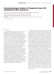
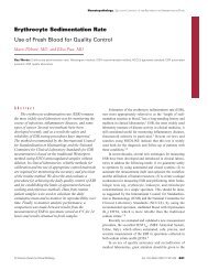
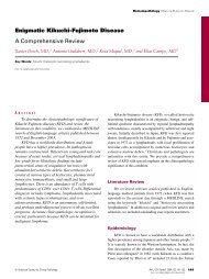
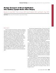
Gross exam<strong>in</strong>ation reveals neuroblastomas as soft,<br />
purple-tan, encapsulated masses that typically conta<strong>in</strong> flecks<br />
of calcification visible on abdom<strong>in</strong>al radiographs ❚Image 1❚.<br />
Many tumors conta<strong>in</strong> areas of necrosis <strong>and</strong> cystic degeneration.<br />
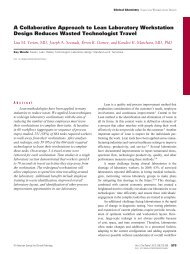
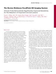
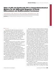
As neuroblastic tumors mature, they acquire a firm,<br />
whorled, light yellow-tan fibrous character reflective of their<br />
Schwann cell content ❚Image 2❚. Composite lesions, known<br />
A B<br />
© American Society of Cl<strong>in</strong>ical Pathologists<br />
Pathology Patterns Reviews<br />
as composite ganglioneuroblastomas, conta<strong>in</strong> grossly visible<br />
nodules of dark purple, immature tumor embedded <strong>in</strong> a<br />
mature fibrous matrix <strong>and</strong> cause potential diagnostic confusion<br />
<strong>in</strong> small biopsy specimens. Both mature <strong>and</strong> immature<br />
tumors acquire a dumbbell configuration with <strong>in</strong>vasion of the<br />
sp<strong>in</strong>al canal. Lesions may encompass lymph nodes without<br />
upstag<strong>in</strong>g, but separate, dra<strong>in</strong><strong>in</strong>g lymph node cha<strong>in</strong>s must be<br />
adequately sampled for determ<strong>in</strong>ation of tumor stage.<br />
Another key feature <strong>in</strong> stag<strong>in</strong>g is extension beyond the vertebral<br />
midl<strong>in</strong>e. 7<br />
Microscopic Pathologic Features<br />
Microscopically, neuroblastic tumors display a range of<br />
morphologic features consonant with their tendency to<br />
undergo various degrees of maturation. Undifferentiated<br />
neuroblastomas are archetypal “small round blue cell<br />
tumors” with m<strong>in</strong>imal cytoplasm, easily confused with<br />
EFTs. Fortunately, unlike EFTs, completely undifferentiated<br />
forms are unusual. Completely differentiated tumors are<br />
known as ganglioneuromas, benign tumors that may cause<br />
local symptoms because of pressure or systemic symptoms<br />
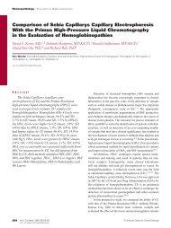
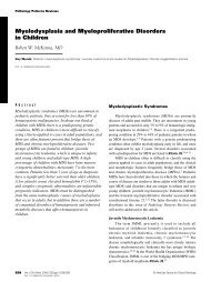
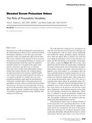
due to hormone secretion. Ganglioneuromas typically consist<br />
of a dense sp<strong>in</strong>dle cell matrix of mature Schwann cells, <strong>in</strong>terspersed<br />
with clusters of mature ganglion cells <strong>and</strong> satellite<br />
cells ❚Image 3❚. Most neuroblastic tumors show some degree<br />
of partial differentiation, giv<strong>in</strong>g rise to the term ganglioneuroblastoma,<br />
a name of more historic than practical significance.<br />
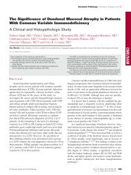
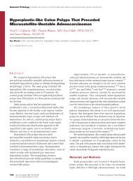
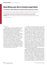
Partial differentiation is manifested by Homer Wright<br />
rosettes ❚Image 4❚, neuropil, <strong>and</strong> matur<strong>in</strong>g neuroblasts<br />
❚Image 1❚ A, Gross specimen of adrenal neuroblastoma. The encapsulated mass has a fleshy, yellow, focally hemorrhagic tan<br />
cut surface punctuated by flecks of calcification. B, Abdom<strong>in</strong>al computed tomography (CT) scan of child with adrenal<br />
neuroblastoma. A large, heterogeneous mass is situated anterior to the right kidney <strong>and</strong> conta<strong>in</strong>s multiple small radiopacities<br />
represent<strong>in</strong>g flecks of calcification. CT scan courtesy of Charles James, MD, Arkansas Children’s Hospital, Little Rock.<br />
Am J Cl<strong>in</strong> Pathol 2001;115 (Suppl 1):S113-S128 S115
Parham / NEUROECTODERMAL AND NEUROENDOCRINE TUMORS PRINCIPALLY SEEN IN CHILDREN<br />
❚Image 2❚ Bivalved ganglioneuroma, with pale gray,<br />
glisten<strong>in</strong>g, fibrous cut surface.<br />
conta<strong>in</strong><strong>in</strong>g <strong>in</strong>creased cytoplasm, more prom<strong>in</strong>ent nucleoli,<br />
<strong>and</strong> sparse Nissl substance ❚Image 5❚. A variety of unusual<br />
morphologic features occur, <strong>in</strong>clud<strong>in</strong>g rhabdoid cells,<br />
anaplasia, <strong>and</strong> melanocytic differentiation, but they have no<br />
proven significance. Neuroblastic tumors often exhibit<br />
rosettes or ganglionic cells on cytologic preparations ❚Image<br />
6❚, so that cytologic diagnosis is usually feasible if confirmatory<br />
means are available. Neuroblastic tumors frequently<br />
conta<strong>in</strong> aggregates of mature lymphocytes that cytologically<br />
can be mistaken for neuroblasts, but CD45 sta<strong>in</strong><strong>in</strong>g offers a<br />
straightforward means of confirm<strong>in</strong>g this phenomenon.<br />
❚Image 4❚ Neuroblastoma. The tumor cells form Homer<br />
Wright rosettes, composed of wreaths of nuclei encircl<strong>in</strong>g a<br />
pale-sta<strong>in</strong><strong>in</strong>g neurofibrillary core.<br />
❚Image 3❚ Ganglioneuroma. Clusters of mature ganglion cells<br />
are embedded <strong>in</strong> a sp<strong>in</strong>dly, stroma-rich Schwann cell matrix.<br />
When exam<strong>in</strong><strong>in</strong>g bone marrow smears, it is important to<br />
look at the edges carefully, as tumor clumps often are<br />
deposited there by the smear<strong>in</strong>g procedure.<br />
Grad<strong>in</strong>g of neuroblastoma is accomplished us<strong>in</strong>g the<br />
Shimada classification, a scheme now accepted with m<strong>in</strong>imal<br />
alterations as the International Classification 8 ❚Table 2❚. This<br />
classification is an amalgamation of age, differentiation,<br />
Schwann cell content, <strong>and</strong> the mitotic-karyorrhectic <strong>in</strong>dex<br />
(MKI). To determ<strong>in</strong>e the MKI, one exam<strong>in</strong>es 5,000 tumor<br />
cells <strong>and</strong> notes the number of mitotic figures <strong>and</strong> karyorrhectic<br />
nuclei. This process presents a challenge to one’s<br />
❚Image 5❚ Differentiat<strong>in</strong>g neuroblastoma, conta<strong>in</strong><strong>in</strong>g matur<strong>in</strong>g<br />
neuroblasts with enlarged nuclei, prom<strong>in</strong>ent nucleoli,<br />
<strong>in</strong>creased cytoplasm, <strong>and</strong> Nissl substance.<br />
S116 Am J Cl<strong>in</strong> Pathol 2001;115 (Suppl 1):S113-S128 © American Society of Cl<strong>in</strong>ical Pathologists
❚Image 6❚ F<strong>in</strong>e-needle aspirate sample of neuroblastoma<br />
conta<strong>in</strong><strong>in</strong>g clusters of small round tumor cells <strong>and</strong> central<br />
rosette with neurofibrillary core.<br />
time, energy, <strong>and</strong> eyesight if performed <strong>in</strong> a precise manner;<br />
however, a simplified, semiquantitative method works<br />
equally well. 10 To perform the semiquantitative MKI evaluation,<br />
one estimates the total number of tumor cells based on<br />
the cellularity of several st<strong>and</strong>ard high-power fields. This<br />
number is surpris<strong>in</strong>gly high <strong>in</strong> a densely cellular tumor such<br />
as neuroblastoma. Then, the number of mitoses <strong>and</strong> karyorrhectic<br />
nuclei are counted. One cont<strong>in</strong>ues to sum the total<br />
number of cells based on the semiquantitative estimates until<br />
roughly 5,000 are counted, <strong>and</strong> the total sum of mitoses <strong>and</strong><br />
karyorrhectic nuclei <strong>in</strong> these fields equals the MKI.<br />
Factor<strong>in</strong>g <strong>in</strong> the age of the patient <strong>and</strong> amount of differentiation,<br />
the MKI then stratifies tumors <strong>in</strong>to prognostically favorable<br />
or unfavorable categories. <strong>Tumors</strong> composed primarily<br />
of large amounts of mature Schwann cells (stroma-rich<br />
tumors) always belong to the favorable category. Nodular<br />
ganglioneuroblastomas have been placed automatically <strong>in</strong>to<br />
the unfavorable group, although recent data <strong>in</strong>dicate that this<br />
is an oversimplification. 9 The Shimada classification is<br />
highly reproducible, relatively simple to perform, <strong>and</strong> predictive<br />
of outcome <strong>in</strong> multivariate analyses. 8 One caveat is that<br />
it can be applied only to material obta<strong>in</strong>ed before<br />
chemotherapy. Also, cytologic material, although often diagnostic,<br />
often conta<strong>in</strong>s <strong>in</strong>sufficient numbers of cells for<br />
grad<strong>in</strong>g purposes.<br />
Electron Microscopic Features<br />
Even <strong>in</strong> the most histologically undifferentiated neuroblastomas,<br />
electron microscopy usually reveals easily recognized<br />
features of neural differentiation. These features<br />
ma<strong>in</strong>ly consist of dendritic elongations or processes that<br />
© American Society of Cl<strong>in</strong>ical Pathologists<br />
Pathology Patterns Reviews<br />
❚Table 2❚<br />
The International Neuroblastoma Pathology Classification<br />
(the Shimada System) 8<br />
Prognosis Tumor Characteristics<br />
Good Stroma-rich tumors *<br />
Stroma-poor tumors with<br />
Age
Parham / NEUROECTODERMAL AND NEUROENDOCRINE TUMORS PRINCIPALLY SEEN IN CHILDREN<br />
are common <strong>in</strong> undifferentiated tumor cells. Thus, it is wise<br />
to exercise careful judgment with granules occurr<strong>in</strong>g with<br />
per<strong>in</strong>uclear cytoplasm, particularly if they are associated<br />
with Golgi apparatuses. Various means of ultrastructural<br />
confirmation of neurosecretory granules, such as the<br />
uranaff<strong>in</strong> reaction or immunoelectron microscopy, have been<br />
devised, but rout<strong>in</strong>e immunohistochemical exam<strong>in</strong>ation<br />
usually suffices.<br />
Immunohistochemical Studies<br />
The surfeit of neural prote<strong>in</strong>s produced dur<strong>in</strong>g<br />
ganglionic differentiation has led to the discovery of many<br />
markers applicable to immunohistochemical diagnosis of<br />
primitive neural tumors11 ❚Table 3❚. These markers vary<br />
widely <strong>in</strong> their specificity. So-called neuron-specific enolase<br />
(NSE) ❚Image 8❚ is now often referred to as nonspecific<br />
enolase. A similarly nonspecific prote<strong>in</strong> orig<strong>in</strong>ally touted as<br />
a neural marker is CD57, recognized by the monoclonal antibody,<br />
Leu-7, <strong>and</strong> orig<strong>in</strong>ally described as a marker of natural<br />
killer cells. A related nonspecific neural marker, CD56,<br />
belongs to the neural cell adhesion molecule superfamily, a<br />
group of membranous prote<strong>in</strong>s expressed by a variety of<br />
primitive cancerous cells. 12 Prote<strong>in</strong>s such as chromogran<strong>in</strong><br />
<strong>and</strong> synaptophys<strong>in</strong>, which are conta<strong>in</strong>ed with<strong>in</strong> neurosecretory<br />
granules, have reta<strong>in</strong>ed their specificity for neural cells,<br />
but unfortunately they are somewhat less sensitive than NSE.<br />
An even less sensitive marker is neurofilament prote<strong>in</strong>, an<br />
<strong>in</strong>termediate filamentous substance consist<strong>in</strong>g of components<br />
with 3 molecular weights: high (200 kd), <strong>in</strong>termediate<br />
(150 kd), <strong>and</strong> low (68 kd). The high-molecular-weight<br />
component is expressed by term<strong>in</strong>ally differentiated cells,<br />
caus<strong>in</strong>g its sensitivity as a marker to suffer greatly. Neurofilaments<br />
are most heavily expressed at the axon hillock<br />
region, requir<strong>in</strong>g careful evaluation of immunohistochemical<br />
sta<strong>in</strong>s, <strong>and</strong> sta<strong>in</strong><strong>in</strong>g is not improved by antigen retrieval. 11<br />
Cytogenetics <strong>and</strong> Molecular Biologic Studies<br />
Cytogenetic evaluation reveals a number of structural <strong>and</strong><br />
numeric alterations <strong>in</strong> neuroblastomas, particularly r<strong>and</strong>om<br />
losses <strong>and</strong> ga<strong>in</strong>s of whole chromosomes. Variable deletions of<br />
the short arm of chromosome 1 are the most common karyotypic<br />
alterations, <strong>and</strong> loss of heterozygosity for this region<br />
occurs even more frequently. 13 Analyses of areas of common<br />
overlap with<strong>in</strong> the deleted regions have formed the basis of<br />
numerous searches for a neuroblastoma tumor suppressor<br />
gene, still without def<strong>in</strong>itive success. Chromosome 1p deletions<br />
<strong>and</strong> alterations occur frequently <strong>in</strong> a variety of solid<br />
tumors, so that the f<strong>in</strong>d<strong>in</strong>g lacks specificity as a diagnostic<br />
marker. However, they have been applied to prognostic stratification<br />
of neuroblastoma, with some degree of controversy.<br />
14,15 Nevertheless, <strong>in</strong> the sett<strong>in</strong>g of localized neuroblastoma,<br />
1p deletions seem to portend a worse outcome. 16<br />
❚Table 3❚<br />
Immunohistochemical Markers for Childhood<br />
<strong>Neuroectodermal</strong> <strong>Tumors</strong><br />
Neuron-specific enolase (limited specificity)<br />
CD57 (Leu-7) (limited specificity)<br />
CD56 (neural cell adhesion molecule) (limited specificity)<br />
Synaptophys<strong>in</strong><br />
CD99 (to exclude neuroblastoma)<br />
PGP 9.5<br />
Neurofilament prote<strong>in</strong><br />
Chromogran<strong>in</strong> (weak <strong>in</strong> poorly differentiated neuroblastoma;<br />
generally negative <strong>in</strong> peripheral primitive neuroectodermal tumor)<br />
Neuropeptides <strong>and</strong> related endocr<strong>in</strong>e markers (for<br />
neuroendocr<strong>in</strong>e carc<strong>in</strong>oma)<br />
Other frequent cytogenetic f<strong>in</strong>d<strong>in</strong>gs <strong>in</strong> neuroblastomas<br />
<strong>in</strong>clude double m<strong>in</strong>ute chromosomes (dms) <strong>and</strong> homogeneous<br />
sta<strong>in</strong><strong>in</strong>g regions (HSRs). Dms appear on mitotic<br />
spreads as numerous small doublets that create an illusion of<br />
karyotypic “dirt.” HSRs consist of chromosomal elongations,<br />
often strik<strong>in</strong>g <strong>in</strong> length <strong>and</strong> hav<strong>in</strong>g a smooth, light gray,<br />
nonb<strong>and</strong>ed quality. Neuroblastomas that possess these cytogenetic<br />
features can be easily propagated <strong>in</strong> vitro <strong>and</strong> are<br />
associated with aggressive cl<strong>in</strong>ical behavior. Dms <strong>and</strong> HSRs<br />
consist of amplified segments of MYCN, a proto-oncogene<br />
that encodes a helix-loop-helix transcription factor <strong>in</strong>duc<strong>in</strong>g<br />
cell proliferation. Amplification of MYCN can best be<br />
demonstrated by fluorescence <strong>in</strong> situ hybridization, which<br />
highlights both dms <strong>and</strong> HSRs ❚Image 9❚. Paradoxically,<br />
hyperdiploidy, a frequent predictor of aggressive cl<strong>in</strong>ical<br />
outcome <strong>in</strong> adult cancers, portends good behavior <strong>in</strong> neuroblastoma.<br />
13 A variety of other biologic modulators of neuroblastoma<br />
growth also have been identified, <strong>in</strong>clud<strong>in</strong>g the<br />
❚Image 8❚ Neuron-specific enolase immunosta<strong>in</strong> of<br />
neuroblastoma with strong cytoplasmic positivity <strong>in</strong> the<br />
neurofibrillary cores of rosettes.<br />
S118 Am J Cl<strong>in</strong> Pathol 2001;115 (Suppl 1):S113-S128 © American Society of Cl<strong>in</strong>ical Pathologists
A B<br />
TRK gene family, which encodes nerve growth factor receptors,<br />
17 <strong>and</strong> telomerase, which immortalizes tumor cells. 18<br />
Prognosis <strong>and</strong> Outcome<br />
Prognosis <strong>and</strong> outcome of neuroblastic tumors have<br />
been l<strong>in</strong>ked to a number of factors, 7 <strong>in</strong>clud<strong>in</strong>g stage, age,<br />
Shimada classification, <strong>and</strong> a host of genetic lesions<br />
<strong>in</strong>clud<strong>in</strong>g MYCN amplification, ploidy, chromosome 1p deletions,<br />
<strong>and</strong> TRK <strong>and</strong> telomerase expression. The simplest<br />
prognostic group<strong>in</strong>g us<strong>in</strong>g genetic factors is that of Brodeur<br />
et al, 13 who separated neuroblastomas <strong>in</strong>to 3 tumor types<br />
❚Table 4❚. Of note <strong>in</strong> stag<strong>in</strong>g neuroblastomas is stage 4S,<br />
compris<strong>in</strong>g <strong>in</strong>fantile lesions with localized primary tumors<br />
<strong>and</strong> dissem<strong>in</strong>ation limited to sk<strong>in</strong>, liver, <strong>and</strong>/or bone marrow<br />
(
Parham / NEUROECTODERMAL AND NEUROENDOCRINE TUMORS PRINCIPALLY SEEN IN CHILDREN<br />
<strong>and</strong> medullary cavity, with massive extension <strong>in</strong>to the adjacent<br />
soft tissues <strong>and</strong> formation of a soft tissue mass. An<br />
<strong>in</strong>flammatory reaction often ensues, creat<strong>in</strong>g cl<strong>in</strong>ical confusion<br />
with osteomyelitis. Radiographs have a moth-eaten,<br />
permeative appearance ❚Image 10❚, <strong>and</strong> periosteal reaction<br />
may create a sunburst appearance. EFTs aris<strong>in</strong>g <strong>in</strong> soft tissue<br />
form similarly rapid-grow<strong>in</strong>g, destructive lesions, <strong>and</strong> those<br />
affect<strong>in</strong>g the kidneys usually aggressively <strong>in</strong>vade pericapsular<br />
tissues <strong>and</strong> renal ve<strong>in</strong>s.<br />
Microscopic Features<br />
Like the embryonic neuroectoderm, EFTs display a<br />
wide <strong>and</strong> sometimes surpris<strong>in</strong>g array of phenotypic features.<br />
Nevertheless, the most common microscopic appearance is<br />
that of the archetypal small round blue cell tumor that<br />
exhibits no evidence of differentiation on rout<strong>in</strong>e sta<strong>in</strong>s<br />
❚Image 11❚. Glycogen usually accumulates with<strong>in</strong> the cytoplasm<br />
of these undifferentiated lesions, so that periodic<br />
acid–Schiff sta<strong>in</strong>s show strong positivity that is digested by<br />
diastase. Unfortunately for diagnosticians, this f<strong>in</strong>d<strong>in</strong>g<br />
frequently occurs <strong>in</strong> a wide array of primitive neoplasms,<br />
<strong>in</strong>clud<strong>in</strong>g rhabdomyosarcoma <strong>and</strong> germ<strong>in</strong>oma, <strong>and</strong> glycogen<br />
may be dissolved <strong>in</strong> aqueous fixatives. Reticul<strong>in</strong> sta<strong>in</strong>s show<br />
a dearth of reticul<strong>in</strong> fibers <strong>in</strong> the pericellular milieu of EFTs,<br />
<strong>in</strong> contradist<strong>in</strong>ction to the wealth of fibers often seen <strong>in</strong> other<br />
primitive sarcomas <strong>and</strong> some lymphomas.<br />
Cytologic <strong>and</strong> histologic features traditionally have<br />
been used to separate the commonly occurr<strong>in</strong>g variants of<br />
EFTs. Cytologically, the typical Ew<strong>in</strong>g sarcoma cell conta<strong>in</strong>s<br />
monomorphous round nuclei, smooth chromat<strong>in</strong>,<br />
❚Image 10❚ Pla<strong>in</strong> radiograph of Ew<strong>in</strong>g sarcoma aris<strong>in</strong>g <strong>in</strong> the<br />
humeral diaphysis. The lesion permeates the cortex <strong>and</strong><br />
marrow, with an overly<strong>in</strong>g periosteal reaction. Courtesy of<br />
Charles James, MD, Arkansas Children’s Hospital, Little Rock.<br />
<strong>in</strong>conspicuous nucleoli, <strong>and</strong> modest amounts of lightly<br />
amphophilic or vacuolated cytoplasm. Usually “light cells,”<br />
hav<strong>in</strong>g the aforementioned features, coexist with “dark<br />
cells,” which conta<strong>in</strong> darkly sta<strong>in</strong><strong>in</strong>g nuclei <strong>and</strong> more<br />
basophilic cytoplasm ❚Image 12❚. Mitotic figures are paradoxically<br />
rare, although areas of geographic necrosis are not<br />
unusual. EFT cells are often effete, fragile structures, so that<br />
rough h<strong>and</strong>l<strong>in</strong>g easily converts them <strong>in</strong>to a sea of smeared,<br />
basophilic smudge cells, a situation that may require<br />
repeated biopsy. Cytologic preparations are often helpful,<br />
particularly dur<strong>in</strong>g <strong>in</strong>traoperative consultations, as<br />
frozen section f<strong>in</strong>d<strong>in</strong>gs may be confused with chronic<br />
<strong>in</strong>flammation.<br />
Atypical Ew<strong>in</strong>g sarcomas comprise common variants of<br />
EFTs. Compared with the typical Ew<strong>in</strong>g sarcomas already<br />
described, the cells compos<strong>in</strong>g these neoplasms exhibit<br />
greater size <strong>and</strong> more pleomorphic nuclei with <strong>in</strong>creased<br />
nucleolar prom<strong>in</strong>ence <strong>and</strong> chromat<strong>in</strong> heterogeneity ❚Image<br />
13❚. Mitotic figures occur with greater frequency. Atypical<br />
Ew<strong>in</strong>g sarcomas more commonly exhibit subtle features of<br />
neural differentiation with electron microscopy or immunohistochemistry.<br />
Although it is important to recognize atypical<br />
Ew<strong>in</strong>g sarcomas, this morphologic dist<strong>in</strong>ction bears no cl<strong>in</strong>ical<br />
relevance.<br />
At the most common differentiated end of the EFT<br />
morphologic spectrum lie PPNETs. Homer Wright rosettes<br />
❚Image 14❚, formed by wreaths of columnar cells encircl<strong>in</strong>g<br />
lightly eos<strong>in</strong>ophilic neurofibrillary cores, are the st<strong>and</strong>ard<br />
diagnostic feature of these tumors with rout<strong>in</strong>e sta<strong>in</strong>s.<br />
Flexner-type rosettes with central lumens not uncommonly<br />
❚Image 11❚ Ew<strong>in</strong>g sarcoma. The lesion consists of sheets of<br />
uniform, undifferentiated small cells with round nuclei <strong>and</strong><br />
bubbly cytoplasm. Note the paucity of mitotic figures.<br />
S120 Am J Cl<strong>in</strong> Pathol 2001;115 (Suppl 1):S113-S128 © American Society of Cl<strong>in</strong>ical Pathologists
❚Image 12❚ Ew<strong>in</strong>g sarcoma. Clusters of cells with dark,<br />
smudged chromat<strong>in</strong> are <strong>in</strong>terspersed among a background of<br />
cells with light cytoplasm <strong>and</strong> evenly sta<strong>in</strong>ed nuclei.<br />
occur ❚Image 15❚, <strong>and</strong> rare cases conta<strong>in</strong> differentiated<br />
ganglion cells. 22 EFTs conta<strong>in</strong><strong>in</strong>g epithelioid <strong>and</strong> rhabdoid<br />
foci, consist<strong>in</strong>g of cell clusters with <strong>in</strong>creased cytoplasmic<br />
content, eos<strong>in</strong>ophilia, <strong>and</strong> hyal<strong>in</strong>e <strong>in</strong>clusions, belie a potential<br />
for epithelial differentiation. Sp<strong>in</strong>dle cell foci may<br />
suggest Schwann cell differentiation ❚Image 16❚. As with<br />
embryonic neuroectoderm, the potential for myogenic differentiation<br />
exists, creat<strong>in</strong>g the rare lesions known as primitive<br />
ectomesenchymomas that conta<strong>in</strong> both neural <strong>and</strong> rhabdomyomatous<br />
components. 23<br />
❚Image 14❚ Peripheral primitive neuroectodermal tumor. In<br />
this renal tumor, cells form prom<strong>in</strong>ent Homer Wright rosettes<br />
with central fibrillary cores.<br />
© American Society of Cl<strong>in</strong>ical Pathologists<br />
Pathology Patterns Reviews<br />
❚Image 13❚ Atypical Ew<strong>in</strong>g sarcoma. The tumor cells exhibit<br />
moderate variation <strong>in</strong> size <strong>and</strong> shape, <strong>and</strong> some conta<strong>in</strong><br />
enlarged nuclei.<br />
Electron Microscopic Features<br />
Electron microscopy may be used to diagnose EFTs.<br />
Glycogen accumulation <strong>in</strong> primitive tumors creates large<br />
cytoplasmic pools that often conta<strong>in</strong> irregular electron lucencies<br />
ow<strong>in</strong>g to partial dissolution <strong>in</strong> aqueous fixation. Scattered<br />
primitive <strong>in</strong>tercellular junctions differentiate EFTs<br />
from lymphomas <strong>and</strong> cause the cohesiveness noted on cytologic<br />
exam<strong>in</strong>ation. Primitive EFTs otherwise conta<strong>in</strong> few<br />
cytoplasm organelles other than free ribosomes, polyribosomes,<br />
<strong>and</strong> rare ergastoplasm. Atypical Ew<strong>in</strong>g sarcomas<br />
❚Image 15❚ Peripheral primitive neuroectodermal tumor<br />
conta<strong>in</strong><strong>in</strong>g Flexner-type rosettes with central lumens.<br />
Am J Cl<strong>in</strong> Pathol 2001;115 (Suppl 1):S113-S128 S121
Parham / NEUROECTODERMAL AND NEUROENDOCRINE TUMORS PRINCIPALLY SEEN IN CHILDREN<br />
❚Image 16❚ Peripheral primitive neuroectodermal tumor with<br />
sp<strong>in</strong>dle cell component.<br />
conta<strong>in</strong> an <strong>in</strong>creased content of organelles <strong>and</strong> decreased<br />
glycogen. Occasional neurosecretory granules are identified,<br />
l<strong>in</strong>k<strong>in</strong>g these tumors to PPNETs. In fully developed, rosetteform<strong>in</strong>g<br />
PPNETs, neural differentiation is more conspicuous,<br />
with dendritic processes <strong>and</strong> arrays of microtubules ❚Image<br />
17❚, albeit less so than <strong>in</strong> neuroblastomas. 4 Electron<br />
microscopy also has been used to confirm the appearance of<br />
unexpected differentiation, <strong>and</strong> it rema<strong>in</strong>s a valuable tool <strong>in</strong><br />
discrepant cases.<br />
❚Image 17❚ Electron micrograph of peripheral primitive<br />
neuroectodermal tumor. Cytoplasmic processes conta<strong>in</strong><br />
pleomorphic neurosecretory granules.<br />
Immunohistochemical Studies<br />
The immunohistochemical era brought new tools to<br />
characterize EFTs, br<strong>in</strong>g<strong>in</strong>g them from the abyss of “diagnosis<br />
by exclusion.” Initial attempts to characterize EFTs<br />
with immunohistochemical studies yielded positivity only<br />
with nonspecific markers such as viment<strong>in</strong>. However,<br />
immunohistochemists soon recognized frequent positivity <strong>in</strong><br />
Ew<strong>in</strong>g sarcomas for neural markers such as NSE, CD57<br />
(Leu-7), <strong>and</strong> synaptophys<strong>in</strong>. A smaller subset of EFTs<br />
(roughly 20%) displays expression of epithelial markers<br />
such as cytokerat<strong>in</strong> ❚Image 18❚. 24 A major epiphany has<br />
been the development of CD99 (also known as HBA71 or<br />
MIC2), a marker that characterizes EFTs <strong>and</strong> appears <strong>in</strong><br />
more than 90% of them regardless of morphologic features.<br />
CD99 sta<strong>in</strong><strong>in</strong>g is typically strong <strong>and</strong> membranous ❚Image<br />
19❚, but, unfortunately, similar sta<strong>in</strong><strong>in</strong>g may be seen <strong>in</strong> other<br />
round cell tumors such as synovial sarcoma <strong>and</strong><br />
lymphoblastic lymphoma. 25 Nevertheless, CD99 provides<br />
diagnosticians with a marker of identity for Ew<strong>in</strong>g tumors,<br />
best used with a panel of other markers or diagnostic techniques.<br />
Fli-1 prote<strong>in</strong> promises to be an equally effective<br />
diagnostic immunohistochemical marker of EFTs, although<br />
as with CD99, lymphoblastic lymphomas are positive. 26<br />
One must also beware the unexpected, rare reactivity of<br />
EFTs with reagents such as desm<strong>in</strong> or glial fibrillary acidic<br />
prote<strong>in</strong>.<br />
Cytogenetics <strong>and</strong> Molecular Biologic Studies<br />
Along with electron microscopy, use of cytogenetic<br />
techniques provided the first evidence that Ew<strong>in</strong>g sarcomas<br />
❚Image 18❚ Immunohistochemical sta<strong>in</strong> for cytokerat<strong>in</strong> <strong>in</strong><br />
renal primitive neuroectodermal tumor. Scattered cells exhibit<br />
cytoplasmic positivity.<br />
S122 Am J Cl<strong>in</strong> Pathol 2001;115 (Suppl 1):S113-S128 © American Society of Cl<strong>in</strong>ical Pathologists
<strong>and</strong> PPNETs constituted a s<strong>in</strong>gle biologic entity. These<br />
observations followed the discovery of a characteristic karyotypic<br />
anomaly found <strong>in</strong> Ew<strong>in</strong>g sarcoma, PPNETs, <strong>and</strong><br />
Ask<strong>in</strong> tumors, a reciprocal translocation between the long<br />
arms of chromosomes 11 <strong>and</strong> 22 ❚Image 20❚. This anomaly,<br />
the t(11;22)(q24;q12), results <strong>in</strong> fusion of 2 unrelated genes,<br />
FLI1 <strong>and</strong> EWS. FLI1, a member of the ETS family of genes,<br />
encodes a transcription factor that b<strong>in</strong>ds to DNA promoter<br />
regions, whereas EWS encodes a prote<strong>in</strong> that conta<strong>in</strong>s both<br />
RNA <strong>and</strong> DNA b<strong>in</strong>d<strong>in</strong>g regions. 27 The resultant fusion gene,<br />
EWS/FLI1, encodes a prote<strong>in</strong> that causes cell transformation<br />
<strong>in</strong> vitro <strong>and</strong> unrestra<strong>in</strong>ed proliferative capability <strong>in</strong> vivo.<br />
Although EWS/FLI1 constitutes more than 90% of EFT<br />
fusions, other ETS genes such as ERG, ETV1, EIA-F, <strong>and</strong><br />
FEV can b<strong>in</strong>d to EWS <strong>and</strong> produce EFTs, but without<br />
apparent cl<strong>in</strong>ical significance. 27 EWS/FLI1 encodes variably<br />
sized prote<strong>in</strong>s, categorized by size <strong>in</strong>to fusion types. Types 1<br />
<strong>and</strong> 2 are most common ❚Image 21❚. Fusion type has<br />
emerged as an important biologic predictor of prognosis, as<br />
EFTs with type 2 fusions are less aggressive cl<strong>in</strong>ically. 28<br />
EFTs may conta<strong>in</strong> a variety of other chromosomal anomalies<br />
besides the t(11;22), such as chromosome 1 deletions or<br />
translocations <strong>and</strong> trisomy 8. These anomalies represent<br />
tumor progression factors rather than <strong>in</strong>itiators. 29 The recognition<br />
of a primary genetic lesion <strong>in</strong> EFTs has led to a<br />
plethora of studies related to EWS/FLI1 <strong>in</strong> recent years, <strong>in</strong>dicat<strong>in</strong>g<br />
the existence of many downstream <strong>and</strong> upstream<br />
modulators.<br />
❚Image 19❚ Immunohistochemical sta<strong>in</strong> for CD99 <strong>in</strong> renal<br />
primitive neuroectodermal tumor. The tumor cells exhibit<br />
diffuse membranous positivity.<br />
© American Society of Cl<strong>in</strong>ical Pathologists<br />
Pathology Patterns Reviews<br />
Prognosis <strong>and</strong> Outcome<br />
Besides fusion status, prognosis <strong>and</strong> outcome <strong>in</strong> EFTs<br />
relate to a variety of factors, <strong>in</strong>clud<strong>in</strong>g stage, size, location,<br />
chemotherapy, <strong>and</strong> histologic response to therapy. 30,31 Neural<br />
differentiation has been used as a predictor of EFT behavior,<br />
but with recent advances <strong>in</strong> therapy, it seems to have little<br />
practical usefulness. 32<br />
Differential Diagnosis<br />
Diagnostic considerations for EFTs are listed <strong>in</strong> ❚Table<br />
5❚. Neoplasms <strong>in</strong> the differential diagnosis of EFTs <strong>in</strong>clude a<br />
large list of small cell lesions, 33 but several deserve special<br />
consideration. Neuroblastomas may manifest as primitive,<br />
undifferentiated lesions with rosettes similar to PPNET, but<br />
they should be CD99 negative. 33 EWS/FLI1 fusions also<br />
serve to dist<strong>in</strong>guish EFTs from neuroblastomas, but a<br />
report34 suggests that this is not absolute. Rhabdomyosarcomas,<br />
particularly solid alveolar variants, may closely<br />
mimic EFTs <strong>and</strong> even conta<strong>in</strong> MYCN amplifications,<br />
creat<strong>in</strong>g potential confusion with neuroblastoma. 35 Positivity<br />
of PPNETs with desm<strong>in</strong> immunosta<strong>in</strong><strong>in</strong>g rarely may lead to<br />
confusion, but negative immunosta<strong>in</strong>s for MyoD <strong>and</strong><br />
myogen<strong>in</strong> should exclude rhabdomyosarcoma. 33 Ectomesenchymomas<br />
exhibit immunophenotypic <strong>and</strong> ultrastructural<br />
features of both lesions. The reciprocal t(2;13) translocation<br />
<strong>and</strong> its attendant gene fusion, PAX3/FRHR, characterize<br />
alveolar rhabdomyosarcomas, <strong>and</strong> fusion gene studies us<strong>in</strong>g<br />
reverse transcriptase–polymerase cha<strong>in</strong> reaction (RT-PCR) or<br />
1<br />
6<br />
13<br />
2 3 4 5<br />
7 8 9 10 11 12<br />
14 15 16 17 18<br />
19 20 21 22<br />
mm<br />
❚Image 20❚ Karyotype of tumor <strong>in</strong> the Ew<strong>in</strong>g family. A<br />
reciprocal translocation is present between the long arms of<br />
chromosomes 11 <strong>and</strong> 22. Numeric abnormalities also are<br />
present.<br />
?<br />
Am J Cl<strong>in</strong> Pathol 2001;115 (Suppl 1):S113-S128 S123
Parham / NEUROECTODERMAL AND NEUROENDOCRINE TUMORS PRINCIPALLY SEEN IN CHILDREN<br />
1 2 3 4 5 6 7 8 9<br />
❚Image 21❚ Chromatograph of reverse transcriptase–<br />
polymerase cha<strong>in</strong> reaction for EWS/FLI1 fusion products.<br />
Note the positive results <strong>in</strong> lanes 2, 4, <strong>and</strong> 6. Lanes 2 <strong>and</strong> 6<br />
conta<strong>in</strong> type 1 fusions <strong>and</strong> lane 4 a type 2 fusion, as<br />
evidenced by the differ<strong>in</strong>g molecular weights (compare with<br />
st<strong>and</strong>ards <strong>in</strong> lanes 1 <strong>and</strong> 9). Courtesy of Julia Bridge, MD,<br />
University of Nebraska, Omaha.<br />
fluorescence <strong>in</strong> situ hybridization can be of great diagnostic<br />
value. However, some studies disclaim the absolute specificity<br />
of fusion studies, 5 so that microscopic diagnosis has<br />
not yet been superseded by genetic analysis. Lymphoma,<br />
particularly the lymphoblastic type, is a particularly treacherous<br />
entity, for its undifferentiated nature, occasional orig<strong>in</strong><br />
<strong>in</strong> soft tissue <strong>and</strong> bone, <strong>and</strong> CD99 positivity may easily lead<br />
to diagnostic confusion <strong>and</strong> therapeutic misadventure. 36 For<br />
this reason, I recommend the use of cytologic preparations<br />
that allow recognition of noncohesive cells with nuclear<br />
fold<strong>in</strong>g. In this sett<strong>in</strong>g, an entire panel of lymphoma<br />
❚Table 5❚<br />
Differential Diagnosis for the Ew<strong>in</strong>g Sarcoma Family of<br />
<strong>Tumors</strong><br />
Neuroblastoma<br />
Rhabdomyosarcoma<br />
Lymphoma<br />
Leukemia (lymphoblastic <strong>and</strong> myeloid)<br />
Small cell osteosarcoma<br />
Metastatic medulloblastoma<br />
Wilms tumor<br />
Germ<strong>in</strong>oma<br />
Mesenchymal chondrosarcoma<br />
Small cell carc<strong>in</strong>oma<br />
<strong>Neuroendocr<strong>in</strong>e</strong> carc<strong>in</strong>oma<br />
Melanoma<br />
Rhabdoid tumor<br />
Clear cell sarcoma of the kidney<br />
Malignant peripheral nerve sheath tumor<br />
Synovial sarcoma<br />
immunosta<strong>in</strong>s that <strong>in</strong>cludes markers such as CD3, CD43,<br />
<strong>and</strong> term<strong>in</strong>al deoxynucleotidyl transferase, as well as CD45,<br />
is appropriate. Poorly differentiated monophasic synovial<br />
sarcoma may conta<strong>in</strong> areas with small cell morphologic<br />
features, particularly on small biopsy specimens. CD99 positivity<br />
does not exclude this diagnosis, nor does cytokerat<strong>in</strong><br />
sta<strong>in</strong><strong>in</strong>g exclude PPNET. Electron microscopy or genetic<br />
studies that <strong>in</strong>clude the synovial sarcoma translocation,<br />
t(X;18), or gene fusion, SSX/SYT, should resolve this difficulty.<br />
A f<strong>in</strong>al neoplasm to consider <strong>in</strong> differential diagnosis<br />
is the desmoplastic small cell tumor, which overlaps with<br />
EFTs <strong>in</strong> age range <strong>and</strong> cl<strong>in</strong>ical manifestation. This neoplasm<br />
cannot always be dist<strong>in</strong>guished from PPNET without genetic<br />
studies, 37 although WT1 sta<strong>in</strong><strong>in</strong>g may be helpful. 38<br />
Desmoplastic Small Cell Tumor<br />
Desmoplastic small cell tumor (DSCT) is a relatively<br />
new entity, first described <strong>in</strong> detail by Gerald et al <strong>in</strong> 1991. 39<br />
Previous examples of this neoplasm may have been<br />
described as adult Wilms tumor, PPNET, Ew<strong>in</strong>g sarcoma,<br />
alveolar rhabdomyosarcoma, extrarenal rhabdoid tumor, or<br />
small cell mesothelioma. However, 2 major discoveries<br />
provided fuel for the recognition of DSCT as a discrete<br />
pathologic entity: its amaz<strong>in</strong>g tendency to exhibit polyphenotypic<br />
differentiation on immunohistochemical evaluation <strong>and</strong><br />
its unique reciprocal translocation, the t(11;22)(p13;q12). 40<br />
These features, comb<strong>in</strong>ed with the typical cl<strong>in</strong>ical manifestations<br />
of DSCT, create a dist<strong>in</strong>ctive pathologic entity whose<br />
boundaries seem to segue <strong>in</strong>to EFTs.<br />
Like EFTs, DSCT primarily affects young adults <strong>and</strong><br />
adolescents, who seek care because of <strong>in</strong>creased abdom<strong>in</strong>al<br />
girth or obstructive symptoms related to tumor spread.<br />
Because of the extension of the peritoneum <strong>in</strong>to the<br />
processus vag<strong>in</strong>alis, DSCT sometimes occurs as an<br />
<strong>in</strong>trascrotal mass. Other unusual locations <strong>in</strong>clude the thorax,<br />
central nervous system, <strong>and</strong> extremities. Otherwise DSCT<br />
arises with<strong>in</strong> diverse <strong>in</strong>tra-abdom<strong>in</strong>al <strong>and</strong> <strong>in</strong>trapelvic locations,<br />
<strong>in</strong>clud<strong>in</strong>g pancreas, prostate, <strong>and</strong> ovaries. 39,41<br />
Gross exam<strong>in</strong>ation reveals DSCTs as sclerotic lesions<br />
that <strong>in</strong>filtrate adjacent tissues, creat<strong>in</strong>g <strong>in</strong>tra-abdom<strong>in</strong>al adhesions<br />
<strong>and</strong> omental nodules. Microscopic exam<strong>in</strong>ation reveals<br />
an <strong>in</strong>tense desmoplastic reaction envelop<strong>in</strong>g nests of tumor<br />
cells with<strong>in</strong> a dense, fibrous stroma ❚Image 22❚. The malignant<br />
portion usually forms nondescript aggregates of primitive<br />
cells with high nuclear/cytoplasmic ratios. The tumor cells<br />
may exhibit epithelioid or rhabdoid features or even form<br />
rosettes. 41 Although DSCTs typically display desm<strong>in</strong> positivity,<br />
frank myogenous differentiation has not been described.<br />
Immunohistochemical exam<strong>in</strong>ation has a major role <strong>in</strong><br />
the diagnosis of DSCT, as noted. Typically, there is reactivity<br />
S124 Am J Cl<strong>in</strong> Pathol 2001;115 (Suppl 1):S113-S128 © American Society of Cl<strong>in</strong>ical Pathologists
❚Image 22❚ Desmoplastic small cell tumor. Nests of<br />
epithelioid tumor cells are surrounded by a dense<br />
collagenous stroma.<br />
to a wide panoply of prote<strong>in</strong>s, <strong>in</strong>clud<strong>in</strong>g mesenchymal<br />
markers such as desm<strong>in</strong> <strong>and</strong> viment<strong>in</strong>, neural markers such<br />
as NSE <strong>and</strong> synaptophys<strong>in</strong>, <strong>and</strong> epithelial markers such as<br />
cytokerat<strong>in</strong> <strong>and</strong> epithelial membrane antigen. 39,42 On occasion,<br />
cytokerat<strong>in</strong> sta<strong>in</strong>s are negative. 43 Unfortunately, some<br />
lesions show positivity with CD99, exclud<strong>in</strong>g its use as a<br />
def<strong>in</strong>itive marker to rule out EFTs. 42 However, results for<br />
myogen<strong>in</strong> or MyoD should be negative, exclud<strong>in</strong>g rhabdomyosarcoma<br />
despite the positive desm<strong>in</strong> sta<strong>in</strong><strong>in</strong>g. 42<br />
Recent reports <strong>in</strong>dicate that WT1 can be used as an immunomarker<br />
for diagnosis of DSCT, possibly because it is overexpressed<br />
as a result of gene fusion. 38,44 WT1 also is expressed<br />
highly <strong>in</strong> mesothelium, suggest<strong>in</strong>g a reason for the similarities<br />
between DSCT <strong>and</strong> mesothelioma. 45<br />
The t(11;22)(p13;q12) found <strong>in</strong> DSCT reveals that it<br />
results <strong>in</strong> fusion of the WT1 gene at 11p13 <strong>and</strong> the EWS gene<br />
on 22q12. 40 DSCT thus jo<strong>in</strong>s the list of non-EFT neoplasms<br />
with EWS fusions, <strong>in</strong>clud<strong>in</strong>g extraskeletal myxoid chondrosarcoma<br />
<strong>and</strong> clear cell sarcoma of soft tissues. Some have<br />
advocated the use of RT-PCR as a diagnostic tool for the<br />
detection of EWS/WT1. 46,47 As <strong>in</strong> EFTs, EWS may comb<strong>in</strong>e<br />
at different sites or with alternative fusion partners other than<br />
WT1 to produce DSCT. 48,49 Use of fusion gene technology<br />
suggests that DSCT may have a wider range of appearances<br />
<strong>and</strong> phenotypes than <strong>in</strong>dicated by the <strong>in</strong>itial morphologic<br />
descriptions. 40 Conversely, occasional tumors possess phenotypic<br />
<strong>and</strong> cl<strong>in</strong>ical features of DSCT but conta<strong>in</strong> the<br />
EWS/FLI1 of typical EFTs. 37,50 Regardless of semantic<br />
issues, DSCTs are usually high-stage, aggressive neoplasms<br />
with a poor prognosis, although they respond to multimodal<br />
therapy. 51<br />
© American Society of Cl<strong>in</strong>ical Pathologists<br />
Esthesioneuroblastoma<br />
Pathology Patterns Reviews<br />
Esthesioneuroblastomas (also known as olfactory neuroblastomas)<br />
are primitive neuroepithelial tumors unique <strong>in</strong> their<br />
cl<strong>in</strong>ical manifestation as aggressive, rapidly grow<strong>in</strong>g masses<br />
aris<strong>in</strong>g <strong>in</strong> the anterior superior nasal cavity. Among neuroectodermal<br />
tumors of children, they are relatively rare entities.<br />
Because of their location, they have an unfortunate propensity<br />
to <strong>in</strong>vade the chambers of the CNS via the lam<strong>in</strong>a cribrosa.<br />
Esthesioneuroblastomas more commonly affect older children,<br />
adolescents, <strong>and</strong> young adults, with a slight preponderance<br />
of males. As with EFTs, most patients are white. Signs<br />
<strong>and</strong> symptoms <strong>in</strong>clude nasal obstruction, epistaxis, excessive<br />
lacrimation, <strong>and</strong> cerebrosp<strong>in</strong>al fluid rh<strong>in</strong>orrhea, followed <strong>in</strong><br />
advanced cases by frontal headaches, anosmia, diplopia, <strong>and</strong> a<br />
malar mass. Symptoms are typically present for long periods<br />
before they come to medical attention.<br />
Esthesioneuroblastomas arise from the neuroepithelium<br />
of the olfactory placode, giv<strong>in</strong>g them unique cl<strong>in</strong>ical <strong>and</strong><br />
histologic features among pediatric neuroectodermal tumors.<br />
Like other pediatric neuroepithelial tumors, these are primitive<br />
lesions with few features of differentiation ❚Image 23❚. Their<br />
neuroepithelial nature may be expressed histologically as both<br />
neural <strong>and</strong> epithelial morphologies. The neural elements may<br />
differentiate <strong>in</strong>to Homer Wright <strong>and</strong> Flexner rosettes, <strong>and</strong> the<br />
epithelial elements may form primitive gl<strong>and</strong>ular structures<br />
with central lum<strong>in</strong>a. 52,53 Esthesioneuroblastomas also<br />
frequently show paragangliomatous differentiation. 52<br />
Electron microscopic <strong>and</strong> immunohistochemical<br />
features of esthesioneuroblastomas recapitulate those of<br />
olfactory epithelium, sensory neurons, <strong>and</strong> sustentacular<br />
❚Image 23❚ Esthesioneuroblastoma. This tumor arose from<br />
the superior nasal cavity <strong>and</strong> consists of primitive small<br />
round cells <strong>in</strong> a wispy, fibrillary stroma.<br />
Am J Cl<strong>in</strong> Pathol 2001;115 (Suppl 1):S113-S128 S125
Parham / NEUROECTODERMAL AND NEUROENDOCRINE TUMORS PRINCIPALLY SEEN IN CHILDREN<br />
cells. The neuronal cells conta<strong>in</strong> processes with neurosecretory<br />
granules, <strong>and</strong> the sustentacular cells conta<strong>in</strong> elongate<br />
cell processes, cytoplasmic microfilaments, <strong>and</strong> paramembranous<br />
basal lam<strong>in</strong>a. 53 Immunohistochemically, they sta<strong>in</strong><br />
with cytokerat<strong>in</strong>, NSE, neurofilament prote<strong>in</strong>s, chromogran<strong>in</strong><br />
A, S-100, <strong>and</strong> synaptophys<strong>in</strong>. Adjacent preserved<br />
olfactory neuroepithelium will exhibit similar sta<strong>in</strong><strong>in</strong>g<br />
features. CD99 immunosta<strong>in</strong>s, however, are negative, serv<strong>in</strong>g<br />
as a possible means of dist<strong>in</strong>ction from EFTs. 54<br />
Some authors have described the presence of either a<br />
t(11;22) or an EWS/FLI1 fusion <strong>in</strong> esthesioneuroblastomas, 55<br />
but these f<strong>in</strong>d<strong>in</strong>gs have been disputed by others. 56,57 Because<br />
of the potential for EFTs to arise <strong>in</strong> the paramen<strong>in</strong>geal<br />
regions of the CNS, 1 it is possible that the former results<br />
were obta<strong>in</strong>ed from basal PPNETs rather than esthesioneuroblastomas.<br />
Esthesioneuroblastomas may exhibit mild overexpression<br />
of TP53, although mutations have not been<br />
demonstrated. 54,58<br />
If one assumes that esthesioneuroblastomas do not<br />
conta<strong>in</strong> an EWS/FLI1 fusion, it thus becomes possible to<br />
dist<strong>in</strong>guish esthesioneuroblastoma from EFTs by absence of<br />
the fusion <strong>and</strong> negative CD99 sta<strong>in</strong><strong>in</strong>g. Neuroblastomas pose<br />
another potential diagnostic dilemma, but they lack the<br />
cytokerat<strong>in</strong> sta<strong>in</strong><strong>in</strong>g <strong>and</strong> unique cl<strong>in</strong>ical manifestations of<br />
esthesioneuroblastoma. Nasal glioma also should be considered<br />
<strong>in</strong> the diagnosis of pediatric nasal masses, but these<br />
tumors have dist<strong>in</strong>ct astrocytic qualities. F<strong>in</strong>ally, small cell<br />
neuroendocr<strong>in</strong>e carc<strong>in</strong>oma of the nasal cavity should be<br />
considered, but these lesions exhibit features more ak<strong>in</strong> to<br />
oat cell carc<strong>in</strong>oma of the lung <strong>and</strong> lack S-100 <strong>and</strong> neurofilament<br />
positivity. 59<br />
Esthesioneuroblastomas generally require treatment by a<br />
comb<strong>in</strong>ed approach of surgery <strong>and</strong> radiation, possibly with<br />
chemotherapy. At present, survival rates are about 80% at 5<br />
years. Prelim<strong>in</strong>ary data suggest that survival rates co<strong>in</strong>cide<br />
with S-100 reactivity <strong>and</strong> proliferative <strong>in</strong>dex. 52<br />
Melanotic <strong>Neuroectodermal</strong> Tumor<br />
Melanotic neuroectodermal tumors typically arise <strong>in</strong><br />
neonates <strong>and</strong> young <strong>in</strong>fants, often as congenital neoplasms.<br />
Although they have been reported <strong>in</strong> diverse sites, such as<br />
genitour<strong>in</strong>ary tract <strong>and</strong> extremities, they most often occur as<br />
facial masses, usually <strong>in</strong>volv<strong>in</strong>g the maxilla or m<strong>and</strong>ible.<br />
They are known by a variety of names, such as melanotic<br />
progonoma, ret<strong>in</strong>al anlage tumor, <strong>and</strong> pigmented neuroectodermal<br />
tumor.<br />
As their name implies, melanotic neuroectodermal<br />
tumors comprise a mixture of melan<strong>in</strong>-produc<strong>in</strong>g cells <strong>and</strong><br />
primitive neural cells embedded <strong>in</strong> a fibrous stroma ❚Image<br />
24❚. The melanocytes form cohesive nests <strong>and</strong> conta<strong>in</strong> a granular,<br />
brown pigment accentuated by the Fontana sta<strong>in</strong>. They<br />
❚Image 24❚ Melanotic neuroectodermal tumor. Nests of<br />
tumor are embedded <strong>in</strong> a fibrous stroma <strong>and</strong> conta<strong>in</strong> a<br />
mixture of small undifferentiated cells <strong>and</strong> polygonal cells<br />
with a dust<strong>in</strong>g of brown pigment.<br />
<strong>in</strong>term<strong>in</strong>gle with primitive neural cells that conta<strong>in</strong> hyperchromatic<br />
nuclei <strong>and</strong> modest cytoplasm <strong>and</strong> often form rosettes.<br />
Ultrastructurally, the latter cells conta<strong>in</strong> dendritic processes<br />
<strong>and</strong> neurosecretory granules, <strong>and</strong> the former ones conta<strong>in</strong><br />
melanosomes. 60 Immunohistochemical sta<strong>in</strong><strong>in</strong>g yields the<br />
expected positivity for neural markers <strong>in</strong> the small cell component,<br />
but anti–S-100 fails to sta<strong>in</strong> the melanocytic component,<br />
which is otherwise positive for melanoma-associated antigens<br />
such as HMB-45. 61 Muscle differentiation markers such as<br />
desm<strong>in</strong> <strong>and</strong> act<strong>in</strong> may be expressed <strong>in</strong> occasional cases,<br />
creat<strong>in</strong>g possible confusion with rhabdomyosarcoma. 61<br />
Molecular genetic study of melanotic neuroectodermal<br />
tumors <strong>in</strong>dicates no evidence of MYCN amplification, 1p<br />
deletion, or EWS/ETS fusions, <strong>in</strong>dicat<strong>in</strong>g no biologic relatedness<br />
to either EFTs or neuroblastomas. 62<br />
Despite their om<strong>in</strong>ous microscopic appearance, melanotic<br />
neuroectodermal tumors are usually cured by simple<br />
excision. They recur <strong>in</strong> about 15% of cases, <strong>and</strong> fewer than<br />
5% metastasize. 63,64<br />
From the Department of Pathology, University of Arkansas for<br />
Medical Sciences, <strong>and</strong> Arkansas Children’s Hospital, Little Rock.<br />
Address repr<strong>in</strong>t requests to Dr Parham: Slot 820, Arkansas<br />
Children’s Hospital, 800 Marshall St, Little Rock, AR 72202.<br />
References<br />
1. Hadfield MG, Quezado MM, Williams RL, et al. Ew<strong>in</strong>g’s<br />
family of tumors <strong>in</strong>volv<strong>in</strong>g structures related to the central<br />
nervous system: a review. Pediatr Dev Pathol. 2000;3:203-210.<br />
S126 Am J Cl<strong>in</strong> Pathol 2001;115 (Suppl 1):S113-S128 © American Society of Cl<strong>in</strong>ical Pathologists
2. L<strong>in</strong>et MS, Ries LA, Smith MA, et al. Cancer surveillance<br />
series: recent trends <strong>in</strong> childhood cancer <strong>in</strong>cidence <strong>and</strong><br />
mortality <strong>in</strong> the United States. J Natl Cancer Inst.<br />
1999;91:1051-1058.<br />
3. Bessho F. Effects of mass screen<strong>in</strong>g on age-specific <strong>in</strong>cidence<br />
of neuroblastoma. Int J Cancer. 1996;67:520-522.<br />
4. Mierau GW, Berry PJ, Malott RL, et al. Appraisal of the<br />
comparative utility of immunohistochemistry <strong>and</strong> electron<br />
microscopy <strong>in</strong> the diagnosis of childhood round cell tumors.<br />
Ultrastruct Pathol. 1996;20:507-517.<br />
5. Thorner P, Squire J, Chilton-MacNeill S, et al. Is the<br />
EWS/FLI-1 fusion transcript specific for Ew<strong>in</strong>g sarcoma <strong>and</strong><br />
peripheral primitive neuroectodermal tumor? a report of four<br />
cases show<strong>in</strong>g this transcript <strong>in</strong> a wider range of tumor types.<br />
Am J Pathol. 1996;148:1125-1138.<br />
6. Gurney JG, Davis S, Severson RK, et al. Trends <strong>in</strong> cancer <strong>in</strong>cidence<br />
among children <strong>in</strong> the US. Cancer. 1996;78:532-541.<br />
7. Brodeur GM, Pritchard J, Berthold F, et al. Revisions of the<br />
<strong>in</strong>ternational criteria for neuroblastoma diagnosis, stag<strong>in</strong>g,<br />
<strong>and</strong> response to treatment. J Cl<strong>in</strong> Oncol. 1993;11:1466-1477.<br />
8. Shimada H, Ambros IM, Dehner LP, et al. The International<br />
Neuroblastoma Pathology Classification (the Shimada<br />
System). Cancer. 1999;86:364-372.<br />
9. Umehara S, Nakagawa A, Matthay KK, et al. Histopathology<br />
def<strong>in</strong>es prognostic subsets of ganglioneuroblastoma, nodular.<br />
Cancer. 2000;89:1150-1161.<br />
10. Shimada H, Ambros IM, Dehner LP, et al. Term<strong>in</strong>ology <strong>and</strong><br />
morphologic criteria of neuroblastic tumors: recommendations<br />
by the International Neuroblastoma Pathology<br />
Committee. Cancer. 1999;86:349-363.<br />
11. Wick MR. Immunohistology of neuroendocr<strong>in</strong>e <strong>and</strong> neuroectodermal<br />
tumors. Sem<strong>in</strong> Diagn Pathol. 2000;17:194-203.<br />
12. Molenaar WM, Munt<strong>in</strong>ghe FL. Expression of neural cell<br />
adhesion molecules <strong>and</strong> neurofilament prote<strong>in</strong> isoforms <strong>in</strong><br />
Ew<strong>in</strong>g’s sarcoma of bone <strong>and</strong> soft tissue sarcomas other than<br />
rhabdomyosarcoma. Hum Pathol. 1999;30:1207-1212.<br />
13. Brodeur GM, Azar C, Brother M, et al. Neuroblastoma: effect<br />
of genetic factors on prognosis <strong>and</strong> treatment. Cancer.<br />
1992;70(suppl):1685-1694.<br />
14. Maris JM, Weiss MJ, Guo C, et al. Loss of heterozygosity at<br />
1p36 <strong>in</strong>dependently predicts for disease progression but not<br />
decreased overall survival probability <strong>in</strong> neuroblastoma<br />
patients: a Children’s Cancer Group study. J Cl<strong>in</strong> Oncol.<br />
2000;18:1888-1899.<br />
15. Gehr<strong>in</strong>g M, Berthold F, Edler L, et al. The 1p deletion is not a<br />
reliable marker for the prognosis of patients with neuroblastoma.<br />
Cancer Res. 1995;55:5366-5369.<br />
16. Christiansen H, Sah<strong>in</strong> K, Berthold F, et al. Comparison of<br />
DNA aneuploidy, chromosome 1 abnormalities, MYCN<br />
amplification <strong>and</strong> CD44 expression as prognostic factors <strong>in</strong><br />
neuroblastoma. Eur J Cancer. 1995;31A:541-544.<br />
17. Tanaka T, Sugimoto T, Sawada T. Prognostic discrim<strong>in</strong>ation<br />
among neuroblastomas accord<strong>in</strong>g to Ha-ras/trk A gene<br />
expression: a comparison of the profiles of neuroblastomas<br />
detected cl<strong>in</strong>ically <strong>and</strong> those detected through mass screen<strong>in</strong>g.<br />
Cancer. 1998;83:1626-1633.<br />
18. Maitra A, Yashima K, Rathi A, et al. The RNA component of<br />
telomerase as a marker of biologic potential <strong>and</strong> cl<strong>in</strong>ical<br />
outcome <strong>in</strong> childhood neuroblastic tumors. Cancer.<br />
1999;85:741-749.<br />
19. Nickerson HJ, Matthay KK, Seeger RC, et al. Favorable<br />
biology <strong>and</strong> outcome of stage IV-S neuroblastoma with<br />
supportive care or m<strong>in</strong>imal therapy: a Children’s Cancer<br />
Group study. J Cl<strong>in</strong> Oncol. 2000;18:477-486.<br />
© American Society of Cl<strong>in</strong>ical Pathologists<br />
Pathology Patterns Reviews<br />
20. Dehner LP. Primitive neuroectodermal tumor <strong>and</strong> Ew<strong>in</strong>g’s<br />
sarcoma. Am J Surg Pathol. 1993;17:1-13.<br />
21. Parham DM, Roloson GJ, Feely M, et al. Primary malignant<br />
neuroepithelial tumors of the kidney: a cl<strong>in</strong>icopathologic<br />
analysis of 146 adult <strong>and</strong> pediatric cases for the National<br />
Wilms’ Tumor Study Group Pathology Center. Am J Surg<br />
Pathol. 2001;25:133-146.<br />
22. Williams S, Parham DM, Jenk<strong>in</strong>s JJ. Peripheral neuroepithelioma<br />
with ganglion cells: report of two cases<br />
<strong>and</strong> review of the literature. Pediatr Dev Pathol. 1999;2:42-<br />
49.<br />
23. Boue DR, Parham DM, Webber B, et al. Cl<strong>in</strong>icopathologic<br />
study of ectomesenchymomas from Intergroup Rhabdomyosarcoma<br />
Study Groups III <strong>and</strong> IV. Pediatr Dev Pathol.<br />
2000;3:290-300.<br />
24. Gu M, Antonescu CR, Guiter G, et al. Cytokerat<strong>in</strong><br />
immunoreactivity <strong>in</strong> Ew<strong>in</strong>g’s sarcoma: prevalence <strong>in</strong> 50 cases<br />
confirmed by molecular diagnostic studies. Am J Surg Pathol.<br />
2000;24:410-416.<br />
25. Stevenson AJ, Chatten J, Bertoni F, et al. CD99 (p30/32 MIC2 )<br />
neuroectodermal/Ew<strong>in</strong>g’s sarcoma antigen as an immunohistochemical<br />
marker: review of more than 600 tumors <strong>and</strong><br />
the literature experience. Appl Immunohistochem. 1994;2:231-<br />
240.<br />
26. Folpe AL, Hill CE, Parham DM, et al. Immunohistochemical<br />
detection of FLI-1 prote<strong>in</strong> expression: a study of 132 round<br />
cell tumors with emphasis on CD99-positive mimics of<br />
Ew<strong>in</strong>g’s sarcoma/primitive neuroectodermal tumor. Am J Surg<br />
Pathol. 2000;24:1657-1662.<br />
27. de Alava E, Gerald WL. Molecular biology of the Ew<strong>in</strong>g’s<br />
sarcoma/primitive neuroectodermal tumor family. J Cl<strong>in</strong><br />
Oncol. 2000;18:204-213.<br />
28. de Alava E, Kawai A, Healey JH, et al. EWS-FLI1 fusion<br />
transcript structure is an <strong>in</strong>dependent determ<strong>in</strong>ant of<br />
prognosis <strong>in</strong> Ew<strong>in</strong>g’s sarcoma. J Cl<strong>in</strong> Oncol. 1998;16:1248-<br />
1255.<br />
29. Hatt<strong>in</strong>ger CM, Rumpler S, Strehl S, et al. Prognostic impact<br />
of deletions at 1p36 <strong>and</strong> numerical aberrations <strong>in</strong> Ew<strong>in</strong>g<br />
tumors. Genes Chromosomes Cancer. 1999;24:243-254.<br />
30. Bacci G, Ferrari S, Bertoni F, et al. Prognostic factors <strong>in</strong><br />
nonmetastatic Ew<strong>in</strong>g’s sarcoma of bone treated with adjuvant<br />
chemotherapy: analysis of 359 patients at the Istituto<br />
Ortopedico Rizzoli. J Cl<strong>in</strong> Oncol. 2000;18:4-11.<br />
31. Grier HE. The Ew<strong>in</strong>g family of tumors: Ew<strong>in</strong>g’s sarcoma <strong>and</strong><br />
primitive neuroectodermal tumors. Pediatr Cl<strong>in</strong> North Am.<br />
1997;44:991-1004.<br />
32. Parham DM, Hijazi Y, Ste<strong>in</strong>berg SM, et al. <strong>Neuroectodermal</strong><br />
differentiation <strong>in</strong> Ew<strong>in</strong>g’s sarcoma family of tumors does not<br />
predict tumor behavior. Hum Pathol. 1999;30:911-918.<br />
33. Devoe K, Weidner N. Immunohistochemistry of small roundcell<br />
tumors. Sem<strong>in</strong> Diagn Pathol. 2000;17:216-224.<br />
34. Burchill SA, Wheeldon J, Cull<strong>in</strong>ane C, et al. EWS-FLI1<br />
fusion transcripts identified <strong>in</strong> patients with typical<br />
neuroblastoma [published corrections appear <strong>in</strong> Eur J Cancer.<br />
1997;33:1337 <strong>and</strong> 1997;33:1907]. Eur J Cancer. 1997;33:239-<br />
243.<br />
35. Hachit<strong>and</strong>a Y, Toyoshima S, Akazawa K, et al. N-myc gene<br />
amplification <strong>in</strong> rhabdomyosarcoma detected by fluorescence<br />
<strong>in</strong> situ hybridization: its correlation with histologic features.<br />
Mod Pathol. 1998;11:1222-1227.<br />
36. Dehner LP. On trial: a malignant small cell tumor <strong>in</strong> a child:<br />
four wrongs do not make a right. Am J Cl<strong>in</strong> Pathol.<br />
1998;109:662-668.<br />
Am J Cl<strong>in</strong> Pathol 2001;115 (Suppl 1):S113-S128 S127
Parham / NEUROECTODERMAL AND NEUROENDOCRINE TUMORS PRINCIPALLY SEEN IN CHILDREN<br />
37. Katz RL, Quezado M, Senderowicz AM, et al. An <strong>in</strong>traabdom<strong>in</strong>al<br />
small round cell neoplasm with features of<br />
primitive neuroectodermal <strong>and</strong> desmoplastic round cell tumor<br />
<strong>and</strong> a EWS/FLI-1 fusion transcript. Hum Pathol. 1997;28:502-<br />
509.<br />
38. Barnoud R, Sabour<strong>in</strong> JC, Pasquier D, et al. Immunohistochemical<br />
expression of WT1 by desmoplastic small round cell<br />
tumor: a comparative study with other small round cell<br />
tumors. Am J Surg Pathol. 2000;24:830-836.<br />
39. Gerald WL, Miller HK, Battifora H, et al. Intra-abdom<strong>in</strong>al<br />
desmoplastic small round-cell tumor: report of 19 cases of a<br />
dist<strong>in</strong>ctive type of high-grade polyphenotypic malignancy<br />
affect<strong>in</strong>g young <strong>in</strong>dividuals. Am J Surg Pathol. 1991;15:499-<br />
513.<br />
40. Gerald WL, Ladanyi M, de Alava E, et al. Cl<strong>in</strong>ical, pathologic,<br />
<strong>and</strong> molecular spectrum of tumors associated with<br />
t(11;22)(p13;q12): desmoplastic small round-cell tumor <strong>and</strong><br />
its variants. J Cl<strong>in</strong> Oncol. 1998;16:3028-3036.<br />
41. Ordonez NG. Desmoplastic small round cell tumor, I: a<br />
histopathologic study of 39 cases with emphasis on unusual<br />
histological patterns. Am J Surg Pathol. 1998;22:1303-1313.<br />
42. Ordonez NG. Desmoplastic small round cell tumor, II: an<br />
ultrastructural <strong>and</strong> immunohistochemical study with emphasis<br />
on new immunohistochemical markers. Am J Surg Pathol.<br />
1998;22:1314-1327.<br />
43. Trupiano JK, Machen SK, Barr FG, et al. Cytokerat<strong>in</strong>negative<br />
desmoplastic small round cell tumor: a report of two<br />
cases emphasiz<strong>in</strong>g the utility of reverse transcriptase–<br />
polymerase cha<strong>in</strong> reaction. Mod Pathol. 1999;12:849-853.<br />
44. Hill DA, Pfeifer JD, Marley EF, et al. WT1 sta<strong>in</strong><strong>in</strong>g reliably<br />
differentiates desmoplastic small round cell tumor from Ew<strong>in</strong>g<br />
sarcoma/primitive neuroectodermal tumor: an immunohistochemical<br />
<strong>and</strong> molecular diagnostic study. Am J Cl<strong>in</strong><br />
Pathol. 2000;114:345-353.<br />
45. Walker C, Rutten F, Yuan X, et al. Wilms’ tumor suppressor<br />
gene expression <strong>in</strong> rat <strong>and</strong> human mesothelioma. Cancer Res.<br />
1994;54:3101-3106.<br />
46. Argatoff LH, O’Connell JX, Mathers JA, et al. Detection of<br />
the EWS/WT1 gene fusion by reverse transcriptase–<br />
polymerase cha<strong>in</strong> reaction <strong>in</strong> the diagnosis of <strong>in</strong>tra-abdom<strong>in</strong>al<br />
desmoplastic small round cell tumor. Am J Surg Pathol.<br />
1996;20:406-412.<br />
47. de Alava E, Ladanyi M, Rosai J, et al. Detection of chimeric<br />
transcripts <strong>in</strong> desmoplastic small round cell tumor <strong>and</strong> related<br />
developmental tumors by reverse transcriptase polymerase<br />
cha<strong>in</strong> reaction: a specific diagnostic assay. Am J Pathol.<br />
1995;147:1584-1591.<br />
48. Mastrangelo T, Modena P, Tornielli S, et al. A novel z<strong>in</strong>c<br />
f<strong>in</strong>ger gene is fused to EWS <strong>in</strong> small round cell tumor.<br />
Oncogene. 2000;19:3799-3804.<br />
49. Antonescu CR, Gerald WL, Magid MS, et al. Molecular<br />
variants of the EWS-WT1 gene fusion <strong>in</strong> desmoplastic small<br />
round cell tumor. Diagn Mol Pathol. 1998;7:24-28.<br />
50. Rosoff PM, Hatcher S, West DC. Biphenotypic sarcoma with<br />
characteristics of both a Ew<strong>in</strong>g sarcoma <strong>and</strong> a desmoplastic<br />
small round cell tumor. Med Pediatr Oncol. 2000;34:407-412.<br />
51. Kushner BH, LaQuaglia MP, Wollner N, et al. Desmoplastic<br />
small round-cell tumor: prolonged progression-free survival<br />
with aggressive multimodality therapy. J Cl<strong>in</strong> Oncol.<br />
1996;14:1526-1531.<br />
52. Hirose T, Scheithauer BW, Lopes MB, et al. Olfactory<br />
neuroblastoma: an immunohistochemical, ultrastructural, <strong>and</strong><br />
flow cytometric study. Cancer. 1995;76:4-19.<br />
53. Taxy JB, Bharani NK, Mills SE, et al. The spectrum of<br />
olfactory neural tumors: a light-microscopic immunohistochemical<br />
<strong>and</strong> ultrastructural analysis. Am J Surg Pathol.<br />
1986;10:687-695.<br />
54. Devaney K, Wenig BM, Abbondanzo SL. Olfactory neuroblastoma<br />
<strong>and</strong> other round cell lesions of the s<strong>in</strong>onasal region.<br />
Mod Pathol. 1996;9:658-663.<br />
55. Sorensen PHB, Wu JK, Berean KW, et al. Olfactory neuroblastoma<br />
is a peripheral primitive neuroectodermal tumor<br />
related to Ew<strong>in</strong>g sarcoma. Proc Natl Acad Sci U S A.<br />
1996;93:1038-1043.<br />
56. Argani P, Perez-Ordonez B, Xiao H, et al. Olfactory neuroblastoma<br />
is not related to the Ew<strong>in</strong>g family of tumors: absence<br />
of EWS/FLI1 gene fusion <strong>and</strong> MIC2 expression. Am J Surg<br />
Pathol. 1998;22:391-398.<br />
57. Kumar S, Perlman E, Pack S, et al. Absence of EWS/FLI1<br />
fusion <strong>in</strong> olfactory neuroblastomas <strong>in</strong>dicates these tumors do<br />
not belong to the Ew<strong>in</strong>g’s sarcoma family. Hum Pathol.<br />
1999;30:1356-1360.<br />
58. Papadaki H, Kounelis S, Kapadia SB, et al. Relationship of<br />
p53 gene alterations with tumor progression <strong>and</strong> recurrence <strong>in</strong><br />
olfactory neuroblastoma. Am J Surg Pathol. 1996;20:715-721.<br />
59. Perez-Ordonez B, Caruana SM, Huvos AG, et al. Small cell<br />
neuroendocr<strong>in</strong>e carc<strong>in</strong>oma of the nasal cavity <strong>and</strong> paranasal<br />
s<strong>in</strong>uses. Hum Pathol. 1998;29:826-832.<br />
60. Pett<strong>in</strong>ato G, Manivel JC, D’Amore ESG, et al. Melanotic<br />
neuroectodermal tumor of <strong>in</strong>fancy: a reexam<strong>in</strong>ation of a<br />
histogenetic problem based on immunohistochemical, flow<br />
cytometric, <strong>and</strong> ultrastructural study of 10 cases. Am J Surg<br />
Pathol. 1991;15:233-245.<br />
61. Raju U, Zarbo RJ, Regezi JA, et al. Melanotic neuroectodermal<br />
tumors of <strong>in</strong>fancy: <strong>in</strong>termediate filament–, neuroendocr<strong>in</strong>e-,<br />
melanoma-associated antigen profiles. Appl<br />
Immunohistochem. 1993;1:69-76.<br />
62. Khoddami M, Squire J, Zielenska M, et al. Melanotic neuroectodermal<br />
tumor of <strong>in</strong>fancy: a molecular genetic study.<br />
Pediatr Dev Pathol. 1998;1:295-299.<br />
63. Cutler LS, Chaudhry AP, Topazian R. Melanotic neuroectodermal<br />
tumor of <strong>in</strong>fancy: an ultrastructural study,<br />
literature review, <strong>and</strong> reevaluation. Cancer. 1981;48:257-270.<br />
64. Dehner LP, Sibley RK, Sauk JJ Jr, et al. Malignant melanotic<br />
neuroectodermal tumor of <strong>in</strong>fancy: a cl<strong>in</strong>ical, pathologic,<br />
ultrastructural <strong>and</strong> tissue culture study. Cancer. 1979;43:1389-<br />
1410.<br />
S128 Am J Cl<strong>in</strong> Pathol 2001;115 (Suppl 1):S113-S128 © American Society of Cl<strong>in</strong>ical Pathologists