combinepdf (5)

You also want an ePaper? Increase the reach of your titles
YUMPU automatically turns print PDFs into web optimized ePapers that Google loves.
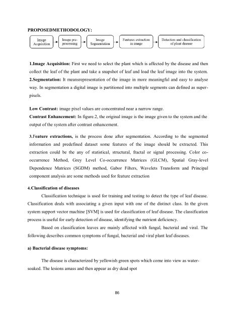
PROPOSEDMETHODOLOGY:<br />
1.Image Acquisition: First we need to select the plant which is affected by the disease and then<br />
collect the leaf of the plant and take a snapshot of leaf and load the leaf image into the system.<br />
2.Segmentation: It meansrepresentation of the image in more meaningful and easy to analyse<br />
way. In segmentation a digital image is partitioned into multiple segments can defined as superpixels.<br />
Low Contrast: image pixel values are concentrated near a narrow range.<br />
Contrast Enhancement: In figure.2, the original image is the image given to the system and the<br />
output of the system after contrast enhancement.<br />
3. Feature extractions, is the process done after segmentation. According to the segmented<br />
information and predefined dataset some features of the image should be extracted. This<br />
extraction could be the any of statistical, structural, fractal or signal processing. Color cooccurrence<br />
Method, Grey Level Co-occurrence Matrices (GLCM), Spatial Gray-level<br />
Dependence Matrices (SGDM) method, Gabor Filters, Wavelets Transform and Principal<br />
component analysis are some methods used for feature extraction<br />
4. Classification of diseases<br />
Classification technique is used for training and testing to detect the type of leaf disease.<br />
Classification deals with associating a given input with one of the distinct class. In the given<br />
system support vector machine [SVM] is used for classification of leaf disease. The classification<br />
process is useful for early detection of disease, identifying the nutrient deficiency.<br />
Based on classification leaves are mainly affected with fungal, bacterial and viral. The<br />
following describes common symptoms of fungal, bacterial and viral plant leaf diseases.<br />
a) Bacterial disease symptoms:<br />
The disease is characterized by yellowish green spots which come into view as watersoaked.<br />
The lesions amass and then appear as dry dead spot<br />
86