pwd sex1-1 sex1-3 - Group of Plant Biochemistry
pwd sex1-1 sex1-3 - Group of Plant Biochemistry
pwd sex1-1 sex1-3 - Group of Plant Biochemistry

Create successful ePaper yourself
Turn your PDF publications into a flip-book with our unique Google optimized e-Paper software.
Identification <strong>of</strong> a Novel Enzyme Required for Starch<br />
Metabolism in Arabidopsis Leaves. The Phosphoglucan,<br />
Water Dikinase 1[w]<br />
Oliver Kötting, Kerstin Pusch, Axel Tiessen, Peter Geigenberger, Martin Steup, and Gerhard Ritte*<br />
<strong>Plant</strong> Physiology, Institute <strong>of</strong> <strong>Biochemistry</strong> and Biology, University <strong>of</strong> Potsdam, 14476 Golm, Germany (O.K.,<br />
K.P., M.S., G.R.); and Max Planck Institute <strong>of</strong> Molecular <strong>Plant</strong> Physiology, 14476 Golm, Germany (A.T., P.G.)<br />
The phosphorylation <strong>of</strong> amylopectin by the glucan, water dikinase (GWD; EC 2.7.9.4) is an essential step within starch<br />
metabolism. This is indicated by the starch excess phenotype <strong>of</strong> GWD-deficient plants, such as the <strong>sex1</strong>-3 mutant <strong>of</strong><br />
Arabidopsis (Arabidopsis thaliana). To identify starch-related enzymes that rely on glucan-bound phosphate, we studied the<br />
binding <strong>of</strong> proteins extracted from Arabidopsis wild-type leaves to either phosphorylated or nonphosphorylated starch<br />
granules. Granules prepared from the <strong>sex1</strong>-3 mutant were prephosphorylated in vitro using recombinant potato (Solanum<br />
tuberosum) GWD. As a control, the unmodified, phosphate free granules were used. An as-yet uncharacterized protein was<br />
identified that preferentially binds to the phosphorylated starch. The C-terminal part <strong>of</strong> this protein exhibits similarity to that<br />
<strong>of</strong> GWD. The novel protein phosphorylates starch granules, but only following prephosphorylation with GWD. The enzyme<br />
transfers the b-P <strong>of</strong> ATP to the phosphoglucan, whereas the g-P is released as orthophosphate. Therefore, the novel protein is<br />
designated as phosphoglucan, water dikinase (PWD). Unlike GWD that phosphorylates preferentially the C6 position <strong>of</strong> the<br />
glucose units, PWD phosphorylates predominantly (or exclusively) the C3 position. Western-blot analysis <strong>of</strong> protoplast and<br />
chloroplast fractions from Arabidopsis leaves reveals a plastidic location <strong>of</strong> PWD. Binding <strong>of</strong> PWD to starch granules strongly<br />
increases during net starch breakdown. Transgenic Arabidopsis plants in which the expression <strong>of</strong> PWD was reduced by either<br />
RNAi or a T-DNA insertion exhibit a starch excess phenotype. Thus, in Arabidopsis leaves starch turnover requires a close<br />
collaboration <strong>of</strong> PWD and GWD.<br />
Starch, as the predominant storage carbohydrate in<br />
plants, is a major constituent <strong>of</strong> human and animal<br />
diets, and it is also an important raw material for<br />
various industrial processes (Slattery et al., 2000).<br />
Amylopectin, the major constituent <strong>of</strong> starch, contains<br />
varying amounts <strong>of</strong> covalently bound phosphate,<br />
depending upon plant species and organ. Phosphate<br />
is monoesterified to the C6 position (approximately<br />
two-thirds) and to the C3 position (approximately onethird).<br />
In addition, approximately 1% <strong>of</strong> the esterified<br />
phosphate may be linked to the C2 position (Tabata<br />
and Hizukuri, 1971). The total phosphorylation level<br />
<strong>of</strong> starch is quite low. In cereal endosperm starch, the<br />
amounts <strong>of</strong> Glc-6-P and Glc-3-P residues are at or<br />
below the detection limit and even in potato (Solanum<br />
tuberosum) tuber starch that is regarded as highly<br />
phosphorylated only 0.2% to 0.5% <strong>of</strong> the Glc residues<br />
are phosphorylated (Tabata and Hizukuri, 1971;<br />
Blennow et al., 2002). In Arabidopsis (Arabidopsis thaliana)<br />
leaf starch approximately 1 in 1,000 Glc residues<br />
is phosphorylated (Yu et al., 2001). Phosphate mono-<br />
1 This work was supported by the Deutsche Forschungsgemeinschaft<br />
(grant nos. SFB 429 TP–B2 to M.S. and TP–B7 to G.R. and P.G.).<br />
* Corresponding author; e-mail ritte@rz.uni-potsdam.de; fax 49–<br />
331–977–2512.<br />
[w] The online version <strong>of</strong> this article contains Web-only data.<br />
Article, publication date, and citation information can be found at<br />
www.plantphysiol.org/cgi/doi/10.1104/pp.104.055954.<br />
esters in amylopectin strongly affect starch functionality,<br />
and high phosphate contents are desirable<br />
for many industrial applications (Slattery et al., 2000;<br />
Blennow et al., 2002).<br />
Phosphorylation <strong>of</strong> starch like polyglucans is catalyzed<br />
by the glucan, water dikinase (GWD, formerly<br />
designated as R1; EC 2.7.9.4; Ritte et al., 2002):<br />
glucan 1 ATP 1 H2O / glucan-P b 1 AMP 1 Pgi:<br />
The catalytic mechanism includes autophosphorylation<br />
<strong>of</strong> the dikinase protein. The b-P <strong>of</strong> ATP is firstly<br />
transferred to a His residue <strong>of</strong> GWD and then to either<br />
the C6 or the C3 position <strong>of</strong> a glucosyl unit (Ritte et al.,<br />
2002; Mikkelsen et al., 2004).<br />
In GWD-deficient plants, not only starch phosphorylation<br />
but also starch breakdown is strongly impaired.<br />
In GWD antisense potato plants (Lorberth<br />
et al., 1998) as well as in the GWD-deficient starchexcess<br />
1 (<strong>sex1</strong>) mutants <strong>of</strong> Arabidopsis (Yu et al., 2001),<br />
leaf starch contents at the end <strong>of</strong> the day are 3 to 5<br />
times higher than those <strong>of</strong> the respective wild-type<br />
plants. More recently, it was shown that transitory<br />
starch in Chlamydomonas and potato is mainly phosphorylated<br />
during degradation (Ritte et al., 2004). This<br />
provides direct evidence for a link between the phosphorylation<br />
<strong>of</strong> starch and its degradation, but the<br />
underlying mechanisms remained obscure. It has been<br />
postulated that the activity <strong>of</strong> certain proteins that are<br />
involved in starch degradation depends on the pres-<br />
242 <strong>Plant</strong> Physiology, January 2005, Vol. 137, pp. 242–252, www.plantphysiol.org Ó 2004 American Society <strong>of</strong> <strong>Plant</strong> Biologists
ence <strong>of</strong> phosphate esters within starch (Yu et al., 2001;<br />
Ritte et al., 2002). However, until now, such enzymatic<br />
activities have never been documented. These proteins<br />
are expected to display a higher affinity to phosphorylated<br />
starch than to nonphosphorylated starch. This<br />
assumption provides a possible strategy for their<br />
identification.<br />
Here we describe the discovery <strong>of</strong> a novel protein,<br />
which preferentially binds to phosphorylated starch.<br />
Its enzymatic function was investigated in vitro using<br />
purified protein and in vivo using transgenic plants.<br />
RESULTS<br />
Identification <strong>of</strong> a Novel Protein That Preferentially<br />
Binds to Phosphorylated Starch Granules in Vitro<br />
To identify proteins whose activity depends on<br />
starch-bound phosphate esters, we compared binding<br />
<strong>of</strong> proteins to phosphorylated or nonphosphorylated<br />
starch granules. Phosphate free starch granules were<br />
isolated from leaves <strong>of</strong> the GWD-deficient Arabidopsis<br />
<strong>sex1</strong>-3 mutant (Yu et al., 2001). Aliquots <strong>of</strong> the starch<br />
preparations were then in vitro phosphorylated using<br />
purified recombinant GWD from potato. Under the<br />
conditions applied, the phosphorylation levels range<br />
between 200 and 400 pmol P/mg starch. All the<br />
phosphate groups are located at the granule surface,<br />
since the recombinant GWD has no access to the<br />
granule interior. Phosphorylated and nonphosphorylated<br />
granules were then incubated with protein<br />
extracts from Arabidopsis wild-type leaves. Proteins<br />
bound to the granules were desorbed by a SDScontaining<br />
buffer and subsequently analyzed using<br />
SDS-PAGE and matrix-assisted laser-desorption ionization<br />
mass spectrometry (Fig. 1). Three proteins did<br />
bind preferentially to the phosphorylated starch.<br />
These were GWD (SEX1; At1g10760), cytosolic phosphorylase<br />
(Pho2; At3g46970), and an as-yet uncharacterized<br />
protein, preliminarily designated as OK1<br />
(At5g26570). In the following we focus on the latter.<br />
OK1 turned out to be one <strong>of</strong> the two putative proteins<br />
with homology to GWD, whose existence was predicted<br />
from the Arabidopsis sequence (Yu et al., 2001).<br />
Database analysis using <strong>Plant</strong>GDB Blast (Dong et al.,<br />
2004) revealed the presence <strong>of</strong> putative OK1 orthologs<br />
in 14 different plant species including potato, tomato,<br />
barley, and rice.<br />
Using primers designed for the At5g26570 gene, the<br />
full-length OK1 cDNA sequence was cloned. In<br />
the sequence thereby derived, 15 additional nucleotides<br />
(1,555–1,569) were found that were not present<br />
in the already existing corresponding National<br />
Center for Biotechnology Information (NCBI) entry<br />
(NM_122538). The OK1 cDNA sequence was submitted<br />
to EMBL (accession no. AJ635427).<br />
OK1 and GWD amino acid sequences were compared<br />
using the BLAST 2 Sequences tool (Tatusova<br />
and Madden, 1999). The C-terminal regions, ranging<br />
from amino acid 611 to 1,196 (OK1), and 860 to 1,398<br />
(GWD), displayed 25% amino acid identity, and 41%<br />
sequence similarity. No similarity could be detected in<br />
the N-terminal regions. The overall amino acid identity,<br />
as analyzed with the AlignX program (Vector NTI,<br />
Invitrogen, Karlsruhe, Germany), is 14%, and sequence<br />
similarity is 24%.<br />
Analysis <strong>of</strong> the OK1 sequence using TargetP<br />
(Emanuelsson et al., 2000) reveals a high probability<br />
for the existence <strong>of</strong> a signal peptide directing the protein<br />
to plastids (score 0.992). Three further domains<br />
could be detected (Fig. 2A). A starch binding domain<br />
(CBM 20) is located at the N-terminal region <strong>of</strong> OK1.<br />
The C terminus <strong>of</strong> OK1 displays homology to the<br />
nucleotide binding domains <strong>of</strong> the dikinases pyruvate,<br />
phosphate dikinase (PPDK; EC 2.7.9.1), phosphoenolpyruvate<br />
(PEP)-synthase (pyruvate, water dikinase;<br />
EC 2.7.9.2), and GWD (Marchler-Bauer et al., 2003). A<br />
region with significant homology to the phosphohistidine<br />
domains <strong>of</strong> these dikinases is also present in the<br />
OK1 sequence (Fig. 2B).<br />
OK1 Is Localized in Plastids<br />
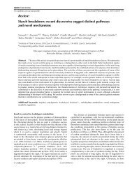
Phosphoglucan, Water Dikinase<br />
Figure 1. In vitro binding <strong>of</strong> proteins to phosphorylated or nonphosphorylated<br />
starch granules. Protein extracts from ecotype Columbia<br />
leaves were incubated with either nonphosphorylated (2) or previously<br />
in vitro phosphorylated (3) starch granules from <strong>sex1</strong>-3 leaves. Granules<br />
were washed, and bound proteins were released with SDS sample<br />
buffer and separated by SDS-PAGE. 1, In vitro phosphorylated starch<br />
granules incubated without extract; M, molecular mass marker. Three<br />
proteins with higher affinity to phosphorylated starch are indicated<br />
(GWD, OK1, and Pho2).<br />
The prediction <strong>of</strong> OK1 being a plastidic protein by<br />
the bioinformatic programs was further strengthened<br />
by western-blot analysis <strong>of</strong> extracts made from Arabidopsis<br />
leaf protoplasts and chloroplasts isolated from<br />
protoplasts. Equal amounts <strong>of</strong> protein were analyzed<br />
by SDS-PAGE and western blot using specific antibodies<br />
raised against ADP-Glc-pyrophosphorylase<br />
(AGPase, plastidic marker), PEP-carboxylase (cytosolic<br />
marker), and OK1. As shown in Figure 3 for<br />
both AGPase and OK1, an immunosignal was obtained<br />
in the chloroplast fraction, whereas there was<br />
hardly any signal in the protoplast fraction. In contrast,<br />
<strong>Plant</strong> Physiol. Vol. 137, 2005 243
Kötting et al.<br />
Figure 2. OK1 contains a starch binding domain and domains typical<br />
for dikinases. A, Protein domains <strong>of</strong> the OK1 protein. The amino acid<br />
number indicates the location <strong>of</strong> the respective domain within the<br />
protein. CTS, Chloroplast targeting signal; SB, starch binding domain<br />
CBM 20; PH, putative phosphohistidine domain; NB, nucleotide<br />
binding domain. B, Alignment <strong>of</strong> the putative phosphohistidine domains<br />
<strong>of</strong> OK1 and different dikinases. The putative phosphohistidine<br />
regions <strong>of</strong> GWDs from Arabidopsis (At), potato (St), and Citrus<br />
reticulata (Cr) were aligned with the putative phosphohistidine regions<br />
<strong>of</strong> PPDKs from Zea mays (Zm) and E. coli, pyruvate, water dikinase<br />
(PPS) from E. coli, and the homologous region <strong>of</strong> OK1. Identical amino<br />
acids in black, conserved in gray boxes. The putative phosphohistidine<br />
is printed in boldface.<br />
the cytosolic marker PEP-carboxylase was exclusively<br />
detected in the protoplast fraction. Nonaqueous fractionation<br />
<strong>of</strong> Arabidopsis leaf material and subsequent<br />
western-blot analysis also indicates that OK1 resides<br />
in plastids (data not shown). Furthermore, OK1 is<br />
found in the Arabidopsis chloroplast protein database<br />
(http://www.pb.ipw.biol.ethz.ch/index.php?toc591;<br />
Kleffmann et al., 2004), thereby providing another line<br />
<strong>of</strong> evidence for plastidic localization <strong>of</strong> this protein.<br />
Thus, the interaction <strong>of</strong> OK1 with phosphorylated<br />
starch may well be <strong>of</strong> physiological relevance.<br />
OK1 Is a Phosphoglucan, Water Dikinase<br />
A vector was constructed allowing the expression <strong>of</strong><br />
OK1 containing a 63His tag at the N terminus in<br />
Escherichia coli and one step purification <strong>of</strong> the recombinant<br />
protein using a Ni-NTA agarose resin. The<br />
full-size OK1 protein is clearly predominant in the<br />
resulting protein fraction (Supplemental Fig. 1). Because<br />
<strong>of</strong> the similarity between OK1 and GWD, we<br />
tested whether or not OK1 also displays starch phosphorylating<br />
activity. As for the in vitro binding assay,<br />
nonphosphorylated or phosphorylated starch granules<br />
served as substrates. OK1 was indeed able to<br />
transfer 33 Pfrom[bg- 33 P]ATP to starch. Most remarkably,<br />
however, the activity strictly depended on a preceding<br />
phosphorylation <strong>of</strong> the granules by recombinant<br />
potato GWD (Fig. 4A). We never observed OK1catalyzed<br />
phosphorylation <strong>of</strong> the unmodified phosphate<br />
free <strong>sex1</strong>-3 starch (n 5 7). Similar to GWD, OK1<br />
transfers the b-P <strong>of</strong> ATP to starch. There was no<br />
significant phosphate incorporation <strong>of</strong> labeled phosphate<br />
into the starch substrate if [g- 33 P]ATP served as<br />
phosphate donor (Fig. 4B).<br />
To study the fate <strong>of</strong> the g-P <strong>of</strong> ATP, we analyzed<br />
OK1-catalyzed incorporation <strong>of</strong> b-P into phosphorylated<br />
starch using [b- 33 P]ATP as phosphate donor as<br />
well as a possible release <strong>of</strong> g-P-ortophosphate into the<br />
soluble phase using [g- 33 P]ATP. For comparison, the<br />
same experiment was also conducted using recombinant<br />
potato GWD, for which we have shown before<br />
that water is the acceptor <strong>of</strong> the g-P (Ritte et al., 2002).<br />
As shown in Table I, in both cases comparable<br />
amounts <strong>of</strong> phosphate were incorporated into starch<br />
or released as inorganic phosphate into the soluble<br />
phase, respectively. In both samples, the amount <strong>of</strong> g-P<br />
released exceeded that <strong>of</strong> b-P detected in the starch<br />
fraction by about 20% to 30%. However, it is possible<br />
that the amount <strong>of</strong> incorporated phosphate is underestimated<br />
due to some loss <strong>of</strong> starch granules during<br />
the extensive washing procedure.<br />
We conclude that OK1 is a phosphoglucan, water<br />
dikinase (PWD) that transfers the b-P <strong>of</strong> ATP to<br />
a phosphoglucan and the g-P <strong>of</strong> ATP to water:<br />
Glucan-P n 1 ATP 1 H 2O / Glucan-P n11 1 AMP 1 P i:<br />
Therefore, we replace the preliminary term OK1<br />
with PWD.<br />
Under standard assay conditions (10 mg phosphorylated<br />
starch granules/mL, 25 mM ATP, 30 min),<br />
a linear correlation between PWD protein amount<br />
and phosphate incorporation into the substrate is only<br />
observed at low protein concentrations (,0.1 mg/mg<br />
starch; Supplemental Fig. 2A). For 0.05 mg PWD/mg<br />
starch, we calculated an activity <strong>of</strong> 2.55 nmol min 21<br />
Figure 3. OK1 is located in chloroplasts. Equal amounts <strong>of</strong> protein<br />
extracted from Arabidopsis leaf protoplasts (P) and chloroplasts (C),<br />
respectively, were separated by SDS-PAGE and examined by immunoblot<br />
analysis using antibodies against OK1, AGPase (plastidic marker),<br />
and PEPCase (cytosolic marker).<br />
244 <strong>Plant</strong> Physiol. Vol. 137, 2005
Figure 4. In vitro activity assay with recombinant OK1 protein. A, OK1<br />
phosphorylates phosphoglucans but not phosphate free glucans. One<br />
microgram recombinant OK1 protein was incubated for 15 min with<br />
25 mM ATP containing 0.5 mCi [bg- 33 P]ATP and 5 mg <strong>of</strong> either nonphosphorylated<br />
(1) or in vitro phosphorylated (2) starch granules from<br />
<strong>sex1</strong>-3 leaves. The radioactivity incorporated into the granules was<br />
counted. -, Control without protein. Bars represent the individual<br />
measurements <strong>of</strong> two parallel samples. B, OK1 transfers the b-P <strong>of</strong> ATP<br />
to phosphoglucans. Recombinant OK1 protein (2.2 mg) was incubated<br />
for 1 h with 4.2 mg in vitro phosphorylated starch granules and either<br />
[bg- 33 P]ATP or solely [g- 33 P]ATP (0.5 mCi in both cases). ATP concentration,<br />
25 mM. The radioactivity incorporated into the granules was<br />
counted. -, Control without protein.<br />
(mg protein) 21 . This value is slightly underestimated<br />
because [bg- 33 P]ATP was used that contains $80% <strong>of</strong><br />
label in the b-position (Ritte et al., 2003). The PWD<br />
activity is similar to the specific activity <strong>of</strong> potato<br />
GWD with soluble potato amylopectin (Ritte et al.,<br />
2002) and also to the activity <strong>of</strong> GWD with unmodified<br />
<strong>sex1</strong>-3 starch granules as substrate (data not shown).<br />
For a more quantitative analysis, starch granules<br />
containing different amounts <strong>of</strong> phosphate esters (9,<br />
49, and 215 pmol P/mg starch) were reacted with<br />
PWD (Supplemental Fig. 2B). The granule preparation<br />
with the highest phosphate content was the most<br />
efficient phosphate acceptor. However, PWD activities<br />
with the different substrates varied about 6-fold,<br />
whereas the level <strong>of</strong> prephosphorylation varied about<br />
24-fold. Thus, there is no linear relation between the<br />
phosphate content <strong>of</strong> a polyglucan and its capacity to<br />
serve as phosphate acceptor for PWD.<br />
As observed with nonphosphorylated <strong>sex1</strong>-3 starch<br />
granules PWD was unable to phosphorylate solubilized<br />
<strong>sex1</strong>-3 starch (Supplemental Fig. 2C). However,<br />
even solubilized <strong>sex1</strong>-3 starch that had been prephosphorylated<br />
by GWD proved to be an extremely poor<br />
substrate for PWD (Supplemental Fig. 2C). The same<br />
holds true for soluble potato starch (Sigma S-2004)<br />
although it contains approximately 15 nmol P/mg<br />
starch (data not shown). Possibly, the structure <strong>of</strong> the<br />
starch granule or <strong>of</strong> the surface <strong>of</strong> the particle is also<br />
important for the PWD activity.<br />
PWD Is Capable <strong>of</strong> Autophosphorylation<br />
Phosphoglucan, Water Dikinase<br />
A phosphohistidine is an intermediate in the dikinase<br />
type reactions catalyzed by PPDK (Goss et al.,<br />
1980), PEP-synthase (Narindrasorasak and Bridger,<br />
1977), and GWD (Ritte et al., 2002; Mikkelsen et al.,<br />
2004). The b-P <strong>of</strong> ATP is firstly transferred to the His<br />
residue and then to pyruvate (in case <strong>of</strong> PPDK and<br />
PEP-synthase) or a glucan (in case <strong>of</strong> GWD). As<br />
mentioned above an amino acid stretch displaying<br />
homology to the phosphohistidine domains <strong>of</strong> these<br />
enzymes is also present in PWD. To study whether<br />
PWD follows a similar mechanism, we incubated the<br />
purified protein with either [bg- 33 P]ATP or [g- 33 P]ATP<br />
in the absence <strong>of</strong> a phosphoglucan and analyzed<br />
a possible autophosphorylation by SDS-PAGE and<br />
autoradiography. Labeling <strong>of</strong> the PWD protein was<br />
observed only in the samples containing [bg- 33 P]ATP<br />
but not in those containing [g- 33 P]ATP (Fig. 5). Thus,<br />
PWD phosphorylates itself with the b-P. The phosphorylation<br />
was heat labile, acid labile but rather<br />
stable in alkali (Fig. 5). This is consistent with a phosphohistidine<br />
being formed, since phosphoserine,<br />
phosphotyrosine, and phosphothreonine are heat stable,<br />
acid stable, but alkali labile (Rosenberg, 1996). The<br />
results strongly indicate that the b-P <strong>of</strong> ATP is firstly<br />
transferred to a His residue <strong>of</strong> PWD and afterward to<br />
the phosphoglucan.<br />
PWD Phosphorylates Preferably the C3 Position <strong>of</strong> the<br />
Glucosyl Residues in Phosphoglucans<br />
To analyze which positions <strong>of</strong> the Glc residues are<br />
phosphorylated by PWD, starch was prephosphory-<br />
Table I. OK1 transfers the g-P <strong>of</strong> ATP to water<br />
Recombinant OK1 or GWD protein (2.2 mg each) were incubated<br />
with in vitro phosphorylated <strong>sex1</strong>-3 starch granules for 80 min. Either<br />
[b- 33 P]ATP or [g- 33 P]ATP <strong>of</strong> identical specific radioactivity served as<br />
phosphate donor. The incorporation <strong>of</strong> b-P into starch (control, buffer<br />
instead <strong>of</strong> protein) and the release <strong>of</strong> g-Pi into the soluble phase<br />
(control, no starch) was determined.<br />
[b- 33 P]-Incorporation [g- 33 P]-Release<br />
pmol pmol<br />
OK1 34.3 42.8<br />
GWD 123.3 172.0<br />
<strong>Plant</strong> Physiol. Vol. 137, 2005 245
Kötting et al.<br />
Figure 5. Autocatalytic phosphorylation <strong>of</strong> recombinant PWD (OK1).<br />
Recombinant PWD protein was incubated in the absence <strong>of</strong> starch<br />
granules with either [bg- 33 P]ATP or solely [g- 33 P]ATP (1.4 mCi/mg<br />
protein each). Heat stability <strong>of</strong> the phosphorylated PWD was tested by<br />
incubation <strong>of</strong> the samples at either 30°C for 30 min or 95°C for 5 min.<br />
Acid or base stability <strong>of</strong> the phosphorylated PWD protein was assayed<br />
by incubation in 0.5 N NaOH or 0.5 N HCl for 30 min at RT, followed by<br />
denaturation in SDS sample buffer for 30 min at 30°C. Samples were<br />
separated by SDS-PAGE (1.8 mg PWD protein/lane) and visualized by<br />
either Coomassie Blue staining (A) or autoradiography (B).<br />
lated by GWD (using unlabeled ATP) and then<br />
phosphorylated by PWD using 33 P-ATP. For comparison,<br />
an experiment in which starch was solely phosphorylated<br />
by GWD (using labeled ATP) was also<br />
performed. Following in vitro phosphorylation, Glc<br />
and Glc-Ps were released from starch by means <strong>of</strong> acid<br />
hydrolysis. To separate Glc-6-P and Glc-3-P, the samples<br />
were subjected to high performance anionexchange<br />
chromatography with pulsed amperometric<br />
detection (HPAEC-PAD). Since the extent <strong>of</strong> in vitro<br />
phosphorylation is too low to allow for reliable<br />
amperometric detection <strong>of</strong> the Glc-Ps thereby formed<br />
we added authentic Glc-6-P and Glc-3-P as standards.<br />
Fractions were collected and the radioactivity was<br />
counted.<br />
In contrast to GWD, which phosphorylates preferably<br />
the C6-position, PWD phosphorylates predominantly<br />
the C3-position (Fig. 6). Approximately 70% <strong>of</strong><br />
the radioactivity incorporated into starch by PWD<br />
coeluted with Glc-3-P in two independent experiments.<br />
It has to be considered that Glc-3-P is a rather<br />
acid labile compound. The investigation <strong>of</strong> starch<br />
bound phosphate esters by acid hydrolysis <strong>of</strong> granular<br />
starch and subsequent analysis <strong>of</strong> the products by<br />
HPAEC-PAD was adapted from Blennow et al. (1998).<br />
They estimated that during 2 h <strong>of</strong> acid hydrolysis<br />
approximately 20% <strong>of</strong> the Glc-3-P is dephosphorylated.<br />
We found that 24% <strong>of</strong> the radioactivity in the<br />
starch hydrolysate was present as inorganic phosphate<br />
using the modified Parvin and Smith method (see<br />
‘‘Materials and Methods’’). In contrast, if starch was<br />
hydrolyzed following in vitro phosphorylation with<br />
GWD less than 5% <strong>of</strong> the label were present as inorganic<br />
phosphate. When 32 P-orthophosphate was<br />
subjected to HPAEC-PAD approximately 20% <strong>of</strong> radioactivity<br />
coeluted with Glc-6-P the remaining 80%<br />
eluted in the fraction between Glc-6-P and Glc-3-P<br />
(data not shown). Thus, in the hydrolyzed PWD<br />
product the radioactivity eluting ahead <strong>of</strong> the<br />
Glc-3-P peak (Fig. 6) can, at least in part, be attributed<br />
to orthophosphate that is released from Glc-3-P during<br />
sample processing.<br />
In Vitro Analysis <strong>of</strong> PWD Purified from Leaves Confirms<br />
the Results with the Heterologously Expressed Protein<br />
Analysis <strong>of</strong> the recombinant PWD has revealed<br />
unique features <strong>of</strong> this enzyme. However, it has to be<br />
kept in mind that the recombinant PWD protein is not<br />
identical to PWD in plants. Since the length <strong>of</strong> the<br />
transit peptide is only predicted but not exactly known<br />
the complete coding sequence <strong>of</strong> PWD was used to<br />
generate the expression vector. Furthermore, the protein<br />
contains an N-terminal His tag and a single amino<br />
acid replacement (see ‘‘Materials and Methods’’).<br />
Therefore, we analyzed the plant-derived protein in<br />
addition to the recombinant PWD. The protein was<br />
partially purified by ammoniumsulfate precipitation<br />
and affinity chromatography using immobilized maltoheptaose<br />
(Supplemental Fig. 3A). As has been<br />
shown for the recombinant protein, activity <strong>of</strong> PWD<br />
purified from leaves strictly depended on a preceding<br />
phosphorylation by GWD (Supplemental Fig. 3B) and<br />
phosphorylation predominantly (or exclusively) occurs<br />
at the C3-position <strong>of</strong> the Glc residues (Supplemental<br />
Fig. 3C).<br />
Reduction <strong>of</strong> PWD in Transgenic <strong>Plant</strong>s Causes a Starch<br />
Excess Phenotype<br />
To investigate the in vivo function <strong>of</strong> PWD, an RNA<br />
interference (RNAi) construct was designed to repress<br />
the activity <strong>of</strong> this protein in Arabidopsis. Fifteen<br />
transgenic lines were analyzed by western blot, all <strong>of</strong><br />
which proved to be strongly reduced in PWD protein<br />
(data not shown). Five <strong>of</strong> these lines were examined in<br />
more detail. As shown in Figure 7A the PWD protein<br />
amount in the RNAi lines 1 to 4 is below the limit <strong>of</strong><br />
Figure 6. PWD (OK1) phosphorylates preferentially the C-3 position <strong>of</strong><br />
the Glc units. Recombinant PWD and GWD protein (first experiment:<br />
25 mg PWD,5mg GWD; second experiment: 4.2 mg PWD, 0.7 mg<br />
GWD), respectively, were incubated with in vitro phosphorylated<br />
<strong>sex1</strong>-3 starch granules and [bg- 33 P]ATP for 1 h. Starch granules were<br />
hydrolyzed. Aliquots <strong>of</strong> the hydrolyzates were supplemented with<br />
Glc-3-P and Glc-6-P as internal standards and subjected to HPAEC-<br />
PAD analysis (top section). Fractions were collected every minute<br />
(1 mL), except for Glc-3-P and Glc-6-P fractions, which were collected<br />
quantitatively in separate fractions. Radioactivity in the collected<br />
fractions was counted. The relative distribution <strong>of</strong> radiolabel was<br />
calculated as percentage <strong>of</strong> total radioactivity in all collected fractions<br />
(bottom section). GWD, White bars; PWD, black bars. Values are<br />
means <strong>of</strong> two independent experiments.<br />
246 <strong>Plant</strong> Physiol. Vol. 137, 2005
Figure 7. Transgenic plants with reduced PWD (OK1) protein exhibit<br />
a starch excess phenotype. A, The PWD protein level is decreased in<br />
PWD RNAi plants. Leaf samples <strong>of</strong> five PWD RNAi lines and wild type<br />
were harvested at the end <strong>of</strong> the light period. Proteins were extracted<br />
and 40 mg each were separated by SDS-PAGE and examined by<br />
immunoblot analysis using an anti-PWD antibody. WT, Wild type; 1 to<br />
5, PWD RNAi lines 1 to 5. B, Starch content in leaves <strong>of</strong> wild-type and<br />
PWD RNAi lines. <strong>Plant</strong>s were grown in a 12-h-light/12-h-dark cycle.<br />
Samples were taken at the end <strong>of</strong> the light (white bars) and dark period<br />
(black bars), respectively. Values are means 6 SE; n 5 4 plants. 1 to 5,<br />
PWD RNAi lines; WT, wild type; FW, fresh weight. C, Starch content in<br />
leaves <strong>of</strong> wild type and PWD knockout mutant. All leaves <strong>of</strong> wild-type<br />
plants (white symbols) or <strong>pwd</strong> plants (black symbols) were harvested at<br />
the times indicated. Values are means 6 SE; n 5 4 plants.<br />
detection using western-blot analysis. In line 5 PWD<br />
expression is reduced by at least 75%. The analysis <strong>of</strong><br />
starch contents in these plants revealed a metabolic<br />
phenotype for PWD. At the end <strong>of</strong> the day, the PWDdeficient<br />
plants contained up to 2 times more starch<br />
than ecotype Columbia wild type (Fig. 7B). Starch is<br />
nearly completely remobilized during night in wildtype<br />
but not in the transgenic plants. In the less<br />
inhibited RNAi-line 5, starch amounts are only slightly<br />
increased compared with wild-type plants. Thus, the<br />
extent <strong>of</strong> PWD inhibition correlates with the effect on<br />
starch content.<br />
We also obtained an insertion mutant<br />
(SALK_110814) and selected a homozygous line for<br />
the At5g26570 gene. No PWD protein could be detected<br />
in the mutant (data not shown). As observed<br />
with the RNAi plants, the PWD knockout mutant<br />
(<strong>pwd</strong>) contains considerably more starch than wildtype<br />
plants throughout the day/night cycle (Fig. 7C).<br />
Starch turnover occurred in the PWD-deficient plants,<br />
but the rate <strong>of</strong> starch degradation in the <strong>pwd</strong> plants<br />
was lower than that <strong>of</strong> the wild-type plants (Fig. 7C).<br />
This effect was also observed using an independent<br />
batch <strong>of</strong> plants grown under a 14-h-light period (data<br />
not shown).<br />
The high starch phenotype resembles that <strong>of</strong> the<br />
GWD-deficient <strong>sex1</strong> mutants. However, when grown<br />
under the same conditions the <strong>sex1</strong> mutants accumulate<br />
more starch than the <strong>pwd</strong> plants. The lack <strong>of</strong> PWD<br />
had a minor effect on plant development, whereas the<br />
GWD knockout mutant <strong>sex1</strong>-3 is strongly retarded in<br />
growth when cultivated under a 12-h-light/12-h-dark<br />
regime (Supplemental Fig. 4).<br />
Starch-bound phosphates in wild-type and PWD<br />
RNAi plants (lines 1–5) were analyzed by HPAEC-<br />
PAD and quantification <strong>of</strong> the peak areas following<br />
acid hydrolysis <strong>of</strong> the starch granules. The Glc-6-P:<br />
Glc-3-P ratio increased from 2.1 in wild type to 2.5 in<br />
the transgenic plants (mean <strong>of</strong> lines 1–5), ranging from<br />
2.2 in line 5 to 2.7 in line 1. This increase, however, was<br />
caused by slightly elevated Glc-6-P levels, whereas the<br />
Glc-3-P content was essentially unchanged (data not<br />
shown). The Glc-6-P:Glc-3-P ratio in starch <strong>of</strong> <strong>pwd</strong><br />
plants was also increased compared with wild type<br />
(data not shown). It has to be considered that the Glc<br />
phosphate levels in starch hydrolyzates mainly reflect<br />
starch phosphorylation during biosynthesis and not<br />
the transient phosphorylation <strong>of</strong> the granule surface<br />
during breakdown (Ritte et al., 2004).<br />
PWD Binds to Leaf Starch Granules during<br />
Their Breakdown<br />
To analyze whether binding <strong>of</strong> PWD to transitory<br />
starch is affected by the physiological state <strong>of</strong> the cell,<br />
granule-bound and soluble protein was extracted from<br />
leaves <strong>of</strong> wild-type plants that had been harvested<br />
either in the light or dark period. As revealed by<br />
western-blot analysis, binding <strong>of</strong> PWD to the surface<br />
<strong>of</strong> transitory starch granules strongly increases during<br />
starch mobilization in darkness (Fig. 8). In contrast, the<br />
PWD level in the buffer soluble fraction was equal<br />
in the light and dark samples. Binding <strong>of</strong> GWD to<br />
transitory starch in Arabidopsis also significantly increases<br />
in darkness (Fig. 8), in agreement with earlier<br />
results using leaves <strong>of</strong> potato and pea, respectively<br />
(Ritte et al., 2000a).<br />
DISCUSSION<br />
Phosphoglucan, Water Dikinase<br />
The factors and mechanisms leading to the degradation<br />
<strong>of</strong> the crystalline starch granule are largely<br />
<strong>Plant</strong> Physiol. Vol. 137, 2005 247
Kötting et al.<br />
Figure 8. PWD (OK1) binds to the surface <strong>of</strong> leaf starch granules during<br />
their degradation. Leaves <strong>of</strong> Arabidopsis wild-type plants were harvested<br />
2 h before (L) and 2 h after the end <strong>of</strong> the light period (D). Soluble<br />
proteins and starch were extracted. Proteins bound to the surface <strong>of</strong><br />
the starch granules were released by incubation in SDS-sample buffer at<br />
RT. Proteins released from 6.5 mg starch (bound) or 40 mg <strong>of</strong> soluble<br />
protein (soluble) were subjected to SDS-PAGE and immunoblot analysis<br />
using antibodies against PWD and GWD. The experiment was<br />
repeated with an independently grown batch <strong>of</strong> plants and yielded the<br />
same result.<br />
unknown, but there is increasing evidence that phosphorylation<br />
<strong>of</strong> starch by GWD is involved (Zeeman<br />
et al., 2004). In a first approach to unravel the link<br />
between the GWD-catalyzed phosphorylation <strong>of</strong><br />
starch and the degradation <strong>of</strong> the polyglucans, we<br />
searched for proteins displaying enhanced affinity to<br />
phosphorylated starch compared with nonphosphorylated<br />
starch. We identified a novel protein (PWD) that<br />
preferentially binds to phosphorylated starch and is<br />
capable <strong>of</strong> glucan phosphorylation. The reaction catalyzed<br />
by PWD differs from that <strong>of</strong> GWD and, thus,<br />
represents a novel enzymatic activity. In contrast to<br />
GWD, phosphorylating activity <strong>of</strong> PWD was only observed<br />
using substrates that already contained glucanbound<br />
phosphate. In vitro phosphorylated <strong>sex1</strong>-3<br />
granules were a suitable substrate for PWD, whereas<br />
we did not detect PWD-catalyzed phosphate incorporation<br />
into the unmodified phosphate free <strong>sex1</strong>-3<br />
starch. Likewise, starch granules from wheat (Sigma<br />
S-5127) were not phosphorylated by PWD, but following<br />
prephosphorylation with GWD they can serve as<br />
substrate for PWD (data not shown).<br />
Why is PWD active only on phosphorylated starch?<br />
It is reasonable to assume that PWD either phosphorylates<br />
glucan chains that were previously phosphorylated<br />
by GWD, or it phosphorylates unphosphorylated<br />
chains within a phosphorylated matrix. Analysis <strong>of</strong><br />
the phosphorylated glucan chains following in vitro<br />
phosphorylation with 33 P-ATP shows that the latter<br />
predominates. Approximately 80% <strong>of</strong> the incorporated<br />
label was recovered in singly phosphorylated glucan<br />
chains; the remaining radioactivity was found in<br />
doubly phosphorylated chains. Probably phosphate<br />
incorporation by GWD locally alters the starch structure<br />
and thereby generates phosphorylation sites that<br />
can be used by PWD.<br />
Whereas activity <strong>of</strong> PWD strictly depends on the<br />
presence <strong>of</strong> glucan bound phosphate binding <strong>of</strong> the<br />
protein to carbohydrates does not. PWD can bind to<br />
the unmodified <strong>sex1</strong>-3 granules, albeit with low efficiency.<br />
The protein also binds to immobilized maltoheptaose<br />
(S. Orzechowski, unpublished data), and we<br />
made use <strong>of</strong> this to enrich the protein from leaf<br />
extracts. These maltoheptaose beads were also not at<br />
all phosphorylated by the recombinant PWD (data not<br />
shown).<br />
An important difference between PWD and GWD is<br />
the site <strong>of</strong> phosphate incorporation. GWD phosphorylates<br />
both the C3 and the C6 position, with a clear<br />
preference <strong>of</strong> the latter. In contrast, PWD phosphorylates<br />
preferably the C3 position (Fig. 6). A low extent<br />
<strong>of</strong> C6-phosphorylation cannot be ruled out; the same<br />
holds true for C2-phosphorylation. It has been suggested<br />
by Tabata and Hizukuri (1971) that about 1% <strong>of</strong><br />
the phosphate in potato tuber starch could be linked to<br />
the C2 position. Glc-2-P is far more acid labile than<br />
Glc-3-P (Tabata and Hizukuri, 1971; Kokesh et al.,<br />
1978), and complete dephosphorylation during acid<br />
hydrolysis as applied here can be expected. Therefore,<br />
we cannot exclude that Glc-2-P residues may also be<br />
formed by PWD (or GWD as well). NMR analysis<br />
could provide further information whether or not C3 is<br />
the single phosphorylation site <strong>of</strong> PWD. However, due<br />
to the low level <strong>of</strong> PWD-catalyzed phosphate incorporation<br />
in vitro and the high background level <strong>of</strong><br />
phosphate esters in the starch substrate, NMR-analysis<br />
<strong>of</strong> the PWD products is not practicable at present.<br />
Starch is phosphorylated during its biosynthesis<br />
(Nielsen et al., 1994; Wischmann et al., 1999) but in<br />
addition also during its degradation (Ritte et al., 2004).<br />
The amounts <strong>of</strong> Glc-6-P and Glc-3-P determined in<br />
hydrolyzed starch mainly reflect starch phosphorylation<br />
during starch synthesis (Ritte et al., 2004). The<br />
absolute level <strong>of</strong> starch-bound Glc-3-P is not significantly<br />
altered in the PWD-deficient plants. This may<br />
indicate that the enzyme is not involved in biosynthesis<br />
associated phosphorylation. However, GWD can<br />
attach phosphate at both C3 and C6 positions <strong>of</strong> Glc<br />
residues in amylopectin, and may thus partly compensate<br />
for lacking PWD activity. The amount <strong>of</strong> GWD<br />
protein is unaltered in the PWD-deficient plants as<br />
revealed by western-blot analysis (data not shown),<br />
but it is possible that more glucan targets become<br />
available for GWD if PWD is lacking.<br />
The increased phosphorylation <strong>of</strong> the granule surface<br />
during breakdown <strong>of</strong> starch in chloroplasts (Ritte<br />
et al., 2004) should favor activity <strong>of</strong> PWD during starch<br />
mobilization. In fact, binding <strong>of</strong> PWD to transitory<br />
starch granules is strongly increased during degradation<br />
<strong>of</strong> the starch particle (Fig. 8). Phosphorylation<br />
during starch mobilization is restricted to the outermost<br />
layer <strong>of</strong> the granule, and the phosphate esters<br />
introduced during breakdown underlie a rapid turnover<br />
(Ritte et al., 2004). Consequently, the reduction <strong>of</strong><br />
C3-phosphorylation during the period <strong>of</strong> starch mobilization<br />
is not expected to noticeably affect the total<br />
Glc-3-P level <strong>of</strong> starch.<br />
The starch excess phenotype observed in the PWDdeficient<br />
plants demonstrates that this enzyme plays<br />
an important metabolic role, and lack <strong>of</strong> PWDcatalyzed<br />
starch phosphorylation cannot be (fully)<br />
compensated for by other enzymes. Since the activity<br />
248 <strong>Plant</strong> Physiol. Vol. 137, 2005
<strong>of</strong> PWD depends on a preceding starch phosphorylation<br />
by GWD, the lack <strong>of</strong> GWD in mutant plants<br />
should also abolish starch phosphorylation by PWD.<br />
Consistently, no Glc-3-P residues could be detected in<br />
starch <strong>of</strong> the GWD-free Arabidopsis mutant <strong>sex1</strong>-3<br />
(Yu et al., 2001). The starch excess phenotype in the<br />
GWD-deficient plants is probably attributable to a<br />
combined reduction <strong>of</strong> GWD and PWD activity, which<br />
might explain the more severe phenotype compared<br />
with PWD mutants.<br />
Further studies are required to explore the link<br />
between phosphorylation and degradation <strong>of</strong> starch.<br />
It has recently been reported that the in vitro degradation<br />
<strong>of</strong> granules isolated from turions <strong>of</strong> the duckweed<br />
by starch associated proteins (including a<br />
putative GWD ortholog) could be increased by addition<br />
<strong>of</strong> ATP, thereby enabling starch phosphorylation<br />
(Reimann et al., 2004). We have obtained similar<br />
results using potato leaf starch. Furthermore, we<br />
know from in vitro studies that as-yet unidentified<br />
proteins exist in Arabidopsis that degrade starch more<br />
efficiently if it is phosphorylated (G. Ritte, unpublished<br />
data). Future work will focus on the characterization<br />
<strong>of</strong> these proteins to evaluate their role in starch<br />
degradation in vivo.<br />
We propose that the newly identified PWD acts<br />
downstream <strong>of</strong> GWD and is involved in starch breakdown<br />
in leaves. Based on molecular modeling it has<br />
been suggested that phosphate linked to the C6<br />
position aligns with the surface <strong>of</strong> double helical<br />
motifs in amylopectin, whereas phosphate esterified<br />
to the C3-position protrudes from the double helical<br />
structure. Thus, double helix packing should be<br />
more affected by C3-phosphorylation (Blennow et al.,<br />
2002; Engelsen et al., 2003). In addition to the GWDcatalyzed<br />
phosphate incorporation, phosphorylation<br />
<strong>of</strong> the C3 position by PWD could play an important<br />
role in rendering the starch granule accessible for<br />
degrading enzymes by disturbing the helix packing<br />
and increasing the hydrophilicity.<br />
MATERIALS AND METHODS<br />
<strong>Plant</strong> Material and Growth Conditions<br />
Arabidopsis (Arabidopsis thaliana) plants were cultivated in a growth<br />
cabinet under controlled conditions (12 h light/12 h dark, 20°C/16°C, 60%/<br />
70% relative humidity [day/night], and approximately 150 mmol quanta m 22<br />
s 21 ). Seeds <strong>of</strong> the mutant SALK_110814 were obtained from the Nottingham<br />
Arabidopsis Stock Center (http://arabidopsis.info, Nottingham, UK). Seeds<br />
<strong>of</strong> the Arabidopsis <strong>sex1</strong>-1 and <strong>sex1</strong>-3 mutants (Yu et al., 2001) were a kind gift<br />
<strong>of</strong> Dr. Samuel Zeeman (University <strong>of</strong> Berne, Switzerland).<br />
Chemicals and Enzymes<br />
[g- 33 P]ATP (10 mCi/mL; 3,000 Ci/mmol), [b- 33 P]ATP (10 mCi/mL; 800 Ci/<br />
mmol), and [ 32 P]phosphoric acid (54 mCi/mL, carrier free) were all purchased<br />
from Hartmann Analytic (Braunschweig, Germany). Glc-3-P was synthesized<br />
as described elsewhere (Ritte et al., 2002).<br />
Preparation <strong>of</strong> Leaf Starch Granules<br />
Leaves (10–30 g) were frozen in liquid nitrogen and homogenized in<br />
a mortar. For the analysis <strong>of</strong> granule-bound proteins in vivo, starch was<br />
extracted as described (Ritte et al., 2000a). For structural analysis <strong>of</strong> starch and<br />
for isolation <strong>of</strong> starch granules serving as raw material for binding or activity<br />
assays, the above method was modified as follows. The extraction buffer<br />
consisted <strong>of</strong> 20 mM HEPES-KOH, pH 8.0, 0.2 mM EDTA, 0.5% (w/v) Triton<br />
X-100. Following passage through a Percoll cushion (Ritte et al., 2000a), the<br />
starch pellet was washed once in extraction buffer. Proteins bound to the<br />
starch granule surface were then removed by incubation with 0.5% (w/v) SDS<br />
on a rotating wheel (approximately 10 mL SDS-solution/mg starch, 3 3 15<br />
min, room temperature [RT]). Subsequently SDS was removed by washing the<br />
granules three times (15 min each) in 50 mM HEPES-KOH, pH 7.2, and once in<br />
water. Starch granules were either used immediately or dried under vacuum<br />
and stored at 220°C.<br />
Radioactive Starch Phosphorylation Assay<br />
Five milligrams starch were resuspended in 50 mM HEPES-KOH, pH 7.5,<br />
1mM EDTA, 6 mM MgCl 2 , 0.025% Triton X-100, and radiolabeled ATP as<br />
indicated in a total volume <strong>of</strong> 0.5 mL if not otherwise stated. The radiolabel<br />
was either [b- 33 P]ATP, [g- 33 P]ATP, or [bg- 33 P]ATP. The latter was obtained by<br />
enzymatic randomization <strong>of</strong> [g- 33 P]ATP according to Ritte et al. (2004) and is<br />
predominantly labeled at the b-position (Ritte et al., 2003). Reactions were<br />
started by addition <strong>of</strong> protein. The samples were agitated on a rotating wheel<br />
at RT for the times indicated, and the reaction was terminated by adding SDS<br />
(final concentration, 2%). The starch was washed two times in water and at<br />
least four times in 2 mM ATP as described (Ritte et al., 2004). Subsequently, the<br />
granules were resuspended in 0.1 mL water, mixed with 3 mL scintillation<br />
fluid, and the radioactivity was counted.<br />
In Vitro Phosphorylation <strong>of</strong> Leaf Starch Granules by<br />
Recombinant GWD<br />
Unless otherwise stated, dried Arabidopsis <strong>sex1</strong>-3 leaf starch granules were<br />
resuspended in 50 mM HEPES-KOH, pH 7.5, 1 mM EDTA, 6 mM MgCl 2 , 0.5 mM<br />
ATP, and purified recombinant potato (Solanum tuberosum) GWD (Ritte et al.,<br />
2002) was added to give final concentrations <strong>of</strong> 10 mg starch/mL buffer and<br />
0.25 mg GWD/mg starch. Following incubation overnight on a rotating wheel,<br />
the reaction was stopped by adding SDS (final concentration, 2%). Removal <strong>of</strong><br />
recombinant protein and washing <strong>of</strong> starch was done as described above<br />
(starch granule preparation). The amount <strong>of</strong> phosphate incorporated was<br />
estimated in parallel samples in which, however, [bg- 33 P]ATP (0.5–1 mCi) was<br />
included under otherwise identical conditions. In the [bg- 33 P]ATP preparation,<br />
$80% <strong>of</strong> the label is normally present in the b-position (Ritte et al., 2003).<br />
Estimations were done as if 100% <strong>of</strong> the label were present in the b-position.<br />
Thus, the indicated phosphate incorporation is slightly underestimated.<br />
Protein Extraction<br />
Arabidopsis leaves were harvested and immediately frozen in liquid<br />
nitrogen. Leaves were ground in a mortar, and 3 to 4 volumes (v/w) binding<br />
buffer (50 mM HEPES-KOH, pH 7.2, 1 mM EDTA, 2 mM dithioerythritol, 2 mM<br />
benzamidine, 2 mM e-aminocaproic acid, 0.5 mM phenylmethylsulfonylfluoride,<br />
0.02% Triton X-100) were added (4°C). All following steps were carried<br />
out at 4°C. <strong>Plant</strong> material was additionally homogenized in an Ultraturrax<br />
(2 3 10 s, maximum speed), passed through a 100-mm nylon mesh, and<br />
centrifuged for 20 min (20,000g). Proteins were precipitated by adding<br />
ammonium sulfate (75% saturation). Following centrifugation, the precipitate<br />
was resolved in binding buffer and desalted using Sephadex G25 (Amersham<br />
Bioscience, Freiburg, Germany).<br />
In Vitro Binding Assay<br />
Phosphoglucan, Water Dikinase<br />
Both in vitro-phosphorylated and nonphosphorylated <strong>sex1</strong>-3 leaf starch<br />
granules (50 mg each) were hydrated in binding buffer and were then mixed<br />
with freshly prepared Arabidopsis protein extract (total volume 0.8 mL,<br />
4–10 mg protein mL 21 ). Following incubation for 15 min at 4°C, unbound proteins<br />
were removed by centrifugation through a 4-mL Percoll-cushion (see<br />
above). The pelleted starch was washed in binding buffer (2 3 5min,4°C).<br />
Bound proteins were solubilized by incubating the starch granules with SDS<br />
sample buffer (62.5 mM Tris-HCl, pH 6.8, 2% [w/v] SDS, 10% [w/v] glycerol,<br />
0.01% [w/v] bromphenol blue) for 15 min at RT with shaking. After centrifu-<br />
<strong>Plant</strong> Physiol. Vol. 137, 2005 249
Kötting et al.<br />
gation (5 min, 20,000g), the supernatant was transferred to a new tube and<br />
incubated at 95°C for 5 min. Equal amounts <strong>of</strong> both samples were separated by<br />
SDS-PAGE (9% acrylamide in the separation gel). Gels were stained with<br />
colloidal Coomassie Blue (Roth, Karlsruhe, Germany), and protein bands<br />
were cut out and subjected to tryptic digestion and matrix-assisted laserdesorption<br />
ionization mass spectrometry analysis as described (Ritte et al.,<br />
2000b).<br />
Antibodies<br />
Antibodies were raised in rabbits. A polyclonal antibody against the<br />
purified recombinant OK1 (PWD) was produced by Eurogentec (Seraing,<br />
Belgium). For AGPase detection, an antibody raised against recombinant<br />
potato AGPase (Tiessen et al., 2002) was used. The PEPCase antibody was<br />
raised against the purified sorghum (Sorghum vulgare) enzyme (Vidal et al.,<br />
1983). The GWD antibody was raised against the purified protein from potato<br />
(Ritte et al., 2000a).<br />
Analysis <strong>of</strong> Protoplast and Chloroplast Extracts<br />
Chloroplasts were isolated from Arabidopsis protoplasts using a protocol<br />
adapted from Kunst (1998). The healthy green leaves <strong>of</strong> two mature Arabidopsis<br />
plants (approximately 4 g fresh weight) were harvested at the end <strong>of</strong><br />
the dark period. Leaves were cut into 1-mm slices and the slices were washed<br />
for 5 min with 20 mL protoplast isolation buffer (PIB; 0.5 M sorbitol, 1 mM<br />
CaCl 2 ,10mM MES-KOH, pH 6.0). Cell wall digestion was done in 10 mL PIB<br />
containing degrading enzymes (100 mg cellulase Onozuka, 25 mg macerozyme<br />
Onozuka) and 100 mg polyviniylpolypyrrolidone. The slices were<br />
vacuum infiltrated and incubated without shaking at RT for 3 h. The solution<br />
was then poured over a common kitchen sieve and the digested leaf material<br />
was carefully washed drop-wise with 30 mL PIB to release the protoplasts.<br />
The protoplast suspension was passed through a nylon net with 100-mm mesh<br />
width and was then centrifuged for 5 min at 40g using a swing-out rotor at<br />
4°C. The supernatant was removed with a pipette and the protoplasts were<br />
carefully resuspended in 5 mL protoplast lysis buffer (PLB; 0.4 M sorbitol,<br />
10 mM NaHCO 3 ,10mM EDTA, 20 mM Tricine-KOH, pH 8.0).<br />
For chloroplast isolation, 25 mL PLB were added to the protoplast<br />
suspension, which was hand shaken vigorously for 1 min and then passed<br />
through a nylon net with 30-mm mesh width to rupture the protoplasts. The<br />
suspension was then centrifuged at 400g for 2 min at 4°C and the supernatant<br />
was removed. The chloroplast pellet was carefully resuspended in 4 mL PLB.<br />
As judged by microscopic inspection, the chloroplasts were highly intact.<br />
Three volumes <strong>of</strong> protoplast or chloroplast suspension were mixed with<br />
1 volume <strong>of</strong> 4-fold concentrated SDS-sample buffer. Equal amounts <strong>of</strong> proteins<br />
extracted from protoplasts or chloroplasts, respectively, and M r marker<br />
proteins were separated by SDS-PAGE (10% polyacrylamide). Western blots<br />
were performed essentially as described by Tiessen et al. (2002).<br />
Cloning <strong>of</strong> the OK1 (PWD) cDNA<br />
RNA was isolated from leaves <strong>of</strong> Arabidopsis wild type (ecotype Columbia)<br />
according to Logemann et al. (1987) and cDNA was prepared with the<br />
SuperScript one-step RT-PCR system (Invitrogen) using OK1rev1 primer<br />
(5#-GACTCAACCACATAACACACAAAGATC-3#). The complete OK1 coding<br />
sequence including 22 bp upstream <strong>of</strong> ATG and 69 bp downstream <strong>of</strong><br />
TAG was amplified in a PCR reaction with the cDNA as template, the<br />
primers OK1fwd2 (5#-ATCTCTTATCACACCACCTCCAATG-3#) and OK1rev2<br />
(5#-TGGTAACGAGGCAAATGCAGA-3#), using the Expand High Fidelity<br />
PCR System (Roche, Mannheim, Germany). The PCR product was<br />
subcloned into a pGEM-T cloning vector (Promega, Mannheim, Germany).<br />
Sequencing <strong>of</strong> OK1pGEM-T-1 revealed an Escherichia coli insertion sequence<br />
(IS1) at 540 bp. Another PCR was done and a second clone (OK1pGEM-T-2)<br />
was sequenced and revealed four base substitutions compared with the<br />
published genomic sequence <strong>of</strong> At5g26570 resulting in one amino acid<br />
substitution (L854/R). A 1,081-bp EcoRI/BamHI fragment from OK1p-<br />
GEM-T-2 was replaced by the corresponding EcoRI/BamHI fragment from<br />
OK1pGEM-T-1. Sequencing <strong>of</strong> the resulting OK1pGEM-T revealed two<br />
remaining base substitutions (G208/A; C1116/G) but no amino acid<br />
replacement.<br />
Cloning <strong>of</strong> the OK1 (PWD) Expression Vector<br />
The expression vector containing the OK1 coding sequence was constructed<br />
by means <strong>of</strong> the GATEWAY technology (Invitrogen) according to<br />
the manufacturer’s protocols. The attB recombination sites were added to the<br />
OK1 cDNA in a PCR with OK1pGEM-T as template and the primers<br />
OK1EntryB1 (5#-GGGGACAAGTTTGTACAAAAAAGCAGGCTCCGAGAG-<br />
CATTGGCAGCCATTG-3#) and OK1EntryB2 (5#-GGGGACCACTTTGTA-<br />
CAAGAAAGCTGGGTCTACAGAGGTTGTGGCCTTGAC-3#), using the<br />
Expand High Fidelity PCR System (Roche). The Entry Clone OK1pSPECTRE<br />
was obtained via the BP reaction with OK1attB PCR product and the Entry<br />
Clone vector pSPECTRE, which was derived from pDONR201 (Invitrogen) by<br />
replacing the PvuI/NruI fragment <strong>of</strong> the kanamycin resistance gene by the<br />
spectinomycin resistance gene. The OK1pDEST17 expression vector was<br />
created in the LR reaction with OK1pSPECTRE and pDEST17 (Invitrogen).<br />
Sequencing <strong>of</strong> OK1pDEST17 revealed a base transposition leading to an<br />
amino acid substitution (D143/N) in the recombinant protein.<br />
Purification <strong>of</strong> Recombinant OK1 (PWD)<br />
E. coli BL21 Star (DE3; Invitrogen) cells were transformed with the<br />
OK1pDEST17 plasmid and incubated in 1L TB medium containing 100 mg/<br />
mL ampicillin overnight (30°C, 250 rpm). Expression <strong>of</strong> the OK1 protein was<br />
induced by adding isopropylthio-b-galactoside to a final concentration <strong>of</strong><br />
1mM at an OD 600 <strong>of</strong> approximately 0.8. Cells were harvested at an OD 600 <strong>of</strong><br />
approximately 1.6 by centrifugation and stored at 280°C until use. For<br />
purification <strong>of</strong> recombinant OK1 protein cells were resuspended (0.25 g/mL)<br />
in lysis buffer (50 mM HEPES, pH 8.0, 300 mM NaCl, 10 mM imidazole, 1 mg/<br />
mL lysozyme; 1 tablet/40 mL Complete EDTA free protease inhibitors,<br />
Roche), incubated for 30 min on ice, and additionally lysed by sonification.<br />
Cell debris was removed by centrifugation and the supernatant was passed<br />
through a 0.45-mm filter. Purification <strong>of</strong> the recombinant protein was achieved<br />
using a column with 1 mL Ni-NTA agarose resin (Qiagen, Hilden, Germany).<br />
After washing with 8 mL lysis buffer, recombinant protein bound to Ni-NTA<br />
agarose was eluted with elution buffers as follows: 2 3 1 mL E1, 1 mL E2,<br />
3 3 1 mL E3 (50 mM HEPES, pH 8.0; 300 mM NaCl; 50, 75, 250 mM imidazole for<br />
E1, E2, and E3, respectively). Fractions with adequate amount and purity <strong>of</strong><br />
recombinant OK1 protein were pooled and concentrated by ultrafiltration<br />
(Diaflo PM30, Amicon, Millipore, Bedford, MA). The buffer in the OK1<br />
preparation was changed using a HiTrap-Desalting column (Amersham)<br />
equilibrated with 50 mM HEPES-KOH, pH 7.5, 1 mM EDTA, 1 mM DTE.<br />
Aliquots were stored at 280°C until use.<br />
RNAi <strong>Plant</strong>s<br />
The RNAi construct for silencing <strong>of</strong> the OK1 (PWD) gene was established<br />
by cloning a pair <strong>of</strong> short PCR-amplified OK1 cDNA fragments in opposite<br />
orientation into pHannibal (Wesley et al., 2001). The two 302-bp fragments,<br />
representing the region 2,153 to 2,454 <strong>of</strong> the complete OK1 cDNA, were<br />
obtained from PCR reactions with two primer pairs: (1) OK1-R1a-fw<br />
(5#-TCCGATGGATCCAGCAACTTCTGGTGGTCCTAT-3#) and OK1-R1a-re<br />
(5#-TTGCGCATCGATGGTCGCACTGGATTTGGAAG-3#); and (2) OK1-R1bfw<br />
(5#-TCCGATCTCGAGACTAGTCCAGCAACTTCTGGTGGTCCT-3#) and<br />
OK1-R1b-re (5#-TTGCGCGGTACCGGTCGCACTGGATTTGGAA-3#). Appropriate<br />
restriction enzyme sites linked to the primers permitted subsequent<br />
two-step cloning <strong>of</strong> the PCR products in opposite directions into pHannibal.<br />
After digestion <strong>of</strong> the resulting vector with NotI restriction enzyme, the RNAi<br />
constructs were ligated into the NotI restriction site <strong>of</strong> the binary vector<br />
pART27 (Gleave, 1992) giving OK1-R1-pART27.<br />
Arabidopsis plants were transformed by the dipping method <strong>of</strong> Clough<br />
and Bent (1998). Fifteen kanamycin resistant T 1 RNAi plants were analyzed by<br />
western blot for reduction <strong>of</strong> OK1 protein levels. All RNAi plants showed<br />
drastically reduced levels <strong>of</strong> OK1 protein compared with the wild type. Seeds<br />
were harvested and further analysis on starch content was done with five lines<br />
<strong>of</strong> the T 2 progeny.<br />
Autocatalytic Phosphorylation<br />
In vitro phosphorylation <strong>of</strong> PWD (OK1) was analyzed essentially as<br />
described for GWD (Ritte et al., 2002).<br />
250 <strong>Plant</strong> Physiol. Vol. 137, 2005
Determination <strong>of</strong> 33 P-Orthophosphate<br />
A photometric assay for the detection <strong>of</strong> orthophosphate (Parvin and<br />
Smith, 1969) was modified to allow for the detection <strong>of</strong> radiolabeled<br />
orthophosphate. This assay bases on the formation <strong>of</strong> a phosphomolybdovanadate<br />
complex, followed by its subsequent extraction into butanol. In the<br />
original protocol, the maximum absorbancy <strong>of</strong> the phosphomolybdovanadate<br />
complex in butanol is measured at 310 nm against a blank, and the amount <strong>of</strong><br />
inorganic phosphate is determined using standard curves (Parvin and Smith,<br />
1969). Since the amounts <strong>of</strong> orthophosphate released in our enzymatic assays<br />
were too low to allow for accurate detection using the photometric assay, the<br />
activities <strong>of</strong> OK1 (PWD) or GWD were analyzed using radiolabeling assays<br />
with 33 P-ATP as substrate (see above) and the radioactivity in the butanol<br />
phase was determined. The activity <strong>of</strong> OK1 and GWD was tested in assays<br />
containing 10 mg starch in 0.5 mL 50 mM HEPES-KOH (pH 7.5), 1 mM EDTA,<br />
6mM MgCl 2 , 0.025% Triton X-100, 10 mM ATP containing 1.65 3 10 6 cpm <strong>of</strong><br />
either [g- 33 P]ATP or [b- 33 P]ATP. Reactions were started by adding 20 mL<br />
(2.2 mg) <strong>of</strong> OK1 or GWD, respectively. After 80 min <strong>of</strong> incubation at RT on<br />
a rotating wheel, the samples were centrifuged (2 min, 13,000g) and 400 mL<strong>of</strong><br />
the supernatant were removed and boiled for 5 min. Forty microliters <strong>of</strong> the<br />
boiled supernatant were diluted with 360 mL water. Subsequently, 800 mL<br />
butanol and 400 mL ammonium metavanadate-molybdate reagent (reagent I;<br />
Parvin and Smith, 1969) were added, the samples were mixed on a vortex for<br />
10 s, and briefly centrifuged to achieve quick phase separation. Aliquots <strong>of</strong> the<br />
upper butanol phase (2 3 200 mL) were immediately mixed with 8 mL<br />
scintillation fluid each and the radioactivity was counted. Under the conditions<br />
applied, the recovery <strong>of</strong> inorganic phosphate in the butanol layer was<br />
$95% as determined in controls with known amounts <strong>of</strong> 32 P orthophosphate<br />
as internal standards. These controls are essential, as the buffer composition<br />
can substantially affect the recovery.<br />
HPAEC-PAD Analysis<br />
HPAEC-PAD analysis was performed essentially as described (Ritte et al.,<br />
2000b). However, a Dionex DX 600 equipped with a CarboPac PA 100 column<br />
was used. Five milligrams <strong>of</strong> in vitro phosphorylated starch were hydrolyzed<br />
with 90 mL <strong>of</strong> 0.7 N HCl for 2 h at 95°C and subsequently neutralized with 0.7 N<br />
NaOH. Prior to HPAEC-PAD analysis, samples were centrifuged through<br />
10-kD membranes (Ultrafree MC, Millipore) that had been washed once with<br />
water.<br />
Analysis <strong>of</strong> Starch-Bound Phosphate in Wild-Type and<br />
Transgenic <strong>Plant</strong>s<br />
All leaves from wild-type and transgenic plants (8–10 plants each) were<br />
harvested at the end <strong>of</strong> the 12-h-light period. Starch granules were isolated.<br />
Seven milligrams starch each were hydrolyzed in 150 mL 0.7 N HCl for 2 h at<br />
95°C. Three aliquots <strong>of</strong> each granule preparation were hydrolyzed. Following<br />
neutralization and filtration through 10-kD membranes (see above), Glc was<br />
determined and samples equivalent to 5 mmol Glc each were analyzed by<br />
HPAEC-PAD. The three different hydrolyzates per starch sample yielded<br />
highly reproducible results. The elution <strong>of</strong> Glc-3-P and Glc-6-P was monitored<br />
using authentic standards.<br />
Purification <strong>of</strong> OK1 (PWD) from Leaves <strong>of</strong> the<br />
Arabidopsis <strong>sex1</strong>-3 Mutant<br />
Leaves <strong>of</strong> the Arabidopsis <strong>sex1</strong>-3 mutant were harvested at the end <strong>of</strong> the<br />
light period (20 g fresh weight), proteins were extracted, precipitated, and<br />
desalted as described. However, ammonium sulfate precipitation was from<br />
0% to 50% saturation. Further purification was achieved by affinity chromatography<br />
using 0.5 mL maltoheptaose immobilized on agarose beads (M-9676,<br />
Sigma-Aldrich, Steinheim, Germany) in a column with gravity flow. All steps<br />
were carried out at 4°C. The column was washed with 10 mL binding buffer,<br />
2.5 mL protein extract (19.5 mg protein) was applied, and the flowthrough was<br />
applied once more. After washing with 10 mL binding buffer, bound proteins<br />
were eluted with Dextri maltose (ICN, Eschwege, Germany) dissolved in<br />
binding buffer (1 mL 10 mg/mL followed by 1 mL 50 mg/mL). Eluted<br />
proteins were further concentrated using spin column filters with an exclusion<br />
limit <strong>of</strong> 10 kD (Amicon YM-10,Microcon, Millipore), washed with 1 volume<br />
binding buffer, and again concentrated 4-fold with spin columns to give a final<br />
volume <strong>of</strong> 0.5 mL with a protein concentration <strong>of</strong> 50 mg/mL.<br />
Analysis <strong>of</strong> Starch Content in Leaves<br />
Measuring <strong>of</strong> the leaf starch content was done basically as described by<br />
Abel et al. (1996). All leaves <strong>of</strong> 4- to 5-week-old plants were harvested, frozen<br />
in liquid nitrogen, and homogenized in a mortar. Samples <strong>of</strong> 40 to 60 mg<br />
homogenized material were extracted 2 times, each with 1 mL 80% (v/v)<br />
ethanol for 20 min at 80°C. Insoluble material was washed in 1 mL water,<br />
vacuum dried, resuspended in 0.5 mL 0.2 N KOH, and incubated at 95°Cor1h.<br />
After neutralization with 1 N acetic acid and centrifugation, 25 mL <strong>of</strong>the<br />
supernatant were mixed with 50 mL amyloglucosidase solution (starch determination<br />
kit, R-Biopharm, Darmstadt, Germany). However, we supplemented<br />
the amyloglucosidase solution with 1 unit <strong>of</strong> a-amylase from Bacillus<br />
amyloliquefaciens (Roche). Enzymatic hydrolysis <strong>of</strong> starch and subsequent<br />
enzymatic determination <strong>of</strong> Glc was performed according to the provider’s<br />
protocol.<br />
Upon request, all novel materials described in this publication will be<br />
made available in a timely manner for noncommercial research purposes,<br />
subject to the requisite permission from any third-party owners <strong>of</strong> all or parts<br />
<strong>of</strong> the material. Obtaining any permissions will be the responsibility <strong>of</strong> the<br />
requestor.<br />
Sequence data from this article have been deposited with the EMBL/<br />
GenBank data libraries under accession number AJ635427.<br />
ACKNOWLEDGMENTS<br />
We thank Anja Fröhlich and Torsten Schulze (MPI, Golm, Germany) for<br />
helping with plant transformation, Silke Gopp for the maintenance <strong>of</strong> plants,<br />
Anke Scharf for technical assistance, and Nora Eckermann (<strong>Plant</strong> Physiology,<br />
University <strong>of</strong> Potsdam, Germany) for advice in the HPAEC-PAD analysis. We<br />
are grateful to Jean Vidal (University <strong>of</strong> Paris) and Maria Ines Zanor (MPI) for<br />
the gift <strong>of</strong> the anti-PEPcarboxylase antibody, and Ben Trevaskis (Commonwealth<br />
Scientific and Industrial Research Organization, Canberra, Australia)<br />
for the gift <strong>of</strong> pSPECTRE vector plasmid. We thank the Salk Institute and the<br />
Nottingham Arabidopsis Stock Center for provision <strong>of</strong> the T-DNA insertion<br />
line.<br />
Received November 3, 2004; returned for revision November 16, 2004;<br />
accepted November 16, 2004.<br />
LITERATURE CITED<br />
Phosphoglucan, Water Dikinase<br />
Abel GJ, Springer F, Willmitzer L, Kossmann J (1996) Cloning and<br />
functional analysis <strong>of</strong> a cDNA encoding a novel 139 kDa starch synthase<br />
from potato (Solanum tuberosum L.). <strong>Plant</strong> J 10: 981–991<br />
Blennow A, Bay-Smidt AM, Olsen CE, Møller BL (1998) Analysis <strong>of</strong><br />
starch-bound glucose 3-phosphate and glucose 6-phosphate using<br />
controlled acid treatment combined with high-performance anionexchange<br />
chromatography. J Chromatogr A 829: 385–391<br />
Blennow A, Nielsen TH, Baunsgaard L, Mikkelsen R, Engelsen SB (2002)<br />
Starch phosphorylation: a new front line in starch research. Trends <strong>Plant</strong><br />
Sci 7: 445–450<br />
Clough SJ, Bent AF (1998) Floral dip: a simplified method for<br />
Agrobacterium-mediated transformation <strong>of</strong> Arabidopsis thaliana. <strong>Plant</strong> J<br />
16: 735–743<br />
Dong Q, Schlueter SD, Brendel V (2004) <strong>Plant</strong>GDB, plant genome<br />
database and analysis tools. Nucleic Acids Res 32: D354–D359<br />
Emanuelsson O, Nielsen H, Brunak S, von Heijne G (2000) Predicting<br />
subcellular localization <strong>of</strong> proteins based on their N-terminal amino<br />
acid sequence. J Mol Biol 300: 1005–1016<br />
Engelsen SB, Madsen AO, Blennow A, Motawia MS, Møller BL, Larsen S<br />
(2003) The phosphorylation site in double helical amylopectin as investigated<br />
by a combined approach using chemical synthesis, crystallography<br />
and molecular modeling. FEBS Lett 541: 137–144<br />
Gleave AP (1992) A versatile binary vector system with a T-DNA organisational<br />
structure conducive to efficient integration <strong>of</strong> cloned DNA into<br />
the plant genome. <strong>Plant</strong> Mol Biol 20: 1203–1207<br />
<strong>Plant</strong> Physiol. Vol. 137, 2005 251
Kötting et al.<br />
Goss NH, Evans CT, Wood HG (1980) Pyruvate phosphate dikinase:<br />
sequence <strong>of</strong> the histidyl peptide, the pyrophosphoryl and phosphoryl<br />
carrier. <strong>Biochemistry</strong> 19: 5805–5809<br />
Kleffmann T, Russenberger D, von Zychlinski A, Christopher W,<br />
Sjölander K, Gruissem W, Baginsky S (2004) The Arabidopsis thaliana<br />
chloroplast proteome reveals pathway abundance and novel protein<br />
functions. Curr Biol 14: 354–362<br />
Kokesh FC, Cameron DA, Kakuda Y, Kuras PV (1978) Hydrolysis <strong>of</strong><br />
alpha-D-glucopyranose 1,2-cyclic phosphate: the effect <strong>of</strong> pH and<br />
temperature on product distribution, and position <strong>of</strong> opening <strong>of</strong><br />
phosphate diester ring in formation <strong>of</strong> D-glucose 2-phosphate. Carbohydr<br />
Res 62: 289–300<br />
Kunst L (1998) Preparation <strong>of</strong> physiologically active chloroplasts from<br />
Arabidopsis. Methods Mol Biol 82: 43–48<br />
Logemann J, Schell J, Willmitzer L (1987) Improved method for the<br />
isolation <strong>of</strong> RNA from plant tissues. Anal Biochem 163: 16–20<br />
Lorberth R, Ritte G, Willmitzer L, Kossmann J (1998) Inhibition <strong>of</strong> a<br />
starch-granule-bound protein leads to modified starch and repression<br />
<strong>of</strong> cold sweetening. Nat Biotechnol 16: 473–477<br />
Marchler-Bauer A, Anderson JB, DeWeese-Scott C, Fedorova ND, Geer<br />
LY, He S, Hurwitz DI, Jackson JD, Jacobs AR, Lanczycki CJ, et al (2003)<br />
CDD: a curated Entrez database <strong>of</strong> conserved domain alignments.<br />
Nucleic Acids Res 31: 383–387<br />
Mikkelsen R, Baunsgaard L, Blennow A (2004) Functional characterization<br />
<strong>of</strong> alpha-glucan,water dikinase, the starch phosphorylating enzyme.<br />
Biochem J 377: 525–532<br />
Narindrasorasak S, Bridger WA (1977) Phosphoenolypyruvate synthetase<br />
<strong>of</strong> Escherichia coli: molecular weight, subunit composition, and identification<br />
<strong>of</strong> phosphohistidine in phosphoenzyme intermediate. J Biol<br />
Chem 252: 3121–3127<br />
Nielsen TH, Wischmann B, Enevoldsen K, Møller BL (1994) Starch<br />
phosphorylation in potato tubers proceeds concurrently with de novo<br />
biosynthesis <strong>of</strong> starch. <strong>Plant</strong> Physiol 105: 111–117<br />
Parvin R, Smith RA (1969) Determination <strong>of</strong> inorganic phosphate in the<br />
presence <strong>of</strong> labile organic phosphates. Anal Biochem 27: 65–72<br />
Reimann R, Hippler M, Machelett B, Appenroth KJ (2004) Light induces<br />
phosphorylation <strong>of</strong> glucan water dikinase, which precedes starch<br />
degradation in turions <strong>of</strong> the duckweed Spirodela polyrhiza. <strong>Plant</strong> Physiol<br />
135: 121–128<br />
Ritte G, Eckermann N, Haebel S, Lorberth R, Steup M (2000b) Compartmentation<br />
<strong>of</strong> the starch-related R1 protein in higher plants. Starch-<br />
Stärke 52: 179–185<br />
RitteG,LloydJR,EckermannN,Rottmann A, Kossmann J, Steup M<br />
(2002) The starch-related R1 protein is an alpha-glucan, water dikinase.<br />
Proc Natl Acad Sci USA 99: 7166–7171<br />
RitteG,LorberthR,SteupM(2000a) Reversible binding <strong>of</strong> the starchrelated<br />
R1 protein to the surface <strong>of</strong> transitory starch granules. <strong>Plant</strong> J 21:<br />
387–391<br />
Ritte G, Scharf A, Eckermann N, Haebel S, Steup M (2004) Phosphorylation<br />
<strong>of</strong> transitory starch is increased during degradation. <strong>Plant</strong> Physiol<br />
135: 2068–2077<br />
Ritte G, Steup M, Kossmann J, Lloyd JR (2003) Determination <strong>of</strong> the<br />
starch-phosphorylating enzyme activity in plant extracts. <strong>Plant</strong>a 216:<br />
798–801<br />
Rosenberg IM (1996) Modified proteins and peptides. B. Phosphorylation.<br />
In IM Rosenberg, ed, Protein Analysis and Purification: Benchtop<br />
Techniques. Birkhäuser, Boston, pp 224–244<br />
Slattery CJ, Kavakli IH, Okita TW (2000) Engineering starch for increased<br />
quantity and quality. Trends <strong>Plant</strong> Sci 5: 291–298<br />
Tabata S, Hizukuri S (1971) Studies on starch phosphate. 2. Isolation <strong>of</strong><br />
glucose 3-phosphate and maltose phosphate by acid hydrolysis <strong>of</strong><br />
potato starch. Starch-Stärke 23: 267–272<br />
Tatusova TA, Madden TL (1999) BLAST 2 Sequences, a new tool for<br />
comparing protein and nucleotide sequences. FEMS Microbiol Lett 174:<br />
247–250<br />
Tiessen A, Hendriks JH, Stitt M, Branscheid A, Gibon Y, Farré EM,<br />
Geigenberger P (2002) Starch synthesis in potato tubers is regulated by<br />
post-translational redox modification <strong>of</strong> ADP-glucose pyrophosphorylase:<br />
a novel regulatory mechanism linking starch synthesis to the<br />
sucrose supply. <strong>Plant</strong> Cell 14: 2191–2213<br />
Vidal J, Godbillon G, Gadal P (1983) Influence <strong>of</strong> light on phosphoenol<br />
pyruvate-carboxylase in sorghum leaves. 2. Immunochemical study.<br />
Physiol <strong>Plant</strong> 57: 124–128<br />
Wesley SV, Helliwell CA, Smith NA, Wang MB, Rouse DT, Liu Q,<br />
Gooding PS, Singh SP, Abbott D, Stoutjesdijk PA, et al (2001)<br />
Construct design for efficient, effective and high-throughput gene<br />
silencing in plants. <strong>Plant</strong> J 27: 581–590<br />
Wischmann B, Nielsen TH, Møller BL (1999) In vitro biosynthesis <strong>of</strong><br />
phosphorylated starch in intact potato amyloplasts. <strong>Plant</strong> Physiol 119:<br />
455–462<br />
Yu TS, K<strong>of</strong>ler H, Häusler RE, Hille D, Flügge UI, Zeeman SC, Smith AM,<br />
Kossmann J, Lloyd J, Ritte G, et al (2001) The Arabidopsis <strong>sex1</strong> mutant<br />
is defective in the R1 protein, a general regulator <strong>of</strong> starch degradation<br />
in plants, and not in the chloroplast hexose transporter. <strong>Plant</strong> Cell 13:<br />
1907–1918<br />
Zeeman SC, Smith SM, Smith AM (2004) The breakdown <strong>of</strong> starch in<br />
leaves. New Phytol 163: 247–261<br />
252 <strong>Plant</strong> Physiol. Vol. 137, 2005
LEGENDS TO THE SUPPLEMENTAL FIGURES<br />
Supplemental Figure 1. Purified recombinant OK1 protein.<br />
Heterologously expressed recombinant OK1 protein was separated by SDS-PAGE and analyzed<br />
both after Coomassie staining (C) and after immunoblotting (B) using a polyclonal anti-OK1<br />
antibody. One µg protein per lane; M, molecular mass marker.<br />
Supplemental Figure 2. Substrate specificity <strong>of</strong> PWD (OK1).<br />
A, Protein amount dependence <strong>of</strong> in vitro phosphorylation <strong>of</strong> phosphorylated <strong>sex1</strong>-3 starch<br />
granules. PWD (OK1) activity assay was performed using five different amounts <strong>of</strong> recombinant<br />
protein (25 µM ATP, 1.6 µCi [βγ- 33 P]ATP, 30 min). The radioactivity incorporated into the<br />
granules was counted. Squares and circles represent two samples <strong>of</strong> one experiment.<br />
B, Activity on starch granules with different phosphate content. Starch granules containing 9, 49,<br />
and 215 pmol P/mg starch were obtained by in vitro phosphorylation <strong>of</strong> <strong>sex1</strong>-3 starch with<br />
different amounts <strong>of</strong> GWD. These granules were then incubated with recombinant PWD and 1<br />
µCi [βγ- 33 P]ATP (25 µM ATP) for 1.5 h. The reaction was started by the addition <strong>of</strong> 1.65 µg<br />
PWD, and the same amount <strong>of</strong> fresh enzyme was added after 1.5 and 3 h. Values are means ± SE,<br />
n = 3.<br />
C, PWD (OK1) phosphorylates preferably granular starch. PWD (4.3 µg) was incubated with 2<br />
mg <strong>of</strong> starch in a total volume <strong>of</strong> 0.4 mL (25 µM ATP, 0.9 µCi [βγ- 33 P]ATP) for 1 h. 1,<br />
solubilized <strong>sex1</strong>-3 starch. 2, solubilzed <strong>sex1</strong>-3 starch that had been phosphorylated by GWD (22<br />
pmol P/mg starch). 3, phosphorylated <strong>sex1</strong>-3 starch granules (approximately 200 pmol P/mg<br />
starch). Starch was solubilized in 0.1 N NaOH for 20 min at 95°C at a concentration <strong>of</strong> 20<br />
mg/mL. The suspension was neutralized with HCl. In all activity assays the concentration <strong>of</strong><br />
NaCl was 91 mM. Incorporation <strong>of</strong> 33 P into solubilized starch was determined as described (Ritte<br />
et al., 2003). Values are means ± SE, n = 3. For comparison: Under identical experimental<br />
conditions, 7950 cpm were incorporated into solubilized non-phosphorylated <strong>sex1</strong>-3 starch by<br />
GWD (4.2 µg).<br />
Supplemental Figure 3. PWD (OK1) protein purified from <strong>sex1</strong>-3 leaves has similar substrate<br />
specificity compared with the recombinant protein.<br />
A, Characterization <strong>of</strong> the PWD-enriched protein fraction. PWD protein was purified from <strong>sex1</strong>-3<br />
leaves as described in Materials and Methods, separated by SDS-PAGE, and analyzed both by<br />
Coomassie staining (1) and immunoblotting (2) using a polyclonal anti-PWD antibody.<br />
Application <strong>of</strong> 1 µg protein per lane. M, molecular mass marker.<br />
B, Activity <strong>of</strong> PWD from leaves strictly depends on a preceding phosphorylation by GWD.<br />
Starch phosphorylating activity <strong>of</strong> PWD purified from <strong>sex1</strong>-3 leaves was tested under standard<br />
reaction conditions (see Materials and Methods) with 2.3 µg protein, 5 µM ATP containing 2.4<br />
µCi [βγ- 33 P]ATP, incubation for 50 min, and either in vitro phosphorylated <strong>sex1</strong>-3 starch<br />
granules (P-starch) or phosphate free <strong>sex1</strong>-3 starch granules (C-starch). Two samples <strong>of</strong> one<br />
experiment are shown. -, control without protein.<br />
C, PWD from leaves phosphorylates preferentially the C3-position <strong>of</strong> glucose units. HPAEC-<br />
PAD analysis <strong>of</strong> hydrolyzed starch following in vitro phosphorylation using [βγ- 33 P]ATP.<br />
1
Internal standards for Glc3P and Glc6P were added to the hydrolysate prior to HPAEC-PAD<br />
analysis (upper panel). Fractions were collected every minute (1 mL), except for Glc3P and<br />
Glc6P fractions, which were collected quantitatively in separate fractions. Radioactivity in the<br />
collected fractions was counted. The relative distribution <strong>of</strong> radiolabel was calculated as % <strong>of</strong><br />
total radioactivity in all collected fractions (lower panel).<br />
Supplemental Figure 4. Comparison <strong>of</strong> wild-type, <strong>pwd</strong> mutant and <strong>sex1</strong> mutants. <strong>Plant</strong>s were<br />
grown in a 12 h light (approximately 100 µmol quanta m -2 s -1 )/12 h dark cycle. At the end <strong>of</strong> the<br />
light period plants were transferred to continuous darkness. <strong>Plant</strong>s were harvested at the indicated<br />
times and chlorophyll was extracted in 80% ethanol. Starch was visualized by iodine staining.<br />
The <strong>sex1</strong>-3 mutant is a GWD knock-out, <strong>sex1</strong>-1 contains a mutated form <strong>of</strong> GWD and<br />
approximately 20-30% <strong>of</strong> starch phosphate compared with wild-type (Yu et al., 2001).<br />
2
Supplemental Figure 1<br />
kDa kD M C B<br />
205<br />
116<br />
97<br />
66<br />
45
A<br />
C<br />
Supplemental Figure 2<br />
33 P incorporation incorporation incorporation (cpm) (cpm) (cpm)<br />
B<br />
33 P incorporation (cpm) (cpm) (cpm)<br />
33 P incorporation (cpm)<br />
6,000<br />
4,000<br />
2,000<br />
- 0 pmol P/mg starch<br />
- 9 pmol P/mg starch<br />
6,000<br />
4,500<br />
3,000<br />
1,500<br />
4,000<br />
3,000<br />
2,000<br />
1,000<br />
0,000<br />
0<br />
0 0.4 0.8 1.2 1.6<br />
2.0<br />
protein amount (µg/mg starch)<br />
- 49 pmol P/mg starch<br />
- 215 pmol P/mg starch<br />
0<br />
0 1.5 3 4.5<br />
time (h)<br />
1 2 3
A<br />
C<br />
kD<br />
205<br />
116<br />
97<br />
66<br />
45<br />
amperometric detection (nC)<br />
M 1 2<br />
Supplemental Figure 3<br />
B<br />
33 P incorporation (cpm)<br />
120 Glc6P<br />
80<br />
40<br />
0<br />
40,000<br />
30,000<br />
20,000<br />
10,000<br />
0<br />
P-starch C-starch<br />
Glc3P<br />
- PWD<br />
0 4 8 12<br />
time (min)<br />
16 20 24<br />
100<br />
80<br />
60<br />
40<br />
20<br />
0<br />
radioactivity (%)
12 h L<br />
12 h D<br />
16 h D<br />
21 h D<br />
42 h D<br />
84 h D<br />
Supplemental Figure 4<br />
Col-0 <strong>pwd</strong> <strong>sex1</strong>-1 <strong>sex1</strong>-3