Cytogenetic Guidelines and Quality Assurance - EuroGentest
Cytogenetic Guidelines and Quality Assurance - EuroGentest
Cytogenetic Guidelines and Quality Assurance - EuroGentest

You also want an ePaper? Increase the reach of your titles
YUMPU automatically turns print PDFs into web optimized ePapers that Google loves.
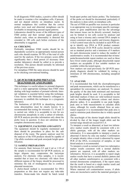
In all diagnostic FISH studies, a positive effort should<br />
be made to examine a few metaphase cells, if present,<br />
<strong>and</strong> not depend entirely on interphase nuclei. In<br />
normal metaphases this confirms that the correct<br />
probes were used <strong>and</strong> abnormal metaphases can be<br />
invaluable in interpreting unusual signal patterns.<br />
Laboratories should be aware of the different types of<br />
FISH probes <strong>and</strong> their normal signal pattern e.g.<br />
checker each, when an abnormality is detected but<br />
extended to 100 cells each before exclusion of the<br />
abnormality is suggested.<br />
6.6 CHECKING<br />
Preferably interphase FISH results should be independently<br />
scored by an appropriately trained person<br />
each who should examine 30-70% of the total of cells<br />
used for the analysis. If the two primary scores differ<br />
significantly then a third person (if necessary from<br />
another laboratory) should be called in to provide a<br />
resolution. This person should normally be informed<br />
of the previous scores.<br />
For metaphase FISH the same process should be used<br />
as for checking conventional b<strong>and</strong>ing.<br />
7. QF-PCR FOR RAPID PRENATAL<br />
DIAGNOSIS OF ANEUPLOIDY<br />
This technique is a useful adjunct to prenatal diagnosis<br />
<strong>and</strong> is a more appropriate technique than FISH when<br />
dealing with large numbers of prenatal referrals. Internal<br />
validation is essential before using this technique.<br />
Close liaison with Molecular Genetics colleagues is<br />
necessary to establish this technique in a cytogenetic<br />
laboratory.<br />
The limitations of QF-PCR in identifying chromosome<br />
abnormalities must be clearly known. It is<br />
recommended that testing for trisomies 13, 18 <strong>and</strong> 21<br />
is carried out, whilst it is acceptable to test for sex<br />
chromosome aneuploidy in only a subset of referrals.<br />
QF-PCR analysis provides information only about the<br />
probe locus in question. It does not substitute for a<br />
complete chromosome analysis.<br />
7.1 EQUIPMENT, FACILITIES AND SAFETY<br />
The equipment should be regularly maintained <strong>and</strong><br />
there should be procedures in place for the safe<br />
disposal of the waste. The genetic analyser used for<br />
the analysis of the STR products should be capable of<br />
2 bp allele resolution <strong>and</strong> peak area/peak height quantification.<br />
7.2 SAMPLE PREPARATION<br />
For amniotic fluid, between 0.5 <strong>and</strong> 4 ml or 1/10 of<br />
the sample is recommended for QF-PCR analysis, as<br />
larger aliquots may compromise the karyotype<br />
analysis. For chorionic villus samples, it is recommended<br />
that at least two chorionic villi taken from<br />
different regions of the biopsy should be processed to<br />
minimise the risk of misdiagnosis due to confined<br />
placental mosaicism.<br />
A chelex-based method is recommended for DNA<br />
preparation as this does not require any tube-tube<br />
transfers. Home-made kits should be batch tested<br />
23<br />
break-apart probes <strong>and</strong> fusion probes. The limitations<br />
of the probe set should be documented, particularly if<br />
the analysis is done solely on interphase cells.<br />
The use of FISH on paraffin wax sections in particular<br />
is an appropriate way to investigate specific rearrangements<br />
or gene amplification <strong>and</strong> has the advantage<br />
that tumour tissue can be directly screened. Analysis<br />
can be limited to ten cells scored by analyser <strong>and</strong><br />
using at least a trisomy <strong>and</strong> a normal DNA sample to<br />
ensure consistent assay quality <strong>and</strong> trisomy diagnosis.<br />
A H2O control must also be included in each PCR setup<br />
to identify any DNA or PCR product contamination.<br />
Between 24-26 PCR cycles should be carried<br />
out as st<strong>and</strong>ard practice <strong>and</strong> a minimum of 4 markers<br />
for each chromosome tested to reduce the number of<br />
uninformative results. It is recommended that tri/tetra/<br />
penta/hexanucleotide repeat markers are used as these<br />
have fewer stutter peaks, although dinucleotide repeat<br />
markers are acceptable if few suitable markers are<br />
available within the tested region.<br />
New markers not used previously for QF-PCR aneuploidy<br />
diagnosis should be validated by testing a<br />
minimum of 100 chromosomes, including aneuploid<br />
samples.<br />
7.3 ANALYSIS<br />
It is recommended that both the electrophoretogram<br />
<strong>and</strong> peak measurements, which can be transferred to a<br />
spreadsheet for convenience, are analysed. To ensure<br />
the quality of the data both minimum <strong>and</strong> maximum<br />
peak heights should be used. It is acceptable to fail<br />
individual markers if there are valid technical reasons<br />
such as bleedthrough between colours <strong>and</strong> electrophoretic<br />
spikes. It is acceptable to use peak height,<br />
peak area or both measurements to calculate allele<br />
ratios, although for results obtained from an automated<br />
sequencer it is recommended that peak area is<br />
used to minimise peak distortion due to widely-spaced<br />
alleles.<br />
The area/height of the shorter length allele should be<br />
divided by that of the longer length allele <strong>and</strong> the<br />
normal range should not exceed 0.8-1.4.<br />
To interpret a result as abnormal, at least two informative<br />
marker results should be consistent with a triallelic<br />
genotype, with all the other markers uninformative.<br />
It is unacceptable to interpret a result as<br />
abnormal if shown by only one marker. Confirmation<br />
of sample identity when a result is abnormal by repeat<br />
PCR of the DNA, re-extraction of samples, or mat<br />
blood analysis is recommended.<br />
To interpret a result as normal at least two informative<br />
marker results consistent with a normal diallelic<br />
pattern are required, with all other markers uninformative.<br />
However, it is acceptable to report single<br />
marker results that have a normal diallelic pattern <strong>and</strong><br />
all other markers uninformative as consistent with a<br />
normal chromosome complement, if the report states<br />
that the result is based on a single marker result <strong>and</strong><br />
that this the result must be confirmed.<br />
Where maternal cell contamination occurs, if allele<br />
ratios are inconclusive <strong>and</strong>/or the maternal genotype is