

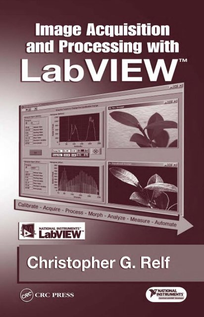
Image Acquisitionand Proces

You also want an ePaper? Increase the reach of your titles
YUMPU automatically turns print PDFs into web optimized ePapers that Google loves.
IMAGE PROCESSING SERIES<br />
Series Editor: Phillip A. Laplante, Pennsylvania Institute of Technology<br />
Published Titles<br />
Adaptive <strong>Image</strong> <strong>Proces</strong>sing: A Computational Intelligence Perspective<br />
Stuart William Perry, Hau-San Wong, and Ling Guan<br />
<strong>Image</strong> Acquisition and <strong>Proces</strong>sing with LabVIEW <br />
Christopher G. Relf<br />
<strong>Image</strong> and Video Compression for Multimedia Engineering<br />
Yun Q. Shi and Huiyang Sun<br />
Multimedia <strong>Image</strong> and Video <strong>Proces</strong>sing<br />
Ling Guan, S.Y. Kung, and Jan Larsen<br />
Shape Analysis and Classification: Theory and Practice<br />
Luciano da Fontoura Costa and Roberto Marcondes Cesar Jr.<br />
Software Engineering for <strong>Image</strong> <strong>Proces</strong>sing Systems<br />
Phillip A. Laplante
Library of Congress Cataloging-in-Publication Data<br />
Relf, Christopher G.<br />
<strong>Image</strong> acquisition and processing with LabVIEW / Christopher G. Relf<br />
p. cm. (<strong>Image</strong> processing series)<br />
Includes bibliographical references and index.<br />
ISBN 0-8493-1480-1<br />
1. <strong>Image</strong> processing--Digital techniques. 2. Engineering instruments--Data processing. 3.<br />
LabVIEW. I. Title. II. Series.<br />
TA1632.R44 2003<br />
621.36’7—dc21 2003046135<br />
CIP<br />
This book contains information obtained from authentic and highly regarded sources. Reprinted material<br />
is quoted with permission, and sources are indicated. A wide variety of references are listed. Reasonable<br />
efforts have been made to publish reliable data and information, but the author and the publisher cannot<br />
assume responsibility for the validity of all materials or for the consequences of their use.<br />
Neither this book nor any part may be reproduced or transmitted in any form or by any means, electronic<br />
or mechanical, including photocopying, microÞlming, and recording, or by any information storage or<br />
retrieval system, without prior permission in writing from the publisher.<br />
The consent of CRC Press LLC does not extend to copying for general distribution, for promotion, for<br />
creating new works, or for resale. SpeciÞc permission must be obtained in writing from CRC Press LLC<br />
for such copying.<br />
Direct all inquiries to CRC Press LLC, 2000 N.W. Corporate Blvd., Boca Raton, Florida 33431.<br />
Trademark Notice: Product or corporate names may be trademarks or registered trademarks, and are<br />
used only for identiÞcation and explanation, without intent to infringe.<br />
Visit the CRC Press Web site at www.crcpress.com<br />
© 2004 by CRC Press LLC<br />
No claim to original U.S. Government works<br />
International Standard Book Number 0-8493-1480-1<br />
Library of Congress Card Number 2003046135<br />
Printed in the United States of America 1 2 3 4 5 6 7 8 9 0<br />
Printed on acid-free paper
Foreword<br />
The introduction of LabVIEW over 16 years ago triggered the Virtual Instrumentation<br />
revolution that is still growing rapidly today. The tremendous advances in<br />
personal computers and consumer electronics continue to fuel this growth. For the<br />
same cost, today’s computers are about 100 times better than the machines of the<br />
LabVIEW 1 days, in CPU clock rate, RAM size, bus speed and disk size. This<br />
trend will likely continue for another 5 to 10 years.<br />
Virtual Instrumentation Þrst brought the connection of electronic instruments to<br />
computers, then later added the ability to plug measurement devices directly into<br />
the computer. Then, almost 7 years ago, National Instruments expanded the vision<br />
of virtual instrumentation when it introduced its Þrst image acquisition hardware<br />
along with the LabVIEW <strong>Image</strong> Analysis library. At the time, image processing on<br />
a personal computer was still a novelty requiring the most powerful machines and<br />
a lot of specialized knowledge on the part of the system developer. Since then,<br />
computer performance and memory size have continued to increase to the point<br />
where image processing is now practical on most modern PCs. In addition, the range<br />
of product offerings has expanded and higher-level software, such as Vision Builder,<br />
has become available to make development of image processing applications much<br />
easier.<br />
Today, image processing is fast becoming a mainstream component of Virtual<br />
Instrumentation. Very few engineers, however, have had experience with image<br />
processing or the lighting techniques required to capture images that can be processed<br />
quickly and accurately. Hence the need for a book like this one. Christopher<br />
Relf has written a very readable and enjoyable introduction to image processing,<br />
with clear and straightforward examples to illustrate the concepts, good references<br />
to more detailed information, and many real-world solutions to show the breadth of<br />
vision applications that are possible. The lucid (pun intended) description of the role<br />
of, and options for, lighting is itself worth the price of the book.<br />
Jeff Kodosky<br />
National Instruments Fellow<br />
Co-inventor of LabView
Preface<br />
<strong>Image</strong> Acquisition and <strong>Proces</strong>sing with LabVIEW Þlls a hole in the LabVIEW<br />
technical publication range. It is intended for competent LabVIEW programmers,<br />
as a general training manual for those new to National Instruments’ (NI) Vision<br />
application development and a reference for more-experienced vision programmers.<br />
It is assumed that readers have attained programming knowledge comparable to that<br />
taught in the NI LabVIEW Basics II course (see http://www.ni.com/training for a<br />
detailed course outline). The book covers introductions and theory of general image<br />
acquisition and processing topics, while providing more in-depth discussions and<br />
examples of speciÞc NI Vision tools.<br />
This book is a comprehensive IMAQ and Vision resource combining reference<br />
material, theory on image processing techniques, information on how LabVIEW and<br />
the NI Vision toolkit handle each technique, examples of each of their uses and realworld<br />
case studies, all in one book.<br />
This is not a “laboratory-style” book, and hence does not contain exercises for<br />
the reader to complete. Instead, the several coding examples, as referenced in the<br />
text, are included on an accompanying CD-ROM in the back of the book.<br />
The information contained in this book refers generally to the National Instruments<br />
Vision Toolkit version 6.1 (Figure 1). Several of the techniques explained herein<br />
may be perfectly functional using previous or future versions of the Vision Toolkit<br />
A glossary has also been compiled to deÞne subject-related words and acronyms.<br />
THE COMPANION CD-ROM<br />
Like most modern computer-related technical books, this one is accompanied<br />
by a companion CD-ROM that contains libraries of example images and code, as<br />
referenced in the text. Every wiring diagram shown in this book has corresponding<br />
source code on the CD-ROM for your convenience — just look in the respective<br />
chapter’s folder for a Þle with the same name as the image’s caption (Figure 2).<br />
To use the code, you will need:<br />
• LabVIEW 6.1 (or higher)<br />
• LabVIEW Vision Toolkit 6.1 (or higher)<br />
• An operating system that supports both of these components<br />
Some of the examples may also require the following components to be installed:<br />
• IMAQ<br />
• Vision Builder 6.1 (or higher)<br />
• IMAQ OCR Toolkit<br />
• IMAQ 1394 Drivers
FIGURE 1 Vision 6.1<br />
FIGURE 2 Companion CD-ROM Contents<br />
The CD-ROM also contains all of the example images used to test the code<br />
featured in the book, a demonstration version of National Instruments LabVIEW<br />
6.0, and a National Instruments IMAQ Demonstration that guides you through some<br />
of the features of NI-IMAQ.
Author<br />
Christopher G. Relf is an Industrial Automation Software<br />
Engineering consultant and LabVIEW specialist. Previously,<br />
his work at JDS Uniphase Pty Ltd (www.jdsu.com) included<br />
the automation of several complex processes, including the<br />
laser writing of optical FBGs (Fiber Bragg Gratings) and<br />
their measurement. Mr. Relf was part of a strong LabVIEW<br />
team that designed and implemented a plug-in-style software<br />
suite that introduced an “any product, any process, anywhere”<br />
paradigm to both the production and R&D phases<br />
of product creation. As a Computational Automation Scientist<br />
with the Division of Telecommunications and Industrial<br />
Physics, CSIRO, (www.tip.csiro.au), Australia’s premier scientiÞc<br />
and industrial research organization, he was the principal<br />
software engineer of several projects including the<br />
automation of thin Þlm Þltered arc deposition and metal shell<br />
eccentricity systems. He has consulted to the New South Wales Institute of Sport<br />
(www.nswis.com.au) and provided advice on the development of automated sport<br />
science data acquisition systems, used to test and train some of Australia’s premier<br />
athletes.<br />
Mr. Relf completed his undergraduate science degree in applied physics at the<br />
University of Technology, Sydney, where he Þrst learned the virtues of LabVIEW<br />
version 3, and has been a strong G programming advocate ever since. He gained his<br />
CertiÞed LabVIEW Developer qualiÞcation from National Instruments in 2002.<br />
Mr. Relf can be contacted via e-mail at Christopher.Relf@mBox.com.au
Acknowledgments<br />
My gratitude goes out to all of the people at CRC Press LLC who have held my<br />
hand through the production of this book. SpeciÞcally, thanks to Nora Konopka, my<br />
Acquisitions Editor, who showed immense faith in taking on and internally promoting<br />
this project; Helena Redshaw, my Editorial Project Development Supervisor,<br />
who kept me on the right track during the book’s development; and Sylvia Wood,<br />
my Project Editor, who worked closely with me to convert my ramblings into<br />
comprehensible format.<br />
The user solutions featured were written by some very talented LabVIEW and<br />
Vision specialists from around the world, most of whom I found lurking either on<br />
the Info-LabVIEW Mailing List (http://www.info-labview.org), or the NI-Zone Discussion<br />
Forum (http://www.zone.ni.com). Theoretical descriptions of the Vision<br />
capabilities of LabVIEW are all very well, but it is only when you build a program<br />
that ties the individual functionalities together to form a useful application that you<br />
realize the true value of their use. I hope their stories help you to see the big picture<br />
(pun intended), and to realize that complete solutions can be created around core<br />
Vision components. Thank you for the stories and examples showing some of the<br />
real-world applications achievable using the techniques covered in the book.<br />
A big thank you goes out to my two very talented reviewers, Edward Lipnicki<br />
and Peter Badcock, whose knowledge of theory often overshadowed my scientiÞc<br />
assumptions — this book would not be anywhere near as comprehensive without<br />
your help. Thanks also to my proofreaders, Adam Batten, Paul Conroy, Archie<br />
Garcia, Walter Kalceff, James McDonald, Andrew Parkinson, Paul Relf, Anthony<br />
Rochford, Nestor Sanchez, Gil Smith, Glen Trudgett and Michael Wallace, who<br />
helped me correct most (hopefully) of the mistakes I made in initial manuscripts.<br />
The section on application lighting is based primarily on information and images<br />
I gathered from NER (a Robotic Vision Systems Incorporated company) — thank<br />
you to Greg Dwyer from the Marketing Communications group for assisting me in<br />
putting it all together. Further information regarding NER’s extensive range of<br />
industrial lighting products can be found at their Web site (http://www.nerlite.com).<br />
I could not have dreamed of writing this book without the assistance of my local<br />
National Instruments staff, Jeremy Carter (Australia and New Zealand Branch Manager)<br />
and Alex Gouliaev (Field Sales Engineer). Your product-related assistance and<br />
depth of knowledge of NI products and related technologies were invaluable and<br />
nowhere short of astounding.<br />
My apologies to my family, my friends and all of my English teachers. This<br />
book is pitched primarily to readers in the United States, so I have had to abandon<br />
many of the strict rules of English spelling and grammar that you tried so hard to<br />
instill in me. I hope that you can forgive me.
Thanks must also go to Ed, Gleno, Jase, Rocko and Adrian, who were always<br />
more than willing to help me relax with a few schooners between (and occasionally<br />
during) chapters.<br />
Those who have written a similar-level technical book understand the draining<br />
exercise that it is, and my love and humble appreciation go to my family and friends,<br />
who supported me in many ways through this project. Ma and Paul, who planted<br />
the “you-can-do-anything” seed, and Ruth, who tends the resulting tree every day:<br />
I couldn’t have done it without you all.<br />
Christopher G. Relf
Dedication<br />
To Don “old fella” Price.<br />
I appreciate your inspiration, support, friendship<br />
and, above all else, your good humor.
Introduction<br />
Of the Þve senses we use daily, we rely most on our sense of sight. Our eyesight<br />
provides 80% of the information we absorb during daylight hours, and it is no<br />
wonder, as nearly three quarters of the sensory receptor cells in our bodies are located<br />
in the back of our eyes, at the retinas. Many of the decisions we make daily are<br />
based on what we can see, and how we in turn interpret that data — an action as<br />
common as driving a car is only possible for those who have eyesight.<br />
As computer-controlled robotics play an ever-increasing role in factory production,<br />
scientiÞc, medical and safety Þelds, surely one of the most important and<br />
intuitive advancements we can exploit is the ability to acquire, process and make<br />
decisions based on image data.<br />
LabVIEW is a popular measurement and automation programming language,<br />
developed by National Instruments (NI). Initially aimed squarely at scientists and<br />
technicians to assist in simple laboratory automation and data acquisition, LabVIEW<br />
has grown steadily to become a complete programming language in its own right,<br />
with add-on toolkits that cover anything from Internet connectivity and database<br />
access to fuzzy logic and image processing. The image-based toolkit (called Vision)<br />
is particularly popular, simplifying the often-complex task of not only downloading<br />
appropriate quality images into a computer, but also making multifarious processing<br />
tasks much easier than many other packages and languages.<br />
This book is intended for competent LabVIEW programmers, as a general<br />
training manual for those new to NI Vision application development and a reference<br />
for more-experienced Vision programmers. It is assumed that you have attained<br />
programming knowledge comparable to that taught in the NI LabVIEW Basics II<br />
course.<br />
I sincerely hope that you will learn much from this book, that it will aid you in<br />
developing and including Vision components conÞdently in your applications, thus<br />
harnessing one of the most complex, yet common data sources — the photon.
Contents<br />
Chapter 1<br />
<strong>Image</strong> Types and File Management.....................................................1<br />
1.1 Types of <strong>Image</strong>s ...............................................................................................1<br />
1.1.1 Grayscale..............................................................................................2<br />
1.1.2 Color.....................................................................................................3<br />
1.1.3 Complex ...............................................................................................4<br />
1.2 File Types .........................................................................................................4<br />
1.2.1 Modern File Formats ...........................................................................5<br />
1.2.1.1 JPEG .....................................................................................7<br />
1.2.1.2 TIFF ......................................................................................7<br />
1.2.1.3 GIF ........................................................................................7<br />
1.2.1.4 PNG.......................................................................................8<br />
1.2.1.5 BMP ......................................................................................8<br />
1.2.1.6 AIPD .....................................................................................8<br />
1.2.1.7 Other Types...........................................................................8<br />
1.3 Working with <strong>Image</strong> Files ...............................................................................9<br />
1.3.1 Standard <strong>Image</strong> Files ...........................................................................9<br />
1.3.2 Custom and Other <strong>Image</strong> Formats.....................................................12<br />
Chapter 2 Setting Up ..........................................................................................15<br />
2.1 Cameras..........................................................................................................15<br />
2.1.1 Scan Types .........................................................................................16<br />
2.1.1.1 Progressive Area Scan ........................................................16<br />
2.1.1.2 Interlaced Area Scan...........................................................16<br />
2.1.1.3 Interlacing Standards ..........................................................17<br />
2.1.1.4 Line Scan ............................................................................18<br />
2.1.1.5 Camera Link .......................................................................19<br />
2.1.1.6 Thermal...............................................................................20<br />
2.1.2 Camera Advisors: Web-Based Resources..........................................20<br />
2.1.3 User Solution: X-Ray Inspection System..........................................22<br />
2.1.3.1 Introduction.........................................................................22<br />
2.1.3.2 Control and Imaging Hardware Configuration ..................23<br />
2.1.3.3 Control Software.................................................................23<br />
2.1.3.4 Imaging Software................................................................24<br />
2.1.3.5 Conclusions.........................................................................25<br />
2.2 <strong>Image</strong> Acquisition Hardware .........................................................................26<br />
2.2.1 National Instruments Frame Grabbers...............................................26
2.2.2 IEEE 1394 (FireWire) Systems .........................................................26<br />
2.2.3 USB Systems......................................................................................29<br />
2.3 From Object to Camera .................................................................................30<br />
2.3.1 Resolution...........................................................................................30<br />
2.3.2 Depth of Field (DOF) ........................................................................31<br />
2.3.3 Contrast (or Modulation) ...................................................................32<br />
2.3.4 Perspective (Parallax).........................................................................33<br />
2.3.4.1 Software Calibration...........................................................34<br />
2.3.4.2 Telecentricity.......................................................................35<br />
2.3.5 National Instruments Lens Partners...................................................37<br />
2.4 Lighting ..........................................................................................................37<br />
2.4.1 Area Arrays ........................................................................................38<br />
2.4.2 Ring ....................................................................................................38<br />
2.4.3 Dark Field ..........................................................................................39<br />
2.4.4 Dome ..................................................................................................40<br />
2.4.5 Backlights...........................................................................................41<br />
2.4.6 Continuous Diffuse Illuminator (CDI) ..........................................42<br />
2.4.7 Diffuse on Axis Lights (DOAL) and Collimated on Axis<br />
Lights (COAL) ...................................................................................43<br />
2.4.8 Square Continuous Diffuse Illuminators (SCDI) ..........................44<br />
Chapter 3 <strong>Image</strong> Acquisition ..............................................................................47<br />
3.1 Configuring Your Camera ..............................................................................47<br />
3.2 Acquisition Types...........................................................................................50<br />
3.2.1 Snap....................................................................................................51<br />
3.2.2 Grab....................................................................................................52<br />
3.2.3 Sequence.............................................................................................52<br />
3.3 NI-IMAQ for IEEE 1394...............................................................................54<br />
3.4 User Solution: Webcam <strong>Image</strong> Acquisition ..................................................56<br />
3.4.1 Introduction ........................................................................................56<br />
3.4.2 Functionality.......................................................................................57<br />
3.4.3 Continuous Capture with <strong>Proces</strong>sing and Display............................58<br />
3.4.4 Future Work........................................................................................58<br />
3.5 Other Third-Party <strong>Image</strong> Acquisition Software ............................................59<br />
3.6 Acquiring A VGA Signal...............................................................................59<br />
3.7 TWAIN <strong>Image</strong> Acquisition ............................................................................59<br />
3.8 User Solution: Combining High-Speed Imaging and Sensors<br />
for Fast Event Measurement..........................................................................60<br />
3.8.1 The Challenge ....................................................................................60<br />
3.8.2 The Solution.......................................................................................60<br />
3.8.3 System Considerations.......................................................................62<br />
3.8.4 System Performance ..........................................................................63<br />
3.8.5 Applications .......................................................................................63<br />
3.8.6 Summary ............................................................................................63
Chapter 4<br />
Displaying <strong>Image</strong>s..............................................................................65<br />
4.1 Simple Display Techniques............................................................................65<br />
4.2 Displaying <strong>Image</strong>s within Your Front Panel .................................................67<br />
4.2.1 The Intensity Graph ...........................................................................67<br />
4.2.2 The Picture Indicator .........................................................................68<br />
4.2.3 Embedding an IMAQ Display Window ............................................71<br />
4.3 The <strong>Image</strong> Browser........................................................................................74<br />
4.4 Overlay Tools .................................................................................................77<br />
4.4.1 Overlaying Text..................................................................................77<br />
4.4.2 Overlaying Shapes .............................................................................77<br />
4.4.3 Overlaying Bitmaps ...........................................................................78<br />
4.4.4 Combining Overlays ..........................................................................79<br />
4.5 The Vision Window Tools Palette..................................................................81<br />
4.5.1 Available Tools...................................................................................81<br />
4.5.2 Vision Tools Palette Hints..................................................................84<br />
Chapter 5 <strong>Image</strong> <strong>Proces</strong>sing ...............................................................................85<br />
5.1 The ROI (Region of Interest) ........................................................................85<br />
5.1.1 Simple ROI Use .................................................................................86<br />
5.1.1.1 Line .....................................................................................86<br />
5.1.1.2 Square and Rectangle .........................................................86<br />
5.1.1.3 Oval.....................................................................................88<br />
5.1.2 Complex ROIs....................................................................................88<br />
5.1.2.1 Rotated Rectangle...............................................................89<br />
5.1.2.2 Annulus Arc........................................................................89<br />
5.1.2.3 ROI Tracing Example.........................................................92<br />
5.1.3 Manually Building a ROI: The “ROI Descriptor” ............................93<br />
5.2 User Solution: Dynamic Microscopy in Brain Research..............................98<br />
5.2.1 Program Description ..........................................................................99<br />
5.2.2 Recording Video Sequences...............................................................99<br />
5.2.2.1 Analysis of Video Sequences ...........................................100<br />
5.2.3 Summary ..........................................................................................101<br />
5.3 Connectivity .................................................................................................102<br />
5.4 Basic Operators............................................................................................103<br />
5.4.1 Add (Combine) ................................................................................103<br />
5.4.2 Subtract (Difference)........................................................................105<br />
5.4.3 Other Basic Operators......................................................................108<br />
5.4.4 Averaging .........................................................................................108<br />
5.5 User Solution: <strong>Image</strong> Averaging with LabVIEW .......................................110<br />
5.6 Other Tools...................................................................................................113<br />
5.6.1 Symmetry (Mirroring an <strong>Image</strong>) .....................................................113<br />
5.6.2 Rotate ...............................................................................................115<br />
5.6.3 Unwrap .............................................................................................116<br />
5.6.4 3D View............................................................................................117<br />
5.6.5 Thresholding.....................................................................................117
5.6.6 Equalization......................................................................................122<br />
5.7 User Solution: QuickTime for LabVIEW ...................................................125<br />
5.7.1 QuickTime Integration to LabVIEW...............................................125<br />
5.7.1.1 QTLib: The QuickTime Library.......................................126<br />
5.7.1.2 Single <strong>Image</strong> Reading and Writing..................................126<br />
5.7.1.3 Reading Movies ................................................................126<br />
5.7.1.4 Writing Movies.................................................................127<br />
5.7.1.5 Video Grabbing (Figure 5.61) ..........................................127<br />
5.7.1.6 <strong>Image</strong> Transformation Functions......................................128<br />
5.7.1.7 Future Versions of QTLib.................................................129<br />
5.7.1.8 For More Information.......................................................129<br />
5.8 Filters............................................................................................................130<br />
5.8.1 Using Filters.....................................................................................131<br />
5.8.2 Predefined Filters .............................................................................132<br />
5.8.2.1 Gaussian............................................................................132<br />
5.8.2.2 Gradient.............................................................................133<br />
5.8.2.3 Laplacian...........................................................................135<br />
5.8.2.4 Smoothing.........................................................................137<br />
5.8.3 Creating Your Own Filter.................................................................137<br />
Chapter 6<br />
Morphology......................................................................................141<br />
6.1 Simple Morphology Theory.........................................................................141<br />
6.1.1 Dilation.............................................................................................141<br />
6.1.2 Erosion .............................................................................................142<br />
6.1.3 Closing .............................................................................................142<br />
6.1.4 Opening ............................................................................................143<br />
6.1.5 Nonbinary Morphology....................................................................143<br />
6.2 Practical Morphology...................................................................................144<br />
6.3 The Structuring Element..............................................................................145<br />
6.4 Specific Morphological Functions...............................................................146<br />
6.4.1 Particle Removal ..............................................................................146<br />
6.4.2 Filling Particle Holes .......................................................................147<br />
6.4.3 Distance Contouring ........................................................................148<br />
6.4.3.1 IMAQ Distance.................................................................148<br />
6.4.3.2 IMAQ Danielsson .............................................................150<br />
6.4.4 Border Rejection ..............................................................................151<br />
6.4.5 Finding and Classifying Circular Objects .......................................153<br />
6.4.6 Particle Separation ...........................................................................156<br />
6.5 Case Study: Find and Classify Irregular Objects........................................157<br />
6.6 User Solution: Digital Imaging and Communications<br />
in Medicine (DICOM) for LabVIEW .........................................................159<br />
6.6.1 The Hardware...................................................................................160<br />
6.6.2 The Software ....................................................................................160<br />
6.6.3 Summary ..........................................................................................163<br />
6.6.4 Acknowledgments ............................................................................163
Chapter 7<br />
<strong>Image</strong> Analysis.................................................................................165<br />
7.1 Searching and Identifying (Pattern Matching) ............................................165<br />
7.1.1 The Template....................................................................................166<br />
7.1.2 The Search........................................................................................166<br />
7.1.2.1 Cross Correlation..............................................................167<br />
7.1.2.2 Scale Invariant and Rotation Invariant Matching ............168<br />
7.1.2.3 Pyramidal Matching..........................................................169<br />
7.1.2.4 Complex Pattern Matching Techniques............................170<br />
7.1.3 A Practical Example ........................................................................171<br />
7.2 User Solution: Connector Pin Inspection Using Vision..............................175<br />
7.2.1 The Software ....................................................................................176<br />
7.3 Mathematical Manipulation of <strong>Image</strong>s........................................................178<br />
7.3.1 <strong>Image</strong> to Two-Dimensional Array ...................................................178<br />
7.3.2 <strong>Image</strong> Portions .................................................................................178<br />
7.3.3 Filling <strong>Image</strong>s ..................................................................................181<br />
7.4 User Solution: <strong>Image</strong> <strong>Proces</strong>sing with Mathematica Link for LabVIEW .183<br />
7.4.1 A Typical LabVIEW/Mathematica Hybrid Workflow.....................183<br />
7.4.2 Taking the Next Step .......................................................................184<br />
7.4.3 LabVIEW/Mathematica Hybrid Applications .................................186<br />
7.4.4 Histograms and Histographs............................................................187<br />
7.5 User Solution: Automated Instrumentation for the Assessment of<br />
Peripheral Vascular Function .......................................................................189<br />
7.5.1 The Hardware...................................................................................190<br />
7.5.2 The Software ....................................................................................190<br />
7.5.2.1 DAQ ..................................................................................190<br />
7.5.2.2 Postprocessing and Analysis ............................................191<br />
7.5.3 Summary ..........................................................................................192<br />
7.6 Intensity Profiles ..........................................................................................194<br />
7.7 Particle Measurements .................................................................................194<br />
7.7.1 Simple Automated Object Inspection..............................................194<br />
7.7.2 Manual Spatial Measurements.........................................................197<br />
7.7.3 Finding Edges Programmatically.....................................................199<br />
7.7.4 Using the Caliper Tool to Measure Features...................................201<br />
7.7.5 Raking an <strong>Image</strong> for Features .........................................................203<br />
7.7.5.1 Parallel Raking..................................................................203<br />
7.7.5.2 Concentric Raking ............................................................204<br />
7.7.5.3 Spoke Raking....................................................................205<br />
7.8 User Solution: Sisson-Ammons Video Analysis (SAVA) of CBFs ............207<br />
7.8.1 Introduction ......................................................................................207<br />
7.8.2 Measurement of Cilia Beat Frequency (CBF) ................................207<br />
7.8.3 Video Recording ..............................................................................208<br />
7.8.4 Single Point Analysis.......................................................................209<br />
7.8.5 Whole <strong>Image</strong> Analysis.....................................................................209<br />
7.8.6 Design Challenges ..........................................................................210<br />
7.8.7 Future Directions..............................................................................210
7.8.8 Results ..............................................................................................210<br />
7.9 Analytical Geometry....................................................................................211<br />
7.9.1 Measuring Between Points ..............................................................211<br />
7.9.2 Lines Intersection.............................................................................211<br />
7.9.3 Line Fitting.......................................................................................213<br />
7.9.4 Circle and Ellipse Fitting.................................................................214<br />
Chapter 8 Machine Vision ................................................................................217<br />
8.1 Optical Character Recognition.....................................................................218<br />
8.1.1 Recognition Configuration...............................................................218<br />
8.1.2 Correction Configuration .................................................................218<br />
8.1.3 OCR <strong>Proces</strong>sing ...............................................................................220<br />
8.1.4 Practical OCR ..................................................................................221<br />
8.1.5 Distributing Executables with Embedded OCR Components ........221<br />
8.2 Parsing Human–Machine Information.........................................................223<br />
8.2.1 7-Segment LCDs..............................................................................223<br />
8.2.2 Barcodes ...........................................................................................224<br />
8.2.2.1 One-Dimensional ..............................................................225<br />
8.2.2.2 Two-Dimensional..............................................................225<br />
8.2.3 Instrument Clusters ..........................................................................227<br />
Glossary ................................................................................................................229<br />
Bibliography .........................................................................................................237<br />
Index......................................................................................................................239
1<br />
<strong>Image</strong> Types and File<br />
Management<br />
Files provide an opportunity to store and retrieve image information using media<br />
such as hard drives, CD-ROMs and ßoppy disks. Storing Þles also allows us a method<br />
of transferring image data from one computer to another, whether by Þrst saving it<br />
to storage media and physically transferring the Þle to another PC, or by e-mailing<br />
the Þles to a remote location.<br />
There are many different image Þle types, all with advantages and disadvantages,<br />
including compression techniques that make traditionally large image Þle footprints<br />
smaller, to the number of colors that the Þle type will permit. Before we learn about<br />
the individual image Þle types that the Vision Toolkit can work with, let us consider<br />
the image types themselves.<br />
1.1 TYPES OF IMAGES<br />
The Vision Toolkit can read and manipulate raster images. A raster image is broken<br />
into cells called pixels, where each pixel contains the color or grayscale intensity<br />
information for its respective spatial position in the image (Figure 1.1).<br />
Machine vision cameras acquire images in raster format as a representation of<br />
the light falling on to a charged-coupled device (CCD; see Chapter 2 for information<br />
regarding image acquisition hardware). This information is then transmitted to the<br />
computer through a standard data bus or frame grabber.<br />
<strong>Image</strong><br />
Pixelated Raster <strong>Image</strong> Representation<br />
FIGURE 1.1 Raster image.<br />
1
2 <strong>Image</strong> Acquisition <strong>Proces</strong>sing with LabVIEW<br />
An image’s type does not necessarily deÞne its image Þle type, or vice versa.<br />
Some image types work well with certain image Þle types, and so it is important to<br />
understand the data format of an image before selecting the Þle type to be used for<br />
storage. The Vision Toolkit is designed to process three image types:<br />
1. Grayscale<br />
2. Color<br />
3. Complex<br />
1.1.1 GRAYSCALE<br />
Grayscale images are the simplest to consider, and are the type most frequently<br />
demonstrated in this book. Grayscale images consist of x and y spatial coordinates<br />
and their respective intensity values. Grayscale images can be thought of as surface<br />
graphs, with the z axis representing the intensity of light. As you can see from Figure<br />
1.2, the brighter areas in the image represent higher z-axis values. The surface plot<br />
has also been artiÞcially shaded to further represent the intensity data.<br />
The image’s intensity data is represented by its depth, which is the range of<br />
intensities that can be represented per pixel. For a bit depth of x, the image is said to<br />
have a depth of 2 x , meaning that each pixel can have an intensity value of 2 x levels.<br />
The Vision Toolkit can manipulate grayscale images with the following depths:<br />
Bit Depth Pixel Depth Intensity Extremities<br />
8 bit<br />
Signed: 0 (dark) to 255 (light)<br />
Unsigned: –127 to 126<br />
8 bits of data (1 byte)<br />
16 bit<br />
Signed: 0 to 65536<br />
Unsigned: –32768 to 32767<br />
1 byte 1 byte<br />
32 bit<br />
Signed: 0 to 4294967296<br />
Unsigned: –2147483648 to 2147483647<br />
1 byte 1 byte 1 byte 1 byte<br />
Differing bit depths exist as a course of what is required to achieve an appropriate<br />
imaging solution. Searching for features in an image is generally achievable using<br />
8-bit images, whereas making accurate intensity measurements requires a higher bit<br />
depth. As you might expect, higher bit depths require more memory (both RAM<br />
and Þxed storage), as the intensity values stored for each pixel require more data<br />
space. The memory required for a raw image is calculated as:<br />
MemoryRequired = Resolution ¥ Resolution ¥ BitDepth<br />
For example, a 1024 ¥ 768 8-bit grayscale would require:<br />
MemoryRequired = 1024 ¥ 768 ¥ 8<br />
= 6291456Bits<br />
= 786432Bytes<br />
= 768kBytes<br />
x<br />
y
<strong>Image</strong> Types and File Management 3<br />
FIGURE 1.2 <strong>Image</strong> data represented as a surface plot.<br />
Increasing the bit depth of the image to 16 bits also increases the amount of memory<br />
required to store the image in a raw format:<br />
MemoryRequired = 1024 ¥ 768 ¥ 16<br />
= 12582912Bits<br />
= 1572864Bytes<br />
= 1.536MBytes<br />
You should therefore select an image depth that corresponds to the next-highest level<br />
above the level required, e.g., if you need to use a 7-bit image, select 8 bits, but not 16 bits.<br />
<strong>Image</strong>s with bit depths that do not correspond to those listed in the previous<br />
table (e.g., 1, 2 and 4-bit images) are converted to the next-highest acceptable bit<br />
depths when they are initially loaded.<br />
1.1.2 COLOR<br />
Color images are represented using either the Red-Green-Blue (RGB) or Hue-<br />
Saturation-Luminance (HSL) models. The Vision Toolkit accepts 32-bit color images<br />
that use either of these models, as four 8-bit channels:<br />
Color<br />
Model<br />
Pixel Depth<br />
Channel Intensity<br />
Extremities<br />
RGB a Red Green Blue 0 to 255<br />
HSL a Hue Saturation Luminance 0 to 255<br />
The alpha (a) component describes the opacity of an image, with zero representing<br />
a clear pixel and 255 representing a fully opaque pixel. This enables an<br />
image to be rendered over another image, with some of the underlying image<br />
showing through. When combining images, alpha-based pixel formats have several<br />
advantages over color-keyed formats, including support for shapes with soft or
4 <strong>Image</strong> Acquisition <strong>Proces</strong>sing with LabVIEW<br />
antialiased edges, and the ability to paste the image over a background with the<br />
foreground information seemingly blending in. Generally, this form of transparency<br />
is of little use to the industrial vision system user, so the Vision Toolkit ignores all<br />
forms of a information.<br />
The size of color images follows the same relationship as grayscale images, so<br />
a 1024 ¥ 768 24-bit color image (which equates to a 32-bit image including 8 bits<br />
for the alpha channel) would require the following amount of memory:<br />
MemoryRequired = 1024 ¥ 768 ¥ 32<br />
= 25165824 Bits<br />
= 3145728 Bytes<br />
= 3.072 MBytes<br />
1.1.3 COMPLEX<br />
Complex images derive their name from the fact that their representation includes<br />
real and complex components. Complex image pixels are stored as 64-bit ßoatingpoint<br />
numbers, which are constructed with 32-bit real and 32-bit imaginary parts.<br />
Pixel Depth<br />
Channel Intensity Extremities<br />
Real (4 bytes) Imaginary (4 bytes) –2147483648 to 2147483647<br />
A complex image contains frequency information representing a grayscale<br />
image, and therefore can be useful when you need to apply frequency domain<br />
processes to the image data. Complex images are created by performing a fast Fourier<br />
transform (FFT) on a grayscale image, and can be converted back to their original<br />
state by applying an inverse FFT. Magnitude and phase relationships can be easily<br />
extracted from complex images.<br />
1.2 FILE TYPES<br />
Some of the earliest image Þle types consisted of ASCII text-delimited strings, with<br />
each delimiter separating relative pixel intensities. Consider the following example:<br />
0 0 0 0 0 0 0 0 0 0 0<br />
0 0 0 125 125 125 125 125 0 0 0<br />
0 0 125 25 25 25 25 25 125 0 0<br />
0 125 25 25 255 25 255 25 25 125 0<br />
0 125 25 25 25 25 25 25 25 125 0<br />
0 125 25 25 25 255 25 25 25 125 0<br />
0 125 25 255 25 25 25 255 25 125 0<br />
0 125 5 25 255 255 255 25 25 125 0<br />
0 0 125 25 25 25 25 25 125 0 0<br />
0 0 0 125 125 125 125 125 0 0 0<br />
0 0 0 0 0 0 0 0 0 0 0
<strong>Image</strong> Types and File Management 5<br />
At Þrst look, these numbers may seem random, but when their respective pixel<br />
intensities are superimposed under them, an image forms:<br />
0 0 0 0 0 0 0 0 0 0 0<br />
0 0 0 125 125 125 125 125 0 0 0<br />
0 0 125 25 25 25 25 25 125 0 0<br />
0 125 25 25 255 25 255 25 25 125 0<br />
0 125 25 25 25 25 25 25 25 125 0<br />
0 125 25 25 25 255 25 25 25 125 0<br />
0 125 25 255 25 25 25 255 25 125 0<br />
0 125 25 25 255 255 255 25 25 125 0<br />
0 0 125 25 25 25 25 25 125 0 0<br />
0 0 0 125 125 125 125 125 0 0 0<br />
0 0 0 0 0 0 0 0 0 0 0<br />
Synonymous with the tab-delimited spreadsheet Þle, this type of image Þle is easy<br />
to understand, and hence simple to manipulate. Unfortunately, Þles that use this<br />
structure tend to be large and, consequently, mathematical manipulations based on<br />
them are slow. The size of this example (11 ¥ 11 pixels with 2-bit color) would<br />
yield a dataset size of:<br />
MemoryRequired = 11 ¥ 11 ¥ 2<br />
= 242Bits<br />
ª 30Bytes + delimter character bytes<br />
In the current era of cheap RAM and when hard drives are commonly available<br />
in the order of hundreds of gigabytes, 30 bytes may not seem excessive, but storing<br />
the intensity information for each pixel individually is quite inefÞcient.<br />
1.2.1 MODERN FILE FORMATS<br />
<strong>Image</strong> Þles can be very large, and they subsequently require excessive amounts of<br />
hard drive and RAM space, more disk access, long transfer times, and slow image<br />
manipulation. Compression is the process by which data is reduced to a form that<br />
minimizes the space required for storage and the bandwidth required for transmittal,<br />
and can be either lossy or lossless. As its name suggests, lossless compression occurs
6 <strong>Image</strong> Acquisition <strong>Proces</strong>sing with LabVIEW<br />
FIGURE 1.3 Lossy spatial compression example.<br />
when the data Þle size is decreased, without the loss of information. Lossless<br />
compression routines scan the input image and calculate a more-efÞcient method of<br />
storing the data, without changing the data’s accuracy. For example, a lossless<br />
compression routine may convert recurring patterns into short abbreviations, or<br />
represent the image based on the change of pixel intensities, rather than each intensity<br />
itself.<br />
Lossy compression discards information based on a set of rules, ultimately<br />
degrading the data. For example, an image that is very large can often be resized to<br />
become a little smaller, effectively decreasing the amount of information, yet retaining<br />
the large features of the original image. Consider the example in Figure 1.3.<br />
The smaller barcode image has been resized to 75% of the original image, but<br />
the resolution is still high enough to read the features required. Some lossy compression<br />
routines store color information at a lower resolution than the source image.<br />
For example, a photo-realistic image contains millions of colors, and a machine<br />
vision system may only need to Þnd edges within the image. Often the number of<br />
colors used to represent the image can be dramatically decreased (some vision<br />
routines require images to be resampled to binary levels — black and white).<br />
FIGURE 1.4 Finding edges: Different number of colors.
<strong>Image</strong> Types and File Management 7<br />
Consider the example in Figure 1.4. An edge detection routine has been<br />
executed on two similar images. The Þrst is an 8-bit grayscale image (which<br />
contains 256 colors), and the second is a binary (2 colors) image. Although the<br />
amount of color data has been signiÞcantly reduced in the second image, the edge<br />
detection routine has completed successfully. In this example, decreasing the<br />
image’s color depth to a binary level may actually improve the detection of edges;<br />
the change between background and object is from one extreme to the other,<br />
whereas the steps in the 256-color image are more gradual, making the detection<br />
of edges more difÞcult.<br />
1.2.1.1 JPEG<br />
Of the Þve image Þle formats that the Vision Toolkit supports by default, JPEG<br />
(Joint Photographic Experts Group) Þles are probably the most common. The JPEG<br />
format is optimized for photographs and similar continuous tone images that contain<br />
a large number of colors, and can achieve astonishing compression ratios, even while<br />
maintaining a high image quality. The JPEG compression technique analyzes images,<br />
removes data that is difÞcult for the human eye to distinguish, and stores the resulting<br />
data as a 24-bit color image. The level of compression used in the JPEG conversion<br />
is deÞnable, and photographs that have been saved with a JPEG compression level<br />
of up to 15 are often difÞcult to distinguish from their source images, even at high<br />
magniÞcation.<br />
1.2.1.2 TIFF<br />
Tagged <strong>Image</strong> File Format (TIFF) Þles are very ßexible, as the routine used to<br />
compress the image Þle is stored within the Þle itself. Although this suggests that<br />
TIFF Þles can undergo lossy compression, most applications that use the format use<br />
only lossless algorithms. Compressing TIFF images with the Lempel-Zev-Welch<br />
(LZW) algorithm (created by Unisys), which requires special licensing, has recently<br />
become popular.<br />
1.2.1.3 GIF<br />
GIF (CompuServe Graphics Interchange File) images use a similar LZW compression<br />
algorithm to that used within TIFF images, except the bytes are reversed and<br />
the string table is upside-down. All GIF Þles have a color palette, and some can be<br />
interlaced so that raster lines can appear as every four lines, then every eight lines,<br />
then every other line. This makes the use of GIFs in a Web page very attractive<br />
when slow download speeds may impede the viewing of an image. Compression<br />
using the GIF format creates a color table of 256 colors; therefore, if the image has<br />
fewer than 256 colors, GIF can render the image exactly. If the image contains more<br />
than 256 colors, the GIF algorithm approximates the colors in the image with the<br />
limited palette of 256 colors available. Conversely, if a source image contains less<br />
than 256 colors, its color table is expanded to cover the full 256 graduations, possibly
8 <strong>Image</strong> Acquisition <strong>Proces</strong>sing with LabVIEW<br />
resulting in a larger Þle size. The GIF compression process also uses repeating pixel<br />
colors to further compress the image. Consider the following row of a raster image:<br />
125 125 125 125 125 125 126 126 126 126 Þ 6(125)4(126)<br />
Instead of represented each pixel’s intensity discreetly, a formula is generated that<br />
takes up much less space.<br />
1.2.1.4 PNG<br />
The Portable Network Graphics Þle format is also a lossless storage format that<br />
analyzes patterns within the image to compress the Þle. PNG is an excellent replacement<br />
for GIF images, and unlike GIF, is patent-free. PNG images can be indexed<br />
color, true color or grayscale, with color depths from 1 to 16 bits, and can support<br />
progressive display, so they are particularly suited to Web pages. Most image formats<br />
contain a header, a small section at the start of the Þle where information regarding<br />
the image, the application that created it, and other nonimage data is stored. The<br />
header that exists in a PNG Þle is programmatically editable using code developed<br />
by a National Instruments’ Applications Engineer. You are able to store multiple<br />
strings (with a maximum length of 64 characters) at an unlimited number of indices.<br />
Using this technique, it is possible to “hide” any type of information in a PNG<br />
image, including text, movies and other images (this is the method used to store<br />
region of interest (ROI) information in a PNG Þle). The source code used to realize<br />
this technique is available at the National Instruments Developer Zone at<br />
http://www.zone.ni.com (enter “Read and Write Custom Strings in a PNG” in the<br />
Search box).<br />
1.2.1.5 BMP<br />
Bitmaps come in two varieties, OS/2 and Windows, although the latter is by far the<br />
most popular. BMP Þles are uncompressed, support both 8-bit grayscale and color,<br />
and can store calibration information about the physical size of the image along<br />
with the intensity data.<br />
1.2.1.6 AIPD<br />
AIPD is a National Instruments uncompressed format used internally by LabVIEW<br />
to store images of Floating Point, Complex and HSL, along with calibration information<br />
and other speciÞcs.<br />
1.2.1.7 Other Types<br />
Although the formats listed in this chapter are the default types that the Vision<br />
Toolkit can read, several third-party libraries exist that extend the range of Þle types<br />
supported. One such library, called “The <strong>Image</strong> Toolbox,” developed by George Zou,<br />
allows your vision code to open icon (.ico), Windows MetaÞle (.wmf), ZSoft Cor-
<strong>Image</strong> Types and File Management 9<br />
poration Picture (.pcx) Þles and more. The <strong>Image</strong> Toolbox also contains other<br />
functions such as high-quality image resizing, color-grayscale and RGB-HSL conversion<br />
utilities. More information (including shareware downloads) about George<br />
Zou’s work can be found at http://gtoolbox.yeah.net.<br />
FIGURE 1.5 AVI external module for IMAQ vision builder.<br />
AllianceVision has released a LabVIEW utility that allows the extraction of<br />
images from an AVI movie Þle. The utility can be used as a stand-alone application,<br />
or can be tightly integrated into the National Instruments Vision Builder<br />
(Figure 1.5).<br />
The utility also allows the creation of AVIs from a series of images, including<br />
movie compression codecs. More information about the Alliance Vision AVI External<br />
Module for IMAQ Vision Builder can be found at http://www.alliancevision.com.<br />
1.3 WORKING WITH IMAGE FILES<br />
Almost all machine vision applications work with image Þles. Whether the images<br />
are acquired, saved and postprocessed, or if images are acquired, processed and<br />
saved for quality assurance purposes, most systems require the saving and loading<br />
of images to and from a Þxed storage device.<br />
1.3.1 STANDARD IMAGE FILES<br />
The simplest method of reading an image Þle from disk is to use IMAQ ReadFile. This<br />
Vision Toolkit VI can open and read standard image Þle types (BMP, TIFF, JPEG, PNG<br />
and AIPD), as well as more-obscure formats (Figure 1.6). Using IMAQ ReadFile<br />
with the standard image types is straightforward, as shown in Figure 1.7. Once you<br />
have created the image data space using IMAQ Create, the image Þle is opened and
10 <strong>Image</strong> Acquisition <strong>Proces</strong>sing with LabVIEW<br />
FIGURE 1.6 IMAQ ReadFile<br />
FIGURE 1.7 Loading a Standard <strong>Image</strong> File<br />
read using IMAQ ReadFile. The image data is parsed and, if required, converted into<br />
the type speciÞed in IMAQ Create. This image is then loaded into memory, and<br />
referenced by its <strong>Image</strong> Out output. Both <strong>Image</strong> In and <strong>Image</strong> Out clusters used<br />
throughout the Vision Toolkit are clusters of a string and numeric; they do not contain<br />
the actual image data, but are references (or pointers) to the data in memory.
<strong>Image</strong> Types and File Management 11<br />
LabVIEW often creates more than one copy of data that is passed into sub-VIs,<br />
sequences, loops and the like, so the technique of passing a pointer instead of the<br />
complete image dataset dramatically decreases the processing and memory required<br />
when completing even the simplest of Vision tasks.<br />
Saving standard image Þles is also straightforward using IMAQ WriteFile, as<br />
shown in Figure 1.8. IMAQ WriteFile has the following inputs:<br />
Input<br />
File type<br />
Color palette<br />
Description<br />
Specify the standard format to use when writing the image to Þle. The<br />
supported values are: AIPD, BMP, JPEG, PNG, TIFF a<br />
Applies colors to a grayscale source image. The color palette is an array<br />
of clusters that can be constructed either programmatically, or<br />
determined using IMAQ GetPalette, and is composed of three-color<br />
planes (red, green and blue), each consisting of 256 elements. A speciÞc<br />
color is achieved by applying a value between 0 and 255 for each of<br />
the planes (for example, light gray has high and identical values in<br />
each of the planes). If the selected image type requires a color palette<br />
and if one has not been supplied, a grayscale color palette is generated<br />
and written to the image Þle.<br />
a<br />
IMAQ WriteFile has a cluster on its front panel where TIFF Þle-saving parameters can be<br />
set (including photometric and byte order options), although this input is not wired to the<br />
connector pane. If you use the TIFF format, you should wire the cluster to the connector pane,<br />
allowing programmatic access to these options.<br />
FIGURE 1.8 Saving a Standard <strong>Image</strong> File
12 <strong>Image</strong> Acquisition <strong>Proces</strong>sing with LabVIEW<br />
1.3.2 CUSTOM AND OTHER IMAGE FORMATS<br />
Working with images Þles that are not included in the Þve standard types supported<br />
by IMAQ ReadFile can be achieved either by using a third-party toolkit, or decoding<br />
the Þle using IMAQ ReadFile’s advanced features. Consider the example in Figure<br />
1.9, which shows an image being saved in a custom Þle format. The size of the<br />
image (x,y) is Þrst determined and saved as a Þle header, and then the image data<br />
is converted to an unsigned 8-bit array and saved as well. Opening the resulting Þle<br />
in an ASCII text editor shows the nature of the data (Figure 1.10)<br />
FIGURE 1.9 Save an image in a custom format — wiring diagram.<br />
FIGURE 1.10 A custom Þle format image.<br />
The Þrst four characters in the Þle specify the x size of the image, followed by<br />
a delimiting space (this is not required, but has been included to demonstrate the<br />
technique), then the four characters that represent the y size of the image. When<br />
reading this custom Þle format, the header is Þrst read and decoded, and then the<br />
remaining Þle data is read using IMAQ ReadFile (Figure 1.11).
<strong>Image</strong> Types and File Management 13<br />
FIGURE 1.11 Load image from a custom format — wiring diagram.<br />
When setting the Þle options cluster and reading custom Þles, it is vitally<br />
important to know the raw Þle’s structure. In the previous example, the values in<br />
Figure 1.12 were used.<br />
Input<br />
Read raw Þle<br />
Description<br />
If set as zero, the remaining cluster options are ignored, and the routine attempts<br />
to automatically determine the type of standard image Þle (AIPD, BMP,<br />
JPEG, PNG and TIFF); otherwise the Þle is considered a custom format<br />
File data type<br />
Offset to data<br />
Use min max<br />
Byte order<br />
Indicates how each pixel of the image is encoded; the following values are<br />
permitted:<br />
• 1 bit<br />
• 2 bits<br />
• 4 bits<br />
• 8 bits<br />
• 16 bits (unsigned)<br />
• 16 bits (signed)<br />
• 16 bits (RGB)<br />
• 24 bits (RGB)<br />
• 32 bits (unsigned)<br />
• 32 bits (signed)<br />
• 32 bits (RGB)<br />
• 32 bits (HSL)<br />
• 32 bits (ßoat)<br />
• 48 bits (complex 2 ¥ 24-bit integers)<br />
The number of bytes to ignore at the start of the Þle before the image data<br />
begins; use this input when a header exists in the custom format Þle<br />
This input allows the user to set a minimum and maximum pixel intensity<br />
range when reading the image; the following settings are permitted:<br />
• Do not use min max (min and max are set automatically based on<br />
the extremes permitted for the Þle data type)<br />
• Use Þle values (minimum and maximum values are determined by<br />
Þrst scanning the Þle, and using the min and max values found; the<br />
range between the min and max values are then linearly interpolated<br />
to create a color table)<br />
• Use optional values (use the values set in the optional min value<br />
and optional max value controls)<br />
Sets whether the byte weight is to be swapped (big or little endian; this<br />
setting is valid only for images with a Þle data type of 8 or more bits)
14 <strong>Image</strong> Acquisition <strong>Proces</strong>sing with LabVIEW<br />
FIGURE 1.12 IMAQ ReadFile - File Options
2<br />
Setting Up<br />
You should never underestimate the importance of your image acquisition hardware.<br />
Choosing the right combination can save hours of development time and strongly<br />
inßuence the performance of your built application. The number of cameras, acquisition<br />
cards and drivers is truly mind-boggling, and can be daunting for the Þrsttime<br />
Vision developer. This book does not try to cover the full range of hardware<br />
available, but instead describes common product families most appropriate to the<br />
LabVIEW Vision developer.<br />
2.1 CAMERAS<br />
Your selection of camera is heavily dependent on your application. If you select an<br />
appropriate camera, lens and lighting setup, your efforts can then be focused (pun<br />
intended!) on developing your solution, rather than wrestling with poor image data;<br />
also, appropriate hardware selection can radically change the image processing that<br />
you may require, often saving processing time at execution.<br />
An electronic camera contains a sensor that maps an array of incident photons<br />
(an optical image) into an electronic signal. Television broadcast cameras were<br />
originally based on expensive and often bulky Vidicon image tubes, but in 1970<br />
Boyle invented the solid state charged-coupled device (CCD). A CCD is a lightsensitive,<br />
integrated circuit that stores irradiated image data in such a way that each<br />
acquired pixel is converted into an electrical charge. CCDs are now commonly found<br />
in digital still and video cameras, telescopes, scanners and barcode readers. A camera<br />
uses an objective lens to focus the incoming light, and if we place a CCD array at<br />
the focused point where this optical image is formed, we can capture a likeness of<br />
this image (Figure 2.1).<br />
FIGURE 2.1 Camera schematic.<br />
15
16 <strong>Image</strong> Acquisition <strong>Proces</strong>sing with LabVIEW<br />
2.1.1 SCAN TYPES<br />
The main types of cameras are broken into three distinct groups: (1) progressive<br />
area scan, (2) interlaced area scan and (3) line scan.<br />
2.1.1.1 Progressive Area Scan<br />
If your object is moving quickly, you should consider using a progressive scan<br />
camera. These cameras operate by transferring an entire captured frame from the<br />
image sensor, and as long as the image is acquired quickly enough, the motion will<br />
be frozen and the image will be a true representation of the object. Progressive-scan<br />
camera designs are gaining in popularity for computer-based applications, because<br />
they incorporate direct digital output and eliminate many time-consuming processing<br />
steps associated with interlacing.<br />
2.1.1.2 Interlaced Area Scan<br />
With the advent of television, techniques needed to be developed to minimize the<br />
amount of video data to be transmitted over the airwaves, while providing a satisfactory<br />
resulting picture. The standard interlaced technique is to transmit the picture<br />
in two pieces (or Þelds), called 2:1 Interlaced Scanning (Figure 2.2). So an image<br />
of the letter “a” broken down into its 2:1 interlace scanning components would yield<br />
the result shown in Figure 2.3.<br />
One artifact of interlaced scanning is that each of the Þelds are acquired at a<br />
slightly different time, but the human brain is able to easily combine the interlaced<br />
Þeld images into a continuous motion sequence. Machine vision systems, on the<br />
other hand, can become easily confused by the two images, especially if there is<br />
excessive motion between them, as they will be sufÞciently different from each<br />
other. If we consider the example in Figure 2.3, when the object has moved to the<br />
right between the acquisition of Field A and Field B, a resulting interlaced image<br />
would not be a true representation of the object (Figure 2.4).<br />
Field B<br />
Line x+1<br />
Field B<br />
Line x+2<br />
Field B<br />
Line x+3<br />
Field A<br />
Line 1<br />
Field A<br />
Line 2<br />
Field A<br />
Line 3<br />
Field A<br />
Line 4<br />
Field B<br />
Line 2x<br />
Field A<br />
Line x<br />
FIGURE 2.2 Interlaced scanning schematic.
Setting Up 17<br />
FIGURE 2.3 (a) Interlaced (b) Field A (c) Field B.<br />
FIGURE 2.4 Interlaced image with object in motion.<br />
2.1.1.3 Interlacing Standards<br />
2.1.1.3.1 NTSC vs. PAL<br />
The Þrst widely implemented color television broadcast system was launched in the<br />
U.S. in 1953. This standard was penned by the National Television System Committee,<br />
and hence called NTSC. The NTSC standard calls for interlaced images to<br />
be broadcast with 525 lines per frame, and at a frequency of 29.97 frames per second.<br />
In the late 1950s, a new standard was born, called PAL (Phase Alternating Line),<br />
which was adopted by most European countries (except France) and other nations,<br />
including Australia. The PAL standard allow for better picture quality than NTSC,<br />
with an increased resolution of 625 lines per frame and frame rates of 25 frames<br />
per second.<br />
Both of the NTSC and PAL standards also deÞne their recorded tape playing<br />
speeds, which is slower in PAL (1.42 m/min) than in NTSC (2.00 m/min), resulting<br />
in a given length of video tape containing a longer time sequence for PAL. For<br />
example, a T-60 tape will indeed provide you with 60 min of recording time when<br />
using the NTSC standard, but you will get an extra 24 min if using it in PAL mode.<br />
2.1.1.3.2 RS-170<br />
RS-170 standard cameras are available in many ßavors, including both monochrome<br />
and color varieties. The monochrome version transmits both image and timing<br />
information along one wire, one line at a time, and is encoded using analog variation.<br />
The timing information consists of horizontal synch signals at the end of every line,<br />
and vertical synch pulses at the end of each Þeld.<br />
The RS-170 standard speciÞes an image with 512 lines, of which the Þrst 485<br />
are displayable (any information outside of this limit is determined to be a “blanking”<br />
period), at a frequency of 30 interlaced frames per second. The horizontal resolution<br />
is dependent on the output of the camera — as it is an analog signal, the exact
18 <strong>Image</strong> Acquisition <strong>Proces</strong>sing with LabVIEW<br />
number of elements is not critical, although typical horizontal resolutions are in the<br />
order of 400 to 700 elements per line. There are three main versions of the color<br />
RS-170 standard, which use one (composite video), two (S-Video) or four (RGBS;<br />
Red, Green, Blue, Synchronization) wires. Similar to the monochrome standard, the<br />
composite video format contains intensity, color and timing information on the same<br />
line. S-Video has one coaxial pair of wires: one carries combined intensity and<br />
timing signals consistent with RS-170 monochrome, and the other carries a separate<br />
color signal, with a color resolution much higher than that of the composite format.<br />
S-Video is usually carried on a single bundled cable with 4-pin connectors on either<br />
end. The RGBS format splits the color signal into three components, each carrying<br />
high-resolution information, while the timing information is provided on a separate<br />
wire — the synch channel.<br />
2.1.1.4 Line Scan<br />
Virtually all scanners and some cameras use image sensors with the CCD pixels<br />
arranged in one row. Line scan cameras work just as their name suggests: a linear<br />
array of sensors scans the image (perhaps focused by a lens system) and builds the<br />
resulting digital image one row at a time (Figure 2.5).<br />
FIGURE 2.5 Line scan progression.<br />
The resolution of a line scan system can be different for each of the two axes.<br />
The resolution of the axis along the linear array is determined by how many pixel<br />
sensors there are per spatial unit, but the resolution perpendicular to the array (i.e.,<br />
the axis of motion) is dependent on how quickly it is physically scanning (or<br />
stepping). If the array is scanned slowly, more lines can be acquired per unit length,<br />
thus providing the capability to acquire a higher resolution image in the<br />
perpendicular axis. When building an image, line scan cameras are generally used<br />
only for still objects.
Setting Up 19<br />
Linear Aray<br />
Light<br />
Source<br />
Prism/Grating<br />
A B C D<br />
A B C D<br />
FIGURE 2.6 Spectrometer schematic.<br />
A special member of the Line Scan family does not scan at all — the line<br />
detector. This type is often used when detecting intensity variations along one axis,<br />
for example, in a spectrometer (Figure 2.6).<br />
As the light diffracts through the prism, the phenomenon measured is the relative<br />
angular dispersion, thus a two-dimensional mapping is not required (although several<br />
such detectors actually use an array a few pixels high, and the adjacent values are<br />
averaged to determine the output for their perpendicular position of the array).<br />
2.1.1.5 Camera Link<br />
A relatively new digital standard, Camera Link was developed by a standards<br />
committee of which National Instruments, Cognex and PULNiX are members.<br />
Camera Link was developed as a scientiÞc and industrial camera standard whereby<br />
users could purchase cameras and frame grabbers from different vendors and use<br />
them together with standard cabling. Camera Link beneÞts include smaller cable<br />
sizes (28 bits of data are transmitted across Þve wire pairs, decreasing connector<br />
sizes, therefore theoretically decreasing camera-housing sizes) and much higher data<br />
transmission rates (theoretically up to 2.38 Gbps).<br />
The Camera Link interface is based on Low Voltage Differential Signaling<br />
(LVDS), a high-speed, low-power, general purpose interface standard<br />
(ANSI/TIA/EIA-644). The actual communication uses differential signaling (which<br />
is less susceptible to noise — the standard allows for up to ± 1V in common mode<br />
noise to be present), with a nominal signal swing of 350 mV. This low signal swing<br />
decreases digital rise and fall times, thus increasing the theoretical throughput. LVDS<br />
uses current-mode drivers, which limit power consumption. There are currently three<br />
conÞgurations of Camera Link interface — Base, Medium and Full — each with<br />
increasing port widths.<br />
The Camera Link wiring standard contains several data lines, not only dedicated<br />
to image information, but also to camera control and serial communication (camera<br />
power is not supplied through the Camera Link cable).<br />
More information regarding the Camera Link standard can be found in the<br />
“SpeciÞcations of the Camera Link Interface Standard for Digital Cameras and<br />
Frame Grabbers” document on the accompanying CD-ROM.
20 <strong>Image</strong> Acquisition <strong>Proces</strong>sing with LabVIEW<br />
2.1.1.6 Thermal<br />
Used primarily in defense, search and rescue and scientiÞc applications, thermal<br />
cameras allow us to “see” heat. Thermal cameras detect temperature changes due<br />
to the absorption of incident heat radiation. Secondary effects that can be used to<br />
measure these temperature ßuctuations include:<br />
Type<br />
Thermoelectric<br />
Thermoconductive<br />
Pyroelectric<br />
Description<br />
Two dissimilar metallic materials joined at two junctions generate a<br />
voltage between them that is proportional to the temperature difference.<br />
One junction is kept at a reference temperature, while the other is<br />
placed in the path of the incident radiation.<br />
A bolometer is an instrument that measures radiant energy by correlating<br />
the radiation-induced change in electrical resistance of a blackened<br />
metal foil with the amount of radiation absorbed. Such bolometers have<br />
been commercially embedded into arrays, creating thermal image<br />
detectors that do not require internal cooling.<br />
Pyroelectric materials are permanently electrically polarized, and<br />
changes in incident heat radiation levels alter the surface charges of<br />
these materials. Such detectors lose their pyroelectric behavior if<br />
subject to temperatures above a certain level (the Curie temperature).<br />
Thermal cameras can be connected to National Instruments image acquisition<br />
hardware with the same conÞguration as visible spectrum cameras. More information<br />
regarding thermal cameras can be found at the FLIR Systems Web site<br />
(http://www.ßir.com).<br />
IndigoSystems also provides a large range of off-the-shelf and custom thermal<br />
cameras that ship with native LabVIEW drivers. More information on their products<br />
can be found at http://www.indigosystems.com.<br />
2.1.2 CAMERA ADVISORS: WEB-BASED RESOURCES<br />
Cameras come in all shapes, sizes and types; choosing one right for your<br />
application can often be overwhelming. National Instruments provides an excellent<br />
online service called “Camera Advisor” at http://www.ni.com (Figure 2.7).<br />
Using this Web page, you can search and compare National Instruments tested<br />
and approved cameras by manufacturer, model, vendor, and speciÞcations. There is<br />
also a link to a list of National Instruments tested and approved cameras from the<br />
“Camera Advisor” page. Although the performance of these cameras is almost<br />
guaranteed to function with your NI-IMAQ hardware, you certainly do not have to<br />
use one from the list. As long as the speciÞcations and performance of your selected<br />
camera is compatible with your hardware, all should be well.<br />
Another excellent resource for cameras and lenses system is the Graftek Web site<br />
(http://www.graftek.com; Figure 2.8). Graftek supplies cameras with NI-IMAQ drivers,<br />
so using their hardware with the LabVIEW Vision Toolkit is very simple. The<br />
Graftek Web site also allows comparisons between compatible products to be made<br />
easily, suggesting alternative hardware options to suit almost every vision system need
Setting Up 21<br />
FIGURE 2.7 National Instruments Camera Advisor.<br />
FIGURE 2.8 The Graftek website.
22 <strong>Image</strong> Acquisition <strong>Proces</strong>sing with LabVIEW<br />
2.1.3 USER SOLUTION: X-RAY INSPECTION SYSTEM<br />
Neal Pederson holds an M.S. in Electrical Engineering from Northwestern Polytechnic<br />
University, and is the president of VI Control Systems Ltd., based in Los Alamos,<br />
NM. His company, an NI Alliance Program Member, offers years of experience in<br />
software and hardware development of controls for complex systems such as linear<br />
accelerators and pulse-power systems, data acquisition in difÞcult environments and<br />
sophisticated data analysis. Neal can be contacted by e-mail at np@vicontrols.com.<br />
2.1.3.1 Introduction<br />
The 1-MeV x-ray inspection system will inspect pallets and air cargo containers in<br />
order to identify contraband (including drugs and weapons) without the need for<br />
manual inspection. While speciÞcally designed to inspect air cargo pallets and air<br />
cargo containers, it can be used to inspect virtually any object that can Þt through<br />
the inspection tunnel (Figure 2.9) such as machinery, cars and vans, and materials<br />
of all types.<br />
The system combines two new technologies: the Nested High Voltage Generator<br />
(NHVG), a proprietary technology of North Star Research Corp., and MeVScan<br />
(U.S. Patent #6009146), a magnetically controlled moving x-ray source with a<br />
stationary collimator. It produces x-ray pencil beams for transmission and scatter<br />
imaging. Two transmission images are produced, 30û apart. This gives the operator<br />
a stereoscopic, three-dimensional view of the inspected cargo and increases the<br />
likelihood of seeing objects hidden behind other objects. Additionally, a backscatter<br />
image is produced. These 3 images are processed and displayed separately at the<br />
operator’s control console.<br />
FIGURE 2.9 X-Ray inspection system.
Setting Up 23<br />
2.1.3.2 Control and Imaging Hardware Configuration<br />
The remote computer control system is mounted in a bay of four 24-in. wide racks.<br />
There are four 21-in. monitors controlled by one computer and a multi-monitor<br />
video card. There is also one CRT with four video sources that is used for video<br />
surveillance of the system. One of the four computer monitors is used for controlling<br />
the scan process and for entering user and pallet information. The other three<br />
monitors are used to display and manipulate the three x-ray images (Transmission<br />
1, Transmission 2 and Backscatter). The images are generated by acquiring PMT<br />
data on a PCI-6052E DAQ board. One thousand vertical pixels are acquired for each<br />
x-ray scan. This is duplicated 1000 times in the horizontal direction while pallets<br />
are moving on the conveyors to create a 1000 ¥ 1000 pixel image for each of the<br />
three images. A PC-TIO-10 timing board carefully times the x-ray system and the<br />
data collection. The Remote Device Access Server (RDA) that is built into NI-DAQ<br />
and a Þber-optic Ethernet link allow the data to be acquired from a remote computer.<br />
This isolates the operator’s console from high voltage and prevents EMI/RFI problems.<br />
RDA provides the full 333 kS/s bandwidth of the PCI-6052E DAQ board in<br />
a double-buffered mode. All DC analog and digital controls use Group3’s Þberoptically<br />
distributed control hardware. The Group3 hardware is speciÞcally designed<br />
for accelerator and high voltage applications where isolation is critical.<br />
FIGURE 2.10 Control screen.<br />
2.1.3.3 Control Software<br />
The control software (Figure 2.10) was designed to allow operators with little or no<br />
computer experience to inspect pallets without having any knowledge of how the<br />
x-ray machine works. This meant that a very complicated process of control, conditioning<br />
and fault detection had to be totally automated. This was accomplished by
24 <strong>Image</strong> Acquisition <strong>Proces</strong>sing with LabVIEW<br />
using a series of LabVIEW state machines. Four state machines were used to handle<br />
the major control processes:<br />
1. Cold start procedure: initial log-on and vacuum system conditioning<br />
2. Warm start procedure: repeat log-ons and NHVG preparation<br />
3. Scan procedure: conveyor automation, x-ray generation and image data<br />
collection<br />
4. Fault/reset procedure: fault handling and system reset<br />
The automatic state machines control approximately 200 I/O points. A maintenance<br />
mode will bypass the automatic state machines and allow maintenance personnel<br />
to control any I/O point individually. This was very useful during system<br />
development and troubleshooting.<br />
A system administrator screen allows the system administrator to provide names,<br />
passwords and access levels to users. User names are included in the header of each<br />
archived image and passwords prevent regular users from going into the maintenance<br />
and administrator modes.<br />
2.1.3.4 Imaging Software<br />
The imaging software handles display and manipulation of x-ray image data. The<br />
imaging functions run on separate screens and take advantage of the multi-threaded<br />
parallel loop capabilities that are built into LabVIEW. There are three screens<br />
dedicated to image manipulation; each screen has an x-ray image, a thumbnail copy<br />
of the x-ray image and a control palette for controlling image functions (Figure 2.11).<br />
When image data is acquired, the data is Þrst processed and then buffered in 16 bit<br />
integer format. A fourth x-ray detector is used to normalize the data while it is being<br />
collected. Because the x-ray detectors pick up cosmic rays from outer space, a<br />
software routine is used to eliminate them before display. By double-buffering the<br />
data while it is being collected, the Transmission 1 image is updated 20 scans at a<br />
time while the images are being acquired. Transmission 2 and Backscatter images<br />
are updated after the scan is complete. The 16 bit data is converted to Unsigned 8<br />
bit integer format before it is displayed on the screen as 256 grayscale.<br />
Data for the x-rays has much higher resolution than can be seen with 256<br />
grayscale. A process called window and level is used to “see into” the x-ray data.<br />
The window and level routine allows the operator to set the upper and lower depths<br />
of x-ray penetration that is displayed for each image. This has the affect of being<br />
able to see through materials of different densities.<br />
IMAQ Vision is used to display and manipulate the image data. These functions<br />
include zoom, pan, reverse video, edge enhance and 3-D position, which is used to<br />
Þnd the vertical height of an object after the operator makes a selection on each of<br />
the two stereoscopic transmission images. A thumbnail of each image displays the<br />
entire image in a 250 ¥ 250 pixel area. If the image is zoomed beyond 1¥, the<br />
operator can simply click on the portion of interest in the thumbnail image and the<br />
zoomed image will pan to this portion of the image.
Setting Up 25<br />
FIGURE 2.11 X-Ray imaging screen (front half of a van).<br />
2.1.3.5 Conclusions<br />
The combination of LabVIEW, IMAQ Vision and NI-DAQ RDA provided a complete<br />
and economical virtual instrumentation solution to the control of a very complex x-<br />
ray machine. Other software and hardware packages could have done each piece of<br />
this system, but it would have been difÞcult and expensive to integrate. LabVIEW<br />
provided fast software development that could leverage off other Windows technologies<br />
such as multi-monitor video cards and TCP/IP communications. IMAQ Vision<br />
provided powerful imaging tools that easily combined the data acquisition and<br />
control capabilities of LabVIEW. NI-DAQ RDA provided a seamless integration<br />
between high-speed DAQ boards and a remotely isolated control location.
26 <strong>Image</strong> Acquisition <strong>Proces</strong>sing with LabVIEW<br />
2.2 IMAGE ACQUISITION HARDWARE<br />
<strong>Image</strong> acquisition hardware is the physical interface between your PC and camera,<br />
such as frame grabbers and embedded interfaces that come standard with most PCs.<br />
2.2.1 NATIONAL INSTRUMENTS FRAME GRABBERS<br />
National Instruments image acquisition hardware (Figure 2.12) is often the Þrst<br />
choice for developers using LabVIEW and the Vision Toolkit. It works seamlessly<br />
with the software, and is speciÞcally designed for integration with other National<br />
Instruments products such as DAQ, motion control and CAN hardware.<br />
FIGURE 2.12 A populated PXI-1020.<br />
National Instruments has a small range of image acquisition cards for both PCI and<br />
PXI (Figure 2.13) architectures, and they all have either the RTSI or PXI trigger buses,<br />
which makes synchronizing IMAQ and DAQ very simple and accurate. Although its<br />
IMAQ hardware is often considered expensive, the quality and functionality of the cards<br />
often outweighs the excessive cost of subsequent development time when a card from<br />
a different vendor is chosen. Comprehensive information on the National Instruments<br />
image acquisition hardware range can be found at http://www.ni.com.<br />
2.2.2 IEEE 1394 (FIREWIRE) SYSTEMS<br />
First conceived in the distant past (as far as PC technology is concerned), the IEEE<br />
1394 standard enables simple, high-bandwidth data interfacing between computers,<br />
peripherals and consumer electronics products.<br />
(a)<br />
(b)<br />
FIGURE 2.13 (a) National Instruments Frame Grabber (NI-1409). (b) IMAQ cards in a PXI rack.
Setting Up 27<br />
Apple Computer technologists (www.apple.com) deÞned the standard in 1986,<br />
naming it FireWire in reference to the high data speeds that it could sustain — far<br />
quicker than any other technology of its type at the time. Adopted by the IEEE in<br />
1995, FireWire has become extremely popular as PC and operating system vendors<br />
include embedded support for it. For example, both Dell Computers (www.dell.com)<br />
and Microsoft ® Windows ® (www.microsoft.com/windows) now include native support<br />
for FireWire hardware, and National Instruments retails a Measurement and<br />
Automation Explorer plug-in that extends its capability to interfacing with FireWire<br />
cameras.<br />
The high data rates of FireWire, the ability to mix real-time and asynchronous<br />
data on a single connection, and simultaneous low-speed and high-speed devices on<br />
the same network are all very strong reasons to use FireWire hardware. The FireWire<br />
bus can have up to 63 devices simultaneously connected, although IEEE 1394.1 bus<br />
bridges will soon be available, extending the capability to more than 60,000 devices.<br />
There are currently three standard IEEE 1394 signaling rates: S100 (98.304 Mbps),<br />
S200 (196.608 Mbps) and S400 (393.216 Mbps), although two new standards that<br />
should be available very soon will reach speeds of approximately 800 and 1200<br />
Mbps. Thankfully, these faster busses are designed to support existing lower speed<br />
devices as well, and even mixed speeds simultaneously.<br />
(a)<br />
FIGURE 2.14 (a) PCI IEEE 1394 card. (b) Integrated laptop IEEE 1394 port.<br />
Getting data into your PC requires an IEEE 1394 adapter, which can either be<br />
a PCI card, or an integrated interface (Figure 2.14). There are two accepted plugsocket<br />
standards: six-pin (shown on the PCI card in Figure 2.14(a)) and four-pin<br />
(Figure 2.14(b)). The IEEE 1394 standard deÞnes that the six-pin systems contain<br />
two separately shielded twisted pairs for data transmission, plus two power conductors<br />
and an overall external shield. These power conductors are used to supply power<br />
to devices on the bus (between 8 and 30 volts DC, with a maximum loading of 1.5<br />
A), whereas the smaller four-pin conductors are for data transmission only. This is<br />
particularly important to remember when attempting to connect devices that draw<br />
their power from the IEEE 1394 bus, as they will not work when using the four-pin<br />
variety.<br />
FireWire cables are limited to 4.5 m between devices, although restricting the<br />
speed of the bus to the S200 standard allows a distance increase to approximately<br />
(b)
28 <strong>Image</strong> Acquisition <strong>Proces</strong>sing with LabVIEW<br />
14 m. The use of IEEE 1394 repeaters (Figure 2.15) has become common when the<br />
full S800 speed is required, and IEEE 1394 transceivers have been announced that<br />
are powered by in-wall wiring and extend the distance between active nodes to at<br />
least 70 m using plastic optical Þber.<br />
FIGURE 2.15 IEEE 1394 repeater.<br />
As noted previously, these quoted distances are between each device, so a standard<br />
network containing many FireWire devices can be up to 63 devices ¥ 4.5 m = 283.5 m.<br />
There are a large number of FireWire cameras currently available (Figure 2.16),<br />
including models from Basler Vision Technologies (www.baslerweb.com), Sony<br />
(www.sony.com), unibrain (www.unibrain.com) and SVS-VISTEK GmbH<br />
(www.svs-vistek.com).<br />
FIGURE 2.16 IEEE 1394 camera with lens.<br />
More information regarding the IEEE 1394 standard and applicable hardware<br />
can be found at the IEEE 1394 Trade Association Web site (http://www.1394ta.org).
Setting Up 29<br />
FIGURE 2.17 An inexpensive webcam with a USB interface.<br />
2.2.3 USB SYSTEMS<br />
Although very inexpensive, universal serial bus (USB) cameras often sufÞce when<br />
your image acquisition needs are simple (Figure 2.17). USB cameras are small,<br />
easily connected to your PC, and do not require a frame grabber card.<br />
There are currently two USB standards: the original version is able to handle<br />
data rates of 1.5 Mbps, whereas the new version (USB 2.0) can transmit at 12 Mbps.<br />
USB 2.0 can theoretically reach transfer rates of 480 Mbps; however, there is a wide<br />
variation in edge rates. With typical line loads, full speed devices usually fall in the<br />
12 to 25 ns range, and low-speed devices typically range 110 to 225 ns.<br />
The USB standard allows for a limit of 127 devices to be connected simultaneously,<br />
although devices often reserve large ranges of the available bandwidth, so<br />
this limit is rarely practically achievable. If a large number of devices are required,<br />
you can install extra USB interfaces in your PC, effectively providing independent<br />
USB buses and raising the number of devices that will function simultaneously. USB<br />
cable lengths are limited to 5 m due to a cable delay speciÞcation of 26 ns that<br />
allows for signal reßections to settle in the cable before any subsequent bits are<br />
transmitted.<br />
Both USB and IEEE 1394 are serial buses; IEEE 1394 can move more data in<br />
a given amount of time, but is considerably more expensive than USB. Applications<br />
that are best suited for FireWire are high-bandwidth applications (e.g., external disk<br />
drives and high-quality video streams), whereas USB Þlls the middle- and lowbandwidth<br />
application niche (including Webcams, audio, scanners and printers).<br />
National Instruments does not provide native support for USB cameras, although<br />
several LabVIEW drivers are available. One such solution is Peter Parente’s Webcam<br />
Library, which is featured in Chapter 3.<br />
More information regarding the USB standards and applicable hardware can be<br />
found at the USB Implementers Forum Web site (http://www.usb.org).
30 <strong>Image</strong> Acquisition <strong>Proces</strong>sing with LabVIEW<br />
2.3 FROM OBJECT TO CAMERA<br />
Often the physical constraints of the object you are monitoring or existing hardware<br />
around it will control the placement of your camera, lens system and lighting. Before<br />
you buy any hardware, you should consider several factors.<br />
2.3.1 RESOLUTION<br />
The resolution of a camera system refers to the smallest feature of the object that<br />
it can distinguish. A common example used to demonstrate resolution is a barcode,<br />
shown in Figure 2.18. As you can see, a barcode is made from black and white bars<br />
FIGURE 2.18 Barcode.<br />
of varying width. If the resolution of your acquisition system is too coarse to<br />
resolve between the smallest black and white bars, then useful data will be lost,<br />
and you will not be able to resolve the barcode. One common solution is to use<br />
the highest resolution system available, and then you will never lose useful data.<br />
However, there are several reasons not to choose this option: it requires more<br />
processing power and memory, is much slower and is much more expensive. The<br />
tricky part is to determine a suitable compromise, and therefore most efÞcient and<br />
economical resolution.<br />
As a general rule, your smallest useful object feature should be at least 2 pixels.<br />
If so, you can be assured of never missing it due to its image falling on an inactive<br />
part (sections between each of the CCD array elements) of your detector. Although<br />
that sounds simple, you need to consider the lens system that you are using to focus<br />
your image on the detector array. For example, using a 2¥ zoom system will<br />
effectively increase the resolution of your image, but decrease its Þeld of vision. To<br />
determine the sensor resolution, you will require to achieve a smallest feature of 2<br />
pixels, you can use the following equation:<br />
È FieldOfView<br />
Sensor Resolution 1 D<br />
= 2<br />
Ê<br />
ˆù<br />
Í<br />
Ë SmallestFeature ¯ú<br />
The sensor resolution is a dimensionless quantity; use the same units for both<br />
the Þeld of view and smallest feature to achieve a useful answer. For example, when<br />
reading a barcode 35 mm long, with a narrow bar of 0.75 mm, through a lens system<br />
of 1¥ magniÞcation, the sensor resolution would need to be at least:<br />
È<br />
Sensor Resolution 1<br />
2 35<br />
D<br />
=<br />
Ê ˆù<br />
Í 94pixels<br />
Ë 075 . ¯ú =
Setting Up 31<br />
Although this would be useful for a barcode (as the information you are attempting<br />
to acquire is basically a one-dimensional line proÞle across the code), most<br />
detector arrays are two-dimensional, so this equation needs to be applied across both<br />
dimensions. Another example is measuring the size of an apple. Consider a particular<br />
variety of apple, which has a maximum horizontal size of 8 cm, its vertical size will<br />
not exceed 10 cm, and the required accuracy of the measurement needs to be 1 mm<br />
(0.001 m). The minimum sensor resolution is determined as:<br />
È m<br />
Sensor Resolution H<br />
=<br />
Ê<br />
ˆ<br />
Í<br />
pixels<br />
2 008 . ù<br />
Ë 0 001m<br />
¯ú = 160<br />
.<br />
indicating a sensor with a resolution of at least 160 ¥ 200 is required.<br />
2.3.2 DEPTH OF FIELD (DOF)<br />
È m<br />
Sensor Resolution V<br />
=<br />
Ê<br />
ˆ<br />
Í<br />
pixels<br />
2 010 . ù<br />
Ë 0 001m<br />
¯ú = 200<br />
.<br />
The DOF of a lens system is the lens-perpendicular spatial range of which objects<br />
remain in focus; any objects outside of the DOF will not be in focus. The DOF is<br />
directly related to the blur diameter, as shown in Figure 2.19. Consider a beam (a<br />
group of adjacent rays) of light traveling from the object through the lens and<br />
focusing on the CCD array of a camera. As the beam travels through the lens, it<br />
undergoes geometric aberration, both spatially and temporally, so the beam defocuses.<br />
If the beam is defocused enough to cover an area of the CCD that is larger<br />
than the required resolution (the blur spot), then the image will be out of focus. This<br />
phenomenon also can occur due to chromatic aberration, which is evident when<br />
different frequencies of light (e.g., colors in the visible spectrum) are diffracted at<br />
different angles.<br />
Blur<br />
Diameter<br />
Object<br />
Depth Of Field<br />
Lens<br />
FIGURE 2.19 Depth of Þeld.<br />
One method of increasing DOF is to place a limiting iris in front of the lens<br />
(Figure 2.20).Unfortunately, using this method causes less light to irradiate the lens,<br />
so you will need to increase the amount of light falling on the object to achieve<br />
similar contrast levels.
32 <strong>Image</strong> Acquisition <strong>Proces</strong>sing with LabVIEW<br />
Blur<br />
Diameter<br />
Object<br />
Original DOF<br />
Iris Extended DOF<br />
Iris<br />
Lens<br />
FIGURE 2.20 Decreased iris.<br />
2.3.3 CONTRAST (OR MODULATION)<br />
Contrast is often referred to as “resolution’s brother.” Resolution is the ability to<br />
resolve spatial differences in your image, while contrast is the ability to resolve<br />
intensity differences. Contrast is therefore a dimensionless quantity that is the difference<br />
between the lightest and darkest features of an image:<br />
I<br />
Contrast =<br />
I<br />
Brightest<br />
Brightest<br />
- I<br />
+ I<br />
Darkest<br />
Darkest<br />
where I > I , I π 0<br />
Brightest Darkest Darkest<br />
As this equation suggests, when the difference between I Brightest and I Darkest is<br />
high, the contrast tends toward 1 (unity), indicating that the image has a large range<br />
of contrast. Conversely, when the image is “ßat” and has very little contrast, the<br />
contrast value approaches 0. In an example image<br />
123 122 117 125 134<br />
132 120 115 122 133<br />
131 121 118 125 132<br />
133 125 119 127 130<br />
132 129 120 122 129<br />
I<br />
Brightest<br />
= 134,<br />
I = 115<br />
Darkest
Setting Up 33<br />
134 -115<br />
\ Contrast =<br />
=<br />
134 + 115<br />
0.<br />
076<br />
indicates a low-contrast image, whereas the following example image:<br />
123 180 193 221 247<br />
186 183 149 173 185<br />
255 208 89 143 136<br />
250 210 11 78 102<br />
246 205 15 101 66<br />
I<br />
Brightest<br />
= 255,<br />
I = 11<br />
Darkest<br />
255 -11<br />
\ Contrast =<br />
=<br />
255 + 11<br />
0.<br />
917<br />
indicates an image with a high-contrast level. A high-contrast image often appears<br />
to be sharper than that of a lower contrast image, even when the acquired image is<br />
of identical resolution.<br />
2.3.4 PERSPECTIVE (PARALLAX)<br />
Perspective errors occur when the object’s surface is not perpendicular to the camera<br />
axis (Figure 2.21). The second image is quite badly distorted (skewed) due to the<br />
high angle between the object’s surface and the camera axis.<br />
Perspective errors occur in just about every lens system that exists, unless the<br />
only object feature that you are interested in is directly below the camera axis, and<br />
is only a few pixels wide. There are two common methods of minimizing perspective<br />
error: software calibration and telecentricity.
34 <strong>Image</strong> Acquisition <strong>Proces</strong>sing with LabVIEW<br />
<strong>Image</strong><br />
<strong>Image</strong><br />
Minimal Perspective Error<br />
High Perspective Error<br />
FIGURE 2.21 Camera perspectives.<br />
2.3.4.1 Software Calibration<br />
Calibration occurs when an object with known features is acquired, and the<br />
differences between the known object and the acquired image are used to process<br />
subsequently acquired images. The Vision Toolkit ships with several intuitive<br />
calibration routines and known grids to help make calibration easier. Consider the<br />
standard Vision Toolkit Calibration Grid in Fi gure 2.22. This grid is provided<br />
FIGURE 2.22 Calibration grid.<br />
with the Vision Toolkit as a PDF document, and can found using the following<br />
path: Start > Programs > National Instruments > Vision > Documentation ><br />
Calibration Grid.<br />
The calibration grid’s dots have radii of 2 mm and center-to-center distances of<br />
1 cm, although this may vary slightly depending on your printer setup (using a high-
Setting Up 35<br />
resolution printer will improve the accuracy of a calibration). National Instruments<br />
suggests that once you have printed it out:<br />
• The displacement in the x and y directions be equal.<br />
• The dots cover the entire desired working area.<br />
• The radius of the dots is 6 to 10 pixels.<br />
• The center-to-center distance between dots range from 18 to 32 pixels.<br />
• The minimum distance between the edges of the dots is 6 pixels.<br />
If you cannot print the grid to the speciÞcations recommended by National<br />
Instruments, you can purchase preprinted grids from several companies such as<br />
Edmund Industrial Optics (go to http://www.edmundoptics.com and type “calibration<br />
target” in the Search box). If we acquire an image of a calibration grid using<br />
the same camera setup as described previously, the perspective error is quite apparent<br />
(Figure 2.23).<br />
FIGURE 2.23 Calibration grid with perspective error.<br />
CCD<br />
Array<br />
Lens<br />
System<br />
FIGURE 2.24 Telecentricity.<br />
2.3.4.2 Telecentricity<br />
Telecentricity occurs in dedicated multi-element lens systems when all of the chief<br />
rays for all points across the object are collimated (parallel to the optical axis) (Figure<br />
2.24). Telecentricity can be difÞcult to visualize, as it provides nearly constant<br />
magniÞcation over a range of working distances, virtually eliminating perspective<br />
angle error across the face of the lens.<br />
Consider a car approaching you. As it gets closer, it looks larger, but if it is<br />
viewed with a telecentric lens, its size will not change while it is in the lens’<br />
telecentric range. Another advantage of using a telecentric lens is the parallel nature
36 <strong>Image</strong> Acquisition <strong>Proces</strong>sing with LabVIEW<br />
of the chief rays: several objects’ endpoints can be easily inspected without their<br />
three-dimensional nature distorting the image.<br />
Consider a camera acquiring an object that is not directly under it (Figure 2.25).<br />
A traditional lens system forms an image with the top of the box distorted, and the<br />
sidewalls visible (also distorted), whereas due to the parallel nature of the chief rays,<br />
the telecentric lens forms the undistorted image of only the top surface (Figure<br />
2.26).There are several disadvantages to using a telecentric lens, including the optics<br />
sizes. When using a doubly telecentric design (which refers to a lens system that is<br />
telecentric in both object and image space), both the front- and rear-most lens groups<br />
must be larger than the object and image, respectively. If you have an object that is<br />
15 cm wide, then a much larger aperture diameter is required to provide a clean<br />
image (free of any retention and mounting hardware spoiling the image).<br />
C<br />
A<br />
B<br />
FIGURE 2.25 Object off camera axis.<br />
A common misconception is that telecentric lenses have very large DOFs; this<br />
is not necessarily true (unless the lens was speciÞcally designed to have a large<br />
DOF), as a lens system’s DOF depends only on its focal length (f-number) and<br />
resolution. With telecentric lenses, objects still blur when they are outside of the<br />
DOF, although any blurring occurs symmetrically across the image.<br />
Further telecentricity information can be found at the Edmund Industrial Optics Web<br />
site (http://www.edmundoptics.com/TechSupport/DisplayArticle.cfm?articleid = 239).<br />
An excellent manufacturer of telecentric lenses is the Liebmann Optical Company<br />
(http://www.jenoptik.com; a part of JENOPTIK AG, which grew out of JENOP-<br />
TIK Carl Zeiss JENA GmbH in 1991). Further product information (as well as
Setting Up 37<br />
C<br />
A<br />
B<br />
C<br />
Traditional<br />
Telecentric<br />
FIGURE 2.26 Traditional vs. telecentric lens systems.<br />
several other brands of lenses and machine vision components) can be found at the<br />
Adept Electronic Solutions Web site: (http://www.adept.net.au).<br />
2.3.5 NATIONAL INSTRUMENTS LENS PARTNERS<br />
National Instruments has several lens partners that have been selected due to their<br />
wide range of quality lens systems designed to assist you in completing a vision<br />
system. These partners include Edmund Industrial Optics (http://www.edmundoptics.com),<br />
Navitar (http://www.navitar.com), InÞnity Photo-Optical<br />
(http://www.inÞnity-usa.com) and Graftek Imaging (http://www.graftek.com).<br />
Graftek Imaging’s Web site has a “lens selector.” You can specify lens parameters,<br />
and it will not only Þnd all possible matches within their product range, but also<br />
display possible compatible products for comparison (Figure 2.27).<br />
FIGURE 2.27 The Graftek lens selector Website.<br />
2.4 LIGHTING<br />
Lighting is an important, yet often overlooked facet of image acquisition — lighting<br />
can make or break your system. Some system integrators have been known to<br />
consider the industrial lighting solution a low-priority task, to be quickly decided
38 <strong>Image</strong> Acquisition <strong>Proces</strong>sing with LabVIEW<br />
upon toward the very end of the project during the implementation phase. Although<br />
this approach can be fruitful, selecting an appropriate lighting apparatus during the<br />
project deÞnition stage can drastically change the path of your development process.<br />
Selecting a different illumination solution, rather than investing in higher resolution<br />
detectors and imaging lenses, can improve often image quality. Although machine<br />
vision lighting hardware can come in many shapes and sizes, the most common<br />
types are area array, backlight, dark Þeld and ring and dome illuminators. Specialized<br />
types include cloudy day, diffuse on axis, collimated on axis and square continuous<br />
diffuse illuminators.<br />
2.4.1 AREA ARRAYS<br />
Area array illuminators are by far the most common machine vision lighting devices,<br />
and are typically used on nonspecular surface objects with low reßection. The<br />
physical makeup of an area array is a matrix of small lights (often LEDs) covering<br />
a large area, thus creating a wide beam of light toward the object. The most common<br />
area array variants include spot and ßat area array sources (as shown in Figure 2.28).<br />
Area array sources are often the cheapest solution to general purpose light applications,<br />
and are especially useful when the light source is required to be some distance<br />
from the object (spot lighting).<br />
Camera<br />
Light Source<br />
Object<br />
(a)<br />
(b)<br />
FIGURE 2.28 (a) Area array schematic. (b) Area array.<br />
2.4.2 RING<br />
Ring illumination is a popular choice for applications that require coaxial lighting,<br />
and can produce uniform illumination when used at appropriate distances. As the<br />
ring can be placed around the viewing axis, the object is irradiated almost as though<br />
the light is projecting from or behind the camera itself (Figure 2.29). When used in<br />
this method, the resulting image generally has few shadows. Ring lighting is only<br />
useful when illuminating nonreßective surfaces, otherwise circular glare patterns are<br />
reßected (Figure 2.30).
Setting Up 39<br />
Camera<br />
Light Source<br />
Object<br />
(a)<br />
(b)<br />
FIGURE 2.29 (a) Ring schematic. (b) Ring<br />
FIGURE 2.30 Reßective object illuminated with a ring light.<br />
Ring lighting is often used in areas such as microscopy, photographic documentation,<br />
medical and biological research, metallurgical and forensic inspection, semiconductor<br />
etching and printing inspection. Ring lights are particularly useful when<br />
illuminating objects at short working distances, otherwise its effects become similar<br />
to a point source or area array.<br />
2.4.3 DARK FIELD<br />
The light emitted from dark Þeld illuminators (Figure 2.31) provides low-angle<br />
illumination, enhancing the contrast of surface features, and displaying the contours<br />
of the object, including engraved marks and surface defects.
40 <strong>Image</strong> Acquisition <strong>Proces</strong>sing with LabVIEW<br />
Camera<br />
Light Source<br />
Object<br />
(a)<br />
(b)<br />
FIGURE 2.31 (a) Dark Field schematic; (b) Dark Þeld.<br />
Dark Þeld lights cast shadows from the object, much like an edge detection<br />
image processing routine will enhance the perceived edges (where there is a large<br />
variation in intensity) in an image. The physical layout of the illuminator is similar<br />
to ring lighting, except that the illuminator is placed closer to the object, and the<br />
light is angled inward (Figure 2.32).<br />
(a)<br />
(b)<br />
FIGURE 2.32 (a) Semireßective object illuminated with a ring light. (b) The same object<br />
illuminated with a dark Þeld light.<br />
2.4.4 DOME<br />
Dome illuminators provide diffused uniform light, evenly illuminating three-dimensional<br />
surfaces, and therefore produce very few shadows. The dome light source is<br />
relatively close to the object under inspection. Domes lack 100% on-axis capability,<br />
leaving the light/camera aperture visible in the center of resulting images. If that<br />
area is not part of the region of interest, domes can offer a cost-effective alternative<br />
to continuous diffuse illuminators (Figure 2.33).
Setting Up 41<br />
Camera<br />
Light Source<br />
(off axis)<br />
Object<br />
(a)<br />
(b)<br />
FIGURE 2.33 (a) Dome schematic. (b) Dome Þeld.<br />
2.4.5 BACKLIGHTS<br />
Backlights are useful in providing silhouettes and enhanced images of internal<br />
components of clear or semiclear objects (Figure 2.34).<br />
Camera<br />
Object<br />
Backlight<br />
(a)<br />
(b)<br />
FIGURE 2.34 (a) Backlight schematic. (b) Backlight.<br />
In Figure 2.35, a lamp globe Þlament is difÞcult to resolve using a ring light<br />
for illumination, as excess light pollution is reßected from the glass surface. This<br />
reßected light is eradicated when the globe is backlit with a diffuse source, allowing<br />
much easier Þlament recognition. Backlights can also be useful in detecting cavities<br />
in semitransparent objects, and surface-to-surface breaches in opaque objects (e.g.,<br />
drilled holes). <strong>Image</strong> processing can then be useful to count, map and classify these<br />
artifacts.
42 <strong>Image</strong> Acquisition <strong>Proces</strong>sing with LabVIEW<br />
FIGURE 2.35 (a) Reßective object illuminated with a ring light. (b) The same object illuminated<br />
with a backlight.<br />
2.4.6 CONTINUOUS DIFFUSE ILLUMINATOR (CDI)<br />
CDI creates almost inÞnitely diffuse light, and is particularly useful when inspecting<br />
highly specular surfaces where any reßections may cause a vision system to see<br />
defects generated by reßected light pollution (Figure 2.36).<br />
Camera<br />
Beam<br />
Splitter<br />
Light<br />
Source<br />
(on-axis)<br />
Light<br />
Source<br />
(off-axis)<br />
Object<br />
(a)<br />
FIGURE 2.36 (a) Cloudy day schematic. (b) Cloudy day illuminator.<br />
CDIs emulate cloudy days where only nonparallel diffuse light rays emanate<br />
from the sky. CDI applications include packaged component inspection, e.g., pharmaceutical<br />
blister-packages and electronic component tubes. The printed foil example<br />
in Figure 2.37 shows the reßected light when the object is illuminated with a<br />
ring light; it makes pattern recognition and therefore character recognition very<br />
difÞcult, whereas the diffuse multi-angled light decreases the glare from light pollution<br />
dramatically when using a CDI.<br />
(b)
Setting Up 43<br />
FIGURE 2.37 (a) An object illuminated with a ring light. (b) The same object illuminated<br />
with a CDI.<br />
2.4.7 DIFFUSE ON AXIS LIGHTS (DOAL) AND COLLIMATED ON<br />
AXIS LIGHTS (COAL)<br />
DOAL and COAL lights use beam splitter technology to illuminate the object as<br />
though the camera is on the same axis as the light source (Figure 2.38).<br />
Camera<br />
Light<br />
Source<br />
Beam<br />
Splitter<br />
Diffuser<br />
Object<br />
FIGURE 2.38 (a) DOAL schematic. (b) DOAL illuminator.<br />
If desired, the light source can seem to be virtually at the same point as the camera.<br />
Specular surfaces perpendicular to the camera appear illuminated, while surfaces at<br />
an angle to the camera appear darker. Nonspecular surfaces absorb the incident light<br />
and appear dark. DOAL illuminators emit diffuse light, whereas COAL illuminators<br />
are designed for applications requiring coaxial bright light. The electronic circuit board<br />
example in Figure2.39 shows that the contrast achieved from the DOAL setup is much<br />
higher than if a ring light was used. This makes pattern recognition and caliper<br />
measurement much more reliable in an automated vision application.
44 <strong>Image</strong> Acquisition <strong>Proces</strong>sing with LabVIEW<br />
Unfortunately, DOALs often have a low reßected light throughput to the camera,<br />
such that you may need to invest in multiple sources to provide sufÞcient detector<br />
irradiation.<br />
FIGURE 2.39 (a) PCB tracks illuminated with a ring light. (b) Illuminated with a DOAL.<br />
2.4.8 SQUARE CONTINUOUS DIFFUSE ILLUMINATORS (SCDI)<br />
SCDI illuminators take the DOAL (described earlier) design to another level, providing<br />
signiÞcantly greater uniformity of diffuse illumination (Figure 2.40).<br />
Camera<br />
Diffuser<br />
Beam<br />
Splitter<br />
Light<br />
Source<br />
Object<br />
(a)<br />
(b)<br />
FIGURE 2.40 (a) SCDI schematic. (b) SCDI illuminator.<br />
SCDIs are particularly suited to moderately faceted and undulating specular<br />
surfaces. Common examples of SCDI applications include compact disk artwork<br />
veriÞcation and the inspection of solder patterns on circuit boards ( Figure 2.41).
Setting Up 45<br />
FIGURE 2.41 (a) PCB pads illuminated using a ring light. (b) Illuminated with a SCDI.
3<br />
<strong>Image</strong> Acquisition<br />
There are two main methods of obtaining images: loading from disk and acquiring<br />
directly using a camera. <strong>Image</strong> acquisition is often considered a complex task, but<br />
it can be simpliÞed for most vision applications. National Instrument’s Measurement<br />
and Automation Explorer (MAX), LabVIEW and the Vision Toolkit make image<br />
acquisition a straightforward and integrated task.<br />
3.1 CONFIGURING YOUR CAMERA<br />
The range of cameras available to use with your vision system is astounding – several<br />
different types exist across brand lines (more information of camera types can be<br />
found in Chapter 2). If you are using a National Instruments image acquisition card,<br />
MAX depends on a camera deÞnition Þle to understand the conÞguration of the<br />
camera attached to the card. ConÞguration Þles for common cameras are included<br />
with a standard NI-IMAQ installation (Figure 3.1)<br />
An icd Þle contains all of the information MAX and subsequently IMAQ needs to<br />
acquire images and control the camera. Figure 3.2 illustrates an example icd Þle. As<br />
shown, the icd Þle is of a human-understandable format, and can be manually edited<br />
using Notepad, or a similar ASCII text editor. As the camera conÞguration Þle extension<br />
is icd, the Windows ® operating system should not have an application registered to<br />
launch the Þle type by default; therefore, you will need to manually open the Þle for<br />
FIGURE 3.1 icd Þle directory.<br />
47
48 <strong>Image</strong> Acquisition <strong>Proces</strong>sing with LabVIEW<br />
FIGURE 3.2 A example icd Þle.<br />
viewing and editing. These Þles reside in the \National Instruments\NI-IMAQ\Data\<br />
directory on your hard disk.<br />
Be sure to select the icd Þle that matches both your camera and frame grabber.<br />
The icd Þle’s name often describes the appropriate interface to use with the Þle; for<br />
example, camera Þles will often have an interface sufÞx denoting the interface type:<br />
Camera_Name.icd<br />
Camera_Name(1409).icd<br />
Camera_Name(BNC).icd<br />
If your camera does not have a corresponding icd Þle listed in the directory<br />
above, camera vendors will often provide one for you, or it may be available from<br />
National Instruments. The National Instruments FTP site stores icd Þles for common<br />
cameras, and can be accessed at ftp://ftp.ni.com/support/imaq/camera_support<br />
(Figure 3.3).<br />
Fortunately, if you cannot obtain an appropriate Þle from the camera vendor,<br />
you can either create one, or copy and modify a Þle with a similar conÞguration to<br />
your camera.<br />
If you are using a IEEE 1394 (FireWire) camera, an icd Þle is not required.<br />
NI-IMAQ will create one when the camera is detected. The created Þles are<br />
placed in the \National Instruments\NI-IMAQ for IEEE 1394\Data directory.<br />
Once you have a camera interface installed, you can set its properties using<br />
MAX (Figure 3.4).<br />
Under Devices and Interfaces, right click on the interface (or camera name<br />
in the case of a FireWire camera), and select properties (Figure 3.5).
<strong>Image</strong> Acquisition 49<br />
FIGURE 3.3 National Instruments icd Þle FTP site.<br />
FIGURE 3.4 MAX camera conÞguration.<br />
Depending on the hardware installed and the capabilities of your camera, you<br />
can set conÞguration parameters such as brightness, autoexposure, sharpness, white<br />
balance, saturation, gamma, shutter speed, gain and iris.
50 <strong>Image</strong> Acquisition <strong>Proces</strong>sing with LabVIEW<br />
FIGURE 3.5 MAX camera properties.<br />
FIGURE 3.6 IMAQ session properties.<br />
3.2 ACQUISITION TYPES<br />
There are four image acquisition types supported in the Vision Toolkit: Snap, Grab,<br />
Sequence and StillColor. Although the type your application needs may at Þrst seem<br />
obvious, taking the time to understand the different types could save unnecessary<br />
development time and computation during postacquisition processing.<br />
NI-IMAQ image acquisition often uses information regarding the open IMAQ<br />
session. To determine a plethora of camera, image and session properties, you can
<strong>Image</strong> Acquisition 51<br />
use a property node 1 (Figure 3.6). The property node example in Figure 3.8 returns<br />
the image type of the session, which is determined by either camera initialization,<br />
or is set dynamically. This image type value can then be passed to IMAQ Create,<br />
reserving the correct number of bytes per pixel for the acquired image data.<br />
FIGURE 3.7 Simple snap example.<br />
3.2.1 SNAP<br />
Acquiring an image using IMAQ Snap is the simplest type of image acquisition<br />
(Figure 3.7). Used for slow acquisition rates, IMAQ Snap requires just three steps<br />
to execute: initialize, acquire and close. To initialize the IMAQ session, the interface<br />
name (or the camera name in the case of an FireWire interface) is input to IMAQ Init,<br />
which opens the camera by loading and parsing its icd Þle from disk (as determined<br />
by the Measurement and Automation Explorer), and opening an IMAQ session<br />
(Figure 3.8).<br />
FIGURE 3.8 Simple snap example — wiring diagram.<br />
The IMAQ session refnum is then fed into a property node, which is set to<br />
determine the image type of the camera interface. The dataspace for the image is<br />
1<br />
The FireWire image acquisition VIs do not use a property node for setting acquisition properties. Use<br />
IMAQ1394 Attribute to set properties, and IMAQ1394 Attribute Inquiry to read properties.
52 <strong>Image</strong> Acquisition <strong>Proces</strong>sing with LabVIEW<br />
created, and the system then waits for the user to click either Acquire or Quit. If the<br />
user clicks Acquire, IMAQ Snap is called (if IMAQ Init has not been previously<br />
called, the image acquisition board is now initialized, increasing the delay before<br />
acquisition), and the next available video frame from the camera is returned as <strong>Image</strong><br />
Out. Finally, the IMAQ session is closed and the camera and interface resources<br />
released by calling IMAQ Close.<br />
3.2.2 GRAB<br />
Depending on the frame rate of the camera, grabbing images from an interface buffer<br />
is a very fast method of image acquisition. Using IMAQ Grab is the best method of<br />
acquiring and displaying live images (Figure 3.9). After the IMAQ session has been<br />
initialized, IMAQ Grab Setup is executed. Describing this VI as a “setup” VI is a<br />
slight misnomer, as it actually starts the camera acquiring images. IMAQ Grab<br />
Acquire is used to grab the current image in the camera buffer. IMAQ Grab Acquire<br />
has a Boolean input that deÞnes when the next image is to be acquired: synchronized<br />
on the next vertical blank (i.e., wait for the next full image), or immediately transfer<br />
the current image (or part thereof) from the buffer.<br />
FIGURE 3.9 Simple grab acquisition – wiring diagram.<br />
FIGURE 3.10 Looped Snap example.<br />
A series of images can also be acquired using IMAQ Snap, but as it initializes<br />
the image interface every time it is called, the acquisition rate can be very slow (in<br />
the order of one frame per second) (Figure 3.10).<br />
3.2.3 SEQUENCE<br />
When you know how many images you want to acquire and at what rate, a sequence<br />
acquisition is often appropriate. You can control the number of frames and which
<strong>Image</strong> Acquisition 53<br />
frames to acquire using IMAQ Sequence. In Figure 3.11, a number of images are<br />
created (in the for loop), and an array of those images is passed to IMAQ Sequence.<br />
Once IMAQ Sequence is called, it acquires the next number of frames as deÞned<br />
by the number of images passed to it. The time taken to acquire the frames is<br />
determined by the frame rate:<br />
NumberOfFrames<br />
AcquisitionTime()<br />
s =<br />
FrameRate( Hz)<br />
For example (Figure 3.12), a unibrain Fire-i400 1394 camera can acquire images<br />
at a frame rate of up to 30 frames per second. If you require 100 frames to be<br />
acquired, the acquisition would take:<br />
100<br />
AcquisitionTime =<br />
30Hz<br />
ª 33 . s<br />
FIGURE 3.11 Simple sequence example – wiring diagram.<br />
FIGURE 3.12 Simple sequence example.<br />
IMAQ Sequence also has a Skip Table array input, which allows you to deÞne<br />
a table of which frames to acquire and those to skip. The Skip Table array is deÞned<br />
as each element representing the number of frames to skip before acquiring each
54 <strong>Image</strong> Acquisition <strong>Proces</strong>sing with LabVIEW<br />
corresponding buffer. Therefore, the number of elements in the Skip Table should<br />
be the same as the number of elements in the <strong>Image</strong>s In array. The number of skipped<br />
frames in the Skip Table should also be considered when calculating the acquisition<br />
time.<br />
3.3 NI-IMAQ FOR IEEE 1394<br />
<strong>Image</strong> acquisition is a little different when using an IEEE 1394 (FireWire) camera<br />
instead of a National Instruments frame grabber. To use a FireWire camera, you will<br />
either need to access third-party camera drivers, or use the National Instruments<br />
generic driver, NI-IMAQ for IEEE 1394 (Figure 3.13). Installing NI-IMAQ for IEEE<br />
1394 enables access to FireWire cameras from MAX, and adds a subpalette to the<br />
Motion and Vision function palette (Figure 3.14).<br />
The IEEE 1394 function palette contains VIs that replace the standard NI-IMAQ<br />
image acquisition VIs. All of the example VIs in this chapter have been replicated<br />
with IEEE 1394 capabilities on the accompanying CD-ROM (Figure 3.15).<br />
FIGURE 3.13 NI-IMAQ for IEEE 1394.<br />
FIGURE 3.14 IMAQ IEEE-1394 Function Palette.
<strong>Image</strong> Acquisition 55<br />
FIGURE 3.15 Simple snap example (IEEE 1394).
56 <strong>Image</strong> Acquisition <strong>Proces</strong>sing with LabVIEW<br />
3.4 USER SOLUTION: WEBCAM IMAGE ACQUISITION<br />
Peter Parente holds his B.S. in computer engineering and computer science from<br />
Rensselaer Polytechnic Institute (RPI). His interest in inexpensive image acquisition<br />
came about as part of his work with the Center for Initiatives in Pre-College Education<br />
(CIPCE) at RPI (see http://www.cipce.rpi.edu for more information). Peter<br />
is currently pursuing an additional degree in computer science with a focus on<br />
computer graphics at the University of North Carolina–Chapel Hill.<br />
Version 1.4 of the Webcam VIs can be found on the CD-ROM accompanying<br />
this book. The most recent version can be found at http://www.mindofpete.org.<br />
3.4.1 INTRODUCTION<br />
Much of the image acquisition functionality of LabVIEW has remained out of the<br />
hands of the typical consumer because of the often prohibitively high prices of professional-grade<br />
image acquisition hardware. In many cases, the image quality provided<br />
by these expensive pieces of equipment goes far beyond the needs of LabVIEW<br />
programmers. Many LabVIEW applications can beneÞt from merely having images<br />
of a process displayed on the front panel, possibly with some simple image enhancement<br />
applied by the Vision Toolkit. In such cases, standard acquisition devices can<br />
seem overly complex, and deÞnitely too pricey for their intended use (Figure 3.16).<br />
FIGURE 3.16 Single Capture with Display.<br />
Several inexpensive solutions currently exist, but generally they are drivers for<br />
particular models and brands of cameras, and are often operating-system dependent.<br />
One attempt at adding such functionality appeared in RoboLab, a system developed<br />
by Lego, National Instruments, and Tufts University that allows users to program the<br />
Lego Robotics from within the LabVIEW environment. Version 2.5 of this application<br />
introduced support for the Lego Vision Command USB camera — a Logitech Quick-<br />
Cam housed in a special Lego casing. While RoboLab now allows the robotics hobbyist<br />
to capture and process image data using LabVIEW tools, it lacks support for many<br />
new versions of Windows, and often captures at a slow rate of about two frames per<br />
second at a resolution of 320 ¥ 240 ¥ 24. Additionally, the functionality is nested deep<br />
within the RoboLab software, proving less useful for those who wish to use the<br />
functionality directly within LabVIEW.<br />
Another attempt has been made at Þlling the niche for LabVIEW support of<br />
inexpensive imaging hardware, and has so far proven to be quite successful. The open<br />
t
<strong>Image</strong> Acquisition 57<br />
source LabVIEW Webcam library, released in July 2002, allows images to be captured<br />
on machines running nearly any version of Windows at speeds of 7 to 10 frames per<br />
second at 320 × 240 × 24 resolution. The library relies on a simple dynamic link library<br />
(DLL) that communicates with the Video for Windows (VFW) programming interface<br />
in order to initiate, configure, begin and end a capture session with an imaging device.<br />
Labeling it a “Webcam library” is a slight misnomer, as VFW allows for communication<br />
with practically any imaging device with a fully supported Windows driver.<br />
3.4.2 FUNCTIONALITY<br />
The Webcam library consists of two sets of VIs built around the low-level functionality<br />
of the DLL. The first set assists in the capturing of images using the DLL<br />
described previously. The second set provides a means of converting captured images<br />
to the IMAQ format for processing, to the LabVIEW picture format for display on<br />
the front panel, and to the bitmap format for file storage. When used together, the<br />
capture and conversion VIs provide a straightforward means of acquiring and working<br />
with image data from consumer-grade devices. A number of example uses of<br />
the Webcam library are provided in the subsequent discussions.<br />
3.4.2.1 Single and Continuous Capture with Display<br />
Only two VIs are required to capture a single image from an imaging device and<br />
display it on the front panel. The Webcam Snapshot VI can be coupled with the<br />
Webcam Flat to Picture VI to accomplish this goal as shown in Figure 3.17.<br />
FIGURE 3.17 Continuous Capture with Display.<br />
A slightly more complex configuration of VIs is required to capture images<br />
continuously. The Webcam Snapshot VI used in the single-shot acquisition shown<br />
in Figure 3.17 initializes a session, grabs a single image, and then immediately closes<br />
the session. A similar approach is taken when performing a continuous capture,<br />
except that the grab, conversion and display operations are repeated inside a loop,<br />
with the necessary initialization and discarding VIs outside, as shown in Figure 3.18.<br />
3.4.2.2 Single Capture with <strong>Proces</strong>sing and Display<br />
Any Webcam library image can easily be converted to the IMAQ format for image<br />
processing and analysis using the LabVIEW Vision Toolkit. The Webcam Flat to<br />
IMAQ VI is capable of creating a new buffer or using an existing IMAQ buffer to
58 <strong>Image</strong> Acquisition <strong>Proces</strong>sing with LabVIEW<br />
FIGURE 3.18 Single Capture with <strong>Proces</strong>sing and Display.<br />
hold image data captured from an imaging device. The resulting IMAQ image can<br />
be used with any of the Vision Toolkit VIs. Likewise, any processed images from<br />
the Toolkit can be converted back into the picture format using the Webcam IMAQ<br />
to Picture VI for display. The block diagram in Figure 3.19 demonstrates conversions<br />
to and from the IMAQ format to perform a binary threshold operation on a single<br />
captured image and display the result.<br />
FIGURE 3.19 Continuous Capture with <strong>Proces</strong>sing and Display.<br />
3.4.2.3 Continuous Capture with <strong>Proces</strong>sing and Display<br />
<strong>Proces</strong>sing can also be done within a continuous capture session by including the<br />
necessary VIs within the capture loop. The one notable difference is that the IMAQ<br />
image is created outside of the loop and passed to the Webcam Flat to IMAQ VI.<br />
The block diagram in Figure 3.20 demonstrates continuous image acquisition, processing<br />
and display loop.<br />
3.4.2.4 Future Work<br />
The Webcam library represents a solid step forward in providing robust, intuitive<br />
and inexpensive image acquisition functionality in LabVIEW. However, the library<br />
itself can still be improved by the addition of error reporting, and extended to run<br />
on operating systems other than Windows. The project is under an open source<br />
license, allowing all those who wish to improve or modify the library the freedom<br />
to do so.
<strong>Image</strong> Acquisition 59<br />
3.5 OTHER THIRD-PARTY IMAGE ACQUISITION<br />
SOFTWARE<br />
In addition to Peter Parente’s Webcam <strong>Image</strong> Acquisition suite, Irene He has developed<br />
a functional toolkit called IVision, which can acquire images from FireWire<br />
and USB cameras. IVision also contains VIs that control some of the camera settings,<br />
including focus, zoom, hue, saturation, brightness, frame rate and image size (providing<br />
your hardware supports these features). The software also allows reading and<br />
writing of several image Þle formats (BMP, DIB, JPEG, PNG, PBM, PGM, PPM,<br />
TIFF, and more), and simple image-manipulation functions. One of the IVision<br />
Toolkit’s best features is the free availability of source code, so recompilation to<br />
previous LabVIEW versions is possible. More information about IVision (including<br />
a downloadable version) is at http://www.geocities.com/irene_he/IVision.html.<br />
3.6 ACQUIRING A VGA SIGNAL<br />
An interesting tidbit of information recently cropped up on the National Instruments<br />
Developer Zone: how to acquire a VGA video signal from a computer’s<br />
video card output. When testing the way your software operates, it may be<br />
advantageous to inspect a screen shot of your application. If you attempt to<br />
acquire an image of your screen using a camera, many unwanted artifacts may<br />
be observed: glare from the screen, mismatched refresh rates causing distortion<br />
bars and limited process repeatability. It is possible to “tap” into the 15-pin VGA<br />
D-sub connector on the back of your video card and use an NI-1409 to acquire<br />
the video signal directly. The following video modes are supported using this<br />
technique:<br />
• 640 ¥ 480 70 Hz<br />
• 640 ¥ 480 60 Hz<br />
More information (including the VGA connector wiring diagram, icd Þles and<br />
further instructions) can be found by visiting http://www.zone.ni.com and entering<br />
“Using the IMAQ 1409 to Acquire a VGA Video Signal from a PC Video Card” in<br />
the Search box.<br />
3.7 TWAIN IMAGE ACQUISITION<br />
TWAIN is an image capture API Þrst released in 1992, and typically used as an<br />
interface between image processing software and a scanner or digital camera. The<br />
word TWAIN is from Kipling’s The Ballad of East and West (“...and never the twain<br />
shall meet...”), reßecting the difÞculty, at the time, of connecting scanners to PCs<br />
(a TWAIN acronym contest was launched, with the de facto winner being Technology<br />
Without An Interesting Name). Graftek and AllianceVision have released LabVIEW<br />
TWAIN drivers that allow direct access to TWAIN-compliant scanners and cameras,<br />
and import of images into LabVIEW and the Vision Toolkit. More information can<br />
be found at http://www.alliancevision.com/net/twain/fr_twain.htm.
60 <strong>Image</strong> Acquisition <strong>Proces</strong>sing with LabVIEW<br />
3.8 USER SOLUTION: COMBINING HIGH-SPEED<br />
IMAGING AND SENSORS FOR FAST EVENT<br />
MEASUREMENT<br />
Bryan Webb<br />
Marketing Manager<br />
Microsys Technologies Inc.<br />
Tel 905 678-3299 x3600<br />
Email: bryanwebb@micro-sys.com<br />
3.8.1 THE CHALLENGE<br />
A series of customers needed systems to control automotive safety for short duration<br />
airbag and seatbelt pretensioner tests. The systems required high-speed cameras and<br />
data acquisition, and needed to generate various control signals. Customers also<br />
asked for the ability to view and analyze the collected test data. The challenge was<br />
to design and construct a highly integrated and automated product based on standard<br />
off-the-shelf hardware that could be easily configured in the field for the different<br />
applications and tests.<br />
3.8.2 THE SOLUTION<br />
National Instruments LabVIEW software was used to control NI-DAQ, NI-DMM and<br />
Field Point hardware in addition to the high-speed imagers and other ancillary equipment<br />
and safety interlocks. A companion program (also written in LabVIEW) provides<br />
sophisticated viewing and analysis capability for both image and sensor data.<br />
The automotive market requires the ability to design and build thousands of<br />
parts that go together to construct a car. Consumers have embraced safety as a<br />
saleable concept, and manufacturers want to provide products that fill that need. In<br />
addition, the U.S. government mandates specific tests and pass-performance levels<br />
that must be observed. Original equipment manufacturers have outsourced some<br />
design work to Tier 1 and Tier 2 companies such as Autoliv, TRW, JCI, Magna/Intier,<br />
Delphi and Visteon, who supply parts and perhaps complete systems or interiors to<br />
companies higher up the production chain. This process has reduced in-house test<br />
staff in many companies, often leading to the purchase of commercial test systems<br />
rather than building complex one-off systems in-house, where learning curves can<br />
be long, steep and therefore costly.<br />
The stages of a product’s life include engineering (design validation, total performance<br />
testing) and production (product verification, lot acceptance testing, or<br />
conformity to production). Overall, testing helps validate appropriate designs, verify<br />
appropriate production, and thus limit waste and liability. The question is, “what to<br />
test?” For example, one could simply perform an electrical or physical inspection<br />
on a crude airbag deployment (without any triggered video or data monitoring) just<br />
to see if the bag deploys. However, there is still a question of determining whether<br />
it deploys correctly, as per the product specification.
<strong>Image</strong> Acquisition 61<br />
FIGURE 3.20 The Operations Console.<br />
FIGURE 3.21 A Typical SureFire Airbag Test System.<br />
To meet these challenges, Microsys introduced SureFire (Figure 3.20), a Þfthgeneration<br />
automated test system based on industry-proven National Instruments<br />
LabVIEW software, data acquisition (DAQ), FieldPoint and digital multimeter<br />
(DMM) hardware. SureFire is capable of performing a comprehensive list of airbag<br />
deployment tests (with multiple squibs), seatbelt pretensioner tests, head impact tests<br />
(drop tower), and handling data in dynamic crash (high-G sled or barrier) applications<br />
(Figure 3.21). PowerPlay provides powerful analysis tools with synchronized<br />
high-speed imager and sensor data displays coupled with image enhancement functions<br />
and sensor Þlters and mathematical functions. To complete the test, PowerPlay
62 <strong>Image</strong> Acquisition <strong>Proces</strong>sing with LabVIEW<br />
supports the creation of multiple image movies in AVI format for distribution and<br />
Excel-based test reports.<br />
3.8.3 SYSTEM CONSIDERATIONS<br />
To ensure that the system can handle different test needs, a modular software<br />
approach is used for hardware and other capabilities, which also minimizes the<br />
integration time and effort for future developments. SureFire calls hardware driver<br />
modules as they are required to perform different functions. A similar approach is<br />
used for other features that may be needed by one application and not another (e.g.,<br />
automatic AVI creation at the end of a test). New hardware modules can be developed<br />
and added to SureFire to meet changing test demands. The system can configure<br />
and control almost any high-speed imager by loading the appropriate driver. For<br />
example, if the customer needs to perform squib resistance measurements (Figure<br />
3.22), the appropriate driver module (and hardware) is added. The test system-type<br />
and application-specific demands dictate the driver modules that accompany Sure-<br />
Fire. This allows us to tailor each system to the customer’s specific requirements at<br />
the time of shipping and for later system upgrades.<br />
FIGURE 3.22 Setting the Squib Outputs.<br />
Each SureFire Test System installation has its own unique combination of hardware<br />
— variable quantities and types of safety interlocks, climactic chamber controls,<br />
imager systems, and sensor channels — which all require system interfacing<br />
and control. It is important in any test scenario to produce repeatable and reliable<br />
results that lead to customers being able to make informed business decisions. One<br />
of our key design objectives was to minimize the time between order and delivery<br />
of the system, while keeping system costs in line with customer budgets by using
<strong>Image</strong> Acquisition 63<br />
industry-standard components. The powerful graphics programming available in<br />
LabVIEW (for DAQ, DMM, and FieldPoint components) was critical to the success<br />
of the system. These components provided the required flexibility and minimized<br />
expensive engineering time. Since 1992, the system has been evolving based on<br />
customer demands and feedback.<br />
3.8.4 SYSTEM PERFORMANCE<br />
The system was designed to dynamically configure its hardware and to control and<br />
monitor all of the events in a specified test sequence, which the customer can design<br />
and modify without the assistance of Microsys engineers. For the most part, each<br />
system is based on a standard platform, which is tailored to meet the specific<br />
customer requirements. During installation, the software is initially configured with<br />
customer specifics, including hardware modules, data entry field names, and sensor<br />
data and calibration information.<br />
One of the unique features of SureFire is its integrated high-speed video image<br />
and DAQ data control capability. The ability to configure and control various highspeed<br />
video imaging and DAQ equipment from a single computer simplifies test<br />
preparation and execution, reducing user error and improving test efficiency. Fewer<br />
tests are performed erroneously, leading to less scrap and lower testing costs.<br />
SureFire is database driven (either Microsoft Access or Oracle), therefore all<br />
test configurations, hardware calibration information and test-result pointers are<br />
stored for each test. This provides management with a tool for traceability.<br />
For analysis, PowerPlay provides integrated video and DAQ displays to give<br />
engineers the ability to easily correlate high-speed video images with sensor data<br />
in a fashion similar to National Instruments DIAdem. Users can also compare and<br />
evaluate multiple test results by displaying both video and sensor data from different<br />
tests. PowerPlay will play back all open data windows in a time-synchronized fashion<br />
so that engineers can compare and confirm visual clues with sensor data. <strong>Image</strong><br />
enhancements include linear and angular measurements, color correction, sharpen<br />
and blur filters and edge detection and enhancement.<br />
3.8.5 APPLICATIONS<br />
Other applications for SureFire include aerospace and defense, testing products such<br />
as ejection seats, landing shocks, missile launch (ground-based tests) and ordnance.<br />
SureFire is flexible enough to meet many applications where there is a need to<br />
integrate and control high-speed imaging and sensors with test control. PowerPlay<br />
can be used as a stand-alone analysis tool to combine computer-generated images<br />
with real test images to tweak a simulation model. PowerPlay can import and export<br />
to data files such as DIAdem, CSV, ISO13499, TIFF, JPEG, BMP, AVI and GIF.<br />
3.8.6 SUMMARY<br />
SureFire performs common automotive safety tests through its modular hardware<br />
and software design. Each test system is tailored to meet customer requirements for<br />
safety interlock control and monitoring, auxiliary equipment control, test trigger,
64 <strong>Image</strong> Acquisition <strong>Proces</strong>sing with LabVIEW<br />
high-speed video capture, and sensor data acquisition. Integrated high-speed video<br />
and DAQ hardware coupled with image enhancement and math functions provide<br />
users with advanced test analysis capabilities.
4<br />
Displaying <strong>Image</strong>s<br />
Although not always necessary, displaying acquired and processed images to the<br />
user is often desirable. If your application calls for the user to be able to review<br />
image data, it is wise to consider performance and user interface designs very<br />
seriously. Your Þrst question should be, “does my application really call for image<br />
data display?” Although we all like to “show off” our programming expertise, and<br />
there is no better way than to show the users (read: managers) the before-and-after<br />
data, displaying images can have much more detrimental effects to system performance<br />
than traditional DAQ systems.<br />
First, image data can often contain much more information than simple DAQ<br />
data — a voltage trace with respect to time may be only an array of a few thousand<br />
doubles, whereas a similar application acquiring images over time could easily Þll<br />
up your RAM (e.g., a 1024 ¥ 768 32-bit image is approximately 3 Mb). Similarly,<br />
image data generally contains more “background” data than tradition methods,<br />
simply due to its two-dimensional nature.<br />
Second, displaying any data on your front panel will take time and memory.<br />
Using a multithreaded version of LabVIEW, whenever you wire data to an indicator<br />
(numeric, string, Boolean, image, etc.) the execution thread is stopped, and the<br />
system switches to the user interface (UI) thread. The UI thread’s copy of the<br />
indicator’s data is then updated (this is generally called the operate data), and if the<br />
panel is open, it is redrawn. Then the data is sent back down to the execution thread,<br />
but in order to do this, it is Þrst sent to a protected area of memory called the transfer<br />
buffer with a mutual exclusion object (MUTEX). After all of this is done, the systems<br />
switches back to the execution thread, and when it next reads data from terminals,<br />
it Þnds any new copies in the transfer buffer and uses these as the new values. As<br />
you can imagine, copying image data between these layers can be quite time and<br />
RAM consuming, especially if the dataset is large or inappropriately formatted.<br />
You need to decide if you are willing to trade off system execution speed for<br />
image display — if you do not really need to display an image on your front panel,<br />
I suggest that you do not (perhaps create a modal sub-VI that the user can view only<br />
when it is required). On the other hand, you may not be able to get away from telling<br />
the user what is going on, but you will need to think carefully about the required<br />
update rate and how you are going to transfer the data.<br />
4.1 SIMPLE DISPLAY TECHNIQUES<br />
If you are reading this book, then you probably have already installed the NI Vision<br />
toolkit, and perhaps even played around with some of the example VIs that come<br />
with it. Figure 4.1 is a simple example that you might recognize (it has been slightly<br />
modiÞed from its original functionality). As you can see from the comment text in<br />
65
66 <strong>Image</strong> Acquisition <strong>Proces</strong>sing with LabVIEW<br />
FIGURE 4.1 Simple image display — wiring diagram.<br />
FIGURE 4.2 Simple image display.<br />
the diagram, this example allows the user to select an image to load from disk, and<br />
displays it in its own ßoating window (Figure 4.2).<br />
This technique is very simple to implement, and displaying multiple ßoating<br />
windows is only a little more complex. The IMAQ WindDraw VI has a window number<br />
input, allowing you to specify which ßoating window you want this operation to be<br />
performed on. By default, this is set to zero, but you can specify up to 16 simultaneous<br />
windows. Figure 4.3 demonstrates Þve simultaneous windows. This is achieved by<br />
looping around both IMAQ WindDraw and IMAQ WindClose (Figure 4.4).<br />
Figure 4.1 and Figure 4.4 both use IMAQ WindClose and IMAQ Dispose to clean<br />
up the ßoating windows and release the image data from memory. Technically, you<br />
can leave the ßoating windows open, even after your VI has Þnished executing.<br />
However, this is bad form. Doing so may lead to poor memory management, with<br />
sections of the RAM continuing to be locked and therefore unusable after your<br />
application has quit, forcing the user to manually close them. When you dispose an<br />
image that is still displayed, if you minimize or drag another window over it, the<br />
system will lose the information of the image, and will not be able to refresh the<br />
screen correctly. Rather than dispose of each window separately, you can set the
Displaying <strong>Image</strong>s 67<br />
FIGURE 4.3 Simple multiple image display.<br />
FIGURE 4.4 Simple multiple image display — wiring diagram.<br />
IMAQ WindClose terminal “Close All Windows?” to true, and all windows currently<br />
open (including any embedded windows, as discussed later) will close.<br />
4.2 DISPLAYING IMAGES WITHIN YOUR FRONT<br />
PANEL<br />
Although displaying your image in ßoating windows can be useful, a common<br />
question asked by beginner Vision Toolkit programmers is, “how do I embed a<br />
dynamic image in my front panel?” There are several methods of doing so, depending<br />
on what you want to display and the speed restrictions you are willing to accept.<br />
4.2.1 THE INTENSITY GRAPH<br />
The simplest method of displaying an image on a VI’s front panel is to convert it<br />
to an intensity graph (Figure 4.5). This method uses IMAQ <strong>Image</strong>ToArray (Figure<br />
4.6) to convert the image data to an array that the intensity graph can plot. Do not<br />
forget to either autoscale the z axis (intensity), or set its upper limit to an appropriate<br />
value (the default is 100, whereas you will probably need to set it to 255, as shown<br />
in Figure 4.5), otherwise your intensity data will be clipped.
68 <strong>Image</strong> Acquisition <strong>Proces</strong>sing with LabVIEW<br />
FIGURE 4.5 Intensity graph image display.<br />
FIGURE 4.6 Intensity graph image display — wiring diagram.<br />
4.2.2 THE PICTURE INDICATOR<br />
Although using the intensity graph can be quite fast, it is limited to displaying 8-<br />
bit images — if you would like to display other image types, you can convert the<br />
image data array into a picture, and subsequently display it in a picture indicator<br />
(Figure 4.7)<br />
You can use Draw 1-bit Pixmap, Draw 4-bit Pixmap, Draw 8-bit Pixmap,<br />
Draw TureColor Pixmap and Draw Flattened Pixmap to convert the appropriate<br />
data array to a picture (Figure 4.8). As you can probably imagine, this process of<br />
Þrst converting the image data to an array and then to a picture can be very slow,<br />
depending on the image type, especially the step from an array to a picture. For this
Displaying <strong>Image</strong>s 69<br />
FIGURE 4.7 Picture control image display.<br />
FIGURE 4.8 Picture control image display — wiring diagram.<br />
reason, displaying images in picture controls is not generally appropriate for frequently<br />
updating data sets (e.g., displaying video sequences).<br />
You need not use the IMAQ functions to load and display JPEG, PNG or BMP<br />
images; the LabVIEW Base Development System comes with VIs to accomplish<br />
this without installing the Vision Toolkit (Figure 4.9).<br />
Use the appropriate VI from the Graphics&Sound>GraphicsFormats function<br />
palette (Read JPG File, Read PNG File or Read BMP File) to return the Þle<br />
as ßattened image data, and then convert it to a picture using Draw Flattened Pixmap<br />
(Figure 4.10). Unfortunately, if the picture indicator is not the correct size, the image<br />
will either be clipped or will not Þll up the entire front panel object. This problem can<br />
be avoided by resizing the indicator to Þt the loaded image (Figure 4.12). Resizing
70 <strong>Image</strong> Acquisition <strong>Proces</strong>sing with LabVIEW<br />
FIGURE 4.9 Picture control image display (without IMAQ).<br />
FIGURE 4.10 Picture control image display (without IMAQ) — wiring diagram.<br />
front panel objects can have undesired side effects. The picture indicator may become<br />
too large for the front panel, and even overlap other front panel objects, as shown<br />
above (Figure 4.11).<br />
You can also use this non-IMAQ Vision Toolkit technique to display images<br />
dynamically from Web addresses. A randomly chosen CRC Press Web page shows<br />
an image of a book cover, which can be displayed in a Picture indicator on a front<br />
panel (Figure 4.13). By using DataSocket Read, the raw ASCII text of the image<br />
Þle is read, and subsequently saved to a temporary text Þle on the local hard drive.<br />
Then using the techniques described earlier, the Þle is opened, and the unßattened<br />
data converted to a picture (Figure 4.14).
Displaying <strong>Image</strong>s 71<br />
FIGURE 4.11 Picture control image display (without IMAQ) — autosize.<br />
FIGURE 4.12 Picture control image display (without IMAQ) — autosize — wiring diagram.<br />
4.2.3 EMBEDDING AN IMAQ DISPLAY WINDOW<br />
The IMAQ display window is by far the fastest way to display images, but having<br />
the data in a ßoating window may not always be appropriate. Embedding a Vision<br />
ßoating window in your front panel is also possible, but unfortunately the method<br />
of doing so is not particularly intuitive (Figure 4.15).<br />
This technique uses the FindWindowA and SetParent prototypes of the<br />
user32.dll Windows ® library to “attach” the ßoating daughter to the nominated<br />
window. Before calling this VI, the parent VI must be loaded into memory. You<br />
should then spawn the ßoating window, set its properties (including moving it to
72 <strong>Image</strong> Acquisition <strong>Proces</strong>sing with LabVIEW<br />
FIGURE 4.13 Displaying an image from a web page.<br />
FIGURE 4.14 Displaying an image from a web page — wiring diagram.<br />
FIGURE 4.15 Embed an IMAQ Vision window in a parent — wiring diagram.<br />
the correct position relative to the parent), and then run this VI to execute the<br />
embedding (Figure 4.16). You are also able to embed the child Þrst, and then move<br />
it to its correct position, decreasing the time that the ßoating child is displayed. This<br />
second method is usually preferred, as subsequent operations on the embedded child<br />
are relative to the parent; for example, if you embed the child, and then execute an
Displaying <strong>Image</strong>s 73<br />
FIGURE 4.16 Embed an IMAQ Vision window in a parent example.<br />
IMAQ WindMove, the coordinates that you enter are now relative to the upper left<br />
corner of the parent VI (including its title bar, toolbar and menu, if any).<br />
National Instrument’s Greg McKaskle recently posted a message to the Info-<br />
LabVIEW mailing list to help clear up questions related to some confusing Windows<br />
operating systems terminology (for the complete transcript of the post, see<br />
http://messages.info-labview.org/2002/10/14/10.html):<br />
Each window can have a parent and an owner…. Each window in LabVIEW is owned<br />
by the LabVIEW taskbar button’s window — which is hidden by the way. Typical toplevel<br />
LabVIEW panel windows do not have a parent, otherwise they would be contained<br />
(clipped and moved relative to the parent). Again, they do have an owner, but this<br />
doesn’t imply anything about their position, or their clipping. The owner is used to<br />
send messages to, primarily during key processing.<br />
Although it may look as though the child window is ßoating on top of the parent<br />
VI in the previous example, it is actually embedded; dragging, minimizing and<br />
resizing the parent affects the embedded child as though it is part of the parent’s<br />
front panel (you can easily remove the child’s title bar by calling IMAQ WindSetup<br />
and setting “Window has Title Bar?” to false). Although the embedded child may<br />
seem to be part of the parent, it is not. LabVIEW does not know (or care) that the<br />
child has been embedded, and does not treat it any differently. Therefore, it is still<br />
important to remember to dispose of both the embedded child window and image<br />
data properly (Figure 4.17).
74 <strong>Image</strong> Acquisition <strong>Proces</strong>sing with LabVIEW<br />
FIGURE 4.17 Embed an IMAQ Vision Window in a parent example — wiring diagram.<br />
Although the child window is embedded in the parent, programming data to it<br />
is almost no different to when it was a ßoating window. However, keep in mind the<br />
relative spatial coordinates as mentioned previously, and that moving the child even<br />
partially outside of the parent’s bounds (Figure 4.18) will mask the image (perhaps<br />
even completely, where it may seem that your image window has disappeared). If<br />
you scroll a front panel while it contains a child window, the image may not be<br />
correctly redrawn. Therefore, it is often a good idea to disable scrolling on a front<br />
panel that will have an embedded IMAQ image.<br />
4.3 THE IMAGE BROWSER<br />
The image browser is a very useful function if you need to display a selection of<br />
thumbnails to a user, perhaps with them selecting one (or more) for further processing.<br />
The image browser itself is displayed as a ßoating IMAQ image window (Figure 4.19).<br />
Loading images into the image browser is quite simple. First, the browser is<br />
conÞgured, including its background color, size, style, and the number of images<br />
you want displayed per line. The image browser is then displayed, like any other<br />
image, and populated with images. Figure 4.20 shows the image browser is updated<br />
after each image is inserted into it, but you could put the IMAQ WindDraw outside<br />
of the for loop, causing the program to wait until all of the images have been inserted,<br />
thus displaying them all simultaneously.<br />
FIGURE 4.18 Embedded window poor move example.
Displaying <strong>Image</strong>s 75<br />
FIGURE 4.19 Simple image browser example.<br />
FIGURE 4.20 Simple image browser example — wiring diagram.<br />
As the image browser is a ßoating IMAQ image window, you can embed it as<br />
a child into a parent VI, as discussed previously (Figure 4.21). Adding this code<br />
embeds the image browser into the parent VI’s front panel (Figure 4.22).<br />
One useful example of image browsers is to allow the user to select a thumbnail,<br />
and then display (and perhaps process) the full image on the front panel of<br />
a VI. This is achieved by embedding both the image browser and preview image<br />
into the parent VI, and using the selected browser thumbnail to dictate which<br />
image is loaded into the preview image (Figure 4.23). As not to continually load<br />
images into the preview image, even when the user has not selected a different<br />
image, IMAQ WindLastEvent is used to monitor if the user has clicked within<br />
the image browser (Figure 4.24)
76 <strong>Image</strong> Acquisition <strong>Proces</strong>sing with LabVIEW<br />
FIGURE 4.21 Simple embedded image browser example — wiring diagram.<br />
FIGURE 4.22 Simple embedded image browser example.<br />
FIGURE 4.23 <strong>Image</strong> browser-selector example.
Displaying <strong>Image</strong>s 77<br />
FIGURE 4.24 <strong>Image</strong> browser-selector example — wiring diagram.<br />
4.4 OVERLAY TOOLS<br />
A picture may be worth a thousand words, but those words are often open to the<br />
viewer’s interpretation. You can give the user an enormous amount of indicative<br />
information by overlaying text and other types of pictorial data over an image.<br />
Overlaying is exactly that — placing information on top of the image. It does not<br />
replace any of the image data (all the underlying pixels remain intact), and overlays can<br />
be moved, changed or even removed completely with no effect on the underlying image.<br />
4.4.1 OVERLAYING TEXT<br />
You can overlay text to describe an image to the user and provide technical,<br />
customer and company information. The fonts available to IMAQ Overlay Text<br />
are limited only to those installed on the local machine (the example in Figure<br />
4.25 uses Arial), although some of the font options (strikeout, italic, underline,<br />
outline, shadow and bold) may not be available for the font you have selected.<br />
You should remember to include any nonstandard fonts that you have used when<br />
distributing your VIs, as they may not be installed on the target machine. If the<br />
VI cannot Þnd a user-speciÞed font, it will default to the last font successfully<br />
selected — often with unimpressive results.<br />
Simply supply the text, its color (you can also specify its background color but<br />
the default is transparent), position coordinates and font attributes to the VI and<br />
your text will be overlaid (Figure 4.26).<br />
4.4.2 OVERLAYING SHAPES<br />
You can demonstrate several points or regions of interest including spatial measurements,<br />
using shape overlays. The default shapes include circles, rectangles and arcs<br />
(Figure 4.27). Each of the shapes have deÞnable colors, drawing bounds and Þll
78 <strong>Image</strong> Acquisition <strong>Proces</strong>sing with LabVIEW<br />
FIGURE 4.25 Overlay example — text.<br />
FIGURE 4.26 Overlay example: text — wiring diagram.<br />
options (either Þll or frame), and the arc shape overlay also requires start and end<br />
angles (Figure 4.28).<br />
4.4.3 OVERLAYING BITMAPS<br />
Bitmap overlaying can be useful to display a company logo, a data graph image, a<br />
custom coordinate scale, or just about any other bitmap over the base image. The<br />
example in Figure 4.29 shows the overlaying of a company logo. The image input of<br />
the IMAQ Overlay Bitmap VI is unfortunately in the form of a U32 array: a standard<br />
image input could have made life a little easier when using this function. This means<br />
that all images must Þrst be converted to an array before they can be overlaid. Once
Displaying <strong>Image</strong>s 79<br />
FIGURE 4.27 Overlay example — shapes.<br />
FIGURE 4.28 Overlay example: shapes — wiring diagram.<br />
you have converted your image, you can also deÞne the positional coordinates of the<br />
overlay, relative to the top left corner of the base image (Figure 4.30).<br />
4.4.4 COMBINING OVERLAYS<br />
As you can see, you can overlay multiple objects on a Vision window, as well as<br />
different types of overlays. Figure 4.31 is an example of overlaying all of the types<br />
discussed previously. Combining overlays allows a highly conÞgurable, dynamic<br />
method for displaying information on images, perhaps for user information, or<br />
inclusion in a printed report.
80 <strong>Image</strong> Acquisition <strong>Proces</strong>sing with LabVIEW<br />
FIGURE 4.29 Overlay example — bitmap.<br />
FIGURE 4.30 Overlay example: bitmap — wiring diagram.<br />
FIGURE 4.31 Overlay example — all.
Displaying <strong>Image</strong>s 81<br />
Vision Tools<br />
Intensity Under Cursor<br />
<strong>Image</strong> Type<br />
Cursor Position<br />
Object Anchor<br />
Object Resize<br />
FIGURE 4.32 Vision tools palette.<br />
4.5 THE VISION WINDOW TOOLS PALETTE<br />
If you need to do more than just display an image, and you are looking for some user<br />
interaction, one often overlooked tool for the job is the Window Tools Palette (Figure<br />
4.32). This ßoating palette allows the user to select one of many tools to measure data,<br />
draw objects, zoom, pan and easily deÞne regions of interest (ROIs), while providing<br />
feedback such as image type and current relative mouse coordinates. You display and<br />
hide the tool palette using the IMAQ WindToolsShow VI, which works in a similar<br />
manner to IMAQ WindShow (Figure 4.33). Even before you have displayed your tool<br />
palette, you can programmatically alter its functionality (Figure 4.34). As you see, the<br />
palette is very customizable using IMAQ WindToolsSetup and IMAQ WindToolsMove,<br />
allowing you to deÞne which tools to display, the position of the palette, whether to show<br />
mouse coordinates, and how many tools are displayed per line (Figure 4.35).<br />
FIGURE 4.33 IMAQ WindToolsShow.<br />
4.5.1 AVAILABLE TOOLS<br />
There are 13 tools available on the Vision Tools Palette (Figure 4.36):<br />
Selection Tool (Tool 0): The selection tool allows the user to select a previously<br />
deÞned ROI or shape on the image.<br />
Point (Tool 1): The point tool allows the user to select one pixel, and can be<br />
useful to nominate previously deÞned shapes, or return information particular<br />
to that pixel (intensity or RGB values, for example).
82 <strong>Image</strong> Acquisition <strong>Proces</strong>sing with LabVIEW<br />
FIGURE 4.34 Vision window tools palette — select tools.<br />
FIGURE 4.35 Vision window tools palette: select tools — wiring diagram.<br />
FIGURE 4.36 Vision window tools palette tools.<br />
Line, Rectangle and Oval (Tools 2, 3 and 4): These tools are similar to the<br />
point tool, except that they offer one- (line) and two-dimensional (rectangle<br />
and oval) area selection. Figure 4.37 shows an inverse mask applied to<br />
several area selections, including a rectangle and oval.
Displaying <strong>Image</strong>s 83<br />
FIGURE 4.37 Vision window tools palette — multiple tools demonstration.<br />
Polygon (Tool 5): The example above includes a demonstration of the polygon<br />
tool — the n-sided polygon begins with a left mouse click, and each<br />
subsequent side is deÞned by the next mouse click. The polygon is then<br />
closed to the Þrst point with a double click.<br />
Free (Tool 6): As its name suggests, this tool allows the user to draw a<br />
freehand ßat-sided region in the image. The Þrst point is deÞned by left<br />
clicking, as are subsequent vertices. Double clicking will close the ROI.<br />
Annulus (Tool 7): An annulus is the area between two concentric sliced<br />
circles. This tool is useful when analyzing parts like spanners, and terminal<br />
clamps. The two circles are initially created with a left click drag (Þrst click<br />
deÞnes the center of the circles, and the second deÞnes the radius of the<br />
outer circle), and then the slice and radius of the inner circle is deÞned by<br />
dragging the appropriate ROI handles.<br />
Zoom and Pan (Tools 8 and 9): These tools allow the user to manipulate<br />
the way an image is displayed, by either magnifying a portion of the image,<br />
to moving within the image. Zoom and pan are very useful when the user<br />
needs to observe small areas of a large image, zooming in and then moving<br />
the viewable area to adjacent areas.<br />
Broken and Free-Hand Lines (Tools 10 and 11): As you can see from<br />
Figure 4.38, broken and free lines allow the user a free way of deÞning<br />
open and closed ROIs. The broken line creates a line as the user draws<br />
(click and drag to create the region), which is useful to trace features in<br />
the image, whereas the free line joins the last point deÞned with the Þrst,<br />
creating a closed region.<br />
Rotated Rectangle (Tool 12): The rotated rectangle tool is very useful when<br />
a rectangular ROI is required, but the part’s acquired image is not exactly<br />
aligned with the coordinate system of the image acquisition system. The<br />
user can Þrst draw the rectangle, and then rotate it by moving one of the<br />
ROI’s rotation handles.
84 <strong>Image</strong> Acquisition <strong>Proces</strong>sing with LabVIEW<br />
Annulus<br />
Free<br />
Line<br />
Broken Line<br />
FIGURE 4.38 Vision tools palette — more tools.<br />
4.5.2 VISION TOOLS PALETTE HINTS<br />
While using the region tools, you can alter their functionality by using a tool by<br />
pressing certain keyboard keys, much like the selection tools when using Windows<br />
Explorer or Microsoft ® Word. If you hold down Shift while drawing a ROI, it<br />
constrains the region’s Dx and Dy dimensions to be equal, forcing free-hand rectangles<br />
to form squares, and ellipses to circles. Interestingly, if you hold down the Shift<br />
key when drawing a line segment, it will be forced to be either horizontal or vertical,<br />
and also constrains the rotation of rectangles and annuluses to multiples of 45∞ from<br />
horizontal.<br />
As is common with Windows ® applications, holding down the Control key while<br />
drawing elements on an image allows you to create multiple instances. To erase a<br />
previously created element, either select it and press the Delete key, or simply create<br />
a new element without holding down the Control key.
5<br />
<strong>Image</strong> <strong>Proces</strong>sing<br />
Once you have acquired images, they are often not in an appropriate format for you<br />
to analyze and must be processed Þrst. The Vision Toolkit contains many tools to<br />
help you clean up and alter images, from deÞning a region of interest to Þltering<br />
and spatial manipulation.<br />
5.1 THE ROI (REGION OF INTEREST)<br />
A ROI is a speciÞcation structure that allows for the deÞnition of arbitrarily shaped<br />
regions within a given image, often called subimages (although a ROI can encompass<br />
the entire image if so deÞned). A ROI contains no image data — it is not an image<br />
itself, but a placeholder that remembers a deÞned location within an image.<br />
Why would you need a ROI within an image? Why not just acquire the region<br />
of interest as the complete image? There are many answers to this question: Þrst,<br />
ROIs can be irregular shapes — not necessarily the rectangle or square that your<br />
camera supplies. ROIs can be rectangles or squares (a square is often called an area<br />
of interest, or AOI), circles, annuluses, polygons and freehand shapes. Secondly, you<br />
may want to perform different image processing routines on different areas of an<br />
image — there is no sense in wasting valuable time on processing the whole raw<br />
image, when you are only interested in a few small parts of it. Another reason for<br />
ROIs is that you can dynamically deÞne and change them — users can draw a ROI<br />
over a portion of an image at run time, selecting their particular region of interest.<br />
The license plate in Figure 5.1 has a deÞned ROI around the alphanumeric<br />
characters, to make it much simpler and faster for an optical character recognition<br />
(OCR) routine to decode. Also, the routine would try to recognize characters where<br />
none are present, perhaps returning erroneous results.<br />
FIGURE 5.1 Car license plate with a deÞned ROI<br />
85
86 <strong>Image</strong> Acquisition <strong>Proces</strong>sing with LabVIEW<br />
By traditional deÞnition, every point of an image is either inside or outside of<br />
a given ROI. The Vision Toolkit takes this deÞnition one step further by adding two<br />
new ROI concepts: the line and the point. Strictly speaking, these ROI types are not<br />
actually lines and points, but a single pixel width rectangle and square respectively.<br />
Points are useful to return single value data about a region of your image (point<br />
intensity, color data, etc) and the line can return one-dimensional information.<br />
5.1.1 SIMPLE ROI USE<br />
5.1.1.1 Line<br />
A simple ROI example is that of an intensity contour measurement, as shown in<br />
Figure 5.2 and Figure 5.3. The IMAQ WindLastEvent VI is used to detect the event<br />
that last occurred in the nominated IMAQ window and looks for a draw event<br />
(suggesting that the user has drawn a ROI of some type). IMAQ WindGetROI then<br />
returns the deÞnition of the created ROI and IMAQ ROI Profile returns the intensity<br />
contours along the length of the ROI (if a two-dimensional ROI is deÞned, the<br />
intensity contours follow the outer shape of the ROI, starting at the Þrst deÞned<br />
point).<br />
FIGURE 5.2 Simple ROI intensity contour — wiring diagram.<br />
As you can see, the initial ROI point is that on the far left and as the line is<br />
followed, the intensity levels are returned (suggesting that the background intensity<br />
of the license plate is approximately 240 units, whereas the alphanumeric characters<br />
are approximately 50 units).<br />
5.1.1.2 Square and Rectangle<br />
A square ROI is the simplest two-dimensional ROI — it allows the user to select a<br />
region with four equal sides, although the rectangle ROI is probably the most popular,<br />
allowing the user more ßexibility in deÞning the required region.
<strong>Image</strong> <strong>Proces</strong>sing 87<br />
FIGURE 5.3 A line ROI, with its calculated intensity contour.<br />
FIGURE 5.4 Rectangle ROI contour example<br />
Figure 5.4 shows intensity contour information for a rectangular ROI — the<br />
contours are plotted from the Þrst deÞned corner of the ROI (the top left corner in<br />
this case), along the ROI in a clockwise direction. As they are two-dimensional<br />
ROIs, they have several other applications, including image portion selection (See<br />
Figure 5.5).
88 <strong>Image</strong> Acquisition <strong>Proces</strong>sing with LabVIEW<br />
FIGURE 5.5 Two-dimensional ROI image portion selection.<br />
<strong>Image</strong> portion selection is achieved by Þrst converting a detected ROI to a<br />
rectangle and then extracting the portion of the source image that is bound by that<br />
rectangle using IMAQ Extract, as shown in Figure 5.6.<br />
FIGURE 5.6 Two-Dimensional ROI image portion selection — wiring diagram.<br />
5.1.1.3 Oval<br />
An oval ROI is deÞned by Þrst holding down the left mouse button to deÞne the<br />
center and then dragging to deÞne the radii of the ROI:<br />
As you can see from Figure 5.7, the position of the mouse cursor during the<br />
drag operation may not be particularly intuitive, but it is a corner of the oval’s<br />
bounding rectangle.<br />
5.1.2 COMPLEX ROIS<br />
Although simple ROIs can be very useful in particular situations, several other types<br />
are available, including the rotated rectangle and annulus arc.
<strong>Image</strong> <strong>Proces</strong>sing 89<br />
FIGURE 5.7 Oval ROI deÞnition.<br />
5.1.2.1 Rotated Rectangle<br />
As its name would suggest, rotated rectangles are similar to their simple counterparts,<br />
but with the added functionality of allowing the user to rotate them. When a rotated<br />
square or rectangle is deÞned, several extra construction lines are evident (Figure 5.8).<br />
The masked rotated rectangle ROI in Figure 5.8 was achieved with the code<br />
shown in Figure 5.9.<br />
5.1.2.2 Annulus Arc<br />
An annulus is deÞned as the Þgure bounded by and containing the area between<br />
two concentric circles and an annulus arc is, as you might expect, an arc component<br />
of that annulus (Figure 5.10).<br />
An annulus arc is very useful when unwrapping text, or any otherwise wrapped<br />
image data, for analysis. DeÞning an annulus arc takes place in four steps, as shown<br />
in Figure 5.11(a) deÞning the outer radius, Figure 5.11(b) the starting edge, Figure<br />
5.11(c) the ending edge and Figure 5.11(d) the inner radius.<br />
Using the Annulus Arc selection tool alone may seem of little or no use, if it is<br />
combined with an unwrapping routine (IMAQ Unwrap, for example) you can unwrap<br />
a semicircular portion of an image, distorting it to a rectangular shape, as shown in<br />
Figure 5.12. This example waits for a Draw Event on either of the IMAQ windows<br />
and once one is detected, that window’s ROI is returned, converted from a raw ROI<br />
to an annulus arc and then IMAQ Unwrap is called to unwrap the annulus arc into a<br />
rectangle.<br />
The unwrapped image (the ßoating window containing the text in Figure 5.13)<br />
can then be fed through optical character recognition (OCR) software, including the
90 <strong>Image</strong> Acquisition <strong>Proces</strong>sing with LabVIEW<br />
FIGURE 5.8 (a) Drag out a standard rectangular ROI. (b) Rotate the ROI with the handles.<br />
(c) Rotated rectangular ROI.<br />
FIGURE 5.9 Rotated rectangle ROI image portion selection — wiring diagram.<br />
National Instruments OCR Toolkit add-on for the Vision Toolkit (see Chapter 8 for<br />
more information about OCR).
<strong>Image</strong> <strong>Proces</strong>sing 91<br />
Outer Radius<br />
Drag Point<br />
Inner<br />
Radius<br />
End Edge<br />
Start Edge<br />
FIGURE 5.10 Annulus arc components.<br />
FIGURE 5.11 (a) DeÞning the outer radius. (b) DeÞning the starting edge. (c) DeÞning the<br />
ending edge. (d) DeÞning the inner radius.
92 <strong>Image</strong> Acquisition <strong>Proces</strong>sing with LabVIEW<br />
FIGURE 5.12 Unwrap annulus arc ROI — wiring diagram.<br />
FIGURE 5.13 Unwrap annulus arc ROI.<br />
5.1.2.3 ROI Tracing Example<br />
A user recently posted a question on the National Instruments Developer’s Zone<br />
discussion forum:<br />
I am currently working on a project using LabVIEW 6.0 and IMAQ Vision [and] I<br />
need to get the … coordinates of the cursor in an IMAQ window…<br />
The project called for the user to drag the mouse pointer over an image and then<br />
track a laser beam along the same coordinates. Although previous examples in this<br />
book have used IMAQ WindLastEvent to determine only the type of event that has<br />
occurred, it can also provide us with some limited information regarding the event’s
<strong>Image</strong> <strong>Proces</strong>sing 93<br />
attributes. The VI has three outputs of interest: event, coordinates and other parameters,<br />
which are populated as shown in Table 5.1.<br />
TABLE 5.1<br />
Event ROI Tool Coordinates [Array] Other Parameters [Array]<br />
None Any NULL NULL<br />
Click Cursor [0,1] xy position of the click [0,1,2] selected pixel intensity<br />
Double click Any [0,1] xy position of the click [0,1,2] selected pixel intensity<br />
Zoom [0,1] xy position of the click<br />
[2,3] image xy center position<br />
[0] new zoom factor<br />
Draw Line [0,1] xy start point<br />
[2,3] xy end point<br />
Rectangle [0,1] xy start point<br />
[2,3] xy end point<br />
Oval [0,1] bounding rectangle start<br />
point<br />
[2,3] bounding rectangle end<br />
point<br />
Polygon [0,1] bounding rectangle start<br />
point<br />
[2,3] bounding rectangle end<br />
point<br />
[4,5], [6,7], [8,9] … vertices<br />
Freehand [0,1] bounding rectangle start<br />
point<br />
[2,3] bounding rectangle end<br />
point<br />
[4,5],[6,7],[8,9]… vertices<br />
Move Any [0,1] new position of the window NULL<br />
Size Any [0] new width of the window NULL<br />
[1] new height of the window<br />
Scroll Any [0,1] new center position of image NULL<br />
[0] bounding rectangle width<br />
[1] bounding rectangle height<br />
[2] vertical segment angle<br />
[0] width<br />
[1] height<br />
[0] bounding rectangle width<br />
[1] bounding rectangle height<br />
[0] bounding rectangle width<br />
[1] bounding rectangle height<br />
[0] bounding rectangle width<br />
[1] bounding rectangle height<br />
In this instance, the user was able to use the draw vertices data returned in the<br />
coordinates array, which was subsequently built into an array of clusters (each<br />
containing the x,y pair of the pixels that were traced over). This allowed the user to<br />
trace the path of the mouse (Figure 5.15).<br />
As you can see in Figure 5.14, the coordinates array returns the start and end<br />
point of the freehand draw operation, as well as the vertices of each point along the<br />
way:<br />
5.1.3 MANUALLY BUILDING A ROI: THE “ROI DESCRIPTOR”<br />
It is often beneÞcial to programmatically deÞne ROIs, especially if there are<br />
several objects of interest that are always in the same place within a series of
94 <strong>Image</strong> Acquisition <strong>Proces</strong>sing with LabVIEW<br />
FIGURE 5.14 Trace mouse cursor example.<br />
FIGURE 5.15 Trace mouse cursor example — wiring diagram.<br />
images (e.g., parts on a production line). Setting the upper left and lower right<br />
corners is not enough to fully deÞne a ROI, as it contains several other attributes.<br />
The ROI itself is carried around LabVIEW diagrams as a special cluster called the<br />
ROI Descriptor (Figure 5.16; Table 5.2).<br />
Figure 5.17 is an example of a dynamically deÞned rectangular ROI. As the user<br />
changes the Manual Rect ROI values, a new ROI Descriptor is calculated with an<br />
internal ID. The image data within the ROI is then extracted and displayed as a new
<strong>Image</strong> <strong>Proces</strong>sing 95<br />
Source <strong>Image</strong><br />
Global Rectangle<br />
ROI<br />
Internal ID<br />
Coordinates<br />
FIGURE 5.16 The ROI descriptor.<br />
TABLE 5.2<br />
Item<br />
Global rectangle<br />
Contours<br />
Description<br />
The bounding box of the ROI — a rectangle whose bottom side is parallel<br />
to the bottom of the source image and encloses the complete ROI,<br />
irrespective of the ROI’s shape.<br />
Accurately deÞne the ROI with the following components:<br />
ID: SpeciÞes whether the ROI is considered to be within or without the<br />
deÞned shape. If the ID is external, the ROI is the complete image<br />
outside of the ROI coordinates and is not bound by the global<br />
rectangle.<br />
Type: SpeciÞes the contour shape (Point, Line, Rectangle, Oval,<br />
Polygon, Free, Broken Line, or Free Hand Line).<br />
Coordinates: SpeciÞes the list of contour points (e.g., the top left and<br />
bottom right points for a rectangle)<br />
image (the example in Figure 5.18 uses the global rectangle as the coordinates, as<br />
the type is a rectangle).<br />
One often-useful application of dynamic ROI deÞnition is an inspection<br />
routine, which allows the user to pan and zoom a source image to locate features<br />
(Figure 5.19).<br />
Although the wiring diagram in Figure 5.20 may look complicated, it is just<br />
an extension of previous examples. An event handler waits on the user pressing<br />
one of the inspection navigation buttons on the front panel and adjusts either<br />
the window zoom or ROI descriptor to suit. The new values are then fed into<br />
IMAQ WindZoom and IMAQ WindSetROI and the inspection window is<br />
redrawn to reßect the changes.
96 <strong>Image</strong> Acquisition <strong>Proces</strong>sing with LabVIEW<br />
FIGURE 5.17 Manually building a rectangular ROI.<br />
FIGURE 5.18 Manually building a rectangular ROI — wiring diagram.<br />
FIGURE 5.19 Manual rectangular ROI application.
<strong>Image</strong> <strong>Proces</strong>sing 97<br />
FIGURE 5.20 Manual rectangular ROI application — wiring diagram.
98 <strong>Image</strong> Acquisition <strong>Proces</strong>sing with LabVIEW<br />
5.2 USER SOLUTION: DYNAMIC MICROSCOPY<br />
IN BRAIN RESEARCH<br />
Michael Noll-Hussong and Arthur Konnerth<br />
Ludwig-Maximilians-University Munich<br />
Institute of Physiology<br />
Pettenkofer Str. 12<br />
D - 80336 Muenchen<br />
Germany<br />
Tel +49 (0)89 5996 571/Fax +49 (0)89 5996 512<br />
Web site: http://www.neural.de<br />
e-mail: minohu@gmx.net<br />
The study of nerve cells is today one of the most exciting challenges in research,<br />
especially because the biology of elementary processes like “learning” and “memory”<br />
are not yet satisfactorily understood. Major progress toward a deeper understanding<br />
of the brain’s functional mechanisms has been achieved with the development<br />
of imaging techniques, both macroscopic (e.g., employing positron-emission<br />
tomography) and microscopic (e.g., confocal and multiphoton microscopy). Latter<br />
mode-locked laser-based microscopy techniques have made it possible to visualize,<br />
with a high temporal and spatial resolution, the dynamics of life processes at the<br />
subcellular level, thus yielding new insights into the function of our brain on the<br />
cellular level. Of particular importance in this respect is the study of the changes in<br />
the intracellular calcium concentration in nerve cells, given calcium’s involvement<br />
in the release of neurotransmitters and in memory processes. SpeciÞc ßuorescent<br />
indicator dyes can be introduced into living cells, which emit light of varying<br />
wavelengths and intensities depending on the intracellular calcium concentration,<br />
thus making life processes accessible to direct visualization.<br />
Our laboratory employs a range of microscopy systems of differing quality and<br />
functional characteristics. They are mostly integrated in electrophysiological workbenches,<br />
so that in the course of an experiment both imaging and conventional<br />
electrophysiological characterization of nerve cells are possible.<br />
Addressing some fundamental questions of the physiology of our nervous<br />
system continually makes new demands on both the hardware (in its broadest<br />
meaning) and the software used for investigations. The limitations encountered in<br />
our earlier studies made it very soon apparent that software development is an<br />
integral component of our research. We soon reached the conclusion that would<br />
have to construct modular software appropriate to our own needs and after an<br />
intensive search we came across the programming language LabVIEW, which, as<br />
far as we were concerned, differed positively from its competitors in both its<br />
orientation towards measurement techniques and its integration of hardware and<br />
software components. Above all, the possibility of developing our own, checkable<br />
program codes and the capacity to adapt ßexibly to new demands spoke strongly<br />
in favor of this development environment.
<strong>Image</strong> <strong>Proces</strong>sing 99<br />
As examples of our developments, we introduced program components for<br />
acquisition (FastRecord module) and analysis (FastAnalysis module) of image<br />
data obtained in the course of our brain research using laser microscopy. The<br />
entire software suite was developed using LabVIEW 6i with IMAQ-Vision 6.0.5<br />
and the hardware employed included image capture cards (NI PCI-1408 and NI<br />
PCI-1409) and a DAQ card (NI PCI-6111E). The image acquisition module allows<br />
lossless recording of video data to a hard disk in real time, with analysis functions<br />
being available simultaneously. A subsequent part of the program allows more<br />
detailed analysis of the experimental data and prepares them for Þnal analysis in<br />
appropriate programs (MS Excel ® , Wavemetric Igor). Further modules of our<br />
software allow networking with other data sources (e.g., electrophysiological<br />
parameters, temperature, etc.) and the integration of control logic (e.g., controlling<br />
microscope motors). Other modules include procedures for image analysis in<br />
static ßuorescence microscopy with an auto-ROI function and an alignment<br />
procedure based on structure recognition for antidrift correction in prolonged<br />
sequences of images.<br />
5.2.1 PROGRAM DESCRIPTION<br />
The modules described were integrated in the program on the basis of a menu-driven<br />
queued state machine and thus could be executed directly via the GUI, or as individual<br />
menu items, whereby a uniform variable space deÞnition ensures data<br />
exchange between the individual program components.<br />
5.2.2 RECORDING VIDEO SEQUENCES<br />
For high spatial and temporal resolution recording of the dynamic changes in the<br />
ßuorescence characteristics of indicator dyes, a recording module (FastRecord)<br />
was developed to achieve the following:<br />
• Lossless, real-time display of the biological object under investigation on<br />
the computer monitor. In this case the (analogue) image delivered by the<br />
microscope is processed directly by the computer program.<br />
• Placement of ROIs over interesting regions of the image, interleaved with<br />
lossless, real-time display of brightness-versus-time courses.<br />
• Direct and lossless storage of image data on the hard disk.<br />
• Microscope control via the serial interface (e.g., laser shutter control,<br />
control of the z-drive).<br />
• Communication with the electrophysiological module, for instance via<br />
trigger connections (circuits) and networking of and with measurement<br />
data from other sources.<br />
• Simplicity and standardization of program operation, particularly with<br />
respect to appropriateness for the task in hand, capacity for self-description<br />
and conformity with expectation (in accordance with EN ISO 9241).<br />
• Modular software concept with the option of expansion of the program’s<br />
functionality at any time.
100 <strong>Image</strong> Acquisition <strong>Proces</strong>sing with LabVIEW<br />
The procedure in the course of an experiment begins with the biological object<br />
of interest (a single cell in a vital brain section) is displayed as a video image on<br />
the PC monitor.<br />
ROIs of any shape can be located at speciÞc positions in the image as desired.<br />
The mean brightness value of these ROIs can be displayed simultaneously beside<br />
the live image as brightness-versus-time courses on the monitor. If required,<br />
pressing a key stores image sequences, theoretically of any desired length, without<br />
loss and in real time. The only limit to the recording duration is the storage capacity<br />
of the hard disk. The possibility for direct-to-disk storage is a major advance,<br />
since this circumvents the limitations inherent in the transient and nonpermanent<br />
storage in the expensive RAM and thus allows direct and permanent storage of<br />
digital Þlms using the economic and theoretically unlimited capacity of the hard<br />
disk. In addition, it is possible to communicate with the various peripheral experimental<br />
instruments (laser shutter, microscope z-drive, etc.) and trigger connections<br />
allow data exchange with a so-called “patch-clamp” ampliÞer, so that the start of<br />
an electrophysiological experiment can initiate the recording of an image sequence<br />
and vice versa.<br />
5.2.2.1 Analysis of Video Sequences<br />
The primary aim of the FastRecord module is the comfortable and comprehensive<br />
analysis of image sequences of various formats (AVI, Bio-Rad-PIC, BIN) obtained<br />
using FastRecord or other recording systems. The main focus thereby is the<br />
FIGURE 5.21 Calculate brightness vs. time.<br />
capacity to produce (after deÞnition of the appropriate ROIs) brightness-versus-time<br />
courses and to store the data in a format that can be read by conventional spreadsheet<br />
programs (Figure 5.21).<br />
The procedure for the calculation of the brightness-versus-time courses in calcium-imaging<br />
experiments is as follows: the mean brightness value (F) of a ROI is
<strong>Image</strong> <strong>Proces</strong>sing 101<br />
calculated dynamically by summing the brightness values of all pixels within the ROI,<br />
dividing the sum by the number of pixels and rounding off mathematically to the<br />
nearest integer. The control brightness value (F 0 ) for the ROI is calculated by summing<br />
the individual images in the video sequence up to the point at which the evoked<br />
(electrophysiological) event commences, dividing the sum by the number of individual<br />
images and then calculating the mean brightness for each ROI. After correcting (by<br />
subtraction) both F and F 0 for the background brightness (background ßuorescence),<br />
the quotient DF/F 0 is obtained and displayed as the result of the analysis in the<br />
corresponding curves, which can also be stored in a conventional spread-sheet format<br />
(e.g., Excel, Wavemetrics Igor) for Þnal analysis, if such is necessary.<br />
0<br />
ROIs superimposed<br />
on the live image<br />
Background-corrected<br />
brightness-versus-time graph<br />
Wave-table<br />
FIGURE 5.22 The FastRecord analysis module.<br />
5.2.3 SUMMARY<br />
The program modules FastRecord and FastAnalysis represent a complete solution<br />
for the recording and analysis of (analogue) ßuorescence data with high temporal<br />
and spatial resolution. FastRecord (Figure 5.22) offers PC-based, real-time and<br />
lossless recoding of image data, with the possibility of the simultaneous monitor<br />
display of the object under investigation as well as the graphical representation of<br />
the live ßuorescence values. The data obtained with FastRecord can be analyzed<br />
and prepared for presentation with FastAnalysis. The internal coordination and<br />
modularity of the program assures simple and intuitive use, with the possibility of<br />
expansion and integration of further functions at any time. The use of the widespread<br />
PC platform makes this program an economic alternative for carrying out imaging<br />
experiments, the qualitative (functional appropriateness) and quantitative (processing<br />
speed) characteristics are similar to those of commercially available imaging systems.<br />
As our experience has shown, the modularity of the above programs allows<br />
for ßexible integration in larger applications that, as LabVIEW-based complete<br />
software, accompany the entire course of an experiment: managing, controlling,<br />
measuring and analyzing.
102 <strong>Image</strong> Acquisition <strong>Proces</strong>sing with LabVIEW<br />
5.3 CONNECTIVITY<br />
The nature of image processing alters a PUI’s (pixel under investigation) value with<br />
respect to its surrounding pixel values and perhaps the PUI itself. This process is<br />
called connectivity — it determines which pixels to use in the computation of new<br />
values. Most Vision Toolkit image processing and morphological routines are based<br />
on 4, 6 or 8 pixel connectivity. Consider an image with its pixels referenced as their<br />
position within the image array:<br />
00 01 02 03 04 05 06<br />
10 11 12 13 14 15 16<br />
20 21 22 23 24 25 26<br />
30 31 32 33 34 35 36<br />
40 41 42 43 44 45 46<br />
Four-pixel connectivity concerns the image processing routine with the cardinal<br />
(i.e., North, East, South and West) pixels, so if the PUI is 23, then the connectivity<br />
partners are 13, 22, 24 and 33:<br />
00 01 02 03 04 05 06<br />
10 11 12 13 14 15 16<br />
20 21 22 23 24 25 26<br />
30 31 32 33 34 35 36<br />
40 41 42 43 44 45 46
<strong>Image</strong> <strong>Proces</strong>sing 103<br />
Six-pixel connectivity represents a hexagonal pixel layout, which can be simulated<br />
on a rectangular grid by shifting each second row by half a pixel:<br />
00 01 02 03 04 05 06 00 01 02 03 04 05 06<br />
10 11 12 13 14 15 16 10 11 12 13 14 15 16<br />
20 21 22 23 24 25 26 Þ 20 21 22 23 24 25 26<br />
30 31 32 33 34 35 36 30 31 32 33 34 35 36<br />
40 41 42 43 44 45 46 40 41 42 43 44 45 46<br />
If PUI were 23, its 6 pixel connectivity partners would be 12, 13, 22, 24, 32 and 33:<br />
Eight-pixel connectivity concerns the image processing routine with the cardinal<br />
and subcardinal (i.e., North East, South East, South West and North West) pixels,<br />
so if the PUI is 23, then the connectivity partners are 12, 13, 14, 22, 24, 32, 33 and 34:<br />
5.4 BASIC OPERATORS<br />
Basic operators execute by performing a pixel-by-pixel transformation on the intensities<br />
throughout the source image(s) and function in two modes: with two source<br />
images, or with one source image and a constant operator.<br />
5.4.1 ADD (COMBINE)<br />
00 01 02 03 04 05 06<br />
10 11 12 13 14 15 16<br />
20 21 22 23 24 25 26<br />
30 31 32 33 34 35 36<br />
40 41 42 43 44 45 46<br />
00 01 02 03 04 05 06<br />
10 11 12 13 14 15 16<br />
20 21 22 23 24 25 26<br />
30 31 32 33 34 35 36<br />
40 41 42 43 44 45 46<br />
Adding two source images is performed as simple matrix addition:<br />
Dst( x, y) = SrcA( x, y) + SrcB( x,<br />
y)<br />
A1 A2 A3 B1 B2 B3 A1+B1 A2+B2 A3+B3<br />
A4 A5 A6 + B4 B5 B6 = A4+B4 A5+B5 A6+B6<br />
A7 A8 A9 B7 B8 B9 A7+B7 A8+B8 A9+B9<br />
Source <strong>Image</strong> A Source <strong>Image</strong> B Combined <strong>Image</strong>s
104 <strong>Image</strong> Acquisition <strong>Proces</strong>sing with LabVIEW<br />
Therefore, an example 3 ¥ 3 image combination would be implemented as follows:<br />
122 127 125 122 0 111 244 127 236<br />
190 120 123 + 187 130 113 = 377 250 236<br />
115 178 160 117 180 167 232 358 327<br />
Source <strong>Image</strong> A Source <strong>Image</strong> B Combined <strong>Image</strong>s<br />
As intensity values greater than 255 are forbidden in an 8-bit image (only values<br />
between 0 and 255 inclusive are permitted), any combined intensity values of 256<br />
or greater (shaded cells in the previous example) are clipped to 255 (white). Therefore,<br />
the true (distorted) output of the combination routine for this example is:<br />
122 127 125 122 0 111 244 127 236<br />
190 120 123 + 187 130 113 = 255 250 236<br />
115 178 160 117 180 167 232 255 255<br />
Source <strong>Image</strong> A Source <strong>Image</strong> B Combined <strong>Image</strong>s<br />
As you can see, the combined image’s intensity values are the added components<br />
and are therefore either larger than (hence brighter) or equal to that of the original<br />
pixels. The example in Figure 5.23 was implemented with the code shown in Figure<br />
5.24, using IMAQ Add. IMAQ Add also allows for constant values to be added<br />
FIGURE 5.23 (a) Source image A (b) Source image B (c) Combined images<br />
to all image pixels — the constant input can be either positive or negative (Figure<br />
5.25). As you might expect, using a constant of 100, adds 100 intensity units to each<br />
pixel of the source image (Figure 5.26).
<strong>Image</strong> <strong>Proces</strong>sing 105<br />
FIGURE 5.24 Add images — wiring diagram.<br />
FIGURE 5.25 Add constant to image — wiring diagram.<br />
FIGURE 5.26 Add constant to image.<br />
5.4.2 SUBTRACT (DIFFERENCE)<br />
Subtraction is also performed as a simple matrix operation:<br />
Dst( x, y) = SrcA( x, y) - SrcB( x,<br />
y)<br />
A1 A2 A3 B1 B2 B3 A1-B1 A2-B2 A3-B3<br />
A4 A5 A6 _ B4 B5 B6 = A4-B4 A5-B5 A6-B6<br />
A7 A8 A9 B7 B8 B9 A7-B7 A8-B8 A9-B9<br />
Source <strong>Image</strong> A Source <strong>Image</strong> B Combined <strong>Image</strong>s
106 <strong>Image</strong> Acquisition <strong>Proces</strong>sing with LabVIEW<br />
Therefore, an example 3 ¥ 3 image combination would be implemented as follows:<br />
122 127 125 122 123 111 0 4 14<br />
190 120 123 _ 187 130 113 = 3 –10 10<br />
115 178 160 117 180 167 2 –2 –7<br />
Source <strong>Image</strong> A Source <strong>Image</strong> B Combined <strong>Image</strong>s<br />
As intensity values less than 0 are forbidden in a 256 level image (only values<br />
between 0 and 255 inclusive are permitted), any combined intensity values of 0 or<br />
less (shaded cells in the previous example) are clipped to 0 (black). Therefore, the<br />
true (distorted) output of the combination routine for this example is:<br />
122 127 125 122 123 111 0 4 14<br />
190 120 123 _ 187 130 113 = 3 0 10<br />
115 178 160 117 180 167 2 0 0<br />
Source <strong>Image</strong> A Source <strong>Image</strong> B Combined <strong>Image</strong>s<br />
The subtracted image’s intensity values are the subtracted components and are<br />
therefore either smaller than (hence darker) or equal to that of the original pixels. The<br />
example in Figure 5.27 shows an image of a digital clock, from which an intensitygraduated<br />
box is subtracted. As you can see, the corresponding portions of the resulting<br />
image located where the gray boxes image is dark are relatively unchanged, as the<br />
subtracting factor is dark (therefore close to zero). Conversely, where the gray boxes<br />
image is bright, the pixel intensity values are relatively high and therefore the resulting<br />
image’s pixels are low. The example in Figure 5.28 shows the opposite scenario —<br />
the clock image is subtracted from the gray boxes image. The examples in Figure 5.28<br />
were implemented using IMAQ Subtract, shown in Figure 5.29.<br />
IMAQ Subtract also allows for constant values to be subtracted from all image<br />
pixels (Figure 5.30). As you might expect, using a constant of 100 subtracts 100<br />
intensity units from each pixel of the source image (Figure 5.31).<br />
(a) (b) (c)<br />
FIGURE 5.27 (a) Clock source (b) Gray boxes source (c) Clock – boxes<br />
(a) (b) (c)<br />
FIGURE 5.28 (a) Gray boxes source (b) Clock source (c) Boxes – clock
<strong>Image</strong> <strong>Proces</strong>sing 107<br />
FIGURE 5.29 Subtract images — wiring diagram.<br />
FIGURE 5.30 Subtract constant from image — wiring diagram.<br />
FIGURE 5.31 Subtract constant from image.
108 <strong>Image</strong> Acquisition <strong>Proces</strong>sing with LabVIEW<br />
5.4.3 OTHER BASIC OPERATORS<br />
Operator<br />
IMAQ Multiply<br />
Description<br />
Dst x, y SrcA x, y SrcB x,<br />
y<br />
( ) = ( ) + ( )<br />
Multiplies the pixels of two source images,<br />
or one source image with a constant. Be<br />
careful to match the source and destination<br />
images so that pixel intensity clipping does<br />
not occur (e.g., decrease the intensity range<br />
of the source images, or increase the<br />
destination image).<br />
( ) =<br />
Dst x,<br />
y<br />
( )<br />
( )<br />
SrcA x,<br />
y<br />
SrcB x,<br />
y<br />
IMAQ Divide<br />
Divides the pixels of the Þrst source image<br />
with the pixels of the second source image<br />
or a constant.<br />
( ) =<br />
Dst x,<br />
y<br />
{ ( )}<br />
( )<br />
Const SrcA x,<br />
y<br />
SrcB x,<br />
y<br />
IMAQ MulDiv<br />
Computes a ratio between the two source<br />
images. Each pixel in Source <strong>Image</strong> A is<br />
multiplied by a constant and then divided by<br />
its equivalent Source <strong>Image</strong> B pixel. Unlike<br />
other basic operators, a temporary variable is<br />
used to perform this operation; so information<br />
is not lost due to compression<br />
FIGURE 5.32<br />
IMAQ Modulo<br />
{ } ¥<br />
( ) = ( ) - ( )<br />
Dst x, y SrcA x, y SrcB x,<br />
y<br />
( )<br />
Í SrcA x,<br />
y ú<br />
Í ú<br />
Î SrcB( x,<br />
y)<br />
û<br />
Executes modulo division (remainder) of one<br />
image by another or an image by a constant<br />
5.4.4 AVERAGING<br />
<strong>Image</strong> averaging is a well-tested method of minimizing acquired random image<br />
noise, hence increasing the signal-to-noise ratio (SNR). A common error when
<strong>Image</strong> <strong>Proces</strong>sing 109<br />
attempting image averaging is to add two images and then divide them by two,<br />
hoping for an image average (Figure 5.33). Although this technique will work when<br />
the added pixel values are not clipped to the maximum intensity, if the addition does<br />
equal more than the maximum, compression will occur, as shown in Figure 5.34.<br />
A better method is to scale down each of the source images before the addition<br />
(Figure 5.35) however, this reduces the dynamic range if rounding occurs, which<br />
achieves the result shown in Figure 5.36.<br />
FIGURE 5.33 Average images: The wrong way — wiring diagram.<br />
FIGURE 5.34 (a) Source image A. (b) Source image B (c) The wrong way.<br />
FIGURE 5.35 Average images: A better way — wiring diagram.<br />
FIGURE 5.36 (a) Source image A (b) Source image B (c) A Better Way
110 <strong>Image</strong> Acquisition <strong>Proces</strong>sing with LabVIEW<br />
5.5 USER SOLUTION: IMAGE AVERAGING WITH<br />
LABVIEW<br />
Don J. Roth<br />
Senior Research Engineer<br />
NASA Glenn Research Center<br />
Cleveland, Ohio<br />
e-mail: don.j.roth@grc.nasa.gov<br />
Many data acquisition processes are besieged by noise, including those that are<br />
image-based. 1 Signal averaging is a simple and popular technique to reduce noise<br />
or increase the signal-to-noise ratio (SNR). SNR is proportional to the square root<br />
of the number of averages. Based on the circular buffer technique, the routines I<br />
present allow the running image average of the most recent x number of images,<br />
with the number being continuously adjustable in real-time, without disruption or<br />
discontinuity of the averaging computation. The advantage of this is that you can<br />
manage the trade-off between noise reduction and the amount of processing in the<br />
time-domain during the test, if so desired. In other words, you can tune to, or “dial<br />
in,” the level of SNR desired while the experiment is running and actually see the<br />
in-situ effect on the image (Figure 5.37).<br />
When dealing with continuous single shot measurements, a one-dimensional<br />
array is needed to hold the collection of point measurements. When dealing with<br />
waveforms, a two-dimensional array is needed to hold the collection of one-dimensional<br />
waveforms. When dealing with images, a three-dimensional array is needed<br />
to hold the two-dimensional image frames. LabVIEW functions (such as initialize<br />
array, array subset and insert into array) need to be expanded to deal<br />
with the different array sizes in each of the averaging schemes, as shown below.<br />
This is achieved by right clicking on the appropriate input terminal of the function<br />
and selecting “add dimension” (Figure 5.38).<br />
<strong>Image</strong> averaging was performed by indexing out the columns of the threedimensional<br />
array and then using the LabVIEW Mean function (from the Mathematics,<br />
Probability and Statistics subfunction palette) on each column (one-dimensional<br />
array) within the three-dimensional array. To better visualize this, consider<br />
each image frame as a checkerboard of squares (pixels). Stack the checkerboards<br />
on top of each other. Now extract out the vertical columns at every location and<br />
calculate the mean of each one. The result is a two-dimensional array of mean values<br />
(or an image average) (Figure 5.39).<br />
The National Instruments Developer Zone shows an example running image<br />
averager using IMAQ Vision functions 2 that operates somewhat differently than the<br />
one presented here. In the National Instruments example, after a speciÞed number<br />
of images are averaged, the averaged image becomes the Þrst image for the new set<br />
of images to be averaged, whereas the routines presented here allow the running<br />
image average of the most recent x number of images.<br />
1<br />
Roth, Don J., LabVIEW Technical Resource, Vol. 8, No. 4, pp. 12–14.<br />
2<br />
NI Developer Zone (http://zone.ni.com/) Example: Real-time <strong>Image</strong> Averaging.vi.
<strong>Image</strong> <strong>Proces</strong>sing 111<br />
FIGURE 5.37 <strong>Image</strong> averaging alternatives — Front panel example.<br />
FIGURE 5.38 <strong>Image</strong> averaging alternatives — comparison with other averagers.
112 <strong>Image</strong> Acquisition <strong>Proces</strong>sing with LabVIEW<br />
FIGURE 5.39 <strong>Image</strong> averaging alternatives — how it works.<br />
In the example shown in Figure 5.37, a low-resolution sample image (89 ¥ 44<br />
pixels) plus simulated noise is provided as the input to demonstrate these routines.<br />
An unsigned 8-bit (U8) format with images scaled between 0 and 255 is used. Array<br />
transposition for the version using intensity graphs was necessary. A two-dimensional<br />
interpolation routine 1 has been integrated to allow enlarging the image while<br />
retaining the smoothing effect. The size of the active plotting area is adjusted in<br />
terms of width and height in pixels based on the interpolation factor chosen (interpolation<br />
factor numeric control will display when interpolation is selected in radio<br />
button control). The interpolation routine is only implemented in this example for<br />
the intensity graph container but can easily be integrated into the examples using<br />
IMAQ and Picture Control display containers.<br />
For practical use of these routines, one would substitute NI-IMAQ routines that<br />
perform continuous live image acquisitions from a CCD or other type of camera.<br />
The frame rate one obtains will be inversely proportional to the number of frames<br />
being averaged (as well as the physical size/number of pixels of the frames and size<br />
of intensity graph indicator). On a Compaq 1.5 GHz Xeon single processor-based<br />
system, I was able to process about 50 fps with 10 frames averaged and a 1-ms<br />
delay in the while loop of the block diagram. Keep in mind a two-dimensional array<br />
of noise is calculated within each loop iteration while for an actual machine vision<br />
situation, frame grabbing would be occurring instead and this will affect the true<br />
frame rate achieved. A color table control routine is also included, allowing the user<br />
to modify the color table during acquisition, although a gray scale color table is<br />
provided as the default.<br />
1<br />
NI Developer Zone Example: 2D Interpolation demo.llb.
<strong>Image</strong> <strong>Proces</strong>sing 113<br />
5.6 OTHER TOOLS<br />
Most of the other tools in the <strong>Image</strong> <strong>Proces</strong>sing palette of the Vision Toolkit speak<br />
for themselves, but there are a few that are not completely intuitive.<br />
5.6.1 SYMMETRY (MIRRORING AN IMAGE)<br />
IMAQ Symmetry transforms an image to mirror along one or more axes (Figure<br />
5.40). Using IMAQ Symmetry is quite simple — all you need to deÞne is which<br />
axes to use (Figure 5.41).<br />
FIGURE 5.40 Symmetry application.<br />
FIGURE 5.41 Symmetry application — wiring diagram.
114 <strong>Image</strong> Acquisition <strong>Proces</strong>sing with LabVIEW<br />
The available types of symmetry are:<br />
Horizontal<br />
Based on a horizontal<br />
axis of the image<br />
Rectangular Source<br />
Horizontal<br />
Vertical<br />
Based on a vertical axis<br />
of the image<br />
Rectangular Source<br />
Vertical<br />
Central<br />
Rectangular Source<br />
Central<br />
Based on the center of<br />
the image (i.e., a<br />
combined symmetry of<br />
both horizontal and<br />
vertical)<br />
First diagonal<br />
Square Source<br />
First diagonal<br />
Based on a symmetry<br />
axis drawn from the top<br />
left corner to the bottom<br />
right corner<br />
Second diagonal<br />
Square Source<br />
Second diagonal<br />
Based on a symmetry<br />
axis drawn from the top<br />
right corner to the bottom<br />
right corner<br />
FIGURE 5.42
<strong>Image</strong> <strong>Proces</strong>sing 115<br />
5.6.2 ROTATE<br />
IMAQ Rotate allows you to rotate an image with single point ßoat accuracy (Figure<br />
5.43). This example (Figure 5.44) was achieved using a modiÞed version of Manual<br />
Rectangular ROI Application.vi, discussed earlier.<br />
As you can see, when image data is rotated, the image window itself does not<br />
rotate, leaving “blank” areas around the newly rotated section. The image data for<br />
these areas is undeÞned, so IMAQ Rotate allows you to specify a replace value to<br />
paint them with. The replace value is speciÞed in intensity units between 0 and 255.<br />
FIGURE 5.43 (a) Rectangular source. (b) Horizontal.<br />
FIGURE 5.44 Rotate image application — wiring diagram.
116 <strong>Image</strong> Acquisition <strong>Proces</strong>sing with LabVIEW<br />
IMAQ Rotate also allows you to specify whether the pixel interpolation used<br />
to complete the rotation be either zero-order or bilinear. As Bruce Ammons from<br />
Ammons Engineering recently pointed out on the National Instruments Developer<br />
Zone Discussion Forum:<br />
[The Vision Toolkit] interpolation is a two-step process. When calculating the intensity<br />
of each new pixel, the Þrst step is to determine the coordinates of the pixel within the<br />
original image. The coordinates are usually between existing pixels. The second step<br />
is [to] do the interpolation. Zero order is just picking whichever pixel is closest to the<br />
coordinates. Bi-linear interpolation uses a weighting function that combines the intensities<br />
of the 2 ¥ 2 grid of pixels surrounding the coordinates. It is just like linear<br />
interpolation between two points, but it is done in 3D.<br />
5.6.3 UNWRAP<br />
Often images contain information that is wrapped in a circular pattern, like text<br />
printed on the inner ring of a CD, or a produce jar lid (Figure 5.45). The inset<br />
window contains the unwrapped image. First, you need to deÞne the annulus arc<br />
within the image that is to be unwrapped, which can be deÞned manually or dynamically<br />
by the user drawing out an annulus arc ROI. Once the annulus arc has been<br />
deÞned, it is then fed into IMAQ Unwrap, along with the original image (Figure<br />
5.46). IMAQ Unwrap then distorts the image data within the annulus arc into a<br />
rectangle, so it is inevitable that information will be changed during this transformation<br />
— the new pixel values are determined by either zero-order or bilinear<br />
interpolation, as described in the Rotate section.<br />
FIGURE 5.45 Unwrap.
<strong>Image</strong> <strong>Proces</strong>sing 117<br />
FIGURE 5.46 Unwrap — wiring diagram.<br />
5.6.4 3D VIEW<br />
A grayscale image has three dimensions — each pixel on a two-dimensional spatial<br />
plane has an intensity component, hence the third dimension. An often-useful method<br />
of displaying these intensity maps is by using the Vision Toolkit’s 3D View, where<br />
each pixel’s intensity value is represented spatially (Figure 5.47). As you can see, the<br />
three-dimensional representation is also artiÞcially shaded. This technique is achieved<br />
using IMAQ 3D View (Figure 5.48). IMAQ 3D View is also useful to plot non-Vision<br />
two-dimensional arrays by Þrst converting them to IMAQ images (Figure 5.49).<br />
FIGURE 5.47 3D view.<br />
The example in Figure 5.50 shows a two-dimensional array dataset plotted Þrst<br />
as a image (in a similar fashion to an intensity graph) and then as a 3D View image.<br />
5.6.5 THRESHOLDING<br />
Thresholding enables you to select ranges (often referred to as the threshold interval)<br />
of pixel intensity values in grayscale and color images, effectively compressing the<br />
values outside of the range to their respective extremes.<br />
I<br />
New<br />
Ï 0 IOld<br />
< RangeLow<br />
Ô<br />
= ÌI Range £ I £ Range<br />
Ô<br />
Ó<br />
255 IOld<br />
> RangeHigh<br />
Old Low Old High
118 <strong>Image</strong> Acquisition <strong>Proces</strong>sing with LabVIEW<br />
FIGURE 5.48 3D view — wiring diagram.<br />
FIGURE 5.49 3D view: plot two-dimensional array — wiring diagram.<br />
FIGURE 5.50 3D view — plot two-dimensional array.
<strong>Image</strong> <strong>Proces</strong>sing 119<br />
Consider a grayscale image:<br />
125 122 111 110 107<br />
130 145 155 134 123<br />
131 156 159 141 136<br />
135 177 180 149 142<br />
150 202 213 170 160<br />
Perhaps you are interested only in intensity values that fall within a particular<br />
range, e.g., 140 and 175 inclusive. Performing a threshold operation on the previous<br />
image would yield the following image:<br />
0 0 0 0 0<br />
0 145 155 134 0<br />
0 156 159 141 0<br />
0 255 255 149 142<br />
150 255 255 170 160<br />
You can also perform a binary threshold by suppressing the intensities outside<br />
of the threshold interval to zero and highlighting those within the interval by setting<br />
their values to the maximum intensity values.<br />
I<br />
New<br />
Ï 0<br />
= Ì<br />
Ó<br />
255<br />
I < Range , I > Range<br />
Old Low Old High<br />
Range £ I £ Range<br />
Low Old High
120 <strong>Image</strong> Acquisition <strong>Proces</strong>sing with LabVIEW<br />
Consider the previous image again. Performing a binary threshold using the same<br />
interval values of 140 and 175 yields:<br />
0 0 0 0 0<br />
0 255 255 255 0<br />
0 255 255 255 0<br />
0 0 0 255 255<br />
255 0 0 255 255<br />
FIGURE 5.51 Power meter with barcode label.<br />
For example, Figure 5.51 contains a barcode label. The label is relatively bright<br />
when compared with the rest of the image, so using a nonbinary threshold routine<br />
that sets pixels with intensities higher than a value that is just lower than the label<br />
will achieve the result shown in Figure 5.52.<br />
When used in binary mode, IMAQ Threshold also has a replace value input,<br />
so you can specify the intensity value to be used when the PUI’s value falls within<br />
the threshold interval (Figure 5.53). For example, setting a custom replace value of<br />
100 to the previous matrix image results in:
<strong>Image</strong> <strong>Proces</strong>sing 121<br />
0 0 0 0 0<br />
0 100 100 100 0<br />
0 100 100 100 0<br />
0 0 0 100 100<br />
100 0 0 100 100<br />
FIGURE 5.52 Threshold example.<br />
Applying the same replace value of 100 to the barcode label image above yields<br />
the result in Figure 5.54.<br />
FIGURE 5.53 Threshold example — wiring diagram.
122 <strong>Image</strong> Acquisition <strong>Proces</strong>sing with LabVIEW<br />
FIGURE 5.54 Threshold example — custom replace value.<br />
5.6.6 EQUALIZATION<br />
Equalization is similar to thresholding an image, except that it also spreads the<br />
threshold interval intensities across the complete range permitted by the image type,<br />
redistributing the pixel values to linearize the accumulated histogram. Equalization<br />
increases the contrast of a particular range of intensities.<br />
FIGURE 5.55 Equalization example.
<strong>Image</strong> <strong>Proces</strong>sing 123<br />
For example, the image below contains a glare spot, where incident light<br />
has reßected from a glossy surface, creating a blind spot. Equalization can be<br />
used to enhance the image, effectively stretching the equalization range to cover<br />
the complete intensity range, making it much easier to read the underlying text.<br />
The equalization example in Figure 5.55 was achieved using the code shown in<br />
Figure 5.56.<br />
FIGURE 5.56 Equalization example — wiring diagram.<br />
TABLE 5.3<br />
Item<br />
Minimum<br />
Maximum<br />
Description<br />
The minimum value is the smallest intensity to use during equalization. After the<br />
equalization is complete, all pixel values that are less than or equal to the minimum<br />
in the original image are set to 0 for an 8-bit image. In 16-bit and ßoating-point<br />
images, these pixel values are set to the smallest pixel value found in the original<br />
image.<br />
The maximum value is the highest intensity to use during equalization. After the<br />
equalization is complete, all pixel values that are greater than or equal to the<br />
maximum in the original image are set to 255 for an 8-bit image. In 16-bit and<br />
ßoating-point images, these pixel values are set to the largest pixel value found<br />
in the original image.<br />
The equalization range is a cluster containing the elements described in Table<br />
5.3. Equalization can also be used to remove background data when an object’s<br />
intensities are very different from that of the background (Figure 5.57). Notice that<br />
the background is completely removed and the contrast of the leaves has been<br />
increased.
124 <strong>Image</strong> Acquisition <strong>Proces</strong>sing with LabVIEW<br />
FIGURE 5.57 Equalization example — removing a background.
<strong>Image</strong> <strong>Proces</strong>sing 125<br />
5.7 USER SOLUTION: QUICKTIME FOR LABVIEW<br />
Christophe Salzmann<br />
Swiss Federal Institute of Technology<br />
Lausanne, Switzerland<br />
e-mail: christophe.salzmann@epß.ch<br />
Web site: http://iawww.epß.ch<br />
QuickTime is a free multimedia package from Apple (a fee-based Pro version is<br />
available, but not required to use the LabVIEW interface). It allows users to<br />
perform a wide variety of operations on images, movies and sounds, of which a<br />
subset has been provided to LabVIEW with the help of a CIN. As both QuickTime<br />
and LabVIEW are platform independent, code written to exploit their strengths is<br />
also cross-platform.<br />
QuickTime can be seen as a set of APIs that supports a wide variety of<br />
multimedia tasks, implemented as a set of extensions under MacOS and DLLs<br />
under Windows. Over the years, QuickTime has been extended to support many<br />
of the current formats for compressing, transporting and storing images, videos<br />
and sounds and also handles 3D and VR objects, cubic panorama, MIDI music,<br />
etc. Many of the QuickTime functions are hardware accelerated, leveraging the<br />
graphics processor as opposed to using the CPU for operations. Hardware acceleration<br />
also permits applications to beneÞt from new hardware without requiring<br />
recompilation of the source code.<br />
5.7.1 QUICKTIME INTEGRATION TO LABVIEW<br />
The output of many QuickTime operations results in an image displayed in the user<br />
interface — the display of a given frame of a movie, for example. There are currently<br />
two cross-platform methods of displaying images in LabVIEW: the use of an external<br />
window and the use of the picture toolkit (although often popular, ActiveX solutions<br />
are not cross-platform and therefore are not considered in this text). The external<br />
window solution is similar to the IMAQ toolkit external window. The management of<br />
these external windows is not trivial and using the LabVIEW picture toolkit instead<br />
can alleviate many interfacing problems. A drawback of using the picture toolkit is<br />
the display speed of the LabVIEW picture indicator, but as the speed of the graphics<br />
card increases, this drawback tends not to be as noticed anymore. This solution also<br />
substantially simpliÞes the internal management of the library, as there is no .c code<br />
involved for the user interface, with native LabVIEW handling all aspects of it.<br />
Great effort has been put in to this library, limiting the use of native QuickTime<br />
dialogs, replacing them with their LabVIEW equivalent, providing a greater ßexibility<br />
and familiarity to the user. The user can ask the library to provide the needed<br />
information in order to build a custom dialog, listing the image formats can be<br />
requested and displaying the results in a dialog ring, for example.
126 <strong>Image</strong> Acquisition <strong>Proces</strong>sing with LabVIEW<br />
5.7.1.1 QTLib: The QuickTime Library<br />
In 1994, the ancestor of the QTLib existed for the Connectix (now Logitech) Quick-<br />
Cam, communicating through a serial link and displaying a grayscale grabbed image<br />
in a LabVIEW intensity graph. Since then, it has evolved to support more cameras<br />
and frame grabbers and the integration of the picture toolkit to the standard version<br />
of LabVIEW has permitted image display to be much faster. The current version of<br />
QTLib adds QuickTime functions to manage grabbed images or other front panel<br />
elements, with the ability to save them as single image or as a movie.<br />
The QTLib is implemented using a Fat-CIN, a technique that permits crossplatform<br />
capabilities by embedding multiple compiled codes into one Þle, with the<br />
appropriate platform speciÞc CIN automatically selected at run time using a dynamic<br />
call (see Cross Platform CIN by Christophe Salzmann, LabVIEW Technical<br />
Resource (LTR), Vol. 10, No. 1, 2002, at www.ltrpub.com for more information<br />
concerning Fat-CINs). There are six main usages for this library depicted by six<br />
examples VIs:<br />
1. Reading single images<br />
2. Writing single images<br />
3. Reading movies (i.e., multiple images)<br />
4. Writing movies<br />
5. Video source frame grabbing<br />
6. <strong>Image</strong> transformation<br />
5.7.1.2 Single <strong>Image</strong> Reading and Writing<br />
QT_ImgOpen.vi (Figure 5.58) attempts to Þnd a QuickTime image importer that<br />
supports the Þles’ encoding and will then decompress and return it in the <strong>Image</strong><br />
Out cluster. Then QT_QT<strong>Image</strong>ToLVPict.vi converts the image to a LabVIEW<br />
picture structure, appropriate to be drawn in a picture indicator. A warning is<br />
displayed if a suitable importer/decompressor was not found. The decompression<br />
operation can take some time and memory depending on the original image size<br />
and although QTLib will do its utmost to decompress the picture within the available<br />
memory, it may fail. If this does occur, the image should be decompressed in parts,<br />
although this option is not yet available.<br />
The Write<strong>Image</strong> example takes an array of bytes representing an image and<br />
writes them to the Þle speciÞed by the user. As QT_ImgSave.vi expects the image<br />
an uncompressed 24-bit RGB image, you can convert a front panel element to this<br />
format using either PictToU8.vi or RefToU8.vi.<br />
5.7.1.3 Reading Movies<br />
The ReadMovie.vi (Figure 5.59) reads a selected movie frame by frame. Besides<br />
the native QuickTime movie format, various others are supported, including animated<br />
GIF, ShockWave Flash and MPEG. Although some movie Þles can also<br />
contain more than one track (an audio, text or second video track for example),<br />
currently QTLib does not support them — only the primary video track is used.
<strong>Image</strong> <strong>Proces</strong>sing 127<br />
FIGURE 5.58 Using Read<strong>Image</strong> and Write<strong>Image</strong>.<br />
FIGURE 5.59 Using ReadMovie.<br />
Also, it might be necessary for the software to drop frames or to extend the time<br />
frames are displayed in order to play the movie in real-time, hence the Play at full speed<br />
button.<br />
5.7.1.4 Writing Movies<br />
Movie writing is similar to multiple sequential images writing (Figure 5.60). Currently,<br />
only spatial compression is supported, as using a format with temporal<br />
compression will trigger a new key frame for each frame. QT_MovieAddFrame.vi<br />
adds an image to the created movie and the frame duration (expressed in the movie<br />
time base ticks, not in seconds) is speciÞed. Once all of the required frames are<br />
added, the movie is closed with QT_MovieClose.vi.<br />
5.7.1.5 Video Grabbing (Figure 5.61)<br />
Video grabbing was the Þrst and only function of the initial version of QTLib and<br />
although the original version supported only one video input at a time, the current<br />
version supports up to 32 devices in parallel (Figure 5.61). QTLib has been successfully<br />
tested with USB, 1394 FireWire, serial and built-in digitizer boards. As<br />
described above, QTLib relies on video digitizer drivers to interface with image<br />
acquisition hardware and although most of them respect the QuickTime standard,<br />
some do so only partially or not at all, so you should conÞrm your hardware will<br />
support the standard fully before purchasing it. Under Windows, the original QTLib
128 <strong>Image</strong> Acquisition <strong>Proces</strong>sing with LabVIEW<br />
FIGURE 5.60 Using WriteMovie.<br />
FIGURE 5.61 Video grabbing example.<br />
supported Video For Windows, which has many limitations including the lack of<br />
concurrent inputs. The use of Tim Molteno’s WinVDIG (see www.vdig.com for<br />
more information) alleviates this problem and permits the use of QuickTime standard<br />
APIs across platforms.<br />
A frame will be grabbed at the speciÞed size and returned as an image.<br />
QT_FGGrab.vi must be called as often has possible to keep up with the source’s<br />
frame rate (each frame has an ID associated to it, so you can see if a frame has been<br />
missed). It is also possible to modify various settings of the video inputs, such as<br />
brightness and contrast, in real-time. Some video digitizer drivers have automatic<br />
settings that overcome user control, while others may not provide control for all the<br />
standard settings at all. The frame grabbing process occurs in parallel of other<br />
LabVIEW tasks and then must stopped by calling QT_FGClose.vi (this VI also<br />
releases all of the internal resources associated with the capture).<br />
5.7.1.6 <strong>Image</strong> Transformation Functions<br />
The image transformation function (Figure 5.62) applies one or more graphical<br />
operations (scale, rotation, skew, etc.) to a LabVIEW picture, or other LabVIEW<br />
front panel elements that can be accessed through the Get <strong>Image</strong> method. This<br />
includes picture indicators, graphs, charts, slides, strings, etc. For example, the<br />
Transform<strong>Image</strong>3.vi displays a 45û rotated thermometer. As picture indicators can<br />
return the mouse coordinates, it is possible to perform the reverse transformation to
<strong>Image</strong> <strong>Proces</strong>sing 129<br />
map the mouse click back in the original coordinate. Then the appropriate action,<br />
like changing a slider value, can be taken to provide a real interactive solution.<br />
FIGURE 5.62 Using Transform<strong>Image</strong>.<br />
5.7.1.7 Future Versions of QTLib<br />
It is planned that QTLib will evolve in the future Þrst to keep pace with new releases<br />
of QuickTime and LabVIEW and to add functions that could not be implemented<br />
in the current release of this library. The current version of the QTLib deals mostly<br />
with Þles stored on the hard drive, although a future release will access memory<br />
image maps as well. Also, more transformations and effects will be added to the<br />
image transformation VI.<br />
5.7.1.8 For More Information<br />
More detailed QuickTime information can be found at the Apple Web site<br />
(http://www.apple.com/quicktime) and an excellent introduction to QuickTime programming<br />
is Discovering Quicktime: An Introduction for Windows and Macintosh<br />
Programmers, by George Towner and Apple Computer, published by Morgan Kaufman,<br />
1999.
130 <strong>Image</strong> Acquisition <strong>Proces</strong>sing with LabVIEW<br />
5.8 FILTERS<br />
<strong>Image</strong> Þlters are routines that either suppress or enhance data to meet a particular<br />
speciÞed criterion. Typical Þlter uses include high contrast area enhancement, edge<br />
detection and smoothing.<br />
The two general types of Þlters that you may come across are linear (often referred<br />
to as a convolution) and nonlinear. Linear Þlters are designed to recalculate the value<br />
of the PUI based on its original value and the values of those surrounding it. This<br />
convolution often weights the effect surrounding pixels have on the PUI, usually with<br />
respect to their distance from it (these weightings are reßected in the coefÞcients of<br />
the convolution kernel). An example convolution kernel is shown below:<br />
0 0 0<br />
0 1 0<br />
0 0 0<br />
The effect of this convolution kernel (hereafter referred to as the Þlter) is nothing.<br />
The recalculated value of the PUI is 1 time its original value, added to 0 times the<br />
values of those around it. A more useful example is:<br />
0 1 0<br />
1 1 1<br />
0 1 0<br />
This Þlter changes the value of the PUI to 1 time its original value, plus the sum<br />
of the North, East, South and West pixel values. Note: unless the intensity values<br />
of the used surrounding pixels are zero, the PUI’s value will become higher<br />
(brighter). If the PUI or its surrounding pixels’ intensities sum to a value higher<br />
than the maximum permitted for the image, intensity clipping will occur. A 256<br />
level grayscale image example using the above Þlter could be:<br />
122 127 125 0 1 0<br />
119 120 123 Æ 1 1 1 Æ 607<br />
115 118 120 0 1 0<br />
<strong>Image</strong> Data Filter New PUI Value<br />
As an intensity value of 607 is forbidden in a 256 level image, the new PUI<br />
value will be clipped to 255 (white), which does not reßect the true nature of the
<strong>Image</strong> <strong>Proces</strong>sing 131<br />
Þlter. To avoid clipping, we can alter the previous kernel example to be a simple<br />
averager:<br />
0 1<br />
/ 8 0<br />
1<br />
/ 8<br />
1<br />
/ 2<br />
1<br />
/ 8<br />
0 1<br />
/ 8 0<br />
This Þlter assumes that the value of the original PUI is the most important<br />
(giving it a weighting of 1 / 2 ) and those at the North, East, South and West each have<br />
equal importance of 1 / 8 , so the same image example would be:<br />
122 127 125 0 1<br />
/ 8 0<br />
119 120 123 Æ 1<br />
/ 8<br />
1<br />
/ 2<br />
1<br />
/ 8 Æ 121<br />
115 118 120 0 1<br />
/ 8 0<br />
<strong>Image</strong> Data Filter New PUI Value<br />
As all of the weightings of the Þlter add up to 1, there is no risk of the resulting<br />
PUI value exceeding the upper intensity limit of the pixel.<br />
A 3 ¥ 3 kernel with positive elements has been used in the examples above, but<br />
a grid of practically any size and shape, with elements positive, zero or negative<br />
values can be deÞned as required by your application. If a kernel contains only<br />
positive elements, such as in the examples above, the effect is that of smoothing<br />
“low pass” Þlter. Conversely, if the elements are mixed positive and negative, a<br />
sharpening “high pass” Þlter is achieved. The Vision Toolkit has a set of predeÞned<br />
kernels for Gaussian, gradient, Laplacian and smoothing Þlters in standard sizes of<br />
3 ¥ 3, 5 ¥ 5 and 7 ¥ 7, although you can easily deÞne your own specialized Þlters<br />
of virtually any size.<br />
5.8.1 USING FILTERS<br />
You can use either the predeÞned Þlters built into the IMAQ Vision Toolkit, or create<br />
your own for specialized applications. To apply a Þlter, you simply select (or deÞne)<br />
the appropriate kernel and then use IMAQ Convolute (Figure 5.63).<br />
Connect your image source and kernel and IMAQ Convolute will do the rest.<br />
You may have also noticed the Divider (kernel sum) connector — it is a factor that<br />
is applied to the sum of the obtained products. Usually, you can leave this terminal<br />
unwired (it defaults to zero), but if it is not equal to 0, the elements internal to the<br />
matrix are summed and then divided by this factor. This is not the same as normalization,<br />
where the output is scaled to fall between deÞned values (0 and 1 for<br />
example), but is a raw operator.
132 <strong>Image</strong> Acquisition <strong>Proces</strong>sing with LabVIEW<br />
FIGURE 5.63 IMAQ convolute function.<br />
5.8.2 PREDEFINED FILTERS<br />
The Vision Toolkit has four built in predeÞned Þlter types: Gaussian, gradient,<br />
Laplacian and smoothing. You may discover that these types will cover most of the<br />
general purpose Þltering that you will require. The IMAQ Vision online help contains<br />
a full list of the predeÞned Þlters available and their kernel deÞnitions(Figure 5.64).<br />
FIGURE 5.64 Online Help — predeÞned Þlters.<br />
5.8.2.1 Gaussian<br />
As an effective noise minimization technique, the Gaussian Þlter is often referred<br />
to as a “shaped smoothing Þlter,” although the blurring achieved is much less<br />
pronounced than the latter. A typical 3 ¥ 3 Gaussian Þlter is:<br />
1 2 1<br />
2 4 2<br />
1 2 1
<strong>Image</strong> <strong>Proces</strong>sing 133<br />
Unlike a smoothing Þlter, the PUI weighting is greater than 1 and also greater<br />
than any of the neighboring pixels. This leads to a stronger inclination to the original<br />
PUI value, which in turn promotes a much weaker smoothing effect. As you would<br />
expect, a larger kernel grid invites the inßuence of pixels farther away, increasing<br />
the blurring effect:<br />
1 2 4 2 1<br />
2 4 8 4 2<br />
4 8 16 8 4<br />
2 4 8 4 2<br />
1 2 1 2 1<br />
5.8.2.2 Gradient<br />
Often one of the most interesting (and fun to play with!) Þlter families is the gradient.<br />
This Þlter is particularly useful when attempting to map intensity variations along<br />
a speciÞc axis of the image. An example of a 45û gradient Þlter is:<br />
0 –1 –1<br />
1 0 –1<br />
1 1 0<br />
This Þlter example seemingly “sweeps” through the image from the top right<br />
corner, highlighting variations in intensity, in the same manner as a Þrst-order<br />
derivative. If you were to imagine this sweeping, as the Þlter detects an increase in<br />
intensity, it is marked lighter and decreases are marked darker (Figure 5.65).<br />
Although I have described this Þlter as having a “direction,” all noncumulative Þlters<br />
are purely directionless — the new image is built from artifacts of the source image<br />
only and therefore mathematically Þltering the pixels in differing directions (or in<br />
a random order) has no effect.<br />
Another useful feature of the gradient Þlter is your ability to increase the apparent<br />
thickness of the edges detected. As you increase the size of your kernel, the thickness<br />
also increases (Figure 5.66).<br />
As you can see from the 7 ¥ 7 kernel example, the image has become quite<br />
distorted as the intensity gradients are more highlighted from the background, so<br />
the value of increasing it to this level is questionable (depending on the software<br />
application’s purpose). In the previous example, the 5 ¥ 5 kernel looks to be a good<br />
choice and the effect of the Þlter is even more apparent when the negative of the<br />
image is taken (Figure 5.67).
134 <strong>Image</strong> Acquisition <strong>Proces</strong>sing with LabVIEW<br />
FIGURE 5.65 (a) Original shrub leaves image. (b) 45û Gradient Þltered image. (c) 125û<br />
Gradient Þltered image.<br />
FIGURE 5.66 (a) 3 ¥ 3 45û Gradient Þltered image. (b) 5 ¥ 5 45û Gradient Þltered image.<br />
(c) 7 ¥ 7 45û Gradient Þltered image.
<strong>Image</strong> <strong>Proces</strong>sing 135<br />
FIGURE 5.67 5 ¥ 5 45û Gradient Þltered negative image.<br />
5.8.2.3 Laplacian<br />
The easiest way to understand a Laplacian Þlter is to consider it an omni-directional<br />
gradient Þlter — rather than deÞning a “direction” of the contour mapping, all<br />
contours are enhanced. Consider a 3 ¥ 3 Laplacian Þlter to be deÞned as:<br />
A B C<br />
D x D<br />
C B A<br />
If x = 2(| A | + | B | + | C | + | D|), the Þlter is a very efÞcient edge detector,<br />
increasing the intensity in areas of high contrast and conversely decreasing in areas<br />
of low contrast. An example kernel is:<br />
–1 –1 –1<br />
–1 8 –1<br />
–1 –1 –1<br />
Therefore, the Þltered image will contain bright lines at well-deÞned edges, (Figure<br />
5.68). Similarly, increasing the size of the kernel enhances the edge detection lines<br />
(Figure 5.69).<br />
On the other hand, if x > 2(| A | + | B | + | C | + | D|), then the Þlter works in the<br />
same way as described above, except that each original PUI is retained (therefore,<br />
the original image is retained). This technique can be useful when a point of reference<br />
is difÞcult to Þnd in the Þltered image (Figure 5.70):
136 <strong>Image</strong> Acquisition <strong>Proces</strong>sing with LabVIEW<br />
–1 –1 –1 –1 –1<br />
–1 –1 –1 –1 –1<br />
–1 –1 29 –1 –1<br />
–1 –1 –1 –1 –1<br />
–1 –1 –1 –1 –1<br />
(a)<br />
FIGURE 5.68 (a) Original crushed terra cotta image. (b) 3x3 Laplacian Þltered image.<br />
(b)<br />
FIGURE 5.69 5 ¥ 5 Laplacian Þltered image.
<strong>Image</strong> <strong>Proces</strong>sing 137<br />
(a)<br />
FIGURE 5.70 (a) Original crushed terra cotta image. (b) 5 ¥ 5 Laplacian Þltered image with PUI.<br />
(b)<br />
5.8.2.4 Smoothing<br />
A smoothing Þlter is simply an averaging Þlter, blurring out features, details and<br />
object shapes and is the most often used to, often inappropriately, minimize noise.<br />
The smoothing kernel assuages the variations in image intensity in the pixel neighboring<br />
the PUI. A simple smoothing kernel is:<br />
0 1 0<br />
1 0 1<br />
0 1 0<br />
As you can see, no weight is placed on the PUI — its calculated value is simply<br />
an average of those around it, which all have the same weight, resulting in a blurred<br />
image. As you would expect, a larger active kernel increases the blurring, as the<br />
distant pixels begin to affect the resulting value of the PUI (Figure 5.71(a,b)).<br />
5.8.3 CREATING YOUR OWN FILTER<br />
Although the four general-purpose Þlter families provided with the Vision Toolkit are<br />
quite useful, they are by no means the only ones available. If you are unable to Þnd a<br />
pre-deÞned Þlter to Þt your needs, designing a custom one is very easy. You can simply<br />
wire your kernel as a two-dimensional SGL array into IMAQ Convolute (Figure 5.72).<br />
You can also use a Kernel string parser called IMAQ BuildKernel to construct<br />
a kernel array from a string. The code shown in Figure 5.73 yields the same result<br />
as the example in Figure 5.72. This technique can be very useful when reading<br />
kernels from a text Þle, perhaps edited by a user not familiar with LabVIEW, or<br />
allowing Þlters to be altered as required after your application has been built,<br />
permitting you to distribute Þlters in a plug-in style.<br />
It is good to model a Þlter before coding it, to get it right before opening<br />
LabVIEW. Find an excellent Web-based image convolution Þlter kernel utility at
138 <strong>Image</strong> Acquisition <strong>Proces</strong>sing with LabVIEW<br />
http://www.cs.brown.edu/exploratory/research/applets/appletDescriptions/ÞlterKernel/home.html<br />
(Figure 5.74). This Java applet, developed by John F. Hughes and Da<br />
Woon Jung at Rhode Island’s Brown University, allows you to select an image,<br />
tweak standard kernels or deÞne your own and then view the effect your Þlter will<br />
have. Using this utility could save you a lot of development time.<br />
FIGURE 5.71 (a) Original King of Hearts image. (b) 5 ¥ 5 smoothing Þltered image.<br />
FIGURE 5.72 Constant custom Þlter convolution kernel — wiring diagram.<br />
FIGURE 5.73 Constant Þlter kernel parser — wiring diagram.
<strong>Image</strong> <strong>Proces</strong>sing 139<br />
FIGURE 5.74 Online Þlter kernel applet.
6 Morphology<br />
When applied to vision, morphology (from the Greek word morph, meaning shape<br />
or form) refers to the alteration of an image using computer routines. A morecommon<br />
use for the term is the changing of an image in graduated steps (similar to<br />
the “morphing” of an image of a baby to an adult), although this is a particular type<br />
of morphology.<br />
Occasionally, acquired images are not quite what we are looking for. They may<br />
contain particle noise, or objects may have holes in them that we do not want our<br />
vision routines noticing. When the acquired image is not suitable in these respects,<br />
then you need to morph.<br />
6.1 SIMPLE MORPHOLOGY THEORY<br />
Morphological operations are generally neighborhood based (the new value for the<br />
pixel under inspection (PUI) is determined from the values of its neighboring pixels).<br />
The two main types of binary morphological operations are erosion and dilation<br />
(other morphological operations exist, but are generally combinations of the two).<br />
Morphological operations are very similar to Þlters, except the kernels used are<br />
dependent on the original value of the PUI.<br />
6.1.1 DILATION<br />
Similar to the biological condition when referring to the human iris, dilation refers<br />
to the spatial expansion of an object with the potential of increasing its size, Þlling<br />
holes and connecting neighboring objects. Consider an image with the following<br />
elements:<br />
NW N NE<br />
W x E<br />
SW S SE<br />
Simple dilation occurs when the following procedure is applied: if the PUI is<br />
1, then it retains its value; if it is 0, it becomes the logical OR of its cardinal (North,<br />
South, East and West) neighbors:<br />
x<br />
New<br />
Ï1 xOld<br />
= 1, $( N, E, S,<br />
W) = 1<br />
= Ì<br />
Ó0<br />
otherwise<br />
141
142 <strong>Image</strong> Acquisition <strong>Proces</strong>sing with LabVIEW<br />
An example is shown below. Note that any PUIs with an original value of 1 are<br />
retained, small holes are Þlled and boundaries are retained:<br />
Dilate<br />
Þ<br />
6.1.2 EROSION<br />
Similar to a small island in the middle of a river during water ßow, erosion wears<br />
away the edges of image features, decreasing its size, and potentially opening up<br />
holes in the object. Consider again the previous image with the following elements:<br />
NW N NE<br />
W x E<br />
SW S SE<br />
Simple erosion occurs when the following is applied: if the PUI is 0, then it<br />
retains its value; if it is 1 and all of its cardinal neighbors are also 1, then set the<br />
new pixel value to 1 (otherwise set it to 0):<br />
x<br />
New<br />
Ï0 xOld<br />
= 0<br />
= Ì<br />
Ó1 xOld<br />
=1 and Ÿ ( N, E, S,<br />
W) = 1<br />
An erosion example is shown as follows:<br />
Erode<br />
Þ<br />
6.1.3 CLOSING<br />
To “close” small holes and other background features in an image, all you need to<br />
do is Þrst dilate the source image, and then erode it:
Morphology 143<br />
I<br />
Closed<br />
{ }<br />
= Erode Dilate( ISource<br />
)<br />
Although performing two seemingly opposite morphs on the source image may<br />
suggest that it will result in the destination image being the same as the source, this<br />
is not the case. Original image data is lost on executing the Þrst operation, and hence<br />
the original image cannot be recovered.<br />
Dilate<br />
Þ<br />
Erode<br />
Þ<br />
As you can see, the small inner holes were Þlled, as was a portion of the upper<br />
inlet.<br />
6.1.4 OPENING<br />
Opening an image (expanding holes and other background features in an image)<br />
requires the opposite sequence to closing: Þrst erode the source image, and then<br />
dilate it:<br />
I<br />
Opened<br />
{ }<br />
= Dilate Erode( ISource<br />
)<br />
Erode<br />
Þ<br />
Dilate<br />
Þ<br />
The opened image may not look much like its source, although the thinner<br />
portions of the object have been successfully opened, and only the bulk area remains.<br />
6.1.5 NONBINARY MORPHOLOGY<br />
Although the simplest method of morphology is to Þrst threshold an image, creating<br />
a binary image (a pixel is either light or dark — there is no in-between), other<br />
methods exist such as gray and color morphology.<br />
Gray morphology is achieved by considering each of the neighboring colors as<br />
binary couples, and performing binary morphology between them. This process is
144 <strong>Image</strong> Acquisition <strong>Proces</strong>sing with LabVIEW<br />
FIGURE 6.1 Simple morphology — erosion and dilation.<br />
repeated for n colors –1 (i.e., the number of color neighboring couples), and then these<br />
morphed images are weighted back using their respective gray levels (each morphed<br />
binary is typically promoted to the next highest gray level) and recombined into an<br />
image with n colors –1 colors.<br />
6.2 PRACTICAL MORPHOLOGY<br />
Simple dilation, erosion, opening and closing can be easily achieved using either<br />
IMAQ Morphology or IMAQ GrayMorphology 1 (Figure 6.1).<br />
The morphological function performed on the source image in Figure 6.1 was<br />
performed using the code shown in Figure 6.2. Similarly, both opening and closing<br />
FIGURE 6.2 Simple morphology: erosion and dilation — wiring diagram.<br />
can be achieved using the same VIs. As you can see in Figure 6.3, the opening<br />
routine discards the small specular objects around the main object, whereas the<br />
closing routine attempts to include the specular data to the east of the circle,<br />
effectively closing the spaces between the solid object and close outlying objects<br />
(Figure 6.4)<br />
1<br />
These vision routines presume that the object of an image is the brightest section, and the background<br />
is the darkest; therefore, you may need to invert an image before attempting practical morphology.
Morphology 145<br />
FIGURE 6.3 Simple morphology — opening and closing.<br />
FIGURE 6.4 Simple morphology: opening and closing — wiring diagram.<br />
6.3 THE STRUCTURING ELEMENT<br />
You can change the way the morphing routines transform an image by deÞning a<br />
custom structuring element. A structuring element is a binary mask that deÞnes<br />
which neighboring pixels are considered when performing a morphological function.<br />
The previous examples have been executed using a standard structuring<br />
element:<br />
0 1 0<br />
1 1 1<br />
0 1 0
146 <strong>Image</strong> Acquisition <strong>Proces</strong>sing with LabVIEW<br />
You can deÞne custom structuring elements by passing in a two-dimensional<br />
32-bit array, as long as both the n and m sizes are equal and odd (Figure 6.5).<br />
Custom structuring elements can be very useful if you need to erode or dilate an<br />
object from a particular angle (similar to angular Þltering). The matrix below<br />
shows some examples of nonstandard structuring elements and their effect on a<br />
morphing operation.<br />
Source <strong>Image</strong> Structuring Element Dilated <strong>Image</strong> Eroded <strong>Image</strong><br />
6.4 SPECIFIC MORPHOLOGICAL FUNCTIONS<br />
6.4.1 PARTICLE REMOVAL<br />
Removing unwanted particles is one of the most-common uses of morphology.<br />
Perhaps you have an acquired image that contains several objects of varying size,<br />
and need to count the number larger than a set point, or maybe the smaller objects
Morphology 147<br />
FIGURE 6.5 Custom structuring element.<br />
will be discarded in a future production process, so should be ignored when building<br />
statistical datasets. IMAQ Filter Particle is a very powerful VI in the Vision<br />
Toolkit, which can be used for particle removal (Figure 6.6).<br />
The Selection Values input is an array of clusters (Figure 6.7).The cluster deÞnes<br />
the particle removal method (Table 6.1). The cluster is placed inside an array, and<br />
it permits multiple selection criterion to be deÞned. For example, you can perform<br />
a particle removal that removes particles smaller than a speciÞc area, and those that<br />
do not conÞrm to a deÞned circularity, all within one VI (each particle removal step<br />
executes in the same order as they are deÞned in the array).<br />
A simple particle removal example is shown in Figure 6.8. In this example, the<br />
selection values array has only one element, with the cluster values deÞned as shown<br />
in Table 6.2. This was achieved using the code shown in Figure 6.9.<br />
IMAQ BasicParticle was used to determine the number of particles both<br />
before and after the routine was completed. This VI is also useful when you are<br />
initially determining the lower and upper values of the particle removal, as it also<br />
returns an array that describes each of the particles detected (Figure 6.10). The<br />
clusters return the pixel and calibrated areas (if calibration data exists) of each<br />
FIGURE 6.6 IMAQ Filter Particle.<br />
detected particle, and its global rectangle (a rectangle that is parallel to the image’s<br />
border and bounds the particle) (Figure 6.11).<br />
6.4.2 FILLING PARTICLE HOLES<br />
Once you have removed any unwanted particles from the image, a good next step<br />
is to Þll any holes in the images objects. Filling holes gives you solid image objects,<br />
which are more easily counted and measured (Figure 6.12). IMAQ FillHole accepts<br />
binary source images and changes the intensity values of any detected hole pixels to 1
148 <strong>Image</strong> Acquisition <strong>Proces</strong>sing with LabVIEW<br />
FIGURE 6.7 Morphology selection values cluster.<br />
TABLE 6.1<br />
Selection Values Cluster<br />
Cluster Item<br />
Parameter<br />
Lower value<br />
Upper value<br />
Interval<br />
Description<br />
DeÞnes the type of removal to be performed. Examples include area, center of<br />
mass position, segment lengths, orientation and circularity.<br />
The lower value which the parameter is based on. For example, when using an<br />
area parameter, the lower value is the smallest area on which to base the area<br />
removal routine; if using the include interval, particles smaller than the lower<br />
value are discarded.<br />
The upper value which the parameter is based on. For example, when using an<br />
area parameter, the upper value is the largest area on which to base the area<br />
removal routine; if using the include interval, particles larger than the lower<br />
value are discarded.<br />
Whether to include the range between the upper and lower values, or exclude<br />
them when applying the parameter routine.<br />
(maximum intensity in a binary image). As seen in Figure 6.12, holes ttouching the edge<br />
of the image are not Þlled, as it is impossible from the information provided to determine<br />
whether they are holes or outside edges of irregularly shaped objects (Figure 6.13).<br />
6.4.3 DISTANCE CONTOURING<br />
6.4.3.1 IMAQ Distance<br />
Distance contouring is a neat function that visually maps the relative distance of a<br />
pixel to its border’s edge. More strictly, distance contouring encodes a pixel value<br />
of a particle as a function of the location of that pixel in relation to the distance to<br />
the border of the particle. As you might expect, the source image must have been created<br />
with a border size of at least 1, and be an 8-bit image. Using IMAQ Distance, you can<br />
also deÞne whether the routine uses a square or hexagonal pixel frame during the<br />
transformation (Figure 6.14).<br />
IMAQ Distance is a very simple VI to use, as shown in Figure 6.15.
Morphology 149<br />
FIGURE 6.8 Particle removal.<br />
TABLE 6.2<br />
Selection Values Cluster Example<br />
Cluster Item Value Description<br />
Parameter Area (pixels) Particle removal is based on the pixel area of detected<br />
particles (a calibrated area routine is also available)<br />
Lower value 0.00<br />
Upper value 3000.00<br />
Interval Include <strong>Proces</strong>s the routine using the values between and including<br />
the lower (zero pixels area) and upper (3000 pixels area)<br />
values<br />
FIGURE 6.9 Particle removal — wiring diagram.
150 <strong>Image</strong> Acquisition <strong>Proces</strong>sing with LabVIEW<br />
FIGURE 6.10 IMAQ basic particle basic reports.<br />
Source <strong>Image</strong><br />
Global Rectangle<br />
Detected Particle<br />
FIGURE 6.11 The global rectangle.<br />
6.4.3.2 IMAQ Danielsson<br />
Although IMAQ Distance is useful for fast distance contouring, as seen from<br />
Figure 6.14, the transform is far from accurate. A much more accurate, yet slower<br />
approach is to use IMAQ Danielsson (Figure 6.16). The Danielsson distance map<br />
algorithm, which was devised by Erik Danielsson, is based on the Euclidian distance<br />
map. The map values are coordinates giving the x and y distances to the nearest<br />
point on the boundary, and are stored as complex valued pixels. Then the absolute<br />
function is used to transform these into the radial distance. More information about<br />
the Danielsson distance map can be found in the paper “Distance Maps for Boundary<br />
Detection and Particle Rebuilding,” by Frédérique and Lefebvre<br />
(http://www.ici.ro/ici/revista/sic97_3/GUY.html). IMAQ Danielsson is even<br />
easier to use than IMAQ Distance, as it is not necessary to specify whether to<br />
use square or hexagonal pixel frames (Figure 6.17).
Morphology 151<br />
FIGURE 6.12 Fill holes.<br />
FIGURE 6.13 Fill holes — wiring diagram.<br />
6.4.4 BORDER REJECTION<br />
Objects are often intersected by the image border, and are therefore difÞcult to<br />
characterize. Figure 6.18 includes an irregular object that could be confused with a<br />
circle, and two circles with faults that could be assumed as holes. Although assump-
152 <strong>Image</strong> Acquisition <strong>Proces</strong>sing with LabVIEW<br />
Source Hexagon Square<br />
FIGURE 6.14 Distance example.<br />
FIGURE 6.15 Distance example — wiring diagram.<br />
FIGURE 6.16 Danielsson example.
Morphology 153<br />
FIGURE 6.17 Danielsson example — wiring diagram.<br />
Irregular Object<br />
Object with a<br />
Section Missing<br />
Object with<br />
a Hole<br />
<strong>Image</strong> Border<br />
FIGURE 6.18 Irregular objects touching the image border.<br />
tions could be made to extend these fragments to full objects, not enough information<br />
is present to accurately do so. This is why it is often appropriate to ignore any objects<br />
that are touching the image border, and perhaps consider them in future acquired<br />
frames if the objects are moving.<br />
FIGURE 6.19 Border rejection example.<br />
The example shown in Figure 6.19 counts and labels the total number of objects<br />
that are wholly within the image. This was achieved using IMAQ RejectBorder,<br />
which has very few inputs: the source and destination image data spaces and the<br />
connectivity (Figure 6.20).<br />
6.4.5 FINDING AND CLASSIFYING CIRCULAR OBJECTS<br />
If the image contains circular objects that you want to classify, a very simple VI to<br />
use is IMAQ Find Circles. This VI will search for circles in the image and return
154 <strong>Image</strong> Acquisition <strong>Proces</strong>sing with LabVIEW<br />
FIGURE 6.20 Find and classify circular objects — wiring diagram.<br />
FIGURE 6.21 Find and classify circular objects .<br />
all sorts of information about them, including the number of circles detected, and<br />
their respective center positions, radii and calculated area (Figure 6.21).<br />
IMAQ Find Circles uses a Danielsson distance map to determine the radius<br />
of each object and can also separate perceived overlapping circles. This is implemented<br />
using the code shown in Figure 6.22, and an example application is shown<br />
in Figure 6.23.<br />
FIGURE 6.22 Find and classify circular objects — wiring diagram.
Morphology 155<br />
Detected circles with radii between 30 and 40 pixels<br />
are included in the search; therefore, the smaller<br />
objects are disregarded.<br />
Detected circles with radii between 10 and 30 pixels<br />
are included in the search; therefore, the larger<br />
objects are disregarded.<br />
All of the circular objects are detected, as the search<br />
radii range is between 10 and 40 pixels. Note that<br />
the two small circles that were overlapping have<br />
been separated and characterized correctly.<br />
Decreasing the lower limit of the radii search range<br />
to 1 pixel has detected two extra circles, both of<br />
which are actually noise in the image.<br />
FIGURE 6.23
156 <strong>Image</strong> Acquisition <strong>Proces</strong>sing with LabVIEW<br />
FIGURE 6.24 Particle separation example.<br />
6.4.6 PARTICLE SEPARATION<br />
If two or more particles are touching or overlapping in an image, you can separate them<br />
using IMAQ Separation. This VI performs a series of morphological erosions and<br />
then reconstructs the image based on any created object divisions (Figure 6.24).<br />
If an isthmus is broken or removed during the erosion process, the particles<br />
are reconstructed without the isthmus. The reconstructed particles generally have<br />
the same size as their respective initial particles, although this can fail when the<br />
number of erosions is high. IMAQ Separation is simple to use (note that the<br />
majority of the code in Figure 6.25 is to label each particle with a number).<br />
If the number of erosions is small, IMAQ BasicParticle can be used to<br />
determine the area of the newly separated particles; if the number of erosions is<br />
high, particle counts andglobal rectangle measurements are generally still quite<br />
accurate.<br />
FIGURE 6.25 Particle separation example — wiring diagram.
Morphology 157<br />
6.5 CASE STUDY: FIND AND CLASSIFY<br />
IRREGULAR OBJECTS<br />
A user recently posted a question on the National Instruments Developers’ Zone<br />
discussion forum:<br />
I want to read a barcode that is [a small part of] a big image…. I am not able to read<br />
the barcode as I have to give the correct ROI, which in my case has to be set<br />
dynamically.<br />
The image in question was indeed large (1600 ¥ 1400 pixels), and the position<br />
that the barcode label was attached to the object was undeÞned. The approach used<br />
was divided into several steps, as shown in Table 6.3.<br />
TABLE 6.3<br />
VI<br />
IMAQ Resample<br />
IMAQ Threshold<br />
IMAQ FillHole<br />
IMAQ Particle Filter<br />
IMAQ BasicParticle<br />
IMAQ Extract<br />
IMAQ Read Barcode<br />
Function<br />
Resize the image to 800 ¥ 600 pixels (the extra image<br />
information was not required)<br />
Threshold the image to highlight the label (it is one of<br />
the brightest areas of the source image)<br />
Fill any holes in the objects that remain (including the<br />
black numbers and bars of the actual barcode)<br />
Filter out any objects that have calibrated areas outside<br />
of a deÞned range (this removes objects that do not have<br />
the same area as the barcode label)<br />
Find the Bounding Rectangle of the remaining object<br />
(the barcode label)<br />
Extract the barcode label from the source image<br />
Parse the label for its value<br />
The VI was coded as shown in Figure 6.26. As IMAQ BasicParticle was<br />
used to Þnd the bounding rectangle of the barcode label, the position of the label<br />
(as long as it was completely within the source image) was irrelevant (Figure<br />
6.27).<br />
FIGURE 6.26 Search for label and read barcode — wiring diagram.
158 <strong>Image</strong> Acquisition <strong>Proces</strong>sing with LabVIEW<br />
FIGURE 6.27 Search for label and read barcode.
Morphology 159<br />
6.6 USER SOLUTION: DIGITAL IMAGING<br />
AND COMMUNICATIONS IN MEDICINE (DICOM)<br />
FOR LABVIEW<br />
Chris Reed<br />
Development Engineer<br />
Department of Medical Physics<br />
Royal Perth Hospital, Australia<br />
Tel +61-8-9224-2953/Fax +61-8-9224-1138<br />
e-mail: Christopher.Reed@health.wa.gov.au<br />
The DICOM Encoder is at the center of a suite of software tools designed for the<br />
acquisition and analysis of ultrasound images in cardiovascular research, although<br />
it can be easily adapted to any of the standard medical areas such as angiography<br />
and nuclear medicine. The main function of the DICOM Encoder is to convert an<br />
analog video signal (PAL or NTSC) into a DICOM-compatible digital Þle and log<br />
it to disk. This is achieved in real-time and at the full frame rate of the incoming<br />
video signal, using the National Instruments range of image acquisition cards. The<br />
highest quality signal is captured directly from the ultrasound machine, although<br />
previous studies on analog tapes can also be converted into digital DICOM format<br />
through a standard VCR. If required, a PCI expansion box can be used with a laptop<br />
to provide a portable solution for video capture and archiving. A standard DAQ card<br />
can also be used to acquire physiological data (e.g., pressure), but the latest approach<br />
is to display this information in the actual image, providing a cheaper solution<br />
without being concerned with synchronizing the acquired data with the video<br />
sequence.<br />
The DICOM Encoder was developed to bridge the gap between the old analog<br />
technology and the new digital world of DICOM and overcomes the following<br />
problems:<br />
• Hardware limitations: ultrasound machines typically have limited ram and<br />
hard drive capacity, unlike that of a high-performance pc, greatly extending<br />
the available capture time of individual studies from a minute or two<br />
to over half an hour or more. The accessing and automated archiving of<br />
such large Þles is also much quicker and easier.<br />
• Pulse wave (pw) mode: common ultrasound machines are unable to capture<br />
and store continuous video in pw mode. As the dicom software<br />
captures the raw signal directly from the ultrasound probe, there are no<br />
limitations on which mode the ultrasound unit is in. This feature is essential<br />
for techniques including blood ßow analysis, which requires the doppler<br />
velocity trace.<br />
• Consistent frame rates: the dicom system can guarantee 25 fps (pal), 30<br />
fps (ntsc) sustained capture rates, regardless of image compression or size.<br />
• <strong>Image</strong> resolution/quality: using the “camera” quality analog output from<br />
the ultrasound machines, it is possible to greatly improve the quality of
160 <strong>Image</strong> Acquisition <strong>Proces</strong>sing with LabVIEW<br />
the digital image stored to Þle as opposed to the traditional analog tape,<br />
currently considered the “gold standard.”<br />
• File formats: the dicom Þle format offers random access to the data, a<br />
worldwide standard Þle format and no image quality degradation over time.<br />
6.6.1 THE HARDWARE<br />
Figure 6.28 shows the hardware system overview.<br />
6.6.2 THE SOFTWARE<br />
DICOM was developed using a number of software tools:<br />
• LabVIEW 6.1<br />
• IMAQ Vision Toolkit 6.0<br />
• Intel JPEG Library v1.51<br />
• DicomObjects 4.1<br />
• LabVIEW <strong>Image</strong> Library v1.01<br />
After a system frame rate hardware/software test is completed, the user is then<br />
allowed to acquire a study. Relevant study information (patient name, hardware,<br />
medical details, etc) have already been entered and encoded in the DICOM Header<br />
Interface (Figure 6.29).<br />
The Settings interface allows the user to select the image format (color/monochrome)<br />
and has other default settings that are used by other modules in the program<br />
(Figure 6.30).<br />
Initially, the user can perform an auto exposure on the image to set the optimal<br />
brightness level, or can manually adjust the white/black reference levels. When the<br />
Record button is pressed, acquired images are encoded into JPEG format, displayed<br />
and logged to disk simultaneously. The study details (Þle size, number of frames,<br />
frame size and study length) are also updated. When the study is complete, the user<br />
can calibrate the image to convert screen pixels into a known length, velocity or any<br />
other physiological signal (pressure, ECG, etc.). All this information is embedded<br />
into the Þle according to the format of the DICOM Standard.<br />
Although typical commercial medical devices may occasionally store a Þle<br />
as large as 20 MB, they are generally only a couple of megabytes. However,<br />
research studies can typically last up to 20 minutes, thus creating a single data<br />
Þle that could be in excess of 1.2 GB. Standard DICOM Þles of this size can<br />
take several minutes to save after acquisition, so a specialized fast save format<br />
was developed, allowing a study of any size to be ready for review immediately<br />
after the acquisition is completed. This new format is not strictly DICOM compatible,<br />
although a conversion utility is provided to convert the fast save data to<br />
the standard format.<br />
Any DICOM Þles can be read and displayed in the Edit/Review Interface,<br />
and all the study information is displayed in a separate window when the Details<br />
button is pressed. A horizontal pointer slide is used to display any frame in the
Morphology 161<br />
ULTRASOUND MACHINE (SCU)<br />
Analog VIDEO<br />
(PAL, NTSC)<br />
IDE/ATA100<br />
Controller Card<br />
Harddrive Data<br />
IDE/ATA100<br />
Up to 2x120GB Drives<br />
2 SLOT PCI<br />
EXPANSION<br />
BOX<br />
(AC Powered)<br />
Video Data<br />
PCI-1407 (8-bit Mono) or<br />
PCI-1411 (8-bit Mono/Colour)<br />
<strong>Image</strong> Acquisition Card<br />
Physiological Data<br />
PCI-6024E (16Ch, 12-bit)<br />
Data Acquisition Card<br />
Digital Data<br />
Manual sVHS VCR<br />
Analog sVHS Tapes<br />
Analog Video<br />
32-bit<br />
CARDBUS<br />
Card<br />
PCI CARDBUS Interface<br />
Digital Data<br />
Digital Data<br />
Software Modules Installed on<br />
Laptop/Workstation Computers<br />
Digital Data<br />
Single/Dual<br />
Screen Monitors<br />
Monitor Signal(s)<br />
Laptop<br />
Computer<br />
Workstation<br />
Computer<br />
Software Modules:<br />
1) Vessel Wall Detection<br />
2) DICOM Encoder<br />
3) Blood Flow on CD-ROMs<br />
PACS Archive<br />
100MBit Ethernet<br />
FIGURE 6.28 Hardware System Overview.
162 <strong>Image</strong> Acquisition <strong>Proces</strong>sing with LabVIEW<br />
FIGURE 6.29 The Settings dialog box.<br />
FIGURE 6.30 The Acquire user interface.<br />
study, and an XY Graph displays the length of the study and markers for the time<br />
the user pressed paused during acquisition. It is also used to mark the start and<br />
end of a sequence, which can be converted into an MPEG or AVI movie using<br />
any of the installed system codecs. The user also has a range of VCR-style buttons<br />
to review the study, with the Play Rate control allowing rates up to eight times<br />
normal speed. By changing the Search Mode from Normal to Index, the user can<br />
instantly jump to the start of each session using the pause markers as a reference<br />
(Figure 6.31).
Morphology 163<br />
FIGURE 6.31 The Edit and Review user interface.<br />
6.6.3 SUMMARY<br />
The major lesson learned from developing this package was to use established<br />
software wherever possible to reduce development time. Using LabVIEW and IMAQ<br />
simpliÞed development and provided a range of advanced features that linked the<br />
hardware and software together, albeit at a high cost. Using these two tools made<br />
this part of the software relatively easy to accomplish. The most difÞcult aspect of<br />
the development was the understanding and interfacing to the DICOM Standard,<br />
which was achieved using an ActiveX toolkit and has so far proved remarkably<br />
robust in the latest version of LabVIEW, considering the problems in earlier versions.<br />
6.6.4 ACKNOWLEDGMENTS<br />
• Dr. David Harvey from Medical Connections, for his DicomObjects<br />
ActiveX component, which handles all aspects of the DICOM Standard<br />
and his expert knowledge of the Þeld (see www.medicalconnections.co.uk<br />
for more information).<br />
• Richard Weatherill for his help in developing the Encoder software.<br />
• Holger Lehnich from the Martin-Luther-University Halle-Wittenberg<br />
Medical Faculty for the use of his Motion JPEG Encoder and its further<br />
development for the DICOM application.<br />
• Icon Technologies for their LabVIEW <strong>Image</strong> Library and valued support<br />
as a National Instruments Select Integrator/Alliance Member. See their<br />
website at http://www.icon-tech.com.au for more information.
7<br />
<strong>Image</strong> Analysis<br />
<strong>Image</strong> analysis is a nondestructive method of computing measurements and statistics<br />
based on the intensity values of an image’s pixels, and their respective spatial location<br />
within that image. Analyzing an image can give you insight to objects that it might<br />
contain, and allows you to perform measurements on those objects, or veriÞcation<br />
on their presence.<br />
7.1 SEARCHING AND IDENTIFYING (PATTERN MATCHING)<br />
If you are looking for a particular object within an image, IMAQ Count<br />
Objects is not really going to help you, unless you speciÞcally know that the<br />
only objects in your images are the type that you are looking for. What you will<br />
need is a template of what you are looking for, a list of degrees of freedom that<br />
you are willing to accept, and a routine to search for the template within the<br />
acquired image.<br />
Consider the image of a PCB shown in Figure 7.1. This PCB design has three<br />
similar circuits laid out on the board in parallel. If you were looking for the<br />
existence of a particular component, an image of that component (or a suitable<br />
portion of it) would form your search template (Figure 7.2). By performing a<br />
grayscale pattern match, you are able to identify possible instances of the template<br />
(Figure 7.3).<br />
FIGURE 7.1 Sample PCB.<br />
165
166 <strong>Image</strong> Acquisition <strong>Proces</strong>sing with LabVIEW<br />
7.1.1 THE TEMPLATE<br />
First you need to deÞne what you want the search routine to look for — the template.<br />
It is important to choose a template with high contrast, as the Vision Toolkit search<br />
routine does not simply try to Þnd exact matches to your template’s grayscale<br />
intensity matrix. Instances of the template in the source image under different<br />
ambient lighting conditions and with different background information can often be<br />
detected as well (Figure 7.4).<br />
Another guideline is to try to include only the information you need in a template,<br />
as including image data around the required object template can cause the search<br />
routines to score a match poorly based on what may be irrelevant background data<br />
(Figure 7.5).<br />
You can deÞne a template in one of two ways: the user can deÞne a template<br />
by drawing an ROI (region of interest) over an existing image, or a template can<br />
previously exist as an image Þle. When you save a template as a PNG Þle, several<br />
other details are embedded in the header of the Þle to aid in future pattern matching.<br />
These templates will open as standard PNG Þles when accessed using imaging<br />
software (e.g., PaintShop Pro), but if you subsequently save an image to the PNG<br />
FIGURE 7.2 Sample template<br />
FIGURE 7.3 Sample template search — 3 matches found.<br />
format, the extra information will be stripped. The image Þle will be valid, but the<br />
template portion of it will be lost.<br />
7.1.2 THE SEARCH<br />
There are several methods of performing a pattern match, and below is some limited<br />
theory on the main methods used in the vision industry.
<strong>Image</strong> Analysis 167<br />
FIGURE 7.4 (a) High contract template. (b) Background variance. (c) Lighting variance.<br />
FIGURE 7.5 (a) Minimal template (b) Two instances found. (c) With background data. (d)<br />
One instance found.<br />
7.1.2.1 Cross Correlation<br />
The mathematical process of image cross correlation is a simple one. Conceptually,<br />
the template is laid over the source image, and the intensity values for each corresponding<br />
pixel are individually multiplied, and then all of them are summed to<br />
produce a single correlation value. The template is then moved one pixel, and the<br />
process is repeated until the whole source image has been covered, and a matrix of<br />
correlation values has been created. The correlation value matrix is then scanned<br />
for its highest value; this position generally refers to the position in the source image<br />
that most closely matches the template. Of course, the correlation matrix can have<br />
several high values in it, corresponding to several instances of the template in the<br />
source image.
168 <strong>Image</strong> Acquisition <strong>Proces</strong>sing with LabVIEW<br />
To explain the concept of cross correlation further, consider a source image<br />
matrix of size a ¥ b, and a template image matrix of size c ¥ d (when c £ a and d<br />
£ b). If both the source image and template are normalized, the cross correlation<br />
matrix is populated using the following equation:<br />
b-1<br />
a-1<br />
= ij , ÂÂ ( )( )<br />
xy , ( i+<br />
x) ,( j+<br />
y)<br />
x = 0 y=<br />
0<br />
CrossCorrelationMatrix Template Source<br />
If the images have not been normalized, then we must normalize them by<br />
dividing each element in the respective images by the square root of the sum of its<br />
squares:<br />
CrossCorrelationMatrix<br />
ij ,<br />
=<br />
Ê<br />
Á<br />
Ë<br />
Â<br />
b-1<br />
a-1<br />
ÂÂ<br />
x = 0 y=<br />
0<br />
b-1<br />
a-1<br />
Â<br />
x = 0 y=<br />
0<br />
( Templatexy , )( Source<br />
( i+<br />
x) ,( j+<br />
y)<br />
)<br />
ˆ Ê b-1<br />
a-1<br />
2<br />
2<br />
ˆ<br />
( Templatexy<br />
, )<br />
Source<br />
˜ ÁÂÂ( xy , )<br />
˜<br />
¯ Ë x = 0 y=<br />
0<br />
¯<br />
Consider a cross correlation example with a 3 ¥ 3 template matrix (Table 7.1).<br />
To make the example simpler, let us assume that both the source S xy and template<br />
T xy images have been normalized. Performing the cross correlation between the<br />
images yields:<br />
TABLE 7.1<br />
S 00 S 01 ... ... S 0b<br />
S 10 ... ... ... S 1b T 00 T 01 T 02<br />
... ... ... ... ... T 10 T 11 T 12<br />
... ... ... ... ... T 20 T 21 T 22<br />
S a0 S a1 ... ... S ab<br />
Some values used in the cross correlation are undeÞned (i.e., those cells on<br />
the right and bottom of the matrix), and are consequently set to zero for calculations.<br />
This makes Þnding matches of a partial template near the edges unlikely.<br />
As you might imagine from the equation matrix shown previously, performing<br />
a cross correlation on large images can take some time, although increasing the size<br />
of the template will make the routine even slower. Also, if the rotation of the template<br />
is not known, the cross correlation will need to be repeated at a range of angles to<br />
achieve rotation-invariant matches.<br />
7.1.2.2 Scale Invariant and Rotation Invariant Matching<br />
One of cross correlation’s biggest ßaws is its inability to match objects in a source<br />
image that are either a different size to the template, or have been rotated. If this
<strong>Image</strong> Analysis 169<br />
( T00 ¥ S00 ) + ( T10 ¥ S10 ) + ( T20 ¥ S20<br />
) +<br />
( T00 ¥ S10 ) + ( T10 ¥ S20 ) + ( T20 ¥ S30<br />
) +<br />
( T00 ¥ Sa0 ) + ( T10 ¥ Sa+ 1, 0 ) + ( T20 ¥ Sa+<br />
2,<br />
0 ) +<br />
( T01 ¥ S01) + ( T11 ¥ S11) + ( T21 ¥ S21) +<br />
( T02 ¥ S02 ) + ( T12 ¥ S12 ) + ( T22 ¥ S22<br />
)<br />
( T01 ¥ S11) + ( T11 ¥ S21) + ( T21 ¥ S31) +<br />
( T02 ¥ S12 ) + ( T12 ¥ S22 ) + ( T22 ¥ S32<br />
)<br />
•••<br />
( T01 ¥ Sa 1) + ( T11 ¥ Sa+ 1, 1) + ( T21 ¥ Sa+<br />
2,<br />
1) +<br />
( T02 ¥ Sa2 ) + ( T12 ¥ Sa+ 1, 2 ) + ( T22 ¥ Sa+<br />
2,<br />
2 )<br />
( T00 ¥ S01) + ( T10 ¥ S11) + ( T20 ¥ S21) +<br />
( T00 ¥ Sa 1) + ( T10 ¥ Sa+ 1, 1) + ( T20 ¥ Sa+<br />
2,<br />
1) +<br />
( T01 ¥ S02 ) + ( T11 ¥ S12 ) + ( T21 ¥ S22<br />
) +<br />
( T02 ¥ S03 ) + ( T12 ¥ S13 ) + ( T22 ¥ S23<br />
)<br />
•••<br />
•••<br />
( T01 ¥ Sa2 ) + ( T11 ¥ Sa+ 1, 2 ) + ( T21 ¥ Sa+<br />
2,<br />
2 ) +<br />
( T02 ¥ Sa3 ) + ( T12 ¥ Sa+ 1, 3 ) + ( T22 ¥ Sa+<br />
2,<br />
3 )<br />
••• ••• ••• •••<br />
( T00 ¥ S0 b) + ( T10 ¥ S1 b) + ( T20 ¥ S2<br />
b) +<br />
( T00 ¥ Sab ) + ( T10 ¥ Sa + 1, b ) + ( T20 ¥ Sa + 2,<br />
b ) +<br />
( T01 ¥ S0, b+ 1) + ( T11 ¥ S1 , b+ 1) + ( T21 ¥ S2,<br />
b+<br />
1) +<br />
( T02 ¥ S0, b+ 2 ) + ( T12 ¥ S1 , b+ 2 ) + ( T22 ¥ S2,<br />
b+<br />
2 )<br />
•••<br />
•••<br />
( T01 ¥ Sab , + 1) + ( T11 ¥ Sa+ 1, b+ 1) + ( T21 ¥ Sa+ 2,<br />
b+<br />
1) +<br />
( T02 ¥ Sab , + 2 ) + ( T12 ¥ Sa+ 1, b+ 2) + ( T22 ¥ Sa+ 2,<br />
b+<br />
2)<br />
issue is to be overcome, the template needs to be rescanned over the source image<br />
using different rotations and sizes (variances in both x and y). This can be extremely<br />
time consuming; consider performing a cross correlation 360 times just to perform<br />
a rotation-invariant match — and that is without subdegree accuracy.<br />
Several techniques exist that can assist in speeding up scale and rotation invariant<br />
matching. The most important method is to scan only for variations that are likely to<br />
exist. If your part is always the same size, and no spatial distortion exists, then do not<br />
scan for size variations. Similarly for rotation variance, if your part will be repeatedly<br />
placed at the same orientation, then you do not need to rescan the source image using<br />
a range of different angles (cross correlation can typically detect object rotations of<br />
around ±5∞ without rescanning). If you predict that spatial distortions may exist, only<br />
scan for those that are likely, as removing scans that are unlikely to occur will decrease<br />
the processing time required to complete a search. When objects may be rotated, it is<br />
common to place a small coordinate marker on the object that can be easily found and<br />
extract the rotation angle from it. Then with the template rotated to the new angle,<br />
perform a standard cross correlation.<br />
7.1.2.3 Pyramidal Matching<br />
Although the accuracy of cross correlation is quite good, it is a very slow technique<br />
to use. Pyramidal matching uses a similar searching technique, but subsamples both<br />
the source image and template to smaller spatial resolutions, effectively reducing<br />
the amount of data to be searched by up to 75%. As the resolutions are lower, more<br />
care must be taken in deÞning what is a potential match, as the search is performed
170 <strong>Image</strong> Acquisition <strong>Proces</strong>sing with LabVIEW<br />
FIGURE 7.6 (a) Original template. (b) <strong>Proces</strong>sed template. (c) Geometrically modelled.<br />
on much less data, therefore only areas with very high matching results will be<br />
considered as correlating.<br />
7.1.2.4 Complex Pattern Matching Techniques<br />
Instead of attempting to simply match the template’s intensity matrix with the<br />
intensity matrix of a source image, other techniques are used such as geometric<br />
modeling, which is the process of breaking down both the source and template<br />
images into simply deÞned geometric objects (Figure 7.6).<br />
The source image is then searched using simpler and much faster binary shape<br />
matching routines. Another technique used is nonuniform spatial sampling. Consider<br />
the following binary image sampled uniformly:<br />
As there are sections of pixels with the same intensities as their neighbors, it<br />
can be resampled nonuniformly:<br />
The image is the same, but it is sampled across fewer irregularly shaped<br />
pixels.
<strong>Image</strong> Analysis 171<br />
7.1.3 A PRACTICAL EXAMPLE<br />
The example in Figure 7.7 uses Interactive Pattern Matching Example.vi<br />
(which is on the accompanying CD-ROM) to:<br />
• Prompt the user to load an image to be searched.<br />
• Interactively deÞne a rectangular ROI (the search template).<br />
• Set search parameters.<br />
• Perform a pattern matching.<br />
Once the source image has been loaded, the user deÞnes a ROI, and then presses<br />
the Learn Template button, which calls the code shown in Figure 7.8.<br />
As you can see, the ROI’s global rectangle is returned, and its corresponding underlying<br />
portion is extracted from the source image — this forms the search template. The<br />
next step is to train the system to recognize it, which is achieved by Þrst deÞning the<br />
learn pattern parameters using IMAQ Setup Learn Pattern. This VI simply creates<br />
a learn pattern parameter string based on one input: learn mode. Learn mode speciÞes<br />
the invariance mode to use when learning the pattern. You can choose from values shown<br />
in Table 7.2. Once the template is learned, the next step is to search for it. First, deÞne<br />
searching criteria with IMAQ Setup Match Pattern. Like IMAQ Setup Learn Pattern,<br />
this VI simply constructs a match pattern parameter string based on its inputs (Table<br />
7.3). The actual searching process is executed in IMAQ Match Pattern. Feed the source<br />
image, template and match pattern parameters (from IMAQ Setup Match Pattern)<br />
into this VI, and it will search the source for the template.<br />
Two further methods of increasing the speed of the search include deÞning a<br />
rectangle within the source image to be searched (if you know that the object is<br />
going to be in a particular area of the source image, then there is no sense in searching<br />
the whole image), and deÞning the number of matches requested. If you are sure<br />
that the image will only have a maximum number of instances of the search object,<br />
once they are found it wastes time looking for further instances (Figure 7.9).<br />
The same pattern match could be even faster. Simply rotate the source image<br />
and template 180∞ before performing the search, which causes even less of the source
172 <strong>Image</strong> Acquisition <strong>Proces</strong>sing with LabVIEW<br />
FIGURE 7.7 Interactive pattern matching example.<br />
FIGURE 7.8 Learn template — wiring diagram.<br />
image to be searched (the searching routine works from left to right, and then<br />
from top to bottom), and the last of the three objects will be closer to the top of<br />
the image.<br />
Simple pattern matching is achieved using the code shown in Figure 7.10.
<strong>Image</strong> Analysis 173<br />
TABLE 7.2<br />
“Learn Mode” Cluster Elements<br />
Value<br />
Shift (default)<br />
Rotation<br />
All<br />
Description<br />
Extracts information to specify shift (position) invariant pattern matching<br />
Extracts information to specify rotation invariant pattern matching<br />
Extracts information to specify both shift and rotation invariant pattern<br />
matching<br />
TABLE 7.3<br />
”Setup Learn Pattern” Cluster Elements<br />
Input<br />
Minimum contrast<br />
Match mode<br />
Subpixel accuracy<br />
Rotation angle ranges<br />
Description<br />
This deÞnes the minimum contrast you expect to Þnd in the<br />
image, which assists the searching when the source image has<br />
lighting variances relative to the template image.<br />
Sets the invariance mode to either shift (default) or rotation<br />
invariant.<br />
Set whether you want the search routines to use interpolation to<br />
attempt searches on a subpixel level. Enabling this option will<br />
increase the accuracy of the searching, but will increase the time<br />
required to complete the process, therefore should be used only<br />
when required.<br />
If you know that the part will be placed in front of the lens with<br />
a rotation of a particular range of angles (e.g., a PCB that will<br />
be orientated on a table at either 0, 90, 180 or 270∞), they can<br />
be deÞned to effectively eliminate searching for angles that do<br />
not exist. DeÞning appropriate rotation angle ranges can<br />
dramatically decrease the search time.<br />
FIGURE 7.9 (a) 10 matches expected, 3 found. (b) Three matches expected, three found — faster.
174 <strong>Image</strong> Acquisition <strong>Proces</strong>sing with LabVIEW<br />
FIGURE 7.10 Search image — wiring diagram.
<strong>Image</strong> Analysis 175<br />
7.2 USER SOLUTION: CONNECTOR PIN INSPECTION<br />
USING VISION<br />
N.D. Buck Smith<br />
Cal-Bay Systems Inc.<br />
3070 Kerner Boulevard, Suite B<br />
San Rafael, CA 94901<br />
Tel (415) 258-9400/Fax (415) 258-9288<br />
e-mail: bsmith@calbay.com<br />
Web site: www.calbay.com<br />
A generic inspection system (Figure 7.11) for verifying defects in pin alignment of<br />
standard male connectors was implemented using National Instruments LabVIEW<br />
and the Vision Toolkit. The optical system includes a Pulnix TM-200 camera with<br />
a Navitar TV-zoom lens and a Fostec ring light. The connector chassis is mounted<br />
on a X-Y stage with stepper motors controlled by a PCI-7344 motion controller card<br />
and a NI MID-7602 motor drive. <strong>Image</strong>s are acquired using a NI PCI-1408 IMAQ<br />
card, and the whole system is shrouded in a Þberglass enclosure mounted on a<br />
aluminum frame. The stage has pushpins on X and Y directions to register the<br />
connector chassis to the same repeatable position on each load with respect to the<br />
camera (Figure 7.12).<br />
The x and y steppers are connected to the NI MID drive, which is connected<br />
to the PCI-7344 motion controller in the PCI slot in the PC. Video output of the<br />
Pulnix camera is fed to the PCI-1408 in the PC via a BNC cable. The Fostec ring<br />
light is mounted on the zoom lens such that it provides uniform illumination for the<br />
whole chassis.<br />
FIGURE 7.11 Connector inspection system demonstration.
176 <strong>Image</strong> Acquisition <strong>Proces</strong>sing with LabVIEW<br />
Fostec<br />
NI MID<br />
Drive<br />
PCI-1408<br />
PCI-7344<br />
FIGURE 7.12 Connector inspection system diagram.<br />
FIGURE 7.13 A bent connector pin.<br />
The connector under test (Figure 7.13) is a 16 ¥ 7 pin connector, with the bent<br />
pin indicated.<br />
7.2.1 THE SOFTWARE<br />
There are two submodes for auto inspection, scan mode and single stepping<br />
mode. In Auto Scan mode, the software will automatically go to each connector,<br />
unto the number of connectors speciÞed and inspect them for any bent pins or<br />
missing pins. The results are shown simultaneously on the display (Figure 7.14).<br />
When in Single Stepping mode, the operator can step through each connector<br />
manually, with the results displayed after each step.<br />
In Manual Mode, the system can be moved to any connector using the arrow<br />
keys. One can also move a speciÞed distance (in inches) by entering the x and y<br />
distances, and then pressing GO. The speed of travel can be adjusted from slow to<br />
fast. After manually moving to a desired location for inspection, the ROI can be<br />
changed by pressing the Set ROI button. Pressing the <strong>Proces</strong>s button analyzes the
<strong>Image</strong> Analysis 177<br />
FIGURE 7.14 Front panel of the PIN alignment inspection software.<br />
FIGURE 7.15 A portion of the image processing wiring diagram.<br />
image for defective pins in the Þeld of view and displays the results on the front<br />
panel in the Results box. The number of bad and good pins is also updated with<br />
every image processed.<br />
Shift-invariant pattern matching is used to Þnd the pins and the surrounding<br />
areas with two bright spots. Using thresholding and morphology, the bright spots<br />
are removed and the coordinates of the pin center are measured and checked for<br />
alignment within the limits of the inspection criteria. The overall result of the<br />
algorithm is a robust detection routine that eliminates false positives in most lighting<br />
conditions (Figure 7.15).<br />
As can be seen from the wiring diagram, an ideal pattern search is implemented<br />
using the IMAQ Match Pattern function from the Vision Toolkit.
178 <strong>Image</strong> Acquisition <strong>Proces</strong>sing with LabVIEW<br />
7.3 MATHEMATICAL MANIPULATION OF IMAGES<br />
If you are not able to Þnd a VI within the Vision Toolkit that will perform the<br />
function that you need, and deÞning a custom Þlter (see Chapter 5) is not appropriate,<br />
then breaking an image into a two-dimensional array of intensity values and performing<br />
a custom mathematical process on them may be the only option available.<br />
7.3.1 IMAGE TO TWO-DIMENSIONAL ARRAY<br />
Converting an image to an array is quite simple using IMAQ <strong>Image</strong>ToArray (Figure<br />
7.16), which parses the image, and returns a two-dimensional array of 8-bit integers<br />
that represent the intensity levels across the image (Figure 7.17). IMAQ <strong>Image</strong>ToArray<br />
also has an optional rectangle input, so the user can deÞne a rectangular ROI<br />
of intensities to be returned.<br />
FIGURE 7.16 Convert grayscale image to two-dimensional array.<br />
7.3.2 IMAGE PORTIONS<br />
Rather than returning a two-dimensional array of a complete image or a rectangular<br />
ROI, IMAQ GetPixelValue, IMAQ GetRowCol and IMAQ GetPixelLine are<br />
useful when you need to extract particular predeÞned portions of an image. In the<br />
example shown in Figure 7.18, the user has selected a pixel in the middle of the<br />
image, and IMAQ GetPixelValue has returned the intensity value of that pixel.<br />
IMAQ GetRowCol has also been used to return intensity contours of the horizontal<br />
row and vertical column that intersect at the PUI, and IMAQ GetPixelLine returns<br />
the intensity values along an arbitrary line from the top left corner of the image to<br />
the bottom right (Figure 7.19).<br />
IMAQ SetPixelValue, IMAQ SetRowCol and IMAQ SetPixelLine accept<br />
new intensity value inputs, allowing you to set the intensity values of image portions<br />
(Figure 7.20). As you can see in Figure 7.21, the user can set the new intensity
<strong>Image</strong> Analysis 179<br />
FIGURE 7.17 Convert grayscale image to two-dimensional array — wiring diagram.<br />
FIGURE 7.18 Grayscale image portions to mathematical values.<br />
value for the PUI. The user can also deÞne a multiplier for the selected row (the<br />
current intensity values are multiplied by this number), and a divider for the selected<br />
column. Notice how the multiplied row seems noisy; this is because the calculated<br />
new intensity values are more than 255 (the data is limited to an unsigned 8 bit<br />
integer), therefore the new values wrap to lower levels.
180 <strong>Image</strong> Acquisition <strong>Proces</strong>sing with LabVIEW<br />
FIGURE 7.19 Grayscale image portions to mathematical values — wiring diagram.<br />
FIGURE 7.20 Mathematical values to grayscale image portions — wiring diagram.<br />
FIGURE 7.21 Mathematical values to grayscale image portions.
<strong>Image</strong> Analysis 181<br />
7.3.3 FILLING IMAGES<br />
You can use IMAQ Fill<strong>Image</strong> to change every pixel in an image to a particular<br />
intensity value. The basic example in Figure 7.22 was achieved using the code shown<br />
in Figure 7.23. Filling an image with one intensity value is analogous to using the<br />
Replace Array Subset primitive, changing every value in a two-dimensional array<br />
to a particular value (Figure 7.24).<br />
Filling images with a particular intensity value may be useful to effectively<br />
destroy the source image’s data, but IMAQ Fill<strong>Image</strong> also has a mask input,<br />
allowing you to mask areas of the image so they are not Þlled. Consider the image<br />
of the power meter in Figure 7.25. If a mask is created that covers the barcode<br />
label, the rest of the image can be easily Þlled. Once the masking has been<br />
performed, only the necessary image data is retained. Filling the image (Figure<br />
7.26) in this way also decreases the image’s Þle size when using a compressing<br />
image Þle type (e.g., JPEG).<br />
FIGURE 7.22 Fill image.<br />
FIGURE 7.23 Fill image – wiring diagram.
182 <strong>Image</strong> Acquisition <strong>Proces</strong>sing with LabVIEW<br />
FIGURE 7.24 Fill Array.<br />
FIGURE 7.25 Fill image with masking.<br />
FIGURE 7.26 Fill image with masking — wiring diagram.
<strong>Image</strong> Analysis 183<br />
7.4 USER SOLUTION: IMAGE PROCESSING WITH<br />
MATHEMATICA LINK FOR LABVIEW<br />
Dave Ritter<br />
BetterVIEW Consulting<br />
13531 19th Avenue<br />
South Surrey, B.C., Canada V4A 6B3<br />
Tel (604) 535 2456/Fax (604) 501 6102<br />
e-mail: solutions@bettervi.com<br />
Web site: http://www.bettervi.com<br />
The IMAQ image processing tools are ideal for quickly developing a turnkey<br />
machine vision solution. However, in the early stages of a project, it is often<br />
convenient to acquire an image and experiment with it interactively. If you would<br />
like to experiment with different processing techniques, or perhaps optimize the<br />
lighting placement and camera position, an interactive and immediate commandline<br />
interface offers some compelling advantages.<br />
One powerful, command-driven request/response interface well suited to interactive<br />
experimentation can be found in Mathematica from Wolfram Research. Mathematica’s<br />
sophisticated symbolic capabilities and interactive “notebook” interface<br />
make it the tool of choice for countless scientists, researchers, and engineers worldwide,<br />
and with the help of BetterVIEW’s Mathematica Link for LabVIEW, you can<br />
integrate Mathematica into your LabVIEW workßow. Not only does this solution<br />
offer you the power and immediacy of a command-line interface during the initial<br />
analysis and design phases, it also permits you to develop turnkey, Mathematicaenabled<br />
VIs with complete access to Mathematica’s advanced symbolic, statistical,<br />
and mathematical capabilities.<br />
Admittedly, this solution is not for everyone. However, if you are comfortable<br />
with technical image processing terminology, and some of the underlying mathematical<br />
principles, introducing Mathematica and the Mathematica Link for Lab-<br />
VIEW into your workßow will permit you a much greater degree of control over<br />
the low-level number-crunching details.<br />
7.4.1 A TYPICAL LABVIEW/MATHEMATICA HYBRID WORKFLOW<br />
In the initial phase, our objective is simply to acquire an image into Mathematica<br />
using LabVIEW and National Instruments hardware. Once the image has been<br />
acquired, we will process it interactively in the Mathematica notebook. When this<br />
initial experimentation phase is complete, we can determine whether to develop the<br />
Þnal application using IMAQ, or if we would prefer the lower-level control offered<br />
by a hybrid LabVIEW/Mathematica solution.<br />
Using Mathematica Link for LabVIEW, we begin by calling a LabVIEW-based<br />
video capture VI to acquire the image, as shown in the notebook in Figure 7.27. We<br />
have the option of either passing the image data across the “link” as numerical data,<br />
or simply saving the image in a Þle. In this example, we have chosen to save the<br />
image in a Þle to simplify the image data manipulations at both ends of the link.
184 <strong>Image</strong> Acquisition <strong>Proces</strong>sing with LabVIEW<br />
(The image acquisition VI is conÞgured to automatically save the acquired images<br />
into the Mathematica “<strong>Image</strong>s” folder.)<br />
Once the image has been acquired, it is loaded into Mathematica for interactive<br />
experimentation, as shown below. While this initially may look like a lot of typing<br />
to do, it is important to note that this notebook can be saved in it entirety. All of<br />
FIGURE 7.27 Mathematica notebook.<br />
the processing steps can be duplicated exactly simply by specifying a new Þle name<br />
and executing the notebook. As the notebook compiles a record of the entire session<br />
as you proceed, you can easily trace the path leading to the desired result. Furthermore,<br />
you can accurately evaluate and compare today’s results with results from<br />
days, weeks, or even months past. All of the pertinent information is in the notebook,<br />
thus eliminating the need to keep accurate notes elsewhere — and much of the<br />
guesswork if others need to recreate your experiments (Figure 7.28).<br />
It should be noted that the processing steps revealed in the Þgure take advantage<br />
of the Mathematica Digital <strong>Image</strong> <strong>Proces</strong>sing toolkit. However, because this toolkit<br />
is built entirely using native Mathematica functions, advanced users have the option<br />
of developing their own sophisticated image processing tools to meet very speciÞc<br />
requirements, or to avoid incurring the extra expense of the toolkit.<br />
7.4.2 TAKING THE NEXT STEP<br />
If you are involved in research, the interactive experimentation phase outlined above<br />
may provide all the results you are seeking. In other situations, this initial phase<br />
will provide useful information if you are planning to process several images in a<br />
similar fashion. Perhaps you ultimately want to develop a stand-alone image processing<br />
solution to automate the process for less sophisticated end users. At this<br />
point, you can turn to the IMAQ tools and develop a standard LabVIEW-based<br />
solution, or you can develop a hybrid Mathematica Link for LabVIEW application.
<strong>Image</strong> Analysis 185<br />
FIGURE 7.28 An interactive experimentation example.
186 <strong>Image</strong> Acquisition <strong>Proces</strong>sing with LabVIEW<br />
Link-based hybrids take advantage of LabVIEW’s easy-to-use GUI and Mathematica’s<br />
advanced mathematical capabilities.<br />
7.4.3 LABVIEW/MATHEMATICA HYBRID APPLICATIONS<br />
A simple example of this hybrid approach is shown in Figure 7.29. This VI was<br />
prepared as a demonstration of image processing using Mathematica Link for Lab-<br />
VIEW for NI Week 2002.<br />
FIGURE 7.29 A hybrid Mathematica-LabVIEW application.<br />
FIGURE 7.30 Histograph example.<br />
Note that the only National Instruments component in this system is LabVIEW<br />
itself. The image acquisition camera was an inexpensive Firewire Webcam, and the<br />
image processing was accomplished using Mathematica and its <strong>Image</strong> <strong>Proces</strong>sing<br />
add-ons. When compared with the cost of NI vision hardware and the IMAQ toolkit,<br />
this demonstrates a lower-cost option for less-critical image processing requirements.<br />
More information about Mathematica can be found at http://www.wolfram.com.<br />
Mathematica Link for LabVIEW is available from Wolfram or directly from BetterVIEW<br />
at http://www.bettervi.com.
<strong>Image</strong> Analysis 187<br />
7.4.4 HISTOGRAMS AND HISTOGRAPHS<br />
A histogram can be a very useful tool for determining the intensity distribution of<br />
an image. The formal deÞnition of a histogram is<br />
…a bar graph of a frequency distribution in which the widths of the bars are proportional<br />
to the classes into which the variable has been divided and the heights of the bars are<br />
proportional to the class frequencies.<br />
A more practical explanation is to consider a histogram as a bar chart of intensity<br />
against intensity occurrence, with uniform intensity bandwidths (Figure 7.30).<br />
The Vision Toolkit has two VIs that compute the histogram of an image:<br />
IMAQ Histograph and IMAQ Histogram. IMAQ Histograph returns a cluster<br />
containing the calculated histogram suitable for wiring directly into a front panel<br />
graph. It also returns the histogram’s mean value and standard deviation (Figure<br />
7.31).<br />
FIGURE 7.31 Histograph example — wiring diagram.<br />
FIGURE 7.32 Histogram example.<br />
IMAQ Histogram returns a cluster of data that includes the same information<br />
as IMAQ Histograph, but also contains the raw histogram as a one-dimensional<br />
array, the minimum and maximum values, and the histogram’s area (in pixels)<br />
(Figure 7.32 and Figure 7.33).
188 <strong>Image</strong> Acquisition <strong>Proces</strong>sing with LabVIEW<br />
FIGURE 7.33 Histogram settings.<br />
There is also the ability to deÞne an interval range over which the histogram is<br />
to be calculated. Both IMAQ Histogram and IMAQ Histograph allow the user to<br />
specify the number of classes to sort the intensity values into, although the number<br />
of obtained classes differs from the speciÞed value when the minimum and maximum<br />
boundaries are overshot in the interval range (Figure 7.34). The default number of<br />
FIGURE 7.34 Histogram settings — wiring diagram.<br />
classes is 256, which is most appropriate for 8-bit images. This value gives a<br />
complete class distribution: one class for each grayscale intensity in an 8-bit image.<br />
If the interval range minimum and maximum values are both set to zero (the default),<br />
the histogram will be calculated between 0 and 255 for 8 bit images. However, other<br />
image types will spread the number of classes between the lowest and highest<br />
detected pixel intensities.
<strong>Image</strong> Analysis 189<br />
7.5 USER SOLUTION: AUTOMATED<br />
INSTRUMENTATION FOR THE ASSESSMENT OF<br />
PERIPHERAL VASCULAR FUNCTION<br />
Anne M. BrumÞeld and Aaron W. Schopper<br />
National Institute of Occupational Safety and Health<br />
1095 Willowdale Road<br />
Morgantown, WV 26505-2888<br />
Tel 304-285-6312/Fax 304-285-6265<br />
Email: Znr1@cdc.gov<br />
Methods for quantifying the injurious effects of vibration are a major focus for those<br />
evaluating hand arm vibration syndrome (HAVS) in the workplace. The physiologic<br />
changes in the microcirculation that occur following vibration exposure are not well<br />
understood; therefore, methods for evaluating blood ßow in exposed worker populations<br />
are of particular interest.<br />
FIGURE 7.35 Initial user interface.<br />
A nail press test (Figure 7.35) has been used by clinicians to assess peripheral circulatory<br />
function. Following a 10-second press of the Þngernail, a visual determination<br />
of the rate of blood ßow return is made, as evidenced by the return of color to the<br />
Þngernail. Of a qualitative and subjective nature, the test has been deemed most<br />
meaningful when used as one among several in a battery of health effect assessment<br />
tests. 1,2 New instrumentation representing an automated version of the nail press has<br />
1<br />
Olsen, N. (2002). Diagnostic aspects of vibration-induced white Þnger. Int. Arch. Occup. Environ.<br />
Health, 75: 6–13.<br />
2<br />
Harada, N. (1987). Esthesiometry, nail compression and other function tests used in Japan for evaluating<br />
the hand–arm vibration syndrome. Scand. J. Work Environ. Health, 13: 330–333.
190 <strong>Image</strong> Acquisition <strong>Proces</strong>sing with LabVIEW<br />
been built utilizing a CMOS camera, and force and photoplethysmograph transducers.<br />
A LabVIEW interface, enabling simultaneous image and data acquisition, provides a<br />
basis for the standardization of the force application and duration. The raw data is<br />
postprocessed and analyzed automatically, thereby eliminating observer subjectivity,<br />
and providing quantitative results with which worker populations can be evaluated.<br />
FIGURE 7.36 <strong>Image</strong> press prototype (rear view).<br />
7.5.1 THE HARDWARE<br />
The following components were incorporated into the system design (Figure 7.36):<br />
• National Instruments AI-16XE-50 DAQ Card<br />
• National Instruments PCI-1411<br />
• National Instruments SC-2043-SG, strain gauge accessory<br />
• Miniature CMOS Camera (1/3 inch format, 510 × 492 array size)<br />
• Load cell<br />
• Photoplethysmograph transducer, power supply, amplifier (Biopac)<br />
7.5.2 THE SOFTWARE<br />
7.5.2.1 DAQ<br />
An interactive front panel (Figure 7.35) provides access to a variety of VIs within<br />
a waiting state machine. Relevant subject data is entered by the operator, including<br />
ID number, finger and room temperature, handedness and finger designation, which<br />
are incorporated into the appropriate file header or directory designation.
<strong>Image</strong> Analysis 191<br />
The Resting Preview VI provides image and transducer verification, and<br />
allows focus and finger placement adjustments to be made while familiarizing the<br />
subject with the procedure. The Resting Data VI collects 30 seconds of analog<br />
input data, providing baseline measurements of the pulse volume and resting finger<br />
force. From these measurements, the Resting Mean global variable is populated,<br />
and target force values for subsequent use during the nail press are computed from<br />
this value. As images are not acquired here, data is gathered at a high collection rate<br />
(1024 Hz) and provides an additional resource of data for any subsequently desired<br />
frequency analysis measurements requiring higher resolution.<br />
The Nail Press VI utilizes its front panel interface to instruct and prompt the<br />
subject accordingly. The test consists of two sequences but starts only after the<br />
occurrence of a hardware trigger. This trigger starts a counter on the DAQ card,<br />
which generates a square pulse wave that serves to initiate each buffer of the image<br />
acquisition. During the initial sequence (the first 10 seconds), the interface provides<br />
an audible prompt and visual instructions to “Rest and Relax” while resting images<br />
and analog input data are gathered at 128 Hz. The subject is then instructed to “press<br />
and hold” in order to reach a desired target force, calculated by adding a load cell<br />
calibrated force to the RESTING MEAN global value. Once the target is exceeded,<br />
a countdown of 10 seconds begins, during which the subject maintains the target<br />
force level. The subject is then prompted to “rest and relax” for 20 additional seconds<br />
of data collection. A browser (Figure 7.37) is created which displays the images<br />
acquired during the press for verification of analysis results, and all images and raw<br />
data are written to file for postprocessing and analysis. The generation of time<br />
stamped data for timing verification purposes was essential, as this VI was expanded<br />
and elaborated upon for functionality. The use of software synchronization was not<br />
problematic at the low image acquisition rate (1 Hz); however, the use of hardware<br />
synchronization, which provides resolution in the order of nanoseconds, will be used<br />
when higher collection rates are desired.<br />
7.5.2.2 Postprocessing and Analysis<br />
Characteristic force and blood volume responses during the nail press are shown in<br />
Figure 7.38. The nail press sequence may be reviewed within the <strong>Image</strong> Analysis<br />
VI, which cycles through the acquired images at a user-defined speed (Figure 7.39).<br />
As each image is displayed, a histogram is generated and data from this report is<br />
unbundled and saved for further analysis. Although acquired as RGB images, the<br />
histograms are generated from their 8-bit representations. From the histogram data,<br />
selections are made for those classes demonstrating the most significant change<br />
during the press, and an evaluation of the pixel numbers within these bins over time<br />
generates a curve which, not surprisingly, resembles the curves of the force and<br />
blood volume results.<br />
Critical to the automation of the nail press was the development of an algorithm<br />
that could be applied in the analysis of all of the data. Such an algorithm was<br />
developed and incorporated into both the <strong>Image</strong> Analysis and Data Analysis<br />
VIs. The typical image, force and blood volume data all revealed steep slopes during<br />
the nail press; therefore, the data were analyzed by taking step derivatives along the
192 <strong>Image</strong> Acquisition <strong>Proces</strong>sing with LabVIEW<br />
FIGURE 7.37 Changes during a nail press.<br />
FIGURE 7.38 Representative force and blood volume data.<br />
course of the press. Plotting the absolute values of these results reveals the areas of<br />
maximal change as defined by sharp peaks above baseline. The times corresponding<br />
to these peaks are calculated, and represent the times of maximal change during the<br />
nail press. The final results following the application of the step differentiation<br />
algorithm to the image data, and the force and blood volume data respectively, are<br />
shown in Figure 7.40.<br />
7.5.3 SUMMARY<br />
Using results gained from the developed instrumentation, it is possible to more<br />
accurately determine the times, which parallel the application of force and the return<br />
of blood flow and color to the finger. As demonstrated, the accuracy of the result is<br />
limited by the resolution of the acquisition system; therefore increasing the image<br />
acquisition rate above 1 Hz will certainly provide more precise determinations. This<br />
automated system provides a valuable method for the evaluation of blood flow in<br />
vibration-exposed worker populations.
<strong>Image</strong> Analysis 193<br />
FIGURE 7.39 Pattern matching image analysis user interface.<br />
FIGURE 7.40 Time extraction — load cell and photoplethysmograph data.
194 <strong>Image</strong> Acquisition <strong>Proces</strong>sing with LabVIEW<br />
7.6 INTENSITY PROFILES<br />
Intensity proÞles are one of the most commonly used vision tools. They allow the<br />
user to determine the pixel intensities and therefore their rates of change about a<br />
ROI (Figure 7.41).<br />
The simplest intensity proÞle is the point — a single<br />
value that represents the intensity of the PUI. IMAQ<br />
Light Meter (Point) is used to return this value<br />
from a deÞned x,y position.<br />
Pixel Contour<br />
A line intensity proÞle is a one-dimensional array of<br />
values that represent the intensity of the pixels<br />
along the line, as drawn from the Þrst to the last<br />
point. This example was achieved using IMAQ<br />
Light Meter (Line).<br />
Line Contour<br />
Closed ROI Contour<br />
FIGURE 7.41<br />
A closed ROI returns a one-dimensional array whose<br />
values trace around the ROI from the initial deÞned<br />
point in a clockwise direction. As shown in the<br />
example, the intensity proÞle midpoint is<br />
approximately 125 pixels of travel from the Þrst<br />
ROI point, therefore the Þrst 125 points of the<br />
proÞle correspond to the upper and right sides of<br />
the ROI. The values after 125 represent the lower<br />
and left sides of the ROI.<br />
IMAQ ROIProfile uses the input image and a deÞned ROI to determine the<br />
intensity contour, which it returns in a format appropriate to wire directly to a front<br />
panel graph (Figure 7.42).<br />
7.7 PARTICLE MEASUREMENTS<br />
7.7.1 SIMPLE AUTOMATED OBJECT INSPECTION<br />
One of the simplest machine vision VIs to begin toying with is IMAQ Count Objects.<br />
Although the name suggests that it will return a number of detected objects, it does<br />
much more than that. Consider an acquired image of some pharmaceutical tablets
<strong>Image</strong> Analysis 195<br />
FIGURE 7.42 Closed ROI contour — wiring diagram.<br />
(Figure 7.43). By using IMAQ Count Objects you can obviously count the number<br />
of tablets, but it also returns quite a lot of information about each of them, including<br />
each tablet’s position with respect to the top left corner of the image, its bounding<br />
box, and how many holes (if any) were detected within it.<br />
FIGURE 7.43 White tablets with a black background.<br />
FIGURE 7.44 Count objects.
196 <strong>Image</strong> Acquisition <strong>Proces</strong>sing with LabVIEW<br />
TABLE 7.4<br />
“Count Objects Settings” Cluster Elements<br />
Objects<br />
Setting<br />
Threshold<br />
Ignore Objects touching<br />
<strong>Image</strong> Borders<br />
Fill Holes within Objects<br />
Minimum Objects Size &<br />
Min Size<br />
Maximum Object Size &<br />
Max Size<br />
Show Search Area<br />
Show Bounding Box<br />
Show Object Center<br />
Description<br />
You can select whether the search routine should look for dark or light<br />
objects (with respect to the background).<br />
This is the grayscale intensity that is used as a threshold level.<br />
If a detected image intersects the deÞned search rectangle, it is not<br />
included as a counted image.<br />
If a detected object contains holes (regions within its bounds that are<br />
on the opposite side of the threshold descriptor) then they are Þlled,<br />
and the object is considered as a single entity.<br />
If an object is detected that is smaller than this size, it is ignored. This<br />
can be very useful when you do not have a perfectly populated image<br />
(e.g., excessive noise, dust or other contaminants are creating false<br />
“objects”).<br />
As you might expect, any detected objects larger than the Max Size<br />
are ignored.<br />
If set to true, a green box is overlaid on the image to show the search<br />
area.<br />
As demonstrated in the example above, the bounding box of detected<br />
objects is shown as an overlaid red box with an integer corresponding<br />
to the object’s number (as referenced by the “objects” output array).<br />
Also as demonstrated above, the center of the Bounding Box is drawn<br />
as a small dot overlaid on the image.<br />
This technique is very easily achieved by deÞning the settings of the search<br />
routine (Table 7.4). Although the search rectangle input suggests that by leaving<br />
it unwired it will search the whole image, this is not the case — you will need to<br />
set some sort of search rectangle either manually or programmatically for the<br />
IMAQ Count Objects routine to work (Figure 7.45).<br />
FIGURE 7.45 Count objects — wiring diagram.<br />
As you can see from the example (Figure 7.44), 11 objects were detected<br />
(numerated from 0 to 10), including the irregularly positioned tablet on the right<br />
side of the image, and the partially obscured tablet on the left. The tablet on its end
<strong>Image</strong> Analysis 197<br />
is detected as an object, because this routine is simply looking for objects that stand<br />
out from the background — not those that conform to particular predeÞned criteria,<br />
such as shape, color or size.<br />
If the search rectangle is limited, IMAQ Count Objects will include objects<br />
partially bound by the search rectangle, and return their positions, bounding boxes,<br />
and number holes with respect to the image within the search rectangle (Figure 7.46).<br />
FIGURE 7.46 Count objects — limited search rectangle.<br />
FIGURE 7.47 Count objects — objects touching search rectangle.<br />
Setting the options to ignore objects that touch the search rectangle will consider<br />
only objects that are wholly within the rectangle (Figure 7.47).<br />
7.7.2 MANUAL SPATIAL MEASUREMENTS<br />
Measuring the distance between two points can be very easily accomplished by<br />
using Pythagoras’ theorem (Figure 7.48). If we can determine the spatial coordinates<br />
of the two points between which we want to measure, then calculating the
198 <strong>Image</strong> Acquisition <strong>Proces</strong>sing with LabVIEW<br />
Y<br />
B xy<br />
2 2<br />
[( A A) ( B B)<br />
] = ( ) + ( )<br />
Distance x y , x y Dx Dy<br />
D xy<br />
dy<br />
A xy<br />
dx<br />
FIGURE 7.48 Pythagoras’ Theorem.<br />
X<br />
FIGURE 7.49 Measure between points.<br />
FIGURE 7.50 Measure between points — wiring diagram.
<strong>Image</strong> Analysis 199<br />
distance between them is trivial. For example, if a user draws a line ROI between<br />
two points, the pixel distance between them can be easily determined (Figure 7.49<br />
and Figure 7.50).<br />
While this is simple when we know the spatial positions of the two points, it<br />
becomes more difÞcult when their coordinates must be determined programmatically.<br />
FIGURE 7.51 Finding edges and their parameters — wiring diagram.<br />
7.7.3 FINDING EDGES PROGRAMMATICALLY<br />
An edge in an image is generally deÞned as a feature interface with high contrast;<br />
if an image’s intensity changes dramatically within a small spatial range, then it can<br />
be considered as an edge. The IMAQ Edge Tool can be used to detect and classify<br />
edges in an image by inspecting the pixel intensity values along a trace applied to<br />
the image. The example in Figure 7.51 obtains the pixel coordinates of points along<br />
a line ROI drawn by the user, and uses them to search for edges.<br />
The pixel coordinates are obtained using IMAQ ROIProfile, and are wired into<br />
IMAQ Edge Tool. IMAQ Edge Tool outputs the number of edges detected, their<br />
coordinates and an array of clusters detailing comprehensive information regarding<br />
each of the edges (Figure 7.52).<br />
FIGURE 7.52 Finding edges and their parameters.<br />
IMAQ Edge Tool is conÞgured by deÞning the edge parameters to use during<br />
the search. Contrast speciÞes the threshold for the edge’s intensity difference<br />
between features — edges with a contrast less than this threshold are not treated as<br />
edges, and are subsequently ignored. The contrast is calculated as the difference
200 <strong>Image</strong> Acquisition <strong>Proces</strong>sing with LabVIEW<br />
between the average pixel intensity before the edge and the average pixel intensity<br />
after the edge over the Þlter width (Figure 7.53), which is the maximum and<br />
minimum number of pixels over which you deÞne an edge can occur. A small Þlter<br />
width ensures that the change in contrast occurs rapidly, whereas a larger Þlter width<br />
allows for a more gradual change.<br />
Filter Width<br />
Pixel Intensity<br />
Contrast<br />
FIGURE 7.53 Edge parameters.<br />
Steepness<br />
Distance Along Pixel Coordinate Path<br />
Consider a pixel position a. The contrast is determined by considering the pixels<br />
along the line of interest, both before and after a, that exist within the Þlter width:<br />
fw<br />
È x a ù Èx a ù<br />
=<br />
2<br />
Í ú Í ú<br />
Contrast =<br />
Í Â I( x) ú<br />
∏ Í Â I( x)<br />
ú<br />
fw<br />
x a<br />
x a<br />
Î<br />
Í = -( ) û<br />
ú Í = ú<br />
2<br />
Î û<br />
= +( )<br />
The edge information array output of IMAQ Edge Tool contains an array of<br />
clusters, each describing respective detected edges. The two elements of the cluster<br />
describe the position, which deÞnes the pixel coordinate location of the detected<br />
edge. This output is not the (x,y) position of the edge, nor does it represent the pixel<br />
number along the ROI where the edge is detected. When IMAQ Edge Tool executes,<br />
it searches the intensities that correspond to the pixel locations deÞned in the pixel<br />
coordinates input. This input in not necessarily linear, nor continuous. It is possible<br />
to deÞne the pixel coordinates, so that IMAQ Edge Tool inspects pixels that are far<br />
from each other. For example, the array shown in Figure 7.54 is an entirely valid<br />
pixel coordinates input.<br />
This example shows a spatial discontinuity between the 3rd and 4th elements.<br />
IMAQ Edge Tool does not interpolate between these coordinates, instead it considers<br />
them as continuous, and applies the edge detection across them.<br />
The edge information array output also contains the contrast of the detected<br />
edge, and the polarity. A polarity of 1 indicates a rising edge (the intensities are<br />
changing from dark to light), whereas a polarity of –1 indicates a falling edge (light<br />
to dark).The Þnal output cluster element is called score, and is unused.
<strong>Image</strong> Analysis 201<br />
FIGURE 7.54 Pixel coordinates array.<br />
A B C D E F<br />
Pixel Intensity<br />
Contrast<br />
Distance Along ROI Path<br />
FIGURE 7.55 Example ROI path intensity plot.<br />
7.7.4 USING THE CALIPER TOOL TO MEASURE FEATURES<br />
IMAQ Edge Tool is very useful in Þnding edges along a ROI, but it is then up to<br />
the programmer to parse the returned information and attempt to understand what<br />
the edges mean with respect to the image. IMAQ Caliper Tool is able to detect<br />
and classify edge pairs, which are two consecutive edges with opposite polarities.<br />
Consider the line ROI intensity plot in Figure 7.55. Edges exist (noted A through<br />
F), and can be classiÞed using their polarities (A, C and E have a polarity of 1,<br />
whereas the polarities of B, D and F are –1). Using IMAQ Caliper Tool, the<br />
distances between the polarity pairs (AB, CD and EF) can be measured.<br />
Consider the example in Figure 7.56. Not only have the obvious primary edge<br />
pairs been detected and measured, data concerning secondary pairs (pairs containing<br />
voids) has also been considered. Considering the ROI intensity plot in Figure 7.55,<br />
the primary pairs are AB, CD and EF, and the secondary pairs are AD, AF and CF.<br />
DeÞning the edge parameters in the same way as IMAQ Edge Tool, the caliper<br />
parameters conÞgures IMAQ Caliper Tool. The caliper parameters cluster contains<br />
three elements: polarity, separation and separation deviation (Table 7.5). The example<br />
in Figure 7.56 was achieved using the code shown in Figure 7.57.<br />
After IMAQ Caliper Tool has detected and classiÞed the edge pairs, their<br />
details are overlaid using Show Caliper Measurements.vi (available on the<br />
accompanying CD-ROM). This VI uses both the caliper report and the edge coordinates<br />
outputs from IMAQ Caliper Tool. The format of the edge coordinates<br />
array is different to that output by IMAQ Edge Tool — it does not represent simply<br />
a list of the detected edges, but each pair of array elements are the edge pairs detected.<br />
Considering the ROI intensity plot example in Figure 7.55, the coordinates contained
202 <strong>Image</strong> Acquisition <strong>Proces</strong>sing with LabVIEW<br />
FIGURE 7.56 Using the caliper.<br />
TABLE 7.5<br />
“Caliper Parameters” Cluster Elements<br />
Element<br />
Polarity<br />
Separation<br />
Separation deviation<br />
Description<br />
This controls what are to be considered the leading and trailing edges<br />
during the search. There are Þve possible polarity settings:<br />
None: All edges are considered as leading and trailing edges.<br />
Dark-Light: Leading=Dark, Trailing=Light<br />
Light-Dark: Leading=Light, Trailing=Dark<br />
Dark-Dark: Both Leading and Trailing=Dark<br />
Light-Light: Both Leading and Trailing=Light<br />
SpeciÞes the expected spatial difference between the edge pairs. Only<br />
edge pairs around this value (within the tolerance deÞned by the<br />
separation deviation) are considered. If separation is set to 0, all edge<br />
pairs detected are considered.<br />
DeÞnes the tolerance value for the separation between the edges.<br />
FIGURE 7.57 Using the caliper — wiring diagram.
<strong>Image</strong> Analysis 203<br />
in the array would represent the edge pairs in the following order: AB, AD, AF, CD,<br />
CF, EF, therefore the array element values would be as shown in Figure 7.58.<br />
FIGURE 7.58 ROI intensity plot edge coordinates.<br />
7.7.5 RAKING AN IMAGE FOR FEATURES<br />
7.7.5.1 Parallel Raking<br />
Just as a garden rake has several tines that create parallel lines in the dirt, an image<br />
rake is a series of parallel line ROIs, deÞned by their start and end points, and the<br />
relative distance between each line (Figure 7.59 and Figure 7.60).<br />
Raking an image can be very useful when trying to Þnd a feature in an image.<br />
Consider the example in Figure 7.59. If the part were shifted vertically, a single<br />
horizontal line ROI may fail to locate the hole in the pin, whereas using a rake can<br />
determine where the hole is, and its dimensions. IMAQ Rake is conÞgured using the<br />
inputs in Table 7.6. The IMAQ Rake function returns an array of clusters, where<br />
each array element corresponds to a rake line. The clusters contain an array of<br />
clusters, each specifying the edge coordinates found along the rake line.<br />
FIGURE 7.59 Parallel raking example.<br />
FIGURE 7.60 Parallel raking example — wiring diagram.
204 <strong>Image</strong> Acquisition <strong>Proces</strong>sing with LabVIEW<br />
TABLE 7.6<br />
“IMAQ Rake” Inputs<br />
Input<br />
Description<br />
Edge Parameters This input deÞnes what should be considered as an edge. See Section 7.7.3,<br />
Finding Edges Programmatically, for more details.<br />
Scan Direction You can deÞne the direction that edges are searched for along the lines. The<br />
following settings are permitted:<br />
Left to right (default)<br />
Right to left<br />
Top to bottom<br />
Bottom to top<br />
Step Size<br />
DeÞnes the distance, in pixels, between the parallel lines inside the<br />
rectangular region.<br />
7.7.5.2 Concentric Raking<br />
Concentric circles share a common origin (as an aside, concentricity is the opposite<br />
to eccentricity), so concentric raking occurs when the rake lines follow circles with<br />
a common origin. Concentric raking is particularly useful when inspecting parts that<br />
have features intersecting with circular features (Figure 7.61).<br />
FIGURE 7.61 Concentric raking example.<br />
In the example (Figure 7.61), IMAQ Concentric Rake has found edges along<br />
concentric circular paths as deÞned by the annulus ROI. The positions of the detected<br />
edges are the output, and can be used to reconstruct a line model of the image, or<br />
detect where sections of the cross beams are missing. IMAQ Concentric Rake use<br />
is similar to IMAQ Rake, except the scan direction is deÞned as either clockwise or<br />
counterclockwise (default) (Figure 7.62).
<strong>Image</strong> Analysis 205<br />
FIGURE 7.62 Concentric raking example — wiring diagram.<br />
FIGURE 7.63 Spoke example.<br />
7.7.5.3 Spoke Raking<br />
When you are searching for circular features in an image, spoke raking can be a<br />
very powerful tool. Similar to parallel raking, spoke raking performs edge detection<br />
along rake lines, although they are not parallel. When spoke raking, the rake lines<br />
are portions of radial lines (Figure 7.63).<br />
An arc or circular object can be mapped using the edge coordinates (Figure<br />
7.64) output of IMAQ Spoke. IMAQ Spoke is conÞgured using the inputs shown in<br />
Table 7.7.
206 <strong>Image</strong> Acquisition <strong>Proces</strong>sing with LabVIEW<br />
TABLE 7.7<br />
Input<br />
Description<br />
Edge Parameters This input deÞnes what should be considered as an edge. See section 7.7.3,<br />
Finding Edges Programmatically, for more details.<br />
Scan Direction You can deÞne the direction that edges are searched for along the lines. The<br />
following settings are permitted:<br />
Outside to inside (default)<br />
Inside to outside<br />
Step Size<br />
DeÞnes the distance, in pixels, between the radial lines inside the region.<br />
FIGURE 7.64 Spoke example — wiring diagram.
<strong>Image</strong> Analysis 207<br />
7.8 USER SOLUTION: SISSON-AMMONS VIDEO<br />
ANALYSIS (SAVA) OF CBFS<br />
Bruce A. Ammons, Ph.D.<br />
President — Ammons Engineering<br />
8114 Flintlock Rd., Mt. Morris, MI 48458<br />
Tel (810) 687-4288/Fax (810) 687-6202<br />
e-mail: bruce@ammonsengineering.com<br />
Web site: http://www.ammonsengineering.com<br />
Joseph H. Sisson, M.D.<br />
Professor of Medicine<br />
University of Nebraska Medical Center<br />
The University of Nebraska Medical Center needed a fast, efÞcient, and easy to<br />
use system to accurately measure the beat frequency of ciliated cells. Ammons<br />
Engineering designed the Sisson-Ammons Video Analysis (SAVA) system using<br />
LabVIEW and IMAQ Vision to record video directly into the computer and analyze<br />
the ciliary beat frequency (CBF), greatly reducing the time required to run each<br />
experiment.<br />
7.8.1 INTRODUCTION<br />
In the lungs, cells that line the trachea and bronchi (which are the conducting tubes<br />
of the lungs) have microscopic, hair-like appendages called cilia. Cilia beat in a<br />
synchronized pattern to propel mucus and inhaled particles out of the lungs. Dr.<br />
Joseph H. Sisson, Professor of Medicine at the University of Nebraska Medical<br />
Center, needed to upgrade a system developed using LabVIEW 2.1 in 1991 to a<br />
faster system using current technology. The system measures the CBF of airway<br />
tissues to determine the effect of various reagents, such as those found in medicines<br />
or in cigarette smoke. By adding the reagents to the cell samples and recording the<br />
change in the CBF over time, the effects of the medicines or toxins can be quantiÞed.<br />
A typical experiment may have four or Þve different conditions with three or four<br />
samples for each condition. The system needed to be capable of recording each of<br />
the samples in half-hour intervals.<br />
7.8.2 MEASUREMENT OF CILIA BEAT FREQUENCY (CBF)<br />
To measure the cilia beat frequency; ciliated cells are grown on tissue culture<br />
dishes and incubated for several days until the cells are stable. This gives the<br />
cells time to group together and attach to the surface of the dishes. Before adding<br />
any reagents, a baseline CBF measurement is made for each sample. After adding<br />
reagents to the samples, brief measurements (approximately 5 seconds in duration)<br />
are recorded periodically for up to a 24-hour period. To measure the beat<br />
frequency, a sample is placed under an inverted phase contrast microscope and<br />
a group of motile cells is located, as shown in Figure 7.65. Video is acquired<br />
using a high-speed digital video camera mounted on the microscope that is capable
208 <strong>Image</strong> Acquisition <strong>Proces</strong>sing with LabVIEW<br />
of capturing digital images at up to 85 fps. At a single point, the light intensity alternates<br />
between light and dark each time cilia passes beneath the point. This variation in the<br />
intensity is used to measure the CBF. The operator selects a region of interest on the<br />
video image, which can be a point, a rectangle, or a line.<br />
For each image in the video, the intensity of the pixels within the region is<br />
averaged and converted to a waveform representing the average intensity over time.<br />
This waveform is analyzed using standard LabVIEW tools to extract the dominant<br />
frequency and its amplitude.<br />
The new system divides the CBF measurement process into two major phases.<br />
The Þrst is the recording of the video, and the second phase consists of two alternative<br />
approaches to measuring the frequencies. Single point analysis allows the operator<br />
to choose the points of interest, and whole image analysis uses all of the points in<br />
the image.<br />
FIGURE 7.65 Locating cells using a microscope.<br />
7.8.3 VIDEO RECORDING<br />
During the video recording phase, a live digitized video image is continuously<br />
displayed on the screen. The operator can adjust the frame rate, gain, and the number<br />
of images to store. From these settings, the maximum analysis frequency, spectrum<br />
frequency resolution, and video length in seconds are calculated and displayed. A<br />
timer displays the amount of time elapsed since the start of the experiment.<br />
The video is acquired directly into the computer’s memory using a circular buffer<br />
that contains a video history of the last few seconds. The size of the buffer is slightly<br />
larger than the number of images that will be recorded to disk. The video history<br />
enables the operator to quickly select various points on the video and instantly<br />
measure the CBF at each point. Once the operator has located an area in the sample<br />
with an adequate quantity of cells, they store the video to disk. The sample being<br />
analyzed is selected from the master list of samples, and the image sequence is<br />
stored along with the sample name, elapsed time and comments.<br />
It is also possible to conÞgure the system to record video at regular intervals<br />
without any operator interaction. The operator can leave one sample of interest on<br />
the microscope for recording over time. This way, the same cells are recorded at<br />
exact time intervals.
<strong>Image</strong> Analysis 209<br />
7.8.4 SINGLE POINT ANALYSIS<br />
After recording the samples, the video is analyzed to determine the beat frequencies<br />
(Figure 7.66). Analysis of recorded samples can be done during the longer intervals<br />
between recordings or after the experiment has been completed. The operator can<br />
select any previously recorded sample from a list, which includes a summary of<br />
analysis results for each sample. The recorded video is displayed in a repeating loop,<br />
along with information about the sample name, date, time, elapsed time, and comments<br />
as shown below. The video can be paused, single stepped forward or backward,<br />
and any image can be exported in a variety of formats for inclusion in reports.<br />
FIGURE 7.66 Single point analysis.<br />
The same analysis tools available during the recording phase are available during<br />
the analysis phase, with a few extra features. The operator can store the results for<br />
multiple regions of interest, for which the mean and standard error are calculated.<br />
The results are automatically exported for graphing and inclusion in reports.<br />
7.8.5 WHOLE IMAGE ANALYSIS<br />
To signiÞcantly extend the capabilities of the system, a method to rapidly analyze<br />
large quantities of data was desired, and whole image analysis was the result. The<br />
image is divided into 4 ¥ 4 pixel blocks, and each block is analyzed using the same<br />
routines as the single point analysis. The result of the analysis is displayed in two<br />
intensity graphs, one for frequency and one for amplitude, as shown in Figure 7.67.<br />
The operator can eliminate values outside the valid range. An intensity graph and a<br />
histograph of the remaining points are displayed, along with a statistical summary<br />
of the data.
210 <strong>Image</strong> Acquisition <strong>Proces</strong>sing with LabVIEW<br />
FIGURE 7.67 Whole image analysis.<br />
<strong>Proces</strong>sing the entire image takes about 2 minutes for each sample. The procedure<br />
is fully automated for multiple samples, so the operator can start the analysis and go<br />
to lunch. When all of the samples have been analyzed, the results can be reviewed.<br />
7.8.6 DESIGN CHALLENGES<br />
One of the design challenges of this project was creating an efÞcient Þle structure<br />
for storing multiple experiments on the hard drive. Each experiment usually has a<br />
dozen or more samples, which results in hundreds of video segments per experiment.<br />
Another design challenge was selecting a method of compressing the video segments<br />
so that they load and save quickly without taking up too much space on the hard<br />
drive, yet retain enough detail to prevent errors in the analysis results.<br />
7.8.7 FUTURE DIRECTIONS<br />
Currently, the ciliated cells must be stored for several days so that they have time<br />
to attach to the tissue culture dish surface before the experiment begins. By adding<br />
the capability to track the position of slowly moving cells, it would be possible to<br />
measure the CBF of cells that are not attached to the dish surface. This would greatly<br />
reduce the amount of preparation time required for an experiment.<br />
7.8.8 RESULTS<br />
Ammons Engineering developed a cost-effective solution based on a National Instruments<br />
hardware and software platform. The system is capable of handling multiple<br />
experiments simultaneously and has a simple, easy to use operator interface. Each<br />
sample can be analyzed in just a few minutes, drastically reducing the amount of<br />
time for recording and analyzing an experiment from 5 to 2 days or less.
<strong>Image</strong> Analysis 211<br />
7.9 ANALYTICAL GEOMETRY<br />
The strict deÞnition of analytic geometry is “the analysis of geometric structures<br />
and properties principally by algebraic operations on variables deÞned in terms of<br />
position coordinates.” Analytical geometry can be used to determine the distance<br />
between points, the extrapolated intersection point of two non-parallel lines, Þt<br />
circles and ellipses to points, and much more.<br />
FIGURE 7.68 Manual measure between points — wiring diagram.<br />
7.9.1 MEASURING BETWEEN POINTS<br />
One can use the Pythagoras’ theorem to determine the distance between two points:<br />
2 2<br />
[( A A) ( B B)<br />
] = ( ) + ( )<br />
Distance x y , x y Dx Dy<br />
This calculation can be performed using a VI similar to the one shown in Figure<br />
7.68. Although this method is valid, the Vision Toolkit contains a VI that will<br />
calculate distances for you: IMAQ Point Distances (Figure 7.69 and Figure 7.70).<br />
IMAQ Point Distances accepts an array input of clusters that describe points<br />
in an image (or any other mathematical space), and calculates the distance between<br />
each consecutive pair, which then can be overlaid on the source image, as shown in<br />
Figure 7.69.<br />
7.9.2 LINES INTERSECTION<br />
It is often useful to extrapolate two lines until they intersect, and then determine the<br />
intersection point and the angle between the lines.<br />
Consider the example in Figure 7.71. IMAQ Lines Intersect has been used<br />
to extrapolate two line ROIs that have been drawn by the user (they could have also<br />
been dynamically created). Once the extrapolation is complete, the intersection point<br />
is determined, and the angle between the lines calculated. These results (the extrapolations,<br />
intersection and angle) have subsequently been overlaid to display the<br />
process (Figure 7.72).<br />
The overlaying routine is completed within Show Lines Intersection.vi,<br />
available on the accompanying CD-ROM.
212 <strong>Image</strong> Acquisition <strong>Proces</strong>sing with LabVIEW<br />
FIGURE 7.69 Point distances example.<br />
FIGURE 7.70 Point distances example — wiring diagram.<br />
FIGURE 7.71 Lines intersection example.
<strong>Image</strong> Analysis 213<br />
FIGURE 7.72 Lines intersection example — wiring diagram.<br />
7.9.3 LINE FITTING<br />
Fitting is a useful mathematical technique when used to discover trends in sample<br />
data sets, and can be just as valuable when examining images. Parts under inspection<br />
often contain regular repeating structures, and line Þtting can be used as a quality<br />
control measure to ensure that parts are consistent. Consider Figure 7.73: A series<br />
of Australian ßags has been printed on a sheet, although one of the ßags has been<br />
printed with an incorrect alignment.<br />
FIGURE 7.73 Fit line example.<br />
Using IMAQ Count Objects and IMAQ Fit Line, it is obvious that the third<br />
ßag from the left is not in the correct position. The Federation star (the seven-pointed<br />
star in the bottom left quadrant of the ßag) has been detected, and their centers<br />
determined. This data has been then passed to IMAQ Fit Line, which determined<br />
the line of best Þt. Features that are outside of a user-deÞned range of distances<br />
from the Þtted line can then be regarded as inappropriate, and the part is failed<br />
(Figure 7.74).<br />
IMAQ Fit Line does not perform a line of best Þt in the true mathematical<br />
sense, as it can disregard some points that you include in the set to be considered.<br />
You are able to deÞne a pixel radius that deÞnes a corridor either side of the line<br />
of best Þt, where only points within the corridor are considered. IMAQ Fit Line<br />
Þnds the line of best Þt by Þrst considering a subset of points that has been<br />
deÞned, and subsequently calculating the mean square distance (MSD) of this<br />
line (Figure 7.75).
214 <strong>Image</strong> Acquisition <strong>Proces</strong>sing with LabVIEW<br />
FIGURE 7.74 Fit line example — wiring diagram.<br />
d 0<br />
Points Considered<br />
in this Set<br />
d 1<br />
d 2<br />
d 3<br />
d...<br />
Points not Considered<br />
in this Set<br />
d n<br />
Line of Best Fit<br />
FIGURE 7.75 Line of best Þt determination.<br />
The mean square distance is calculated using the following equation:<br />
MSD<br />
n 1<br />
Âd<br />
2<br />
a<br />
a 0<br />
n<br />
= = -<br />
Once the MSD has been calculated, a new line of best Þt is determined using a<br />
different set of points, and its MSD is also calculated. This process is repeated until<br />
all of the points in the input dataset have been considered (except those discarded<br />
for lying outside of the pixel radius range), and the set with the lowest MSD is<br />
returned as the line of best Þt.<br />
The MSD approach of line Þtting was selected to minimize the impact of outlying<br />
points. Consider the parallel rake application shown in Figure 7.76. If the application<br />
requirement is to Þt a line to the edge of the spanner head blade, the four extreme<br />
right detected edge coordinates are erroneous, although they are perfectly valid<br />
detected edges, they do not form part of the feature that we are looking for, and are<br />
ignored if the IMAQ Fit Line parameters are set appropriately.<br />
7.9.4 CIRCLE AND ELLIPSE FITTING<br />
If you are searching an image for a regular feature, it is often much faster to use a<br />
rake and reconstruct shapes from the detected edges rather than using a pattern<br />
match template. If you want either a circle or ellipse, the IMAQ Fit Circle and<br />
IMAQ Fit Ellipse analytical geometric tools can be helpful. IMAQ Fit Circle<br />
requires a minimum of three points to function, although extra valid points will<br />
increase its accuracy.
<strong>Image</strong> Analysis 215<br />
FIGURE 7.76 Wrench head blade parallel rake.<br />
Consider the example in Figure 7.77. A parallel rake has detected edges that<br />
conform to speciÞc edge parameters (contrast, Þlter width and steepness), which all<br />
lie on the inner and outer edges of a circle. Feeding the detected edges into IMAQ<br />
Fit Circle returns the center, radius, area and perimeter of the circle Þtted to the<br />
edges. Spatially warping the image to create an oval displays how IMAQ Fit Circle<br />
still attempts to Þt a circle to the detected edges (Figure 7.78)<br />
IMAQ Fit Ellipse functions in a similar method to IMAQ Fit Circle, and<br />
returns the Þtted ellipse’s center position, the end points of both the major and minor<br />
axes, and the area. Inputting the minimum four points required into the IMAQ Fit<br />
Ellipse routine deÞnes an ellipse, although using more points causes the routine<br />
to attempt to Þt an ellipse.<br />
FIGURE 7.77 Fit Circle example.<br />
FIGURE 7.78 Skewed Fit Circle example.
8<br />
Machine Vision<br />
A generally accepted deÞnition of machine vision is “…the analysis of images to<br />
extract data for controlling a process or activity.” Machine vision is a subdiscipline<br />
of artiÞcial intelligence that uses video cameras or scanners to obtain information<br />
about a given environment, and to extract data from digital images about objects in<br />
the image. Machine vision takes an image in and outputs some level of description<br />
about the objects in it (i.e., size, position, correlation, etc.). Although applications<br />
for machine vision vary, there are generally only a few main categories:<br />
• Quality assurance<br />
• Sorting<br />
• Material handling<br />
• Robot guidance<br />
• Calibration<br />
Quality assurance represents probably the largest number of machine vision<br />
applications in use today. All that quality assurance machine vision systems do is<br />
inspect an image and decide whether the part should pass or fail, based on a<br />
predetermined set of rules. Although humans can be excellent quality assurance<br />
inspectors, vision systems are appropriate when images need to be inspected at<br />
high speed (often production lines require part inspection rates up to many complex<br />
parts per second), and for monotonous work. Vision systems do not become bored,<br />
or lack concentration; therefore, quality assurance can be much more highly<br />
controlled. Conversely, human inspectors can think and possibly expand or change<br />
the pass/fail rules — this can be both advantageous and disadvantageous. If the<br />
human Þnds a ßaw in the product that is not part of the pass/fail criteria, then it<br />
can be added. On the contrary, humans can interpret rules incorrectly, often leading<br />
to quality assurance issues.<br />
As its name suggests, sorting is the categorization of parts. Vision systems can<br />
detect part types based on predeÞned criteria and sort the parts accordingly. This<br />
detection can also be an extension of quality assurance, where pass and fail parts<br />
are segregated, and the production process continues without human interaction.<br />
Sorting application examples include the sizing of apples in a packing plant, the<br />
removal and remelting of excessively eccentric extruded copper tubes and the scrapping<br />
of Þber Bragg gratings that do not Þt a bandwidth template.<br />
Materials handling in hazardous environments can be simpliÞed and automated<br />
using machine vision. Often, materials need to be handled quickly, and in areas that<br />
do not conform to desirable working conditions for humans. When considering the<br />
storage of products, warehouses populated by humans need to be well lit, and often<br />
use climate control to keep workers comfortable, which could be inappropriate for<br />
the product. Depending on the system used, machine vision systems can require<br />
217
218 <strong>Image</strong> Acquisition <strong>Proces</strong>sing with LabVIEW<br />
minimal lighting or climate control to perform similar functions. Also, environments<br />
that are extremely dangerous for humans such as bomb disposal, toxic waste handling,<br />
high radiation and high temperature environments can be managed using<br />
machine vision process control.<br />
Robot guidance often uses a close coupling of machine vision and robot control<br />
in industries such as arc welding for car construction, spray-painting and machine<br />
tool loading and unloading. Robot guidance routines can also encapsulate learning<br />
functions based on images acquired using machine vision.<br />
Calibration of parts and assemblies can be automated using machine vision.<br />
Mechanical systems can be controlled, and their response monitored by a camera.<br />
A machine vision system can then analyze the part’s response to the mechanical<br />
stimulus, and adjust the part to perform within a speciÞed range. Companies that<br />
manufacture calculators, thermostats and keyboards often use machine vision systems<br />
to perform calibration.<br />
National Instrument’s Vision Toolkit implementation of machine vision comprises<br />
a varied tool palette, including functions that count and measure objects, distances and<br />
intensities; perform pattern matching; and read physical instrument displays.<br />
Further information regarding machine vision theory can be found in the free<br />
online course Introduction to Machine Vision offered by Automated Vision Systems<br />
(http://www.autovis.com/courslst.html).<br />
8.1 OPTICAL CHARACTER RECOGNITION<br />
Optical character recognition (OCR) is the process of extracting textual data from<br />
an image, and is often used in the industrial world. From resolving the letters and<br />
numbers on a license plate of a moving car to scanning a document and saving the<br />
text to a Þle, OCR has become a widely accepted concept.<br />
The IMAQ Vision OCR software is a toolkit purchased separately from the<br />
Vision Toolkit, and adds several very powerful character recognition routines that<br />
are able to quickly resolve text in a variety of ambient lighting conditions, fonts and<br />
printing qualities.<br />
Although the toolkit is particularly suited to reading alphanumeric characters<br />
that have been produced using conventional typesetting and printing, it is also<br />
able to read common fonts used in automotive, electronics and pharmaceutical<br />
Þelds.<br />
8.1.1 RECOGNITION CONFIGURATION<br />
To perform a simple OCR, start by calling IMAQ OCR Recognition Config to<br />
set the recognition parameters (there are quite a lot of them, as shown in Table 8.1).<br />
8.1.2 CORRECTION CONFIGURATION<br />
After you have deÞned the recognition parameters, you can also invoke a correction<br />
routine called IMAQ OCR Correction Config. Using this VI will attempt to correct<br />
common textual errors based on whether you want to give preference to a particular<br />
character type.
Machine Vision 219<br />
TABLE 8.1<br />
“IMAQ OCR Recognition Config” Parameters<br />
Input<br />
Maximum point size<br />
Language<br />
Character set<br />
Character type<br />
Recognition mode<br />
Auto correction<br />
Output delimiter<br />
Description<br />
The OCR routine will only recognize text with<br />
a point size of this value or less (this value<br />
should be set to a value slightly above your<br />
expected point size).<br />
The OCR routine can recognize characters in<br />
English, French and Spanish.<br />
If you know that the text you are trying to return<br />
has only uppercase letters, then you can instruct<br />
the OCR routine not to try and Þnd other<br />
characters (lowercase letters, numbers,<br />
punctuation, etc). Limiting the character set will<br />
not only increase the speed of the OCR routine<br />
dramatically, but also inhibit common errors<br />
(for example, mistaking I for l).<br />
This input instructs the OCR routine to whether<br />
the expected text is normal, italic or a mixture<br />
of both (note: type checking is performed on a<br />
word-level, so mixing normal and italic<br />
characters within a word may cause the OCR<br />
routine to fail).<br />
There are four recognition modes:<br />
High Speed (Very fast, only good for low noise<br />
images with well spaced and deÞned<br />
characters).<br />
Nominal (Down-samples images to 200 dpi<br />
before scanning to improve speed — nominal<br />
and balanced are identical when source images<br />
that are already 200 dpi or less).<br />
Balanced (Good for a wide range of typeface<br />
variations and character qualities).<br />
Unoptimized (Disables all recognition<br />
optimizations).<br />
When enabled, auto correction will cause the<br />
OCR routine to attempt to Þnd the closest match<br />
to characters that it does not recognize at all. If<br />
disabled, unrecognized characters are ignored.<br />
This setting is only useful if you want to change<br />
the character that will appear between detected<br />
words. The available settings are None (disables<br />
the delimiter), Space (default), Tab, Comma and<br />
Full Stop (“Dot”).
220 <strong>Image</strong> Acquisition <strong>Proces</strong>sing with LabVIEW<br />
Several letters and numbers in the OCR’s character library look similar:<br />
Character<br />
Similar Characters<br />
The letter I<br />
1 l i ! | / \ [ ] '<br />
The letter S 5 $<br />
The letter O<br />
0 Ø W o<br />
Using these example characters, you can see how simple text can be incorrectly<br />
parsed:<br />
Printed String<br />
Possible OCR Results<br />
ISO 9001 ISO 9001 1SO 900I !SO 900[<br />
I$Ø 9WW| [50 9OO] \$Ø 9WW/<br />
|S0 900| iSo 9oo! ...<br />
Using IMAQ OCR Correction Config, you can set the way ambiguously<br />
detected characters are interpreted. The VI has the inputs shown in Table 8.2.<br />
TABLE 8.2<br />
“IMAQ OCR Correction Config” Inputs<br />
Input<br />
Character preference<br />
Method<br />
Aggressiveness<br />
Description<br />
This input depends on whether you want the OCR routines to<br />
favor alphabetic or numeric characters, or neither. For example,<br />
setting the character preference to alphabetic will prevent<br />
errors such as ISO Þ 1S0.<br />
The method input is similar to the recognition mode input for<br />
IMAQ OCR Recognition ConÞg. Using this input, you are<br />
able to place more emphasis on whether the correction is faster<br />
or more accurate (default).<br />
Aggressiveness deÞnes to what extent the OCR search will<br />
attempt to parse a character that is ambiguous. When the OCR<br />
search detects a character that it is not sure about, it will compare<br />
it to a list of characters to Þnd a possible match, and setting the<br />
aggressiveness changes the list of characters to be searched:<br />
Conservative List:<br />
Aggressive List:<br />
1I l | ! ] [ 0 O<br />
1 I l | ! ] [ 0 O 2 Z 5 S 8 B<br />
As you might expect, using the aggressive list will slow down<br />
OCR searching of ambiguous text.<br />
8.1.3 OCR PROCESSING<br />
Once you have set both the OCR recognition and correction parameters, all that<br />
remains to do is to perform the OCR search. There are two OCR searchVIs available<br />
in the Vision Toolkit: IMAQ Basic OCR and IMAQ Complex OCR. The only difference
Machine Vision 221<br />
TABLE 8.3<br />
Input<br />
Optional rectangle<br />
Rotation angle<br />
Auto invert mode<br />
Description<br />
This input instructs the OCR search routine to<br />
only search a particular portion of the source<br />
image. If the user deÞnes a region of interest at<br />
run time, then it can be advantageous to pass it<br />
to this input, as it can dramatically speed up the<br />
search process.<br />
If you know that the text you are looking for will<br />
be rotated at a particular angle, then it is a good<br />
idea to pass that value into the rotation angle<br />
input, otherwise the search engine may not<br />
detect the text.<br />
By default, the OCR search engine will detect<br />
both dark text on a light background and light<br />
text on a dark background,<br />
but doing so<br />
requires two sequential searches. If you know that<br />
your text will be of one particular type, or you<br />
speciÞcally want to ignore text of one of the types,<br />
you can set the invert mode using this input.<br />
Setting an invert mode will also increase the speed<br />
of the OCR processing.<br />
between them is that IMAQ Complex OCR will output a report on each of the detected<br />
characters — the actual OCR search and recognition routines are identical.<br />
When performing the OCR, you can deÞne a few more parameters to make the<br />
search easier and faster. Both IMAQ Basic OCR and IMAQ Complex OCR have the<br />
inputs shown in Table 8.3.<br />
8.1.4 PRACTICAL OCR<br />
Figure 8.1 shows a VI that has performed an OCR on an image of a ßoppy disk. As<br />
you can see, the user has deÞned a region of interest (ROI) around some text on the<br />
source image, and the OCR Text output on the front panel of the VI has detected<br />
and resolved the text to read LabVIEW. This example was achieved using the OCR<br />
VIs as described previously, using the code shown in Figure 8.2.<br />
Figure 8.3 shows an image with both alphabetic and numeric characters successfully<br />
detected.<br />
8.1.5 DISTRIBUTING EXECUTABLES WITH EMBEDDED OCR<br />
COMPONENTS<br />
If you intend to distribute your code as a stand-alone executable with OCR<br />
software embedded, you must copy the \OCR folder and its contents from<br />
\LabVIEW\Vi.lib\Vision\addons to the folder where the executable program<br />
will be located on any target machines, and copy Imaqocr.dll, Kitres.dll,
222 <strong>Image</strong> Acquisition <strong>Proces</strong>sing with LabVIEW<br />
FIGURE 8.1 Interactive OCR example.<br />
FIGURE 8.2 Interactive OCR example — wiring diagram.<br />
FIGURE 8.3 Mixed alphabetic and numeric text.<br />
and Neurokit.dll to the Windows ® system directory. Fortunately, this process<br />
can be automated with the use of commonly available software installation
Machine Vision 223<br />
packages, including the LabVIEW Application Builder and SDS Software’s<br />
Setup2Go (http://www.dev4pc.com). You are also required to purchase an OCR<br />
Software Run-Time License from National Instruments for each target PC.<br />
8.2 PARSING HUMAN–MACHINE INFORMATION<br />
You may have an experiment that uses physical displays that cannot be easily<br />
replaced with traditional data acquisition; therefore, acquiring an image of the<br />
physical display and using machine vision to interpret it into a data space can be<br />
useful. Machine vision is also helpful in the Þeld of quality, where physical instruments<br />
can be controlled by a DAQ system, and their results monitored with imagebased<br />
quality control software, or when the incidence of human error is high where<br />
operators enter information by hand.<br />
8.2.1 7-SEGMENT LCDS<br />
Reading 7-segment displays (either LCD or LED based) is faster and simpler than<br />
optical character recognition as a much smaller number of characters need to be<br />
recognized — numerical digits only. The Vision Toolkit ships with VIs that read 7-<br />
segment displays, and are very easy to use. Resolving 7-segment displays takes place<br />
in two steps: learning and reading.<br />
Because an LCD or LED display may contain more than one digit, the learning<br />
routines may need to detect several digits and return their respective ROIs; therefore,<br />
the Þrst step is call IMAQ Get LCD ROI. Subsequently use IMAQ Read LCD to<br />
search for 7-segment digits within the generated ROIs. IMAQ Read LCD also has<br />
an output called segment status, which is an array of clusters that is populated with<br />
seven Boolean indicators arranged to simulate a 7-segment LCD digit. This output<br />
can be very useful if IMAQ Read LCD is not returning the values you expect, as it<br />
represents the segments that the VI could detect (Figure 8.4).<br />
FIGURE 8.4 The segment status array.<br />
As you can see, the third digit is erroneous, so IMAQ Read LCD attempts to<br />
match the pattern with that of known digits. In this case, if we assume that the fault<br />
is that the upper segment has not been detected, IMAQ Read LCD will automatically<br />
correct the output from 28?6 to 2886.<br />
IMAQ Read LCD is incredibly tolerant to poor-quality source images, and is<br />
able to overcome variable lighting across the image and noise issues. As long as the<br />
light drift (the difference between the average of the background pixel intensities in<br />
the top left and bottom right corners) is below 90, the contrast between the segments
224 <strong>Image</strong> Acquisition <strong>Proces</strong>sing with LabVIEW<br />
and background is at least 30, and each digit to be detected is larger than 18 ¥ 12<br />
pixels, IMAQ Read LCD is generally successful.<br />
As described previously, using the Vision Toolkit LCD VIs is quite simple. Figure<br />
8.5 is an example application. As you can see, the user has deÞned the ROI to<br />
encompass the two digits on the right, and IMAQ Get LCD ROI has detected two<br />
digits within the ROI. IMAQ Read LCD has then successfully determined that the<br />
two digits are both eights. This example was achieved using the code shown in<br />
Figure 8.6.<br />
FIGURE 8.5 Interactive 7-segment LCD example.<br />
FIGURE 8.6 Interactive 7 segment LCD example — wiring diagram.<br />
8.2.2 BARCODES<br />
Labeling parts with machine-readable codes has become extremely common in<br />
recent years. From every packaged product in a supermarket, to library books, and<br />
even employee cards, barcodes are one of the cheapest and easiest systems that we<br />
can introduce. LED or laser handheld barcode scanners are inexpensive and are<br />
simple to interface to computers either through the PC’s serial port or in line with<br />
the output of the keyboard (often known as a keyboard wedge). The Vision Toolkit<br />
includes VIs that can resolve and read barcodes that are part of an image. If you are<br />
already acquiring an image of an object for other vision-based reasons, it makes
Machine Vision 225<br />
good sense to add barcode-reading code to limit the inßuence of human error when<br />
tracking work in progress (WIP).<br />
8.2.2.1 One-Dimensional<br />
The standard one-dimensional barcodes are the ones we are most accustomed to: a<br />
row of alternating black and white bars of varying widths representing the code. An<br />
example one-dimensional barcode is shown in Figure 8.7.<br />
Reading one-dimensional barcodes with LabVIEW is achieved by using only<br />
one VI: IMAQ Read Barcode. Although most barcodes look quite similar, there<br />
are many different standards currently in use. IMAQ Read Barcode can read the<br />
following types: 1<br />
• Codabar<br />
• Code 39: often referred to Code 3 of 9<br />
• Interleaved 2 of 5<br />
• Code 93<br />
• Code 128: the most commonly used standard<br />
• EAN 8<br />
• EAN 13<br />
• MSI<br />
• UPCA<br />
FIGURE 8.7 An example one-dimensional barcode.<br />
Figure 8.8 is a simple barcode reader example. This example is based on the<br />
code in Figure 8.9. As you can see, IMAQ Read Barcode returns a cluster output<br />
called extra info. This cluster contains the elements shown in Table 8.4.<br />
8.2.2.2 Two-Dimensional<br />
As the name suggests, two-dimensional barcodes allow you to represent data in a<br />
two-dimensional matrix printed on a page. Data Matrix is a very common and area<br />
efÞcient two-dimensional barcode symbology that uses a unique perimeter pattern<br />
to help the barcode scanner determine each of the barcode’s cell locations (Figure<br />
8.10).<br />
1<br />
The Þrst three barcode types contain validation characters (similar to cyclic redundancy check error<br />
characters), which can be subsequently used to validate the barcode when using the Vision Toolkit VIs.<br />
The other barcode types either have no such validation codes embedded in them, or any validation is<br />
performed automatically because the barcode type requires the parsing routine to perform the decoding.
226 <strong>Image</strong> Acquisition <strong>Proces</strong>sing with LabVIEW<br />
The Data Matrix standard can encode letters, numbers and raw bytes of data,<br />
including extended characters and Unicode. Large amounts of text and data can be<br />
stored when using two-dimensional barcodes: more than 800 ASCII characters can<br />
be encoded in a single barcode. For example, encoding the text Barcode12345678<br />
yields the results shown in Table 8.5 when using different barcode types.<br />
FIGURE 8.8 Interactive barcode reader example.<br />
FIGURE 8.9 Interactive barcode reader example — wiring diagram.<br />
The National Instruments Vision Toolkit does not offer support for two-dimensional<br />
barcodes by default, but Ammons Engineering has developed a toolkit for reading Data<br />
Matrix ECC200 two-dimensional barcodes from images in LabVIEW. This toolkit’s only<br />
required parameters are the dimensions of the barcode, and is very fast (typical read<br />
times are under 100 ms). More information on the Ammon’s two-dimensional barcode<br />
toolkit can be found at their Web site (http://www.ammonsengineering.com).<br />
Dynamically creating two-dimensional barcodes using LabVIEW is also not<br />
supported by default, although ID Automation has an excellent ActiveX barcode<br />
creation toolkit available on their Web site (http://www.idautomation.com/activex).
Machine Vision 227<br />
TABLE 8.4<br />
“IMAQ Read Barcode–Extra Info” Cluster Elements<br />
Cluster Element<br />
Special character 1<br />
Description<br />
Particular barcode types contain special leading<br />
and trailing characters, as deÞned below (if<br />
special characters do not exist, these values are<br />
returned as 0):<br />
• Codabar: start character<br />
• Code 128: function character (FNC)<br />
• EAN 8: Þrst country code<br />
• EAN 13: Þrst country code<br />
Special character 2 This variable is similar to special character 1:<br />
• Codabar: stop character<br />
• EAN 8: second country code<br />
• EAN 13: second country code<br />
• UPCA: system number<br />
ConÞdence level<br />
When IMAQ Read Barcode executes, it<br />
measures the relative widths of the dark and<br />
light bars in the barcode, and the conÞdence<br />
level reßects the level of error in these width<br />
measurements. ConÞdence level returns values<br />
between 0 (poor conÞdence) and 1000 (high<br />
conÞdence); generally conÞdence levels over<br />
700 are acceptable.<br />
FIGURE 8.10 An example of a two-dimensional barcode.<br />
8.2.3 INSTRUMENT CLUSTERS<br />
Soliton Automation has developed a Vision Toolkit-based inspection system that<br />
inspects automotive instrument clusters for color, illumination and defects. The PCbased<br />
machine vision system uses National Instruments’ image acquisition hardware<br />
to capture images, and customized application software inspects the quality of<br />
illumination, colors, sequences and currents. More information about this solution
228 <strong>Image</strong> Acquisition <strong>Proces</strong>sing with LabVIEW<br />
and methods of including similar code into your own application can be found at<br />
Soliton Automation’s Web site (http://www.solitonautomation.com).<br />
Barcode<br />
Barcode Type<br />
Code 39 (3 of 9)<br />
Code 128<br />
Data Matrix<br />
(c)<br />
FIGURE 8.11
Glossary<br />
1-Bit <strong>Image</strong> An image comprising pixels that contain only a single bit of information<br />
— each pixel is either on or off. Usually, “on” refers to white,<br />
while “off” is black.<br />
4-Bit <strong>Image</strong> An image Þle format that allows for 4 bits of image-based data per<br />
pixel. Such an image can contain up to 16 different colors or levels of gray.<br />
8-Bit <strong>Image</strong> An image where each pixel contains 8 bits of information. An 8-<br />
bit pixel can take on one of 256 possible values. There are two common<br />
types of 8-bit images: grayscale and indexed color. In grayscale images,<br />
each pixel is represented as one of 256 shades linearly distributed of gray<br />
from 0 (usually black) to 256 (usually white), and therefore does not<br />
require a palette (but often contains one anyway). Indexed color images<br />
always contain a palette, and each pixel is an index to a color in the palette<br />
of 256 colors.<br />
24-Bit <strong>Image</strong> A 24-bit image contains pixels made up of RGB byte triplets.<br />
Additive Primary Colors Red, green and blue; the colors used to create all other<br />
colors when direct or transmitted light is used. They are called “additive<br />
primaries” because they produce white when superimposed.<br />
Antialiasing A method of Þlling data that has been missed due to undersampling.<br />
Antialiasing is often used to remove jaggies by interpolating values<br />
between pixels, reducing the stairstepping artifact often found in highcontrast<br />
digital images.<br />
AOI Area of interest; a rectangle within an image deÞned as two points within<br />
the image, with each side parallel with one of the image’s axes. See also<br />
ROI.<br />
Aspect Ratio The proportion of an image’s size given in terms of the horizontal<br />
length vs. the vertical height. An aspect ratio of 4:3 indicates that the<br />
image is 4/3 times as wide as it is high.<br />
Bezier Curve A curve created from endpoints and two or more control points<br />
that serve as positions for the shape of the curve. Originated by P. Bezier.<br />
Binary A type of image in which the pixel values represent either light (1) or<br />
dark (0), with no intermediate shades of color.<br />
Bit Block Transfer A raster operation that moves a block of bits representing<br />
some portion of an image or scene from one location in the frame buffer<br />
to another.<br />
Bitmap An image that contains a value for each of its pixels. See also Vector<br />
<strong>Image</strong>.<br />
Bit Plane A theoretical two-dimensional plane containing a single bit of memory<br />
for each pixel in an image.<br />
Bounding Rectangle The smallest rectangle that Þts around a given object, often<br />
rotationally restricted to be parallel to both image axes.<br />
229
230 <strong>Image</strong> Acquisition <strong>Proces</strong>sing with LabVIEW<br />
BMP Microsoft bitmap image Þle format extension.<br />
Camera An image sensing device where an image is formed on a plane, and the<br />
energy of the image is sensed point by point.<br />
Cartesian Coordinates An equally spaced grid that uniquely assigns every point<br />
in the space as a coordinate pair (two-dimensional: x, y) or triplet (threedimensional:<br />
x, y, z). In imaging, each point is usually referred to as a<br />
pixel. Most images use the top-left as their origin (0,0). See also Coordinates.<br />
Chroma-key An image blending function which replaces pixels of a speciÞed<br />
hue range with pixels from a secondary image. This is often referred to<br />
as the weatherman effect, as weather forecasters often stand in front of a<br />
solid blue or green background, which is transformed into a weather map.<br />
CIE Commission Internationale de l’Eclairage. The International Commission<br />
of Illumination. The CIE is a standards organization that provides speci-<br />
Þcations for the description of device-independent color.<br />
CMY/CMYK Cyan, magenta, yellow, black. Printers coat paper with colored<br />
pigments that remove speciÞc colors from the illumination light. CMY is<br />
the subtractive color model that corresponds to the additive RGB model.<br />
Cyan, magenta and yellow are the color complements of red, green and<br />
blue. Due to the difÞculties of manufacturing pigments that will produce<br />
black when mixed together, a separate black ink is often used and is<br />
referred to as K (The symbol B cannot be used, as it represents blue).<br />
Color Model/Space A mathematical coordinate system used to assign numerical<br />
values to colors. There are many ways to deÞne such spaces, including<br />
CMY, CMYK, IHS, HSL, HSV and RGB.<br />
Compression An image processing method of saving disk and memory requirements<br />
by reducing the amount of space required to save a digital image.<br />
<strong>Image</strong> data is rewritten so that it is represented by a smaller set of data.<br />
See also Lossless and Lossy Compression.<br />
Compression Ratio The ratio of a Þle’s uncompressed size and its compressed<br />
size.<br />
Convolution An image processing operation used to spatially Þlter an image,<br />
deÞned as a kernel. The size of the kernel, the numbers within it, and<br />
often a single normalizer value deÞne the operation that will be applied<br />
to the image. The kernel is applied to the image by placing the kernel<br />
over the image to be convolved and moving it through the image so that<br />
it is individually centered over every pixel in the original image. See also<br />
Kernel.<br />
Crop An image processing method of removing a region near the edge of the<br />
image, retaining a central area.<br />
Decompression Returning an image from a compressed format. See also Compression.<br />
Dilation See Morphology.<br />
Dithering The method of using neighborhoods of display pixels to represent one<br />
image intensity or color. This method allows low intensity resolution<br />
display devices to simulate higher resolution images. For example, a
Glossary 231<br />
binary display device can use block patterns to simulate grayscale images.<br />
See also Halftone.<br />
DLL Dynamic linked library. A compiled and linked collection of computer<br />
functions that are not directly bound to an executable. These libraries are<br />
linked at run time by the operating system. Each executable can link to<br />
a commonly shared DLL, saving memory by avoiding redundant functions<br />
from coexisting.<br />
DPI Dots per inch. The number of printer dots that can be printed or displayed<br />
along a 1 in., one-dimensional line. Higher DPI values represent higher<br />
resolutions.<br />
Edge A region of contrast or color change. Edges are often useful in machine<br />
vision because optical edges often mark the boundary of physical objects.<br />
Edge Detection A method of isolating and locating an optical edge in a given<br />
digital image. See also Edge.<br />
Encoding The manner in which data is stored when uncompressed (binary,<br />
ASCII, etc.), how it is packed (e.g., 4-bit pixels may be packed at a rate<br />
of 2 pixels per byte), and the unique set of symbols used to represent the<br />
range of data items.<br />
EPS Adobe encapsulated postscript Þle format extension.<br />
Equalize An image processing algorithm that redistributes the frequency of<br />
image pixel values so that any given continuous range of values is equally<br />
represented.<br />
Erosion See Morphology.<br />
File Format A speciÞcation for encoding data in a disk Þle. The format dictates<br />
what information is present in the Þle and how it is organized within it.<br />
Filter An image processing transform that removes a speciÞed quantity from an<br />
image.<br />
Frame A single picture, usually taken from a collection of images such as in a<br />
movie or video stream.<br />
Frame Buffer A computer peripheral that is dedicated to storing digital images.<br />
Gain and Level Gain and level are image processing terms that roughly correspond<br />
to the brightness (gain) and contrast (level) control on a television.<br />
By altering the gain, the entire range of pixel values are linearly shifted<br />
brighter or darker, whereas level linearly stretches or shrinks the intensity<br />
range.<br />
Gamma Correction A nonlinear function that is used to correct the inherent<br />
nonlinearity of cameras and monitors.<br />
Geometric Transform A class of image processing transforms that alter the<br />
location of pixels.<br />
GIF CompuServe Graphics Interchange File format extension. Uses the LZW<br />
compression created by Unisys, which requires special licensing. All GIF<br />
Þles have a palette.<br />
GUI Graphical user interface. A computer–user interface that substitutes graphical<br />
objects for code to enhance user interaction.<br />
Gray Level A shade of gray assigned to a pixel.
232 <strong>Image</strong> Acquisition <strong>Proces</strong>sing with LabVIEW<br />
Grayscale A type of image in which the pixel values are proportional to the<br />
light intensity corresponding to a small spot in the image. Zero is usually<br />
black and higher numbers indicate brighter pixels.<br />
Halftone The reproduction of a continuous-tone image on a device that does not<br />
directly support continuous output. This is done by displaying or printing<br />
pattern of small dots that from a distance can simulate the desired output<br />
color or intensity.<br />
Histogram A tabulation of pixel value populations usually displayed as a bar<br />
chart where the x axis represents all the possible pixel values and the y<br />
axis is the total image count of each given pixel value. Each histogram<br />
intensity value or range of values is called a bin. Each bin contains a<br />
positive number that represents the number of pixels in the image that<br />
fall within the bin’s range. When the collection of bins is charted, the<br />
graph displays the intensity distributions of all the images pixels.<br />
HSL Hue, saturation and lightness. A method of describing colors as a triplet of<br />
real values. The hue represents the wavelength of the color, the saturation<br />
is the depth, and the lightness refers to how black or white a color is.<br />
Huffman Encoding A method of encoding symbols that varies the length of the<br />
code in proportion to its information content. Groups of pixels that appear<br />
frequently in an image are coded with fewer bits than those of lower<br />
occurrence.<br />
IHS Intensity, hue and saturation.<br />
<strong>Image</strong> Format Refers to the speciÞcation under which an image has been saved<br />
to disk or in which it resides in computer memory. There are many<br />
commonly used digital image formats in use, including TIFF, BMP, GIF,<br />
and JPEG. The image format speciÞcation dictates what image information<br />
is present and how it is organized in memory. Many formats support<br />
various subformats.<br />
<strong>Image</strong> <strong>Proces</strong>sing The general term refers to a computer discipline wherein<br />
digital images are the main data object. This type of processing can be<br />
broken down into several subcategories, including compression, image<br />
enhancement, image Þltering, image distortion, image display and coloring<br />
and image editing. See also Machine Vision.<br />
Intensity Graph A native LabVIEW front panel indicator, which can display<br />
256 colors (selected, via a color table, from the available 16 million). The<br />
value that is input to each pixel determines both the color and the intensity<br />
that is displayed.<br />
Jaggies A term used to describe the visual appearance of lines and shapes in<br />
raster pictures that results from using a drawing grid of insufÞcient spatial<br />
resolution.<br />
JPEG Joint Photographic Experts Group image compression capable of both<br />
lossy and lossless compression.<br />
JPG Joint Photographic Experts Group Þle format extension. See JPEG.<br />
Kernel A matrix of pixels that is used as an operator during an image convolution.<br />
The kernel is set prior to the convolution in a fashion that will<br />
emphasize a particular feature of the image. Kernels are often used as
Glossary 233<br />
spatial Þlters, each one tuned to a speciÞc spatial frequency that the<br />
convolution is intended to highlight. See also Convolution.<br />
LZW Lempel-Zev-Welch. An image compression method with lossless performance.<br />
Level See Gain and Level.<br />
LUT Look up table. A continuous block of computer memory that is initialized<br />
in such a fashion that it can be used to compute the values of a function<br />
of one variable. The LUT is set up so that the functions variable is used<br />
as an address or offset into the memory block. The value that resides at<br />
this memory location becomes the functions output. Because the LUT<br />
values need only be initialized once, they are useful for image processing<br />
because of their inherent high speed. See also Palette.<br />
Lossless Compression A method of image compression where there is no loss<br />
in quality when the image is subsequently uncompressed (the uncompressed<br />
image is identical to its original). Lossless compression usually<br />
results in a lower in compression ratio than lossy compression.<br />
Lossy Compression A method of image compression where some image quality<br />
is sacriÞced in exchange for higher compression ratios. The amount of<br />
quality degradation depends on the compression algorithm used, and its<br />
settings.<br />
Machine Vision A subdiscipline of artiÞcial intelligence that uses video cameras<br />
or scanners to obtain information about a given environment. Machine<br />
vision processes extract information from digital images about objects in<br />
the image. Machine vision takes an image in and outputs some level of<br />
description about the objects in it (i.e., color, size, brightness). See also<br />
<strong>Image</strong> <strong>Proces</strong>sing.<br />
Morphing An imaging process where one image is gradually transformed into<br />
a second image, where both images previously exist. The result is a<br />
sequence of in-between images which when played sequentially give the<br />
appearance of the starting image being transformed to the second image.<br />
Morphing is made up of a collection of image processing algorithms. The<br />
two major groups are warps and blends.<br />
Morphology A neighborhood image processing algorithm similar to image convolution<br />
except that Boolean logic is applied instead of arithmetic. There<br />
are two types of morphology: binary and grayscale. The four major morphology<br />
operations are:<br />
• Erosion: a Þlter which tends to make bright objects smaller.<br />
• Dilation: a Þlter which tends to make bright objects larger.<br />
• Opening: an erosion followed by a dilation.<br />
• Closing: a dilation followed by an erosion.<br />
Neighborhood <strong>Proces</strong>s A class of image processing routines that works on<br />
neighborhoods of pixels at a time. Each pixel in the new image is computed<br />
as a function of the neighborhood of the pixel from the original<br />
pixel. See also Point <strong>Proces</strong>s.<br />
MPEG Moving Pictures Experts Group image compression.
234 <strong>Image</strong> Acquisition <strong>Proces</strong>sing with LabVIEW<br />
MUTEX Mutual exclusion object. A program object that allows multiple program<br />
threads to share the same resource, such as Þle access, but not<br />
simultaneously. When a program is started, a MUTEX is created with a<br />
unique name, and then any thread that needs the resource must lock the<br />
MUTEX from other threads while it is using the resource. The MUTEX<br />
is set to unlock when the data is no longer required or the routine is<br />
Þnished.<br />
Overlay An object, image or subimage that can be placed over a given image.<br />
The pixels from the original image are not altered, but the overlay can be<br />
viewed as if they had been. Usually used to place temporary text and<br />
annotation marks such as arrows and company logos over an image.<br />
Packed Bits When a binary image is usually stored in computer memory 8 pixels<br />
per byte. Using this technique saves memory, but makes reading and<br />
writing any individual pixel somewhat more difÞcult, as most PCs cannot<br />
directly access memory in chunks smaller than a byte.<br />
Palette A digital images palette is a collection of 3 LUTs, which are used to<br />
deÞne a given pixels display color; one LUT for red, one for green and<br />
one for blue. A palette image requires its palette in order to be displayed<br />
in a fashion that makes sense to the viewer (without a palette describing<br />
what color each pixel is to be displayed with, images are most likely<br />
displayed as randomly selected noise). A grayscale palette occurs when<br />
each of the 3 LUTs are linear, as since each color component (R, G, B)<br />
will be an equal value, any pixels input to them will be displayed in a<br />
varying shade of gray. See also LUT.<br />
Pattern Recognition/Recognition A subdiscipline of machine vision where<br />
images are searched for speciÞc patterns. Optical character recognition<br />
(OCR) is one type of pattern recognition, where images are searched for<br />
the letters of the alphabet.<br />
PCX ZSoft Corporation picture Þle format extension.<br />
Picture Indicator A native LabVIEW front panel indicator that can display 32-<br />
bit color, using 8 bits for each color, R, G and B, with the left-most byte<br />
(a) unused. The intensity of each pixel in the picture control’s colored<br />
image is proportional (for a linear gamma curve) to the sum of the RGB<br />
values. The color of each pixel is determined by the ratio of the RBG<br />
values for that pixel.<br />
Pixel Picture element. The most fundamental element of a bitmap image.<br />
Point <strong>Proces</strong>s A class of image processing that mathematically transforms each<br />
pixel individually, with no weighting from neighboring pixel intensities.<br />
Posterize A special effect that decreases the number of colors or grayscales in<br />
an image.<br />
Pseudocolor A method of assigning color to ranges of a grayscale image’s pixel<br />
values. Most often used to highlight subtle contrast gradients or for visually<br />
quantifying pixel values. The applied color usually has no correspondence<br />
to the original, physical imaged scene.<br />
Render The process of displaying an image.
Glossary 235<br />
Resolution There are two types of resolution in digital images: spatial and<br />
intensity. Spatial resolution is the number of pixels per unit of length<br />
along the x and y axis, whereas intensity resolution is the number of<br />
quantized brightness levels that a pixel can have.<br />
RGB Red, green, blue. A triplet of numeric values used to describe a color.<br />
ROI Region of interest. A speciÞcation structure that allows for the deÞnition<br />
of arbitrarily shaped regions within a given image. A ROI is a placeholder<br />
that remembers a location within an image. The ROI can encompass the<br />
entire image. See also AOI.<br />
Scan line See Raster.<br />
Screen Coordinates Screen coordinates are those of the actual graphics display<br />
controller, with its origin at the upper lefthand corner of the display. See<br />
also Coordinates.<br />
Segment A contiguous section of a raster line deÞned in physical coordinates<br />
by the triplet of its left-most point and length (x, y, l).<br />
Systems Integrator A service provider who can deliver a custom hardware and<br />
software system, integrating various components from different manufacturers,<br />
often writing custom software and fabricating custom hardware.<br />
TGA TrueVision Corporation Þle format extension.<br />
TIFF Tagged <strong>Image</strong> File Format extension.<br />
Thumbnail A small copy of an image usually used to display many images on<br />
the screen at once in a browser conÞguration.<br />
Transform An algorithm that alters an image, often written as xform. See also<br />
Point Transform, Neighborhood Transform and Geometric Transform.<br />
Triplet Three numbers which when used together represent a single quantity or<br />
location (e.g., RGB or x, y, z.)<br />
Umbras Portions of a shadow that are neither totally light nor totally dark.<br />
Video Signal A time varying signal where the amplitude of the signal is proportional<br />
to the light sensed on a point in the image, and the elapsed time<br />
identiÞes the location of the sensed point in an image.<br />
Video Stream A sequence of still images that are transmitted and displayed in<br />
synchronous order given the appearance of live motion.<br />
WMF Microsoft Corporation Windows MetaFile Format Þle format extension.<br />
WPG Word Perfect Corporation Þle format extension.
Bibliography<br />
TEXTS<br />
IMAQ Vision Concepts Manual, 2002, National Instruments<br />
LabVIEW Advanced Programming Techniques, Bitter, Mohiuddin, and<br />
Nawrocki, 2001, CRC Press LLC<br />
LabVIEW Graphical Programming (2nd ed.), Gary W. Johnson, 1997,<br />
McGraw Hill<br />
LabVIEW Power Programming, Gary W. Johnson, 1998, McGraw Hill<br />
National Instruments Developer Zone, http://zone.ni.com<br />
National Instruments LabVIEW Zone, http://www.ni.com/devzone/lvzone<br />
Shape Analysis and ClassiÞcation, Luciano da Fontoura Costa and Roberto<br />
Marcondes Cesar Jr., 2001, CRC Press LLC<br />
The Engineering Handbook, Richard C. Dorf, 2003, CRC Press LLC<br />
The <strong>Image</strong> <strong>Proces</strong>sing Handbook (4th ed.), John C. Russ, 2002, CRC Press<br />
ONLINE RESOURCES<br />
On a General Theory of Connectivity in <strong>Image</strong> Analysis, Ulisses Braga-Neto<br />
and John Goutsias, Center for Imaging Science and Department of Electrical<br />
and Computer Engineering, The Johns Hopkins University,<br />
http://cis.jhu.edu/~ulisses/papers/01-nsip.pdf<br />
OpenG.org, the largest Open Source LabVIEW development community,<br />
http://www.OpenG.org<br />
The Info-LabVIEW Mailing List, http://www.info-labview.org<br />
Pattern Recognition in One Dimension, http://www.math-2000.com<br />
SearchVIEW.net, “The Starting Point for LabVIEW on the Net,”<br />
http://www.searchview.net<br />
The Find All VIs Project, David A. Moore, http://www.mooregoodideas.com<br />
What is Telecentricity?, Jim Michalski, Edmund Optics,<br />
http://www.edmundoptics.com/techsupport/DisplayArticle.cfm?articleid=261<br />
COMPLETELY IRRELEVANT RESOURCES<br />
Calvin and Hobbes, Bill Watterson, http://www.ucomics.com/calvinandhobbes<br />
Industrious 2001, Mono*Crafts, http://www.yugop.com/ver3/stuff/03/ßa.html<br />
Melvin Kaminsky’s biography, “I cut my Þnger: that’s tragedy. A man walks<br />
into an open sewer and dies: that’s comedy!”<br />
http://us.imdb.com/M/person-biography?Mel+Brooks<br />
237
238 <strong>Image</strong> Acquisition <strong>Proces</strong>sing with LabVIEW<br />
Tooheys Old Black Ale,<br />
http://www.australianbeers.com/beers/ tooheys_old/tooheys_old.htm<br />
When in the USA, the author prefers Shiner Bock, http://www.shiner.com
Index<br />
A<br />
aberration, see DOF<br />
acquisition types, 50-55<br />
grab, 52<br />
sequence, 53<br />
snap, 51<br />
time, 53<br />
Adding images, see IMAQ Add<br />
AIPD, see file types<br />
AllianceVision, see AVI External Module<br />
alpha color, 3<br />
antialias,<br />
alpha, 3<br />
analytical geometry, 211-216<br />
circle fitting, 214-215<br />
distance measurement, 211-212<br />
ellipse fitting, 214-215<br />
line fitting, 213-214<br />
lines intersection, 211-213<br />
AOI (Area Of Interest), see ROI:Square<br />
area array lighting, see lighting<br />
ASCII,<br />
file type, 4-5<br />
average images, 108-109<br />
user solution, 110-112<br />
AVI External Module, 9<br />
B<br />
backlighting, see lighting<br />
bandwidth, 5<br />
barcode,<br />
reading, see machine vision<br />
resolution compression, 6<br />
blur diameter, see DOF<br />
BMP, see file types<br />
border rejection, 151<br />
browser, see image:display:browser<br />
C<br />
calibration grid, 34<br />
calibration target, 35<br />
caliper tool, 201-203<br />
camera,<br />
advisors (web based), 20-21<br />
Camera Link, 19<br />
configuration, 49<br />
introduction, 15<br />
interlaced area scan, 16<br />
line scan, 18<br />
progressive area scan, 16<br />
thermal, 20<br />
CCD (charged-coupled device)<br />
introduction, 15<br />
resolution, see resolution<br />
CDI (Continuous Diffuse Illuminator), see<br />
lighting<br />
circles,<br />
finding, 153<br />
fitting, 214-215<br />
class (histogram), 188<br />
COAL (Collimated On Axis Light), see<br />
lighting<br />
compression,<br />
formula, 8<br />
level (JPEG), 7<br />
lossless, 5-8<br />
lossy, 5-8<br />
LZW (Lempel-Zev-Welch), 7<br />
concentric raking, see raking<br />
connectivity, 102-103<br />
4-pixel, 102<br />
6-pixel, 103<br />
8-pixel, 103<br />
convolution, see filter<br />
contrast, 31<br />
edge detection, 200<br />
cross correlation (pattern matching), 167-<br />
168<br />
D<br />
Danielsson distance mapping, 150<br />
dark field lighting, see lighting<br />
DataMatrix (2 dimensional barcode), 225-<br />
226<br />
239
240 <strong>Image</strong> Acquisition <strong>Proces</strong>sing with LabVIEW<br />
DataSocket Read,<br />
rip image from web page, 70<br />
distance contouring, 148-151<br />
distance measurement, see analytical<br />
geometry<br />
divider (kernel sum), see filter<br />
DOAL (Diffuse On Axis Light), see lighting<br />
DOF (Depth Of Field), 31<br />
dome lighting, see lighting<br />
E<br />
edge detection, 199-206<br />
edge pairs, see caliper tool<br />
ellipse (fitting), 214-215<br />
equalization, 122-124<br />
extrapolation of lines, see analytical<br />
geometry<br />
F<br />
FFT (Fast Fourier transform),<br />
complex image types, 4<br />
FOV (Field Of View), 30<br />
file types, 4-5<br />
AIPD, 8<br />
BMP, 8<br />
Custom, 12<br />
GIF, 7<br />
JPEG, 7<br />
other, 8<br />
PNG, 8<br />
prototyping, 137-139<br />
TIFF, 7<br />
fill,<br />
image portions, 181<br />
overlaid shapes, 78<br />
filter, 130-139<br />
custom, 137<br />
directional, see gradient<br />
directionless, see Laplacian<br />
edge detection, 135<br />
Gaussian, 132<br />
gradient, 133<br />
Laplacian, 135<br />
linear, 130<br />
low pass, 131<br />
high pass, 131<br />
nonlinear, 130<br />
smoothing, 137<br />
width, see edge detection<br />
FireWire, see IEEE 1394<br />
frame overlaid shapes, 78<br />
frame grabber, see<br />
image:acquisition:hardware<br />
frequency,<br />
complex image types, 4<br />
fitting, see analytical geometry<br />
G<br />
geometric modeling (pattern matching), 170<br />
geometery, see analytical geometery<br />
GIF, see file types<br />
global rectangle,<br />
ROI creation, 95<br />
H<br />
histograph, 187-188<br />
histogram, 187-188<br />
HSL (Hue-Saturation-Luminance)<br />
color representation, 2<br />
I<br />
ICO, see file types (other)<br />
IEEE 1394 (FireWire),<br />
bus, 27-28<br />
cameras, 28<br />
camera configuration, 48<br />
introduction, 26<br />
NI-IMAQ for IEE 1394, 54<br />
repeater, 28<br />
image,<br />
acquisition,<br />
configuration, 47<br />
hardware, 25<br />
TWAIN, 59<br />
USB, see USB<br />
VGA signal, 59<br />
analysis,<br />
data clusters, 10<br />
depth, 2<br />
display, 65-84<br />
browser, 74-77<br />
embed in panel, 71<br />
picture indicator, 68-70<br />
intensity graph, 67<br />
introduction, 65<br />
simple, 65-67
Index 241<br />
types, 1<br />
<strong>Image</strong> Toolbox, The, see file types (other)<br />
IMAQ Add, 103-105<br />
IMAQ BasicOCR, 220<br />
IMAQ BuildKernel, 137<br />
IMAQ CaliperTool, 201-203<br />
IMAQ ComplexOCR, 220<br />
IMAQ ConcentricRake, 204-205<br />
IMAQ Convolute, 131<br />
IMAQ CountObjects, 194-197<br />
IMAQ Danielsson, 150<br />
IMAQ Dispose, 66<br />
IMAQ Distance, 148<br />
IMAQ Divide, 108<br />
IMAQ EdgeTool, 199<br />
IMAQ Equalize, 123<br />
IMAQ Extract, 88<br />
IMAQ Fill<strong>Image</strong>, 181<br />
IMAQ FindCircles, 153<br />
IMAQ FitCircle, 214-215<br />
IMAQ FitEllipse, 214-215<br />
IMAQ FitLine, 213-214<br />
IMAQ GetLCDROI, 223<br />
IMAQ GetPalette,<br />
saving custom file formats, 11<br />
IMAQ GetPixelLine, 178<br />
IMAQ GetPixelRow, 178<br />
IMAQ GetRowCol, 178<br />
IMAQ Grab, 52<br />
IMAQ GrayMorphology, 144<br />
IMAQ Histograph, 187-188<br />
IMAQ Histogram, 187-188<br />
IMAQ <strong>Image</strong>ToArray,<br />
intensity graph, 67<br />
mathematical manipulation of<br />
images, 178<br />
IMAQ LightMeter(Line), 194<br />
IMAQ LightMeter(Point), 194<br />
IMAQ LinesIntersect, see analytical<br />
geometry<br />
IMAQ Modulo, 108<br />
IMAQ Morphology, 144<br />
IMAQ MulDiv, 108<br />
IMAQ Multiply, 108<br />
IMAQ OCRCorrectionConfig, 220-221<br />
IMAQ OCRRecognitionConfig, 218-219<br />
IMAQ Overlay Bitmap, 78<br />
IMAQ Overlay Text, 77<br />
IMAQ PointDistances, see analytical<br />
geometry<br />
IMAQ Rake, 203-204<br />
IMAQ ReadBarcode, 225-226<br />
IMAQ Readfile,<br />
reading image files, 9-13<br />
IMAQ ReadLCD, 223<br />
IMAQ RejectBorder, 153<br />
IMAQ ROI Profile, 86, 194, 199<br />
IMAQ Rotate, 115-116<br />
interpolation, 116<br />
replace value, 115<br />
IMAQ SetPixelLine, 178<br />
IMAQ SetPixelRow, 178<br />
IMAQ SetRowCol, 178<br />
IMAQ SetupLearnPattern, 171<br />
IMAQ SetupMatchPattern, 171<br />
IMAQ Sequence, 53<br />
IMAQ Snap, 51<br />
IMAQ Spoke, see raking<br />
IMAQ Subtract, 105-107<br />
IMAQ Symmetry, 113-114<br />
IMAQ 3D View, 117<br />
IMAQ Threshold, 120-121<br />
IMAQ WindClose, 66<br />
IMAQ WindDraw, 66<br />
IMAQ WindLastEvent,<br />
ROI tracing, 92<br />
use, 93<br />
IMAQ WindSetup, 73<br />
IMAQ WindToolsMove, 81<br />
IMAQ WindToolsSetup, 81<br />
IMAQ WindToolsShow, 81<br />
IMAQ Writefile,<br />
saving custom file formats, 11<br />
IMAQ Unwrap, 89<br />
application, 116<br />
intensity graph, see image:display:intensity<br />
graph<br />
intensity profile, 195-195<br />
interlace fields, 16<br />
interlaced area scan, see scan types<br />
interlacing standards, 17-18<br />
interpolation,<br />
bi-linear, 116<br />
zero-order, 116<br />
interval range (histogram), 188<br />
iris, see DOF<br />
IVision Toolkit, 59
242 <strong>Image</strong> Acquisition <strong>Proces</strong>sing with LabVIEW<br />
J<br />
JPEG, see file types<br />
K<br />
Kaminsky, Melvin, 238<br />
kernel, see filter<br />
L<br />
lens,<br />
National Instruments partners, 37<br />
telecentric, 35-37<br />
light drift (OCR), 223<br />
lighting, 37-44<br />
area array, 38<br />
backlighs, 41<br />
CDI (Continuous Diffuse<br />
Illuminator), 42<br />
COAL (Collimated On Axis<br />
Light), 43<br />
dark field, 39<br />
DOAL (Diffuse On Axis Light),<br />
43<br />
dome, 40<br />
SCDI (Square Continuous Diffuse<br />
Illuminator), 44<br />
line fitting, see analytical geometry<br />
line scan, see scan types<br />
linear array, see scan types<br />
lines intersection, see analytical geometry<br />
LZW (Lempel-Zev-Welch), see<br />
compression<br />
M<br />
machine vision, 217-228<br />
barcode reading, 224-227<br />
one dimensional, 225-<br />
226<br />
two dimensional, 225-<br />
227<br />
optical character recognition<br />
(OCR), 218-223<br />
7 segment LCD display<br />
recognition, 223-224<br />
magnitude,<br />
complex image types, 4<br />
mask,<br />
filling image portions, 181<br />
MAX (Measurement & Automation<br />
Explorer), 47<br />
mean square distance (line fitting), 213-214<br />
memory,<br />
bit depth, 2<br />
mirror image, see IMAQ Symmetry<br />
modulation, see contrast<br />
morphology, 141-164<br />
closing, 142<br />
dilation, 141<br />
erosion, 142<br />
non binary, 143<br />
opening, 143<br />
N<br />
NTSC (National Television System<br />
Committee),<br />
see interlacing standards<br />
O<br />
objective lens, 15<br />
optical character recognition (OCR), see<br />
machine vision<br />
application distribution, 221-222<br />
OS/2 image, see BMP<br />
overlay, 77-80<br />
bitmaps, 78<br />
shapes, 77<br />
text, 77<br />
P<br />
PAL (Phase Alternating Line),<br />
see interlacing standards<br />
parallax, see perspective<br />
particle,<br />
classification, 157<br />
measurement, 194-197<br />
separation, 156<br />
pattern matching, 165-174<br />
PCX, see file types (other)<br />
perspective, 33<br />
calibration, 34<br />
telecentricity, 34<br />
phase,<br />
complex image types, 4<br />
picture indicator,<br />
see image:display:picture<br />
indicator<br />
pixel,
Index 243<br />
depth, 2<br />
radius (line fitting), 214<br />
PNG, see files types<br />
file header, 8<br />
ROI information, 8<br />
pointer, see image data clusters<br />
profile, see intensity profile<br />
progressive area scan, see scan types<br />
polarity (edge detection), 200<br />
PXI (Portable eXtensions for<br />
Instrumentation), 26<br />
pyramidal pattern matching, 169<br />
pyroelectric, see camera:thermal<br />
Q<br />
QuickTime, 125<br />
R<br />
raking, 203-206<br />
concentric, 204-205<br />
parallel, 203-204<br />
spoke, 205-206<br />
raster, 1<br />
recognition configuration (OCR), 218-219<br />
reflect image, see IMAQ Symmetry<br />
resolution, 30-31<br />
calculation example, 31<br />
RGB (Red-Green-Blue)<br />
color representation, 2<br />
ROI (Region Of Interest), 85-97<br />
annulus arc ROI, 89<br />
descriptor, building manually, 93<br />
introduction, 85<br />
line ROI, 86<br />
oval ROI, 88<br />
rotated rectangle ROI, 89<br />
square ROI, 86<br />
tracing, 92<br />
rectangular ROI, 86<br />
rotate image, see IMAQ Rotate<br />
rotational invariant pattern matching, 168-<br />
169<br />
RS-170, see interlacing standards<br />
S<br />
scale invariant pattern matching, 168-169<br />
scan types, 16<br />
interlaced area scan, 16<br />
line scan, 18<br />
progressive area scan, 16<br />
SCDI (Square Continuous Diffuse<br />
Illuminator),<br />
see lighting<br />
Setup2Go application installation utility,<br />
223<br />
7 segment LCD recognition, see machine<br />
vision<br />
skip table, 53<br />
spatial measurements (manual), 197-199<br />
spectrometer, see linear array<br />
spoke raking, see raking<br />
structuring element, see morphology<br />
subtract images, see IMAQ Subtract<br />
surface graph, 2<br />
surface plot, 2<br />
S-Video, see interlacing standards<br />
T<br />
tab-delimited,<br />
image file type, 5<br />
telecentricity, 35-37<br />
template (pattern matching), 166<br />
thermoconductive, see camera:thermal<br />
thermoelectric, see camera:thermal<br />
3D data representation, see IMAQ 3D View<br />
thresholding an image, 118-122<br />
thumbnail, see image:display:browser<br />
TIFF, see file types<br />
tools palette, see vision window tools<br />
palette<br />
transparency, 3<br />
TWAIN, 59<br />
type,<br />
color, 3<br />
complex, 4<br />
grayscale, 2<br />
image, 1<br />
U<br />
Unwrap image, see IMAQ Unwrap<br />
USB (Universal Serial Bus),<br />
acquiring images, 56<br />
bus, 29<br />
cameras, 29<br />
introduction, 29<br />
user solutions,
244 <strong>Image</strong> Acquisition <strong>Proces</strong>sing with LabVIEW<br />
Automated Instrumentation for<br />
the<br />
Assessment of Peripheral<br />
Vascular<br />
Function, 189<br />
Combining High-Speed Imaging<br />
and<br />
Sensors for Fast Event<br />
Measurement, 60<br />
Connector Pin Inspection using<br />
Vision, 175<br />
Digital Imaging and<br />
Communications in<br />
Medicine (DICOM) for<br />
LabVIEW<br />
Dynamic Microscopy in Brain<br />
Research, 98<br />
<strong>Image</strong> Averaging with LabVIEW,<br />
110<br />
<strong>Image</strong> <strong>Proces</strong>sing with<br />
Mathematica Link for<br />
LabVIEW, 183<br />
QuickTime for LabVIEW, 125<br />
Sisson-Ammons Video Analysis<br />
(SAVA) of<br />
CBFs, 207<br />
Webcam <strong>Image</strong> Acquisition, 56<br />
X-Ray Inspection System, 22<br />
V<br />
VFW (Video For Windows) image<br />
acquisition,<br />
see IVision Toolkit<br />
VGA signal, acquiring, 59<br />
Vidicon tube, 15<br />
vision window tools palette, 81-84<br />
annulus tool, 83<br />
broken line tool, 83<br />
free tool, 83<br />
freehand line tool, 83<br />
line tool, 82<br />
oval tool, 82<br />
pan tool, 83<br />
point tool, 81<br />
polygon tool, 83<br />
rectangle tool, 82<br />
rotated rectangle tool, 83<br />
selection tool, 81<br />
zoom tool, 83<br />
W<br />
window,<br />
parent-child relationship, 73<br />
WMF, see file types (other)<br />
Z<br />
Zou, George, see file types (other)