Cordyceps sphecocephala and a Hymenostilbe sp. infecting wasps
Cordyceps sphecocephala and a Hymenostilbe sp. infecting wasps
Cordyceps sphecocephala and a Hymenostilbe sp. infecting wasps

Create successful ePaper yourself
Turn your PDF publications into a flip-book with our unique Google optimized e-Paper software.
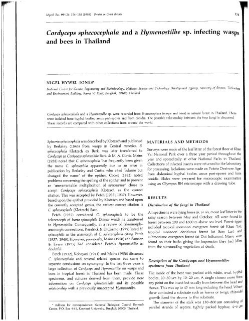
Mycol. Res. 99 (2): 154-158 (1995) Printed in Great Britain<br />
--<br />
154<br />
<strong>Cordyceps</strong> <strong><strong>sp</strong>hecocephala</strong> <strong>and</strong> a <strong>Hymenostilbe</strong> <strong>sp</strong>. <strong>infecting</strong> wa<strong>sp</strong>s<br />
<strong>and</strong> bees in Thail<strong>and</strong><br />
NIGEL HYWEL-JONES*<br />
National Centre for Genetic Engineering <strong>and</strong> Biotechnology, National Science <strong>and</strong> Technology Development Agency, Minislry of Science, Technology<br />
<strong>and</strong> Environment Building, Rama VI Road, Bangkok, 10400, Thail<strong>and</strong><br />
<strong>Cordyceps</strong> <strong><strong>sp</strong>hecocephala</strong> <strong>and</strong> a <strong>Hymenostilbe</strong> <strong>sp</strong>. were recorded from Hymenoptera (wa<strong>sp</strong>s <strong>and</strong> bees) in natural forest in Thail<strong>and</strong>. These<br />
were isolated from hyphal bodies, ascus part-<strong>sp</strong>ores <strong>and</strong> from conidia. The possible relationship between the two fungi is discussed.<br />
These records are compared with other collections from around the world.<br />
Sphaeria <strong><strong>sp</strong>hecocephala</strong> was described by Klotzsch <strong>and</strong> published<br />
by Berkeley (1843) from wa<strong>sp</strong>s in Central America. S.<br />
<strong><strong>sp</strong>hecocephala</strong> Klotzsch ex Berk. was later transferred to<br />
<strong>Cordyceps</strong> as <strong>Cordyceps</strong> <strong>sp</strong>hecophila Berk. & M. A. Curtis. Mains<br />
(1958) noted that C. <strong><strong>sp</strong>hecocephala</strong> 'has frequently been given<br />
the name C. <strong>sp</strong>hecophila apparently due to an error in<br />
publication by Berkeley <strong>and</strong> Curtis, who cited Tulasne but<br />
changed the name' of the epithet. Cooke (1892) noted<br />
problems concerning the <strong>sp</strong>elling of the epithet <strong>and</strong> to prevent<br />
an 'unwarrantable multiplication of synonymy' chose to<br />
accept <strong>Cordyceps</strong> <strong><strong>sp</strong>hecocephala</strong> Klotzsch as the correct<br />
citation. This was accepted by Petch (1933; 1937). However,<br />
based upon the epithet provided by Klotzsch <strong>and</strong> based upon<br />
the currently accepted genus, the earliest correct citation is<br />
C. <strong><strong>sp</strong>hecocephala</strong> (Klotzsch) SacCo<br />
Petch (1937) considered C. <strong><strong>sp</strong>hecocephala</strong> to be the<br />
teleomorph of Isaria <strong>sp</strong>hecophila Ditmar which he transferred<br />
to <strong>Hymenostilbe</strong>. Consequently, in a review of teleomorphanamorph<br />
connections, Kendrick & DiCosmo (1979) listed H.<br />
<strong>sp</strong>hecophila as the anamorph of C. <strong><strong>sp</strong>hecocephala</strong> citing Petch<br />
(1937; 1948). However, previously, Mains (1950) <strong>and</strong> Samson<br />
& Evans' (1975) had considered Petch's <strong>Hymenostilbe</strong> as<br />
doubtfuL.<br />
Petch (1932), Kobayasi (1941) <strong>and</strong> Mains (1958) discussed<br />
C. <strong><strong>sp</strong>hecocephala</strong> <strong>and</strong> several related <strong>sp</strong>ecies but came to<br />
separate conclusions on synonymy, In the last three years a<br />
large collection of <strong>Cordyceps</strong> <strong>and</strong> Hymenostibe on wa<strong>sp</strong>s <strong>and</strong><br />
bees in tropical forest in Thail<strong>and</strong> has been made. These<br />
<strong>sp</strong>ecimens, <strong>and</strong> cultures derived from them, provide new<br />
information on <strong>Cordyceps</strong> <strong><strong>sp</strong>hecocephala</strong> <strong>and</strong> its possible<br />
relationship with a previously unaccepted <strong>Hymenostilbe</strong>,<br />
. Address for corre<strong>sp</strong>ondence: National Biological Control Research<br />
Centre, P.O. Box 9-52, Kasetsart University, Bangkok 10900, Thail<strong>and</strong>.<br />
MA TERIALS AND METHODS<br />
Surveys were made of the leaf litter of the forest floor at Khao<br />
Yai National Park over a three year period throughout the<br />
year <strong>and</strong> <strong>sp</strong>oradically at other National Parks in Thail<strong>and</strong>.<br />
Collections of infected insects were returned to the laboratory<br />
for processing. Isolations were made on Potato Dextrose Agar<br />
from abdominal hyphal bodies, ascus part-<strong>sp</strong>ores <strong>and</strong> from<br />
conidia. Slides were prepared for microscopic examination<br />
using an Olympus BH microscope with a drawing tube.<br />
RESUL TS<br />
Distribution of the fungi in Thail<strong>and</strong><br />
All <strong>sp</strong>ecimens were lying loose in, or on, moist leaf litter in the<br />
rainy season between May <strong>and</strong> October. All were found in<br />
forest between 100 <strong>and</strong> 1600 m above sea leveL. Forest types<br />
included tropical monsoon evergreen forest (at Khao Yai),<br />
tropical monsoon deciduous forest (at Sam Lan) <strong>and</strong><br />
submontane evergreen forest (at Doi Inthanon). Many were<br />
found on their backs giving the impression they had fallen<br />
from the surrounding vegetation at death,<br />
Description of the <strong>Cordyceps</strong> <strong>and</strong> HymenostiIbe<br />
<strong>sp</strong>ecimens from Thail<strong>and</strong><br />
The inside of the host was packed with white, ovaL, hypha!<br />
bodies, 20-30 ¡Jm by 10-20 ¡Jm. A single stroma arose from<br />
any point on the insect but usually from between the head <strong>and</strong><br />
thorax. This was up to 45 mm long including the head. Where<br />
these contacted a substrate such as leaves or twigs, rhizoidal<br />
growth fixed the stroma to this substrate.<br />
The diameter of the stalk was 150-800 ¡Jm consisting of<br />
parallel str<strong>and</strong>s of septate, tightly packed hyphae, 4-6 L.m
--<br />
se<br />
154 N. Hywel-Jones 155<br />
<strong>sp</strong>s<br />
nology<br />
(hao<br />
t the<br />
l<strong>and</strong>.<br />
ItOry<br />
A.gar<br />
from<br />
¡bon<br />
i the<br />
din<br />
'pes<br />
Ýai),<br />
<strong>and</strong><br />
vere<br />
illen<br />
,hal<br />
'om<br />
<strong>and</strong><br />
iere<br />
idal<br />
of<br />
¡Jm<br />
Fig. 1. Three fertile heads of C. <strong><strong>sp</strong>hecocephala</strong> showing the extemal<br />
appearance (left side) <strong>and</strong> internal appearance (right side). Scale bar,<br />
1mm.<br />
across. There was no evidence of branching. Fertie heads<br />
were terminaL, variable in size <strong>and</strong> shape, the smallest being<br />
2'2 x 1'2 mm <strong>and</strong> the largest II x 1'9 mm (Fig. I) but mostly<br />
in the range 4-8 X 1'4-1'8 mm. Heads were citriform to<br />
cylindric in shape, attenuating to a bent tip. Both stalk <strong>and</strong><br />
head were ochraceous to cream yellow.<br />
Dissection of the head showed the colour was confied to<br />
the outer walls. The interior contained perithecia with walls of<br />
tightly interwoven, hyaline hyphae in a loose context of<br />
hyaline cells around a central core which was an extension of<br />
the stalk (Fig. I). Perithecia were oblique to the walls of the<br />
fertile head but with a distinctive curve on the neck ending<br />
almost at right-angles to the outer wall of the head. Perithecia<br />
were large 880-1000 x 200-260 ¡Jm with no evidence of a<br />
hamathecium. All seemed to mature at the same time.<br />
However, within each perithecium asci were found at different<br />
stages of development.<br />
Asci were hyaline, fiiform, at least 700 ¡Jm long <strong>and</strong> up to<br />
7 i.m diameter with a prominent apical cap (Fig. 2). A<br />
thickened plug was sometimes seen at the tip of the cap above<br />
the central canal (Fig. 2). This feature was not constant within<br />
or between <strong>sp</strong>ecimens <strong>and</strong> did not appear to be related to the<br />
state of ascus development. At maturity, as co <strong>sp</strong>ores broke<br />
readily into hyaline, fusoid part-<strong>sp</strong>ores, 10-14 x 1'5-2'5 i.m<br />
(Fig. 2).<br />
Most <strong>sp</strong>ecimens examined, <strong>and</strong> e<strong>sp</strong>ecially larger forms,<br />
were very mature <strong>and</strong> devoid of part-<strong>sp</strong>ores. For others,<br />
pressure applied to the fertile head was often enough to<br />
release part-<strong>sp</strong>ores into water. Natural release of part-<strong>sp</strong>ores<br />
was not observed.<br />
On some <strong>sp</strong>ecimens there were eggs on the stroma <strong>and</strong><br />
dipteran larvae within the <strong>Cordyceps</strong> head. Larvae fed upon<br />
peri the cia <strong>and</strong> stromatal contents within the fertile head.<br />
When this happened it was not possible to fid any healthy<br />
perithecia or any <strong>sp</strong>ores.<br />
When the <strong>Cordyceps</strong> head was damaged, it was possible for<br />
new growth to occur. This was either from the point of<br />
Fig. 2. Tips of seven immature asci <strong>and</strong> three mature asci with examples of part <strong>sp</strong>ores. Scale bar, 10 ¡J.
<strong>Cordyceps</strong> <strong>infecting</strong> wa<strong>sp</strong>s<br />
o 00<br />
~<br />
Fig. 3. Part of the hymeniallayer showing the conidiogenous cells <strong>and</strong> conidia of H. <strong><strong>sp</strong>hecocephala</strong>. Scale bar, 10 il.<br />
damage or from the base of the fertile head. The new growth,<br />
however, appeared to be sterile <strong>and</strong> there was no evidence of<br />
a regrowth of the teleomorph or the development of an<br />
anamorph.<br />
No evidence was found of an anamorph on the same<br />
stroma as the teleomorph. Occasionally, <strong>sp</strong>ecimens were<br />
found which in all re<strong>sp</strong>ects of habit matched the <strong>Cordyceps</strong><br />
form. These differed in not producing a fertile teleomorph but<br />
in producing a stroma which remained the same diameter<br />
throughout rounding only at the tip. Examination revealed<br />
the presence of a <strong>Hymenostilbe</strong> state on the terminal part of the<br />
stroma.<br />
The conidiogenous cells were very variable in size <strong>and</strong><br />
shape as is typical of the genus. They were cylindricaL,<br />
10-20 x 3-6 i.m with one to three stout denticles (Fig. 3). A<br />
notable feature of the conidiogenous cells were long sterile<br />
extensions up to 25 i.m which gave the stroma a markedly<br />
setulose appearance at low power. Conidia were broadly<br />
clavate, strongly apiculate <strong>and</strong> 3-10 x 3-4' 5 i.m.<br />
Specimens exa~jned : AIl <strong>sp</strong>ecimens were on adult wa<strong>sp</strong>s<br />
(Hymenoptera) except for one record on a bee. These were deposited<br />
in the insect-fugus collection at NBCRC with the author's codes.<br />
Teleomorph: NHJ595.01, Khao Yai - Phakajai, 24 Sept. 1991, N. L.<br />
Hywel-Jones; NHJ623.04, Khao Yai - Heo Sawat, 15 Oct. 1991, N.<br />
L. Hywel-Jones & K. Jones; NHJ804.02, Khao Yai - Heo Sawat road<br />
marker Ia 44'8, 25 June 1992, N. L. Hywel-Jones, L. Manoch, A.<br />
Rongchitprapas & S. Sivichai; NHJ858.04, Khao Yai - Gong Giao,<br />
28 Aug. 1992, N. L. Hywel-Jones, A. Rongchitprapas & S. Sivichai;<br />
NHJ933.02, Khao Yai road marker Ia 29'2, 13 Oct. 1992, N. L.<br />
Hywel-Jones; NHJ948, NHJ953.03 & NHJ953.04, Sam LanwaterfalL,<br />
15 Oct. 1992, N. L. Hywel-Jones, A. Rongchitprapas & S.<br />
Sivichai; NHJI070, NHJI075, Khao Yai - Phakajai, 27 May 1993,<br />
N. L. Hywel-Jones & R. Nasit; NHJ1206, Khao Yai - Heo Narok, 22<br />
June 1993, N. L. Hywel-Jones & R. Nasit; NHJ1224, Khao Yai - Heo<br />
Narok, 29 June 1993, N. L. Hywel-Jones, R. NasH, R. Plomhan & S.<br />
Thienhiru; NHJ1316 & NHJ1743 & NHJI752, Khao Yai - Gong<br />
Giao, 6 Aug. 1993, N. L. Hywel-Jones, R. Nasit, S. Sivichai & R.<br />
Plomhan; NHJ1800, NHJ1801, NHJ1807 & NHJI822, Khao Yai-<br />
Heo Narok, 10 Aug. 1993, N. L. Hywel-Jones, R. Nasit & S. Sivichai;<br />
NHJ2133, Khao Yai - Gong Giao, 9 Sept. 1993, N. L. Hywel-Jones,<br />
R. NasH, S. Sivichai & S. Thienhiru; NHJ2199, Doi Inthanon road<br />
marker Ia 25'5, 26 Sept. 1993, N. L. Hywel-Jones, K. Auncam, R.<br />
Nasit, S. Thienhiru & A. J. S. Whalley.<br />
Anamorph: NHJ1332, Khao Yai - Phakrajai on an adult bee, 6 July<br />
1993, S. Sivichai, L. Tangchit & c. Yomsopit; NHJ1741, Khao Yai-<br />
Phakajai, 6 Aug. 1993, N. L. Hywel-Jones, R. Nasit & S. Sivichai;<br />
NHJI858, Khao Yai road marker Ia 44'8, 17 Aug. 1993, N. L.<br />
Hywel-Jones, R. Nasit & S. Sivichai.<br />
Immature: NHJ787~01, Khao Yai road marker Ia 29'2, 6 June 1992,<br />
N. L. Hywel-Jones.<br />
Culture of the fungi<br />
Cultures were started from hyphal bodies, part-<strong>sp</strong>ores, <strong>and</strong><br />
from conidia. Initial isolations were on PDA. Part-<strong>sp</strong>ores <strong>and</strong><br />
conidia germinated in the dark at 22 DC within 20 h. Hyphal<br />
bodies germinated after 5 d under the same conditions apart<br />
from brief periods of light each day for examination.<br />
Isolates are stored in the NCGEB insect-fugus collection.<br />
Isolate numbers coincide with the herbarium numbers given to<br />
<strong>sp</strong>ecimens. Isolates derived from part-<strong>sp</strong>ores were NHJ595.01,<br />
623.04, 804.02, 948, 953.02, 953.03, 1743 <strong>and</strong> 1800. Isolates<br />
derived from conidia were NHJI332, 1741, 1752 <strong>and</strong> 1858.<br />
Isolate NHJ787.01 was derived from abdominal hyphal<br />
bodies.<br />
Isolations from all three sources were sterile but morphologically<br />
indistinguishable. Growth was very slow <strong>and</strong><br />
156
;6<br />
eo<br />
S.<br />
rrg<br />
R.<br />
i-<br />
ai .<br />
es,<br />
ad<br />
R.<br />
tly<br />
iai;<br />
L.<br />
)2,<br />
,d<br />
,d<br />
ial<br />
1ft<br />
m.<br />
to<br />
11,<br />
es<br />
i8.<br />
ial<br />
0-<br />
,d<br />
N. Hywel-Jones<br />
stromatic on PDA reaching 33 mm in 14 wk at 22°. Cultues<br />
were hyaline to pale grey brown with only <strong>sp</strong>arse aerial<br />
mycelium. Some isolates developed an irregular, immersed,<br />
hyaline margin in contrast to the regular, immersed, hyaline<br />
margin that was also noted. The reverse was grey-white to<br />
pale grey brown. Several stout synnemata developed after<br />
3 wk. They were silky pale cream <strong>and</strong> up to 400 i.m diam. <strong>and</strong><br />
appeared similar to the stromata on the hosts. There was no<br />
sign of either a teleomorph or an anamorph developing on<br />
these stromata.<br />
DISCUSSION<br />
Since T orrbia fist noted the appearance of infected wa<strong>sp</strong>s in<br />
Cuba in 1749 (see Samson, Evans & Latgé, 1988) there have<br />
been many reports of <strong>Cordyceps</strong> <strong>infecting</strong> wa<strong>sp</strong>s around the<br />
world. Petch (1932) noted <strong>Cordyceps</strong> gentilis (Ces.) SacCo on a<br />
hornet sent to him from Thail<strong>and</strong>. He considered C. gentilis<br />
distinct from C. <strong><strong>sp</strong>hecocephala</strong> (= C. <strong>sp</strong>hecophila in Petch's early<br />
wrtings) noting the 'longer, more acute head' of C. gentilis<br />
which contrasted with the' ovoid or narrow-oval, <strong>and</strong> acute'<br />
head of C. <strong><strong>sp</strong>hecocephala</strong>.<br />
Petch (1933) later described the shape of the head of C.<br />
<strong><strong>sp</strong>hecocephala</strong> as variable <strong>and</strong> used the description <strong>and</strong><br />
measurements of the Thai <strong>sp</strong>ecimen of C. gentilis in support.<br />
In this paper, he considered C. gentilis synonymous with C.<br />
<strong><strong>sp</strong>hecocephala</strong> along with C. lachnopoda Penz. & Sacc., C.<br />
o:ccephala Penz. & Sacc., C. puiggarii Speg. <strong>and</strong> C. thyrsoides<br />
Möller. Kobayasi (1941) described C. <strong><strong>sp</strong>hecocephala</strong> from<br />
Japan. He regarded C. oxycephala to be distinct from C.<br />
<strong><strong>sp</strong>hecocephala</strong> (based on the shape of the head) <strong>and</strong> considered<br />
C. gentilis a synonym of the former <strong>sp</strong>ecies along with C.<br />
puiggarii. Later, Mains (1948) showed that C. puiggarii was not<br />
the same as these wa<strong>sp</strong> <strong>Cordyceps</strong>.<br />
Mains (1958) re-described <strong>Cordyceps</strong> <strong><strong>sp</strong>hecocephala</strong> from a<br />
substantial collection of <strong>sp</strong>ecimens from North America <strong>and</strong><br />
concluded that there was much variation in the overall<br />
morphology which manifested itself in the many different<br />
names given to what, essentially was a single <strong>sp</strong>ecies. All early<br />
observations put weight upon gross morphology as a separator<br />
of <strong>sp</strong>ecies with litHe attention being paid to microscopic<br />
features.<br />
The work of Mains (1958) <strong>and</strong> the present observations<br />
from Thail<strong>and</strong>, based upon substantial collections within welldefined<br />
geographical regions, suggest there is a single <strong>sp</strong>ecies,<br />
C. <strong><strong>sp</strong>hecocephala</strong> which shows gross morphological variation<br />
but is constant in microscopic detaiL. While the microscopic<br />
characteristics of C. <strong><strong>sp</strong>hecocephala</strong> in Thail<strong>and</strong> showed litHe<br />
variation there were large differences in the shape <strong>and</strong> size of<br />
the fertile head (Fig. I). The Thai records suggest that<br />
maturity of the <strong>sp</strong>ecimen, size of the host, micro-habitat<br />
conditions such as humidity <strong>and</strong> damage re-growth may all<br />
afect the form of the head.<br />
Ditmar (1817) described <strong>and</strong> figured Isaria <strong>sp</strong>hecophila from<br />
a hornet in Germany noting that the upper part of the<br />
simple, brown synnema was pilose <strong>and</strong> darkened. His figures<br />
indicate a minutely setulose condition with globose, hyaline<br />
conidia. Speare (1920) discussed Ditmar's fugus <strong>and</strong>, on the<br />
basis of the ilustrations, suggested that this should be placed<br />
157<br />
in Hirsutella. Petch (1932) agreed noting that it (Ditmar's<br />
fugus) 'does not agree with a <strong>Hymenostilbe</strong>'.<br />
Later, Petch (1937) found a conidial state on an ichneumon<br />
wa<strong>sp</strong> in Engl<strong>and</strong> <strong>and</strong> considered this identical to Ditmar's<br />
fugus <strong>and</strong> concluded 'that Jsaria <strong>sp</strong>hecophila Ditm. is a<br />
<strong>Hymenostilbe</strong> <strong>and</strong> that it is the conidial stage of <strong>Cordyceps</strong><br />
<strong><strong>sp</strong>hecocephala</strong> (Kl.) Cooke'. The description given by Petch<br />
(1937) was brief with no ilustrations. However, he noted the<br />
synnemata were cream-coloured <strong>and</strong> 1'75 mm long. Ditmar's<br />
fugus had brown synnemata which were 90 mm long.<br />
Specimens examined by Petch (1937) had clavate or narrowoval<br />
conidia in contrast to the globose conidia of Ditmar's<br />
fugus.<br />
Mains (1950) noted for H. <strong>sp</strong>hecophila that it 'is doubtfu<br />
whether it is Ditmar's fugus'. Samson & Evans (1975)<br />
accepted nine <strong>sp</strong>ecies of <strong>Hymenostilbe</strong> <strong>and</strong>, like Mains (1950),<br />
they also regarded H. <strong>sp</strong>hecophila as doubtfu. No type<br />
<strong>sp</strong>ecimen remais of Ditmar's fugus but it is possible this is<br />
Hirsutella saussurei Speare which also has long, brown<br />
synnemata <strong>and</strong> conidia that appear globose in a mucous coat.<br />
In <strong>sp</strong>ite of these doubtfu records, Kendrick & DiCosmo<br />
(1979) listed H. <strong>sp</strong>hecophila as the anamorph of C. <strong><strong>sp</strong>hecocephala</strong><br />
citing Petch (1937; 1948). Furthermore, in their scheme the C.<br />
<strong><strong>sp</strong>hecocephala</strong>-H. <strong>sp</strong>hecophila association was coded 2.3.1<br />
suggesting the link was documented with experimental<br />
evidence of one morph developing from the other in culture.<br />
There is no evidence in either of the cited works by Petch that<br />
he ever attempted to culture either fugus. Indeed, Petch<br />
(1948) was merely a listing of British entomogenous fugi in<br />
which he noted the association based upon his 1937 study of<br />
herbarium materiaL. Furthermore, it seems as though there<br />
have been no attempts to isolate either fugus until now.<br />
As Petch (1937) cites only a single record associating the<br />
two <strong>sp</strong>ecies, under the Kendrick & DiCosmo scheme it would<br />
have been better to code this association at a lower level of<br />
2.2.1 - documented but circumstantial evidence of association<br />
based on a single observation of co-habitation. But as earlier<br />
workers (Mains, 1958; Samson & Evans, 1975) had already<br />
raised doubts as to whether Ditmar's fugus could be<br />
considered the same as Petch's <strong>Hymenostilbe</strong> records associated<br />
with C. <strong><strong>sp</strong>hecocephala</strong>, it would be prudent to say that a<br />
<strong>Hymenostilbe</strong> may be associated with C. <strong><strong>sp</strong>hecocephala</strong> based<br />
upon Petch's 1937 writings.<br />
The <strong>Hymenostilbe</strong> <strong>sp</strong>. on wa<strong>sp</strong>s from Thail<strong>and</strong> had a setulose<br />
appearance due to sterile extensions of the conidiogenous<br />
cells. This feature has not been described before for<br />
<strong>Hymenostilbe</strong> (Samson & Evans, 1975) but the conidial state is<br />
clearly assignable to <strong>Hymenostilbe</strong> <strong>and</strong> not Hirsutella. The<br />
current work showed that C. <strong><strong>sp</strong>hecocephala</strong> is present on wa<strong>sp</strong>s<br />
in Thail<strong>and</strong> <strong>and</strong> that a <strong>Hymenostilbe</strong> <strong>sp</strong>. not considered by<br />
Samson & Evans (1975) is also present on wa<strong>sp</strong>s <strong>and</strong> bees in<br />
the same micro-habitats. Furthermore, it showed that isolations<br />
from the <strong>Cordyceps</strong> <strong>and</strong> <strong>Hymenostilbe</strong> are morphologically<br />
similar, suggesting they may be linked.<br />
Until more collections can be made it is proposed that the<br />
<strong>Hymenostilbe</strong> <strong>sp</strong>. found in Thail<strong>and</strong> is possibly an anamorph of<br />
C. <strong><strong>sp</strong>hecocephala</strong> based upon its co-habitation <strong>and</strong> morphological<br />
similarity in cuture <strong>and</strong> may be the same as the<br />
<strong>Hymenostilbe</strong> <strong>sp</strong>ecimens examined by Petch (1937) <strong>and</strong> Mains
",,!,'<br />
Ic~-c,~_ n<br />
<strong>Cordyceps</strong> infeding wa<strong>sp</strong>s<br />
(1950). It is unlikely that the <strong>Hymenostilbe</strong> from Thail<strong>and</strong> is the<br />
same as Ditmar's fugus from Germany.<br />
The National Research Council of Thail<strong>and</strong> is thaned for<br />
providing the opportunity to do this work. Dr Banpot<br />
Napompeth <strong>and</strong> his staff at the NBCRC are e<strong>sp</strong>ecially thanked<br />
for providing me with a friendly atmo<strong>sp</strong>here in which to<br />
work. Miss Rungtip Nasit <strong>and</strong> Somsak Sivichai proved adept<br />
at finding many of these <strong>sp</strong>ecimens. Mrs Surang Thienhiru of<br />
the Royal Forestry Department is thanked for her assistance.<br />
REFERENCES<br />
Berkeley, M. ¡. (1843). On some entomogenous Sphaeriae. Hooker's London<br />
Journal of Botany 2, 205-211.<br />
Cooke, M. C. (1892). Vegetable Wa<strong>sp</strong>s <strong>and</strong> Plant Worm. Society for Promoting<br />
Christian Knowledge: London.<br />
Ditmar, L. P. F. (1817). Die Pilze Deutschl<strong>and</strong>s. Flora Abb nach der Nalur van<br />
Jacob Sturm 3 Heft 4, 1-130.<br />
(Accepted 8 June 1994)<br />
158<br />
Kendrick, W. B. & DiCosmo, F. (1979). Teleomorph-anamorph connections in<br />
Ascomycetes. In The Whole Fungus: The Sexual-Asexual Synthesis, vol. 1<br />
(ed. W. B. Kendrick), pp. 283-395. National Museum of Canada: Toronto.<br />
Kobayasi, Y. (1941). The genus <strong>Cordyceps</strong> <strong>and</strong> its allies. Science Report of the<br />
Tokyo Bunrika Daigaku (Section B. no. 84) 5, 53-260.<br />
Mains, E. B. (1948). Entomogenous fungi. Mycologia 40, 402-416.<br />
Mains, E. B. (1950). Entomogenous <strong>sp</strong>ecies of Akanthomyces, Hymenoslibe <strong>and</strong><br />
Insecticola in North America. Mycologia 42, 566-589.<br />
Mains, E. B. (1958). North American entomogenous <strong>sp</strong>ecies of <strong>Cordyceps</strong>.<br />
Mycologia 50, 169-222.<br />
Petch, T. (1932). Notes on entomogenous fungi, 22-48. Transactions of the<br />
British Mycological Society 16, 209-245.<br />
Petch, T. (1933). Notes on entomogenous fugi, 49-75. Transactions of the<br />
British Mycological Society 18, 48-75.<br />
Petch, T. (1937). Notes on entomogenous fungi, 101-134. Transactions of the<br />
Brilish Mycological Society 21, 34-67.<br />
Petch, T. (1948) A revised list of British entomogenous fugi. Transactions of<br />
the British Mycological Society 31, 286-304.<br />
Samson, R. A & Evans, H. C. (1975). Notes on entomogenous fungi from<br />
Ghana. 3. The genus <strong>Hymenostilbe</strong>. Proceedings, Koninklijke Nederl<strong>and</strong>se<br />
Akadamie van Wetenschappen, Series C 78, 73-80.<br />
Samson, R. A, Evans, H. C. & Latgé, ¡. P. (1988). Aiias of Entomopathogenic<br />
Fungi. Springer, Heidelberg. 189pp.<br />
Speare, AT. (1920). On certain entomogenous fungi. Mycologia 12, 62-76.