

The Biochemical Institute Christian-Albrechts-University of Kiel
The Biochemical Institute Christian-Albrechts-University of Kiel
The Biochemical Institute Christian-Albrechts-University of Kiel

Create successful ePaper yourself
Turn your PDF publications into a flip-book with our unique Google optimized e-Paper software.
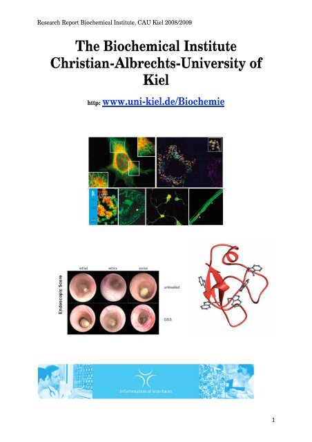
Research Report <strong>Biochemical</strong> <strong>Institute</strong>, CAU <strong>Kiel</strong> 2008/2009<br />
<strong>The</strong> <strong>Biochemical</strong> <strong>Institute</strong><br />
<strong>Christian</strong>-<strong>Albrechts</strong>-<strong>University</strong> <strong>of</strong><br />
<strong>Kiel</strong><br />
http: www.uni-kiel.de/Biochemie<br />
1
Research Report <strong>Biochemical</strong> <strong>Institute</strong>, CAU <strong>Kiel</strong> 2008/2009<br />
<strong>Biochemical</strong> <strong>Institute</strong><br />
<strong>Christian</strong>-<strong>Albrechts</strong>-<strong>University</strong> <strong>of</strong> <strong>Kiel</strong><br />
Research Report 2008/2009<br />
2
Research Report <strong>Biochemical</strong> <strong>Institute</strong>, CAU <strong>Kiel</strong> 2008/2009<br />
Contens<br />
Scientific Staff Members 2008/2009.......................................................................................... 4 <br />
Pr<strong>of</strong>. Dr. rer. nat. Stefan Rose-John – Executive Director ..................................................... 4 <br />
Pr<strong>of</strong>. Dr. rer. nat. Paul Saftig – Director ............................................................................... 4 <br />
Pr<strong>of</strong>. Dr. med. Ursula Just – Director.................................................................................... 4 <br />
Pr<strong>of</strong>. Dr. med. Roland Schauer – Emeritus ............................................................................ 4 <br />
Post-Docs: .............................................................................................................................. 4 <br />
Doctoral students:................................................................................................................... 4 <br />
MD Students:.......................................................................................................................... 5 <br />
Contact ....................................................................................................................................... 6 <br />
Editorial...................................................................................................................................... 7 <br />
1. Research Group Pr<strong>of</strong>. Dr. Stefan Rose-John.................................................................. 8 <br />
2. Research Group Pr<strong>of</strong>. Dr. Hilmar Lemke..................................................................... 13 <br />
3. Research Group Pr<strong>of</strong>. Dr. Joachim Grötzinger ............................................................ 15 <br />
4. Research Group Pr<strong>of</strong>. Dr. Jürgen Scheller ................................................................... 19 <br />
5. Research Group Pr<strong>of</strong>. Dr. Paul Saftig .......................................................................... 23 <br />
6. Research Group Pr<strong>of</strong>. Dr. Karina Reiss........................................................................ 32 <br />
7. Research Group PD Dr. Michael Schwake .................................................................. 35 <br />
8. Research Group Dr. Judith Blanz................................................................................. 37 <br />
9. Research Group Dr. Bernd Schröder............................................................................ 41 <br />
10. Research Group Pr<strong>of</strong>. Dr. Ursula Just .......................................................................... 44 <br />
11. Research Group Dr. Thomas Höfken ........................................................................... 48 <br />
12. Research Group Dr. Ralf Schwanbeck......................................................................... 50 <br />
13. Research Group Pr<strong>of</strong>. Dr. Roland Schauer................................................................... 52 <br />
Seminars 2008/2009................................................................................................................. 57 <br />
Publications 2008/2009 ............................................................................................................ 59 <br />
Accumulated Impact Factors.................................................................................................... 65 <br />
3
Research Report <strong>Biochemical</strong> <strong>Institute</strong>, CAU <strong>Kiel</strong> 2008/2009<br />
Scientific Staff Members 2008/2009<br />
Scientists:<br />
Pr<strong>of</strong>. Dr. rer. nat. Stefan Rose-John – Executive Director<br />
Pr<strong>of</strong>. Dr. rer. nat. Hilmar Lemke<br />
Pr<strong>of</strong>. Dr. rer. nat. Joachim Grötzinger<br />
Pr<strong>of</strong>. Dr. rer. nat. Jürgen Scheller<br />
Pr<strong>of</strong>. Dr. rer. nat. Paul Saftig – Director<br />
Pr<strong>of</strong>. Dr. rer. nat.Karina Reiß<br />
PD Dr. rer. nat. Michael Schwake<br />
Dr. rer. physiol. Bernd Schröder<br />
Pr<strong>of</strong>. Dr. med. Ursula Just – Director<br />
Dr. rer. nat. Ralf Schwanbeck<br />
Dr. rer. nat. Thomas Höfken<br />
Pr<strong>of</strong>. Dr. med. Roland Schauer – Emeritus<br />
Post-Docs:<br />
Dr. rer. nat. Athena Chalaris<br />
Dr. rer. nat. Claudia Drucker<br />
Dr. rer. nat. Doreen Floss<br />
Dr. rer. nat. Nathalie Jänner<br />
Dr. rer. nat. Sascha Jung<br />
Dr. rer. nat. Inken Lorenzen<br />
Dr. rer. nat. Franziska Meier-Stiegen<br />
Dr. rer. nat. Andrea Rittger<br />
Dr. rer. nat. Alex Schneede<br />
Dr. rer. nat. Jenny Schröder<br />
Dr. rer. nat. Stephanie Tenhumberg<br />
Dr. rer. nat. Ahmad Trad<br />
Dr. rer. nat. Kozuke Yamamoto<br />
Doctoral students:<br />
Nina Adam<br />
Kristina Bernoth<br />
Olga Braun<br />
Michelle Danahar<br />
Christin Dewitz<br />
Christoph Garbers<br />
Jessica Gewiese<br />
Vinayaga Srinivasan Gnanapragassam<br />
Johann Groth<br />
Meng Lin<br />
Sven Malchow<br />
Simone Martini<br />
Ulrike May<br />
Matthias Michalek<br />
4
Research Report <strong>Biochemical</strong> <strong>Institute</strong>, CAU <strong>Kiel</strong> 2008/2009<br />
Daniel Milkereit<br />
Swantje Mindorf<br />
Madlen Mohr<br />
Katja Müller<br />
Verena Pawlak<br />
Johannes Prox<br />
Judith Pohanke<br />
<strong>Christian</strong> Raab<br />
Katharina Rothe<br />
Telly Savallas<br />
Janna Schneppenheim<br />
Antje Schütt<br />
Mohammad Shomali<br />
Björn Spudy<br />
Jan Suthaus<br />
Christopher Tiedje<br />
Silvio Weber<br />
Christina Wehling<br />
Marion Willenborg<br />
Christina Zachos<br />
MD Students:<br />
Meike Lüdemann<br />
Anne Oberdörster<br />
Ingo Plagmann<br />
Wolfgang Thaiss<br />
MiriamWagner<br />
5
Research Report <strong>Biochemical</strong> <strong>Institute</strong>, CAU <strong>Kiel</strong> 2008/2009<br />
Contact<br />
<strong>Biochemical</strong> <strong>Institute</strong><br />
<strong>Christian</strong>-<strong>Albrechts</strong>-<strong>University</strong> <strong>of</strong> <strong>Kiel</strong><br />
Olshausenstr. 40<br />
D-24098 <strong>Kiel</strong><br />
Germany<br />
phone: +49 (0) 431/880-2211<br />
fax: +49 (0) 431/880-2007<br />
email: mkanter@biochem.uni-kiel.de<br />
http: www.uni-kiel.de/Biochemie<br />
6
Research Report <strong>Biochemical</strong> <strong>Institute</strong>, CAU <strong>Kiel</strong> 2008/2009<br />
Editorial<br />
<strong>The</strong> positive feedback we received after our three preceding bi-annual reports has encouraged<br />
us to now publish our fourth report for the years 2008/2009. After the <strong>Institute</strong> <strong>of</strong><br />
Biochemistry at the <strong>Christian</strong>-<strong>Albrechts</strong>-Universität, <strong>Kiel</strong> has undergone fundamental changes<br />
in the years 2000-2002, we are now in a phase <strong>of</strong> consolidation and expansion. Renovations<br />
in the <strong>Institute</strong> are close to completion and all the laboratories and teaching areas <strong>of</strong> the<br />
institute have been fully modernized.<br />
In the years 2008 and 2009, members <strong>of</strong> the Biochemistry <strong>Institute</strong> were represented in all<br />
three biomedical collaborative research centers (SFB415: "Specifity and Pathophysiology <strong>of</strong><br />
Signal Transduction Pathways", SFB617, "Molecular mechanisms <strong>of</strong> epithelial defense" and<br />
SFB654: "Plasticity and Sleep"), in which the <strong>Christian</strong>-<strong>Albrechts</strong>-<strong>University</strong> <strong>of</strong> <strong>Kiel</strong> is<br />
involved. Late in 2009, a new SFB841 "Liver inflammation: infection, immune regulation<br />
und consequences" was approved by the Deutsche Forschungsgemeinschaft and is going to<br />
start in January 2010. This new SFB is coordinated by the <strong>University</strong> Hospital Hamburg-<br />
Eppendorf with participation <strong>of</strong> the Biochemistry <strong>Institute</strong> in <strong>Kiel</strong>.<br />
Since the SFB415 will end in summer 2010, the Biochemistry <strong>Institute</strong> has taken the initiative<br />
to apply for a new SFB with the title 'Proteolysis as a regulatory event in pathophysiology'.<br />
This initiative was pre-reviewed by a board <strong>of</strong> the Deutsche Forschungsgemeinschaft in 2009<br />
and we were invited to prepare a full proposal. Our initiative will be visited by a reviewing<br />
panel <strong>of</strong> the Deutsche Forschungsgemeinschaft in March 2010.<br />
Furthermore, the scientific work <strong>of</strong> members <strong>of</strong> the <strong>Institute</strong> <strong>of</strong> Biochemistry is supported by<br />
additional grants from the Deutsche Forschungsgemeinschaft and the European Union.<br />
Since 2008, the cluster <strong>of</strong> excellence ‘Inflammation at Interfaces’ formed by scientists from<br />
the <strong>University</strong> <strong>of</strong> <strong>Kiel</strong>, the <strong>University</strong> zu Lübeck and the Research Center in Borstel is funded<br />
for five years. Scientists from the Biochemistry <strong>Institute</strong> are prominently involved in the<br />
cluster <strong>of</strong> excellence and one <strong>of</strong> the three major research projects <strong>of</strong> the cluster is coordinated<br />
at our <strong>Institute</strong>. Consequently, a new junior research group and a new pr<strong>of</strong>essorship have been<br />
established at the <strong>Institute</strong> <strong>of</strong> Biochemistry.<br />
Thus, our <strong>Institute</strong> continues to represent a spearhead in biomedical research in the Northern<br />
Germany landscape <strong>of</strong> science. In this brochure you can find an overview <strong>of</strong> the projects<br />
being carried out in the <strong>Institute</strong> <strong>of</strong> Biochemistry in 2008 and 2009, along with internet<br />
addresses where you can find more detailed information.<br />
<strong>Kiel</strong>, January 2010<br />
Stefan Rose-John<br />
7
Research Report <strong>Biochemical</strong> <strong>Institute</strong>, CAU <strong>Kiel</strong> 2008/2009<br />
1. Research Group Pr<strong>of</strong>. Dr. Stefan Rose-John<br />
A Group Leader: Pr<strong>of</strong>. Dr. Stefan Rose-John<br />
B Lab Members: Post-Docs:<br />
Dr. Nathalie Jänner<br />
Dr. Stephanie Tenhumberg<br />
Dr. Claudia Drucker<br />
Dr. Athena Chalaris<br />
Dr. Kozuke Yamamoto<br />
C Research Report<br />
Doctoral Students:<br />
Nina Adam<br />
Olga Braun<br />
Christin Dewitz<br />
Christoph Garbers<br />
Jessica Gewiese<br />
Sven Malchow<br />
Ulrike May<br />
Antje Schütt<br />
Jan Suthaus<br />
Ulrike May<br />
Technicians:<br />
Stephanie Schnell<br />
Michaela Jahn<br />
Inez Götting<br />
C.1. <strong>The</strong> soluble interleukin-6 receptor: generation and physiologic function<br />
<strong>The</strong> soluble interleukin-6 receptor (IL-6R) in complex with interleukin-6 (IL-6) stimulates cells, which express<br />
the signaling receptor subunit gp130 but no ligand binding IL-6R. Such cells in the absence <strong>of</strong> the soluble IL-6R<br />
are unable to respond to IL-6. This process has been named 'trans-signaling'. Trans-signaling has been shown to<br />
be important for inflammation reactions, neuronal survival, hematopoiesis and tumor rejection.<br />
We have characterized the metalloproteinase, which is responsible for the release <strong>of</strong> the soluble IL-6R by<br />
biochemical and genetic means. This protease belongs to the Metalloproteinase with Disintegrin Domain<br />
(ADAM) family <strong>of</strong> metalloproteinases. We plan to perform experiments to better understand the biochemical<br />
and structural prerequisites <strong>of</strong> limited proteolysis <strong>of</strong> the IL-6R by members <strong>of</strong> the ADAM family. We also want<br />
to understand the regulation <strong>of</strong> the cleavage reaction. In this respect it is interesting that we could recently show<br />
that the induction <strong>of</strong> apoptosis leads to activation <strong>of</strong> the shedding protease ADAM17 (see below). This seems to<br />
8
Research Report <strong>Biochemical</strong> <strong>Institute</strong>, CAU <strong>Kiel</strong> 2008/2009<br />
be a general phenomenon, which might play an important role in the regulation <strong>of</strong> the inflammatory process. We<br />
could show that neutrophils, which are the first line <strong>of</strong> defense <strong>of</strong> the body during infection and inflammation,<br />
are a major source <strong>of</strong> the soluble IL-6R in vivo. Interestingly, induction <strong>of</strong> apoptosis leads to a selective<br />
activation <strong>of</strong> ADAM17, which in turn is responsible for shedding <strong>of</strong> the IL-6R.<br />
As mentioned above, we could show that the protease ADAM-17 (also called TACE), which is responsible for<br />
cleavage <strong>of</strong> TNFα is also strongly involved in shedding <strong>of</strong> the IL-6R. Using a novel homologous recombination<br />
strategy, we have recently constructed ADAM17 hypomorphic mice (called ADAM17 ex/ex mice ) to analyze the<br />
involvement <strong>of</strong> this protease in various shedding processes. Using these ADAM17 ex/ex mice we currently study<br />
the physiologic role <strong>of</strong> cleavage <strong>of</strong> proteins <strong>of</strong> the ligands <strong>of</strong> the EGF-Receptor and <strong>of</strong> the notch protein.<br />
<strong>The</strong> phenotype <strong>of</strong> the ADAM17 hypomorphic mice clearly shows an involvement <strong>of</strong> ADAM17 in inflammatory<br />
processes. <strong>The</strong>refore these mice are an excellent experimental tool to study the overall physiologic role <strong>of</strong> the<br />
ADAM17 metalloprotease and its involvement in inflammation and cancer.<br />
C.2. Viral Interleukin-6: Strukture, Pathophysiology and Strategies <strong>of</strong> Neutralisation<br />
On target cell Interleukin-6 (IL-6) binds to a receptor complex consisting <strong>of</strong> the ligand binding subunit<br />
Interleukin-6-receptor (IL-6R) and the signal transducing subunit gp130. <strong>The</strong> complex <strong>of</strong> soluble IL-6R and IL-6<br />
acts on cells, which do not express IL-6R. Such cells would not be able to respond to IL-6 alone. Such cells<br />
comprise hematopoietic progenitor cells, endothelial cells, smooth muscle cells, T-cells and neural cells.<br />
Interestingly, the recently identified viral IL-6 (vIL-6) encoded by Human Herpes Virus 8 (HHV8) binds to and<br />
activates gp130 directly. Such, vIL-6 activates a significant larger spectrum <strong>of</strong> target cells than human IL-6. We<br />
want to characterize the biochemical and physiological properties <strong>of</strong> vIL-6. Furthermore, we are in the process <strong>of</strong><br />
generating transgenic mice, which overexpress vIL-6. Using these mice we currently evaluate the involvement <strong>of</strong><br />
vIL-6 in human diseases like Castleman disease, primary effusion lymphoma and multiple myeloma.<br />
Using a novel strategy, we used our recently generated recombinant antibodies against vIL-6 to target the vIL-6<br />
within cells expressing the protein. <strong>The</strong> underlying principle <strong>of</strong> this strategy is to anchor the recombinant vIL-6<br />
antibodies within the endoplasmic reticulum (ER) with the help <strong>of</strong> the canonical ER retention sequence KDEL.<br />
Indeed we could demonstrate that vIL-6 can incuce signaling from within the cell and that such signaling can be<br />
completely blocked from within the cell.<br />
We have performed structure-function analysis to clarify how vIL-6 can bind directly to gp130 whereas human<br />
IL-6 needs the IL-6R in order to bind to and activate gp130. <strong>The</strong>se data clearly show that the so-called site III <strong>of</strong><br />
vIL-6 is responsible for this property and that the ability to directly bind to gp130 can be transferred to human<br />
IL-6 by transferring site III.<br />
Transgenic mice, which over-express the vIL-6 protein show a phenotype ressembling human Castleman<br />
disease. <strong>The</strong>se data indicate that vIL-6 is strongly involved in the phathophysiology <strong>of</strong> HHV8.<br />
C.3. Development <strong>of</strong> the IL-6 trans-signaling antagonist sgp130Fc<br />
We could show in the past years that IL-6 trans-signaling can specifically be inhibited by the sgp130Fc protein<br />
without affecting IL-6 signaling via the membrane bound IL-6R. <strong>The</strong>se data established the sgp130Fc protein<br />
not only as a molecular tool to experimentally distinguish between classic- and trans-signaling. <strong>The</strong> sgp130Fc<br />
protein can also be used to block the course <strong>of</strong> inflammatory diseases in animal models <strong>of</strong> rheumatoid arthritis,<br />
peritonitis, inflammatory bowel disease and inflammation induced colon cancer.<br />
One goal <strong>of</strong> the next years will be to improve the properties <strong>of</strong> the sgp130Fc protein in terms <strong>of</strong> protein stability,<br />
affinity towards the IL-6/sIL-6R complex and feasibility <strong>of</strong> production. Furthermore, we are also engaged in a<br />
study to solve the three dimensional structure <strong>of</strong> the sgp130Fc bound to the IL-6/sIL-6R complex.<br />
Since the sgp130Fc protein has a considerable therapeutic potential, Stefan Rose-John together with Pr<strong>of</strong>. Stefan<br />
Schreiber (Director <strong>of</strong> the <strong>Institute</strong> <strong>of</strong> Clinical Molecular Biology at the <strong>University</strong> Hospital in <strong>Kiel</strong>) founded a<br />
biotechnology company (Conaris Research <strong>Institute</strong>), which develops the sgp130Fc protein into a drug. In<br />
December 2008, Conaris Researcha <strong>Institute</strong> and the company Ferring have completed an exclusive worldwide<br />
license agreement for the development <strong>of</strong> sgp130Fc for inflammatory conditions such as IBD and rheumatoid<br />
arthritis. Sgp130Fc is undergoing pre-clinical testing prior to moving in to Phase I in 2010.<br />
9
Research Report <strong>Biochemical</strong> <strong>Institute</strong>, CAU <strong>Kiel</strong> 2008/2009<br />
D Publications 2008/2009<br />
1. Linker LA, Lühder F, Kallen K-J, Lee D-H, Engelhardt B, Rose-John S and<br />
Gold R (2008) IL-6 transsignalling modulates the early effector phase <strong>of</strong> EAE<br />
and targets the blood-brain-barrier. J Neuroimmunol, 205: 64-72.<br />
2. Tenhumberg S, Waetzig GH, Chalaris A, Rabe B, Seegert D, Scheller J, Rose-<br />
John S and Grötzinger J (2008) Structure guided optimization <strong>of</strong> the IL-6<br />
transsignaling antagonist sgp130Fc. J Biol Chem. 283: 27200-27207<br />
3. Weigmann B, Lehr HA, Yancopoulos G, Valenzuela D, Murphy A, Stevens S,<br />
Schmidt J, Galle PR, Rose-John S, and Neurath MF (2008) <strong>The</strong> transcription<br />
factor NFATc2 controls IL-6-dependent T cell activation in experimental<br />
colitis. J Exp Med. 205: 2099-2110<br />
4. Von Bismarck P, Claass A, Schickor C, Krause MF and Rose-John S (2008)<br />
Altered pulmonary interleukin-6 signaling in preterm infants developing<br />
bronchopulmonary dysplasia. Exp Lung Res. 34: 694-706<br />
5. West MA, Prescott AR, Chan KM, Zhou Z, Rose-John S, Scheller J, and Watts<br />
C (2008) TLR-ligand induced podosome disassembly is ADAM17 dependent.<br />
J Cell Biol. 182: 993-1005<br />
6. Mudter J, Amoussina L, Schenk M, Bruestle A, Weigmann B, Yu J, Wirtz S,<br />
Biesterfeld S, Galle PR, Lehr HA, Rose-John S, Mueller C, Loh<strong>of</strong>f M, and<br />
Neurath MF (2008) <strong>The</strong> transcription factor Interferon regulatory factor-4<br />
controls experimental colitis via T cell derived interleukin-6. J Clin Invest.<br />
118: 2415-2426<br />
7. Sudarman E, Bollati M, Hafner M, Müller W, Scheller J, Rose-John S and<br />
Eichler J (2008) Synthetic Mimetics <strong>of</strong> the gp130-Binding Site for Viral<br />
Interleukin-6 as Inhibitors <strong>of</strong> the vIL-6 - gp130 Interaction. Chem Biol Drug<br />
Des, 71: 494-500<br />
8. Marino M, Scuderi F, Provenzano C, Scheller J, Rose-John S, and Bartoccioni E<br />
(2008) IL-6 regulates MCP-1, ICAM-1 and IL-6 expression in human<br />
myoblasts. J Neuroimmunol. 196: 41-48<br />
9. Benrick A, Jirholt P, Wernstedt I, Gustafsson M, Scheller J, Eriksson A-L,<br />
Borén J, Hedner T, Ohlsson C, Härd T, Rose-John S, Jansson J-O (2008) A<br />
non-conservative polymorphism in the IL-6 signal transducer (IL6ST)/gp130<br />
is associated with myocardial infarction in a hypertensive population. Regul<br />
Peptides 146: 189-196<br />
10. Rabe B, Chalaris A, May U, Waetzig GH, Seegert D, Williams AS, Jones S,<br />
Rose-John S, Scheller J (2008) Transgenic blockade <strong>of</strong> Interleukin-6-transsignaling<br />
abrogates inflammation. Blood 111: 1021-1028<br />
11. Hieronymus T, Ruau D, Ober-Blöbaum J, Baek J-H, Rolletschek A, Rose-John<br />
S, Wobus AM, Müller AM, and Zenke M (2008) <strong>The</strong> transcription factor<br />
repertoire <strong>of</strong> Flt3+ hematopoietic stem cells. Cells Tissues Organs. 188: 103-<br />
115<br />
12. Walker F, Zhang H-H, Matthews V, Weinstock J, Nice EC, Ernst M, Rose-<br />
John S and Burgess AW (2008) IL6/sIL6R complex amplifies emergency<br />
granulopoietic response in the absence <strong>of</strong> G-CSF and GM-CSF.<br />
Blood,111:3978-85<br />
Publikationen 2008 Impact Factor<br />
13. Nechemia-Arbely Y, Barkan D, Pizov G Shriki A, Rose-John S, Galun E and<br />
Axelrod JH (2008) IL-6/IL-6R axis plays a critical role in acute kidney injury.<br />
J Am Soc Nephrol. 19:1106-1115<br />
1. Sina C, Gavrilova O, Förster M, Till, A, Derer S, Hildebrand F, Rabe B, Scheller<br />
J, Chalaris A, Scheller J, Rehmann A, Franke A, Ott S, Häsler R, Nikolaus S,<br />
Fölsch UR, Rose-John S, Jiang H-P, Li J, Schreiber S and Rosenstiel P (2009)<br />
G-protein coupled receptor 43 (GPR43) is essential for neutrophil recruitment<br />
during intestinal inflammation. J Immunol<br />
3.159<br />
5,520<br />
15.219<br />
1.618<br />
9.120<br />
16.559<br />
2.375<br />
3.159<br />
2.276<br />
10.432<br />
2.376<br />
10.432<br />
7.505<br />
Publications 2009 Impact Factor<br />
6.000<br />
10
Research Report <strong>Biochemical</strong> <strong>Institute</strong>, CAU <strong>Kiel</strong> 2008/2009<br />
2. Drucker C, Gewiese J, Malchow S, Scheller J, and Rose-John S (2009) <strong>The</strong><br />
impact <strong>of</strong> Interleukin-6 classic- and trans-signaling on liver damage and<br />
regeneration. J Autoimmun, in press<br />
3. Hösel M, Quasdorff M, Webb D, Zedler U, Esser K, Arzberger S, Wiegmann K,<br />
Kirschning K, Langenkamp A, Rose-John S and Protzer U (2009) Not<br />
interferon, but IL-6 controls early gene expression in Hepatitis B virus (HBV)<br />
infection. Hepatology<br />
4. Andratsch M, Mair N, Constantin CE, Scherbakov N, Benetti C, Vogl C, Sailer<br />
CA, Üceyler N, Brockhaus J, Martini R, Sommer C, Zeilh<strong>of</strong>er HU, Müller W,<br />
Kuner R, Davis JB, Rose-John S, Kress M (2009) Reversal <strong>of</strong> Cancer Pain<br />
through Anti-Interleukin-6 Treatment. J Neurosci<br />
5. Benedict C, Scheller J, Rose-John S, Born S and Marshall L (2009) Enhancing<br />
Influence <strong>of</strong> Intranasal Interleukin-6 on Slow Wave Activity and Memory<br />
Consolidation During Sleep. FASEB J 23:3629-36<br />
6. Drucker C, Rabe, Chalaris A, Schulz E, Scheller J, Rose-John S (2009)<br />
Interleukin-6 Trans-Signaling regulates glycogen consumption after D-<br />
Galactosamine induced liver damage. J Interf Cyt Res<br />
7. May U, Schiffelholz T, Baier PC, Krueger JM, Rose-John S and Scheller J<br />
(2009) IL6-trans-signalling increases rapid-eye-movement sleep in rats. Eur J<br />
Pharmacol 613:141-145<br />
8. Plagmann I, Chalaris A, Kruglov AA; Nedospasov S, Rosenstiel P, Stefan<br />
Rose-John S, Scheller J (2009) Transglutaminase-catalyzed covalent<br />
multimerization <strong>of</strong> camelidae anti-human TNF single domain antibodies<br />
improves neutralizing activity. J Biotechnol 142:170-178<br />
9. Adam N, Rabe B, Suthaus J, Grötzinger J, Rose-John S and Scheller J (2009)<br />
Understanding receptor independent gp130 activation <strong>of</strong> viral interleukin-6. J<br />
Virol 83: 5117-5126<br />
10. Nowell MA, Williams AS, Carty SA, Scheller J, Hayes AJ, Jones GW,<br />
Richards PJ, Slinn S, Ernst M, Jenkins BJ, Topley N, Rose-John S and Jones<br />
SA (2009) <strong>The</strong>rapeutic targeting <strong>of</strong> IL-6 trans-signaling counteracts STAT3<br />
control <strong>of</strong> the inflammatory infiltrate in experimental arthritis. J Immunol,<br />
182: 613-622<br />
11. Chen L, Frister A, Wang S, Ludwig A, Behr H, Pippig S, Li B, Simm A,<br />
H<strong>of</strong>mann B, Pilowski C, Koch S, Buerke M, Rose-John S, Werdan K, and<br />
Loppnow H (2009) Interaction <strong>of</strong> vascular smooth muscle cells and<br />
monocytes by soluble factors synergistically enhances interleukin-6 and MCP-<br />
1 production. Am J Physiol-Heart C. 296: H987-996<br />
12. Baier PC, May U, Scheller J, Rose-John S, Schiffelholz T (2009) Impaired<br />
hippocampus-dependent and -independent learning in IL-6 deficient mice.<br />
Behav Brain Res. 200: 192-196<br />
13. Grivennikov S, Terzic J, Karin E, Mucida D, Yu G-Y, Vallabhapurapu S,<br />
Rose-John S, Cheroutre H, Eckmann L and Karin M (2009) IL-6 and STAT3<br />
signaling is required for survival <strong>of</strong> intestinal epithelial cells and colitis<br />
associated cancer. Cancer Cell, 15: 103-113<br />
14. Islam O, Gong AX, Rose-John S and Heese K (2009) Interleukin-6 and Neural<br />
Stem Cells - More Than Gliogenesis. Mol Biol Cell, 20: 188-199<br />
Impact factors 2008: 89. 750<br />
Impact factors 2009: 95.688<br />
Total impact factors 2008/2009: 185.438<br />
E Grants<br />
7.881<br />
11.355<br />
7.452<br />
7.049<br />
1.774<br />
2.787<br />
2.748<br />
5.308<br />
6.000<br />
3.643<br />
3.171<br />
24.962<br />
5.558<br />
E.1 Stefan Rose-John<br />
Die Rolle von gp130-Trans-Signaling bei der Leber-Regeneration und -Krebs: therapeutische<br />
Perspektiven<br />
DFG RO 632/13-1, Fördersumme (2006 – 2010) 261.400 €<br />
11
Research Report <strong>Biochemical</strong> <strong>Institute</strong>, CAU <strong>Kiel</strong> 2008/2009<br />
E.2 Jürgen Scheller und Stefan Rose-John<br />
Der lösliche Interleukin-6-Rezeptor: Entstehung und physiologische Bedeutungen<br />
Sonderforschungsbereich 415, Teilprojekt B5, Fördersumme (2007 – 2010) 248.700 €<br />
E.3 Stefan Rose-John<br />
Virales Interleukin-6: Struktur, Pathophysiologie und Strategien zu seiner Neutralisierung<br />
Sonderforschungsbereich 415, Teilprojekt C6, Fördersumme (2007 – 2010) 268.800 €<br />
E.4 Stefan Rose-John, Jürgen Scheller, Jan Born, Dunja Hinze-Selch<br />
<strong>The</strong> function <strong>of</strong> the gp130-signaling-family for sleep and plasticity<br />
Sonderforschungsbereich 654, Teilprojekt C5, Fördersumme (2005 – 2009) 444.600 €<br />
E.5 Stefan Rose-John and Jürgen Scheller<br />
<strong>The</strong> function <strong>of</strong> the gp130-signaling-family for sleep and plasticity<br />
Sonderforschungsbereich 654, Teilprojekt C5, Fördersumme (2009 – 2013) 444.600 €<br />
E.5. Excellenzcluster Inflammation at Interfaces, Integrated Research Network F: gp130 signalling,<br />
Fördersumme (2008-2012) 2.749.800 €; Miniproposal (2009): 50.000 €<br />
E.6. Excellenzcluster Inflammation at Interfaces, Integrated Research Network F: gp130 signalling,<br />
Fördersumme (2008-2012) 2.749.800 €; Miniproposal (2009): 50.000 €<br />
12
Research Report <strong>Biochemical</strong> <strong>Institute</strong>, CAU <strong>Kiel</strong> 2008/2009<br />
2. Research Group Pr<strong>of</strong>. Dr. Hilmar Lemke<br />
A Group Leader: Pr<strong>of</strong>. Dr. Hilmar Lemke<br />
B Lab Members: Post-Doc<br />
Dr. rer. nat. Ahmad Trad<br />
C Research Report<br />
C.1. <strong>The</strong> cells <strong>of</strong> the adaptive immune system, B and T lymphocytes, with their clonally distributed receptors<br />
form a web <strong>of</strong> interacting variable domains, known as the idiotypic network. This exerts 2 functions. First, antigen-induced<br />
cellular activations by idiotypic cross-reactivity enable a generalization <strong>of</strong> single antigenic experiences<br />
so that the immune systems in its entirety benefits for its battle with environmental microbes. Second,<br />
idiotypic network interactions allow the transfer <strong>of</strong> the collective maternal immunological experience to the <strong>of</strong>fspring<br />
thus enabling a maternally mediated immunological education before the first encounter with external<br />
antigens.<br />
In the latter context, it was a remarkable finding that maternally-derived immune or monoclonal IgG antibodies<br />
(idiotypes) reactive with suitable model allergens like ovalbumin or bee venom phospholipase A2 (bvPLA2) are<br />
able to induce long-lasting IgE unresponsiveness in the <strong>of</strong>fspring. Our latest investigations have shown that antiidiotypic<br />
antibodies to the transgenerational IgE-suppressive IgG are likewise able to induce such IgE suppression.<br />
Since the effective anti-idiotype did not mimic epitopes <strong>of</strong> the allergen (bvPLA2), we conclude that idiotypic<br />
interactions are mechanistically responsible to the transgenerational IgE-suppression by maternal IgG antibodies.<br />
<strong>The</strong>se results may <strong>of</strong>fer new ways for a prophylactic and possibly therapeutic treatment <strong>of</strong> human IgE-mediated<br />
allergies.<br />
C.2. In this project, we analyze the model immune response to the hapten 2-phnel-oxazolone (phOx) and obtained<br />
some interesting results which are <strong>of</strong> general importance.<br />
1.) <strong>The</strong> analysis <strong>of</strong> the thymus-dependent (TD) anti-phOx response following a pre-immunization with the thymus-independent<br />
type 2 (TI-2) form <strong>of</strong> the antigen demonstrated that the latter, in contrast to current understanding,<br />
induces a special sort <strong>of</strong> immunological memory which we named network memory. In addition, evi-<br />
13
Research Report <strong>Biochemical</strong> <strong>Institute</strong>, CAU <strong>Kiel</strong> 2008/2009<br />
dence was obtained for the first time that receptor revision by VH replacement may occur during immune maturation<br />
in genetically non-engineered wildtype mice.<br />
2.) We compared the phOx-specific repertoires <strong>of</strong> `natural´ antibodies, i.e. phOx-reactive antibodies from nonimmunized<br />
mice, with those <strong>of</strong> TI-2-induced primary and TD-induced primary, secondary, tertiary and quaternary<br />
antibodies. <strong>The</strong> analysis these IgM and IgG antibodies revealed that a) `natural´, TI-2- and TD-induced IgM<br />
antibodies used a huge repertoire <strong>of</strong> VH/VL genes, b) a restriction <strong>of</strong> the repertoire to a few V genes was first<br />
observed after class switch recombination to IgG, and c) IgM antibodies encoded by genes <strong>of</strong> the VH1 family<br />
hardly ever switched to IgG (Lange et al, in preparation).<br />
3.) In addition, the complete anti-phOx IgM/IgG repertoire allowed an analysis <strong>of</strong> the amino acid sequences <strong>of</strong><br />
the most variable part <strong>of</strong> antibodies, the third hypervariable region, VHCDR3. It has been concluded that the extreme<br />
variability <strong>of</strong> this region is sufficient to generate a full antibody repertoire, even when only 1 VH and 1 VL<br />
gene are available (Xu & Davis, 2000, Immunity 13: 37-45). In this report, antibodies with a particular specificity<br />
exhibited similar VHCDR3 sequences. In contrast, anti-phOx antibodies <strong>of</strong> our analysis did not show<br />
similar or identical motives in VHCDR3 (Lange et al, in preparation).<br />
C.3. It is assumed that the VHCDR3 has 2 functions: on the one hand, it is <strong>of</strong> particular importance for binding<br />
<strong>of</strong> the epitope and, on the other, it is the primary target for idiotypic regulation, not only in B but also in T<br />
cells with their analogous VßCDR3. However, the significance <strong>of</strong> VHCDR3 for the regulation <strong>of</strong> an immune response<br />
is not known. Previous results had indicated that there might be a correlation between immune maturation<br />
and VHCDR3-dependent idiotypic network regulation (Lange et al, 1996, Eur J Immunol 26: 2234-42).<br />
For a further investigation <strong>of</strong> this subject, we started to analyze the anti-phOx response in mutant mice which<br />
lack most <strong>of</strong> the 13 D gene segments <strong>of</strong> BALB/c mice and express only one <strong>of</strong> them. Three different strains are<br />
available: Strain ΔD-DFL contains the normal DFL-D-segment DFL16.1 which preferentially codes for neutral<br />
amino acids. Strain ΔD-iD contains a D gene segment with an inverted reading frame (`inverted D´ = iD) coding<br />
preferentially for charged amino acids. Strain ΔD-rf2 contains one D gene segment in the second reading frame<br />
(`reading frame´ 2 = rf2) which preferentially codes for hydrophobic amino acids. This project is done in collaboration<br />
with Dr. Michael Zemlin (Pediatric Clinics, <strong>University</strong> <strong>of</strong> Marburg, Germany) who participated in the<br />
generation <strong>of</strong> these mice in the laboratory <strong>of</strong> Pr<strong>of</strong>. Dr. Harry Schroeder (Birmingham, USA).<br />
<strong>The</strong> hitherto obtained results in strains ΔD-iD and ΔD-rf2 show already a) that the full repertoire <strong>of</strong> D gene segments<br />
is essential for a good immune response and b) that the normally dominant Ox1 idiotype is only <strong>of</strong> minor<br />
importance in these mice although the typical VHCDR3 sequence DRG can principally, but rarely be generated.<br />
Further investigations will show how the immune maturation develops in these mice (Ahmad Trad, Doctoral<br />
<strong>The</strong>sis, <strong>Kiel</strong> 2009).<br />
D Publications 2008/2009<br />
Publications 2008 Impact Factor<br />
1. Lange H, Zemlin M, Tanasa RI, Trad A, Weiss T, Menning H, Lemke H (2008)<br />
Thymus-independent type 2 antigen induces a long-term IgG-related network<br />
memory. Mol. Immunol. 45: 2847-2860.<br />
3.555<br />
Publications 2009 Impact Factor<br />
1. Tanasa RI, Trad A, Lange H, Grötzinger J, Lemke H (2009) Allergen IgEisotype-specific<br />
suppression by maternally derived monoclonal anti-IgGidiotype.<br />
Allergy. (in press)<br />
6.204<br />
2. Lemke H, Tanasa RI, Trad A, Lange H (2009) Benefits and burden <strong>of</strong> the<br />
maternally-mediated immunological imprinting. Autoimmun. Rev. 8: 394-399. 5.371<br />
Impact factors 2008: 3. 555<br />
Impact factors 2007: 11.575<br />
Total impact factors 2008/2009: 15.13<br />
E Grants<br />
Currently no grants<br />
14
Research Report <strong>Biochemical</strong> <strong>Institute</strong>, CAU <strong>Kiel</strong> 2008/2009<br />
3. Research Group Pr<strong>of</strong>. Dr. Joachim Grötzinger<br />
A. Group Leader: Pr<strong>of</strong> Dr. rer. nat. Joachim Grötzinger<br />
B. Lab Members: Post-Docs<br />
Dr. rer. nat. Sascha Jung<br />
Dr. rer. nat. Inken Lorenzen<br />
C. Research Report<br />
Doctoral Students:<br />
Matthias Michalek<br />
Madlen Mohr<br />
Verena Pawlak<br />
Mohammad Shomali<br />
Björn Spudy<br />
Diploma students:<br />
Laura Ruschkies<br />
Technicians:<br />
Britta Hansen<br />
Ursula Mundt<br />
Jessica Schneider (part-time)<br />
Janina Schröder (Trainee Lab. Technician)<br />
C.1. Cytokines and their receptors<br />
Interleukin-27 is a heterodimeric protein, which consists <strong>of</strong> the two proteins p28 and EBI-3. Whereas p28 is<br />
thought to belong to the four-helix bundle family <strong>of</strong> cytokines, EBI-3 is a soluble cytokine receptor containing<br />
the typical signatures <strong>of</strong> this family. <strong>The</strong> signaltransducers <strong>of</strong> IL-27 are gp130 and WSX-1, which are activated<br />
by IL-27 upon binding to their extracellular parts. EBI-3 as well as WSX-1 contains a so called cytokine binding<br />
region, which consists <strong>of</strong> two domains with the signature <strong>of</strong> two conserved cysteine bridges in the N-terminal<br />
and a conserved WSXWS motive in the C-terminal domain. Like interleukin-6 and interleukin-15, p28 is able to<br />
interact with three different receptor chains. <strong>The</strong> structure, the location and the affinity <strong>of</strong> the different receptorbinding<br />
epitopes <strong>of</strong> p28 to their respective receptor chains are not known. <strong>The</strong> goal <strong>of</strong> the planned work is to<br />
establish a structure/function relationship for p28 and its receptors EBI-3 and WSX-1. <strong>The</strong>refore, we will solve<br />
the structure <strong>of</strong> p28 and the cytokine binding regions <strong>of</strong> EBI-3 and WSX-1 by multidimensional heteronuclear<br />
NMR spectroscopy. By using one 15 N labeled component and a second unlabeled component we will identify the<br />
amino-acid residues involved in the interaction between the two molecules by 15 N- 1 H correlated NMR spectra.<br />
<strong>The</strong> identification <strong>of</strong> the residues crucial for the ligand/receptor interaction and the three-dimensional structure<br />
15
Research Report <strong>Biochemical</strong> <strong>Institute</strong>, CAU <strong>Kiel</strong> 2008/2009<br />
<strong>of</strong> these molecules is the prerequisite for the development <strong>of</strong> therapeutically interesting antagonists and/or<br />
agonists.<br />
C.2. Antimicrobial proteins<br />
<strong>The</strong> primary defence <strong>of</strong> an organism against pathogens is performed by the physical barrier <strong>of</strong> the epidermis and<br />
the epithel. <strong>The</strong> secondary defence is mediated by the epidermal cells, by their secretion <strong>of</strong> antimicrobial<br />
peptides and proteins, like defensins and pore forming proteins. <strong>The</strong> molecular mechanisms <strong>of</strong> their interaction<br />
with the pathogens are not well understood. <strong>The</strong> enlightening <strong>of</strong> the three dimensional structure <strong>of</strong> these proteins<br />
is the basis for understanding their mechanisms <strong>of</strong> action. In this project the three-dimensional structures <strong>of</strong> the<br />
newly discovered antimicrobial peptides <strong>of</strong> Caenorhabditis elegans, Ciona intestinalis and Hydra were solved<br />
by NMR spectroscopy.<br />
D. Publications 2008/2009<br />
1. Xun Y, Tremouilhac P, Carraher C, Gelhaus C, Ozawa K, Dixon N.E,<br />
Leippe M, Grötzinger J, Dingley AJ, Kralicek AV. (2009) Cell-free<br />
synthesis and combinatorial selective 15 N labeling <strong>of</strong> the cytotoxic protein<br />
amoebapore A from Entamoeba histolytica. Prot Express and Purification<br />
68, 22-27.<br />
2. Dhir V, Reisch N, Bleicken CM, Lebl J., Kamrath C, Schwarz HP,<br />
Grötzinger J, Sippell WG, Riepe FG, Arlt W, Krone N. (2009) Steroid 17-<br />
Hydroxylase Deficiency: Functional Characterization <strong>of</strong> Four Mutations<br />
(A174E, V178D, R440C, L465P) in the CYP17A1 Gene. J Clin Endocrinol<br />
Metab 94, 3058-3064.<br />
3. Lange W, Geißendörfer J, Schenzer A, Grötzinger J, Seebohm G, Friedric T,<br />
Schwake M. (2009) Refinement <strong>of</strong> the binding site and mode <strong>of</strong> action <strong>of</strong><br />
the anticonvulsant retigabine on KCNQ K + channels. Mol Pharmacol 75,<br />
1–9.<br />
4. Bosch TCG, Augustin R, Anton-Erxleben F, Fraune S, Hemmrich G, Zill H,<br />
Rosenstiel P, Jacobs G, Schreiber S, Leippe M, Stanisak M, Grötzinger J,<br />
Jung S, Podschun R, Bartels J, Harder J, Schröder JM. (2009) Uncovering<br />
the evolutionary history <strong>of</strong> innate immunity: the simple metazoan hydra<br />
uses epithelial cells for host defence. Dev Comp Immun 33, 559-569.<br />
5. Andrä J, Hammer MU, Grötzinger J, Jakovkin I, Lindner B, Vollmer E,<br />
Fedders H, Leippe M, Gutsmann T. (2009) Significance <strong>of</strong> loop structure<br />
and arginine residues for the antibacterial activity <strong>of</strong> arenicin-1 and its<br />
interaction with phospholipid and lipopolysaccharide model membranes.<br />
Biol Chem 390, 337-340.<br />
6. Bruhn O, Cauchard J, Gelhaus C, Thaller G, Leippe M, Grötzinger J. (2009)<br />
Antimicrobial properties <strong>of</strong> the equine α-defensin DEFA1 against<br />
pathogenic bacteria <strong>of</strong> the horse. Vet Immunol Immunop 130, 102-10.<br />
7. Jung S, Dingley AJ, Augustin R, Anton-Erxleben F, Stanisak M, Gelhaus C,<br />
Gutsmann T, Hammer MU, Podschun R, Leippe M, Bosch TCG,<br />
Grötzinger J. (2009) Hydramacin-1: Structure and antibacterial activity <strong>of</strong><br />
a peptide from the basal metazoan Hydra. J Biol Chem 284, 1896–1905.<br />
8. Michalek M, Gelhaus C, Hecht O, Podschun R, Schröder JM, Leippe M,<br />
Grötzinger J. (2009) <strong>The</strong> human antimicrobial protein Psoriasin acts by<br />
permeabilization <strong>of</strong> bacterial membranes. Dev Comp Immun 33, 740-746.<br />
9. Adam N, Rabe B, Suthaus J, Grötzinger J, Rose-John S, Scheller J. (2009)<br />
Understanding receptor independent gp130 activation by viral interleukin-<br />
6. J Virol 83, 5117-5126.<br />
10. Krone N, Grötzinger J, Holterhus PM, SippellWG, Schwarz HP, Riepe FG.<br />
(2009) Congenital adrenal hyperplasia due to 11-hydroxylase deficiency –<br />
insights from two novel CYP11B1 mutations (p.M92X, p.R453Q) . Horm<br />
Res 72, 281—286.<br />
Publications 2009 Impact Factor<br />
11. Tanasa RI, Trad A, Lange H, Grötzinger J, Lemke H. (2009) Allergen IgEisotype-specific<br />
suppression by maternally derived monoclonal anti-IgG-<br />
1.621<br />
6.325<br />
4.711<br />
2.833<br />
3.035<br />
1.907<br />
5.520<br />
2.833<br />
5.308<br />
2.285<br />
6.204<br />
16
Research Report <strong>Biochemical</strong> <strong>Institute</strong>, CAU <strong>Kiel</strong> 2008/2009<br />
idiotype. Allergy (in press).<br />
12. Regenhard R, Leippe M, Schubert S, Podschun R, Kalm E, Grötzinger J,<br />
Lo<strong>of</strong>t C. (2009) Antimicrobial Activity <strong>of</strong> Bovine Psoriasin. Veterinary<br />
Microbiology 136, 335-340.<br />
13. Bleicken C, Loidi L, Dhir V, Parajes S, Quinteiro C, Dominguez F,<br />
Grötzinger J, Sippell WG, Riepe FG, Arlt W, Krone N. (2009) Functional<br />
characterization <strong>of</strong> three CYP21A2 sequence variants employing a yeast<br />
co-expression system. Human Mutation 30, 443-450.<br />
1. Tenhumberg S, Waetzig GH, Chalaris A, Rabe B, Seegert D, Scheller J,<br />
Rose-John S, Grötzinger J. (2008) Structure guided optimization <strong>of</strong> the<br />
interleukin-6 transsignalling antagonist sgp130. J Biol Chem 283, 27200–<br />
27207.<br />
2. Riepe FG, Hiort O, Grötzinger J, Sippell WG, Krone N, Holterhus PM.<br />
(2008) Functional and structural consequences <strong>of</strong> a novel point mutation in<br />
the CYP21A2 gene causing Congenital Adrenal Hyperplasia: Potential<br />
relevance <strong>of</strong> helix C for POR-CYP21 interaction. J Clin Endocrinol Metab.<br />
93, 2891-2896.<br />
3. Dingley AJ, Lorenzen I, Grötzinger J. (2008) NMR analysis <strong>of</strong> viral protein<br />
structures. Methods Mol Biol. 451:441-62.<br />
4. Bräsen C, Schmidt M, Grötzinger J, Schönheit P. (2008) Reaction<br />
Mechanism and structural model <strong>of</strong> ADP-forming acetyl-CoA synthease<br />
from the hyperthermophilic archaeon pyrococcus furiosus: Evidence for a<br />
second active site histidine residue. J Biol Chem 283, 15409-15418.<br />
5. Fedders H, Michalek M, Grötzinger J, Leippe M. (2008) An exceptional salt<br />
tolerant antimicrobial peptide derived from a novel gene family <strong>of</strong><br />
hemocytes <strong>of</strong> the marine invertebrate Ciona intestinalis. Biochem. J. 416,<br />
65-75.<br />
6. Welzel M, Wüstemann N, Simic-Schleicher G, Dörr HG, Schulze E, Shaikh<br />
G, Clayton P, Grötzinger J, Holterhus PM, Riepe FG. (2008)<br />
Carboxyterminal mutations in 3b-hydroxysteroid dehydrogenase cause<br />
congenital adrenal hyperplasia due to 3beta-hydroxysteroid dehydrogenase<br />
deficiency. J Clin Endocrinol Metab. 93, 1418-1425.<br />
7. Andrä J, Jakovkin I, Grötzinger J, Hecht O, Krasnosdembskaya AD,<br />
Goldmann T, Gutsmann T, Leippe M. (2008) Structure and mode <strong>of</strong> action<br />
<strong>of</strong> the antimicrobial peptide arenicin. Biochem J 410, 113-122.<br />
Impact factors 2009: 51.985<br />
Impact factors 2008: 32.432<br />
Total impact factors 2008/2009: 84.417<br />
E. Grants<br />
2.370<br />
7.033<br />
Publications 2008 Impact Factor<br />
5.520<br />
6.325<br />
5.520<br />
4.371<br />
6.325<br />
4.371<br />
E.1. Strukturaufklärung antimikrobieller Peptide und Proteine mit Hilfe der NMR-Spektroskopie<br />
DFG SFB 617-A9, Fördersumme (2005 – 2009) 274,800 €<br />
E.2. Rekombinante Synthese humaner antimikrobieller Peptide und Proteine<br />
DFG SFB 617-Z2, Fördersumme (2005 – 2009 ) 238,800 €<br />
E.3. Strukturaufklärung des Interleukin-15 / Interleukin-15-Rezeptor Komplexes und extrazellulärer gp130<br />
Domänen<br />
DFG SFB 415-B7, Fördersumme (2007 – 2010) 222,400 €<br />
E.4. Ursprüngliche cytolytische und antimokrobielle Mechanismen von humanpathogenen und freilebenden<br />
Protozoen im Vergleich zu höheren Organismen<br />
DFG Einzelantrag Leippe/Grötzinger: Fördersumme (2007 – 2008) 82,000 €<br />
17
Research Report <strong>Biochemical</strong> <strong>Institute</strong>, CAU <strong>Kiel</strong> 2008/2009<br />
E.5. Excellenzcluster Inflammation at Interfaces, Integrated Research Network G: NOD-like receptors, TP5:<br />
NOD2 structure, Fördersumme (2008-209); 170.000 €;<br />
E.6. Excellenzcluster Inflammation at Interfaces, Integrated Research Network F: gp130 signalling, TP1:<br />
Structural and molecular desing <strong>of</strong> novel cytokine antagonists, Fördersumme (2008-209); 200.000 €;<br />
18
Research Report <strong>Biochemical</strong> <strong>Institute</strong>, CAU <strong>Kiel</strong> 2008/2009<br />
4. Research Group Pr<strong>of</strong>. Dr. Jürgen Scheller<br />
A Group Leader: Pr<strong>of</strong>. Dr. Jürgen Scheller<br />
B Lab Members: Post-Doc:<br />
Dr. Doreen Floss (since 2009)<br />
C Research Report<br />
Doctoral Students:<br />
Christin Dewitz<br />
Christoph Garbers<br />
Katja Müller<br />
Nina Adam<br />
Olga Braun<br />
Ulrike May (until 2009)<br />
Medical doctoral students<br />
Anne Oberdörster<br />
Ingo Plagmann (2007-2008)<br />
Wolfgang Thaiss<br />
Diploma students<br />
Anna Tillmann (2008)<br />
Timo Effenberger (2009)<br />
Jan Sommer (2009/2010)<br />
Technicians:<br />
Stefanie Schnell (until 2009)<br />
Annett Lickert (since 2009)<br />
Jessica Schneider (part-time)<br />
Thies Reick (Trainee Lab. Technician)<br />
C.1. Role <strong>of</strong> Interleukin 6 in vitro and in vivo<br />
<strong>The</strong> immunoregulatory cytokine Interleukin-6 (IL6) acts in a pro- and anti-inflammatory fashion. Synthesized by<br />
myeloid cells, fibroblasts and endothelial cells, IL6 on target cells, binds to the IL6 receptor (IL6R) and signals<br />
via complex formation with the ubiquitously expressed gp130 receptor. Paradoxically, most cells, which respond<br />
to IL6 during inflammatory states do not express the IL6R and are themselves not responsive to the cytokine. A<br />
naturally occurring soluble form <strong>of</strong> the IL6R renders all cells responsive to IL6. Interleukin 6 (IL6) transsignaling<br />
has emerged as a prominent regulator <strong>of</strong> immune responses during both innate and acquired immunity.<br />
Regulation <strong>of</strong> IL6 trans-signaling is reliant upon the release <strong>of</strong> soluble IL6 receptor (sIL6R), which binds IL6 to<br />
19
Research Report <strong>Biochemical</strong> <strong>Institute</strong>, CAU <strong>Kiel</strong> 2008/2009<br />
create an agonistic IL6/sIL6R complex capable <strong>of</strong> activating cell types that would not normally respond to IL6<br />
itself.<br />
Here we developed a transgenic strategy based on the overexpression <strong>of</strong> the soluble form <strong>of</strong> gp130, which<br />
specifically blocks all IL6 responses mediated by the soluble IL6R but does not affect IL6 responses via<br />
themembraneboundIL6R. In these mice, inflammatory processes are blocked as in IL6_/_ mice, strongly arguing<br />
for a major role <strong>of</strong> the soluble IL6R during inflammation in vivo.<br />
Colitis-associated cancer (CAC) is the most serious complication <strong>of</strong> inflammatory bowel disease.<br />
Proinflammatory cytokines have been suggested to regulate preneoplastic growth during CAC tumorigenesis.<br />
Interleukin 6 (IL-6) is a multifunctional NF-kB-regulated cytokine that acts on epithelial and immune cells.<br />
Using genetic tools, we now demonstrate that IL-6 is a critical tumor promoter during early CAC tumorigenesis.<br />
In addition to enhancing proliferation <strong>of</strong> tumor-initiating cells, IL-6 produced by lamina propria myeloid cells<br />
protects normal and premalignant intestinal epithelial cells (IECs) from apoptosis. <strong>The</strong> proliferative and survival<br />
effects <strong>of</strong> IL-6 are largely mediated by the transcription factor Stat3, whose IEC-specific ablation has pr<strong>of</strong>ound<br />
impact on CAC tumorigenesis. Thus, the NF-kB-IL-6-Stat3 cascade is an important regulator <strong>of</strong> the proliferation<br />
and survival <strong>of</strong> tumor-initiating IECs.<br />
Human herpesvirus 8 encodes a viral version <strong>of</strong> interleukin-6 (vIL-6) which shows 25% sequence homology<br />
with human IL-6. In contrast to human IL-6, which first binds to the IL-6 receptor (IL-6R) and only<br />
subsequently associates with the signal transducing receptor subunit gp130, vIL-6 has been shown to directly<br />
bind to gp130 without the need <strong>of</strong> IL-6R. As a functional consequence, vIL-6 can activate far more target cells in<br />
the body since all cells express gp130, but only cells such as hepatocytes and some leukocytes express IL-6R.<br />
We sought to understand which amino acid sequences within the vIL-6 protein were responsible for its ability to<br />
bind and activate gp130 independent <strong>of</strong> IL-6R. As a first approach, we constructed chimeric IL-6 proteins in<br />
which all known gp130 interacting sites (sites II and III) were sequentially transferred from vIL-6 into the human<br />
IL-6 protein. To our surprise, human IL-6 carrying all gp130 interacting sites from vIL-6 did not show IL-6Rindependent<br />
gp130 activation. Even more surprisingly, the loop between helix B and C <strong>of</strong> vIL-6, clearly shown<br />
in the crystal structure not to be in contact with gp130, is indispensable for direct binding to and activation <strong>of</strong><br />
gp130. This points to an IL-6R induced change <strong>of</strong> site III conformation in human IL-6, which is already<br />
preformed in vIL-6. <strong>The</strong>se data indicate a novel activation mechanism <strong>of</strong> human IL-6 by the IL-6R that will be<br />
important for the construction <strong>of</strong> novel hyperactive cytokine variants.<br />
C.2. Single domain antibodies<br />
Tumor necrosis factor (TNF) plays an important role in chronic inflammatory disorders, such as Rheumatoid<br />
Arthritis and Crohn’s disease. Recently, monoclonal Camelidae variable heavy-chain domain-only antibodies<br />
(VHH) were developed to antagonize the action <strong>of</strong> human TNF (hTNF). Here, we show that hTNF–VHH does<br />
not interfere with hTNF trimerization, but competes with hTNF for hTNF-receptor binding. Moreover, we<br />
describe posttranslational dimerization and multimerization <strong>of</strong> hTNF–VHH molecules in vitro catalyzed by<br />
microbial transglutaminases (MTG). <strong>The</strong> ribonuclease S-tag-peptidewas shownto act as a peptidyl substrate in<br />
covalent protein cross-linking reactions catalyzed by MTG from Streptomyces mobaraensis. <strong>The</strong> S-tag sequence<br />
was C-terminally fused to the hTNF–VHH and the fusion protein was expressed and purified from Escherichia<br />
coli culture supernatants. hTNF–VHH-S-tag fusion proteins were efficiently dimerized and multimerized<br />
byMTG whereas hTNF–VHHwas not susceptible to protein crosslinking. Cell cytotoxicity assays, using hTNF<br />
as apoptosis inducing cytokine, revealed that dimerized and multimerized hTNF–VHH proteins were much more<br />
active than the monomeric hTNF–VHH.We hypothesize that improved inhibition by dimeric and multimeric<br />
single chain hTNF–VHH proteins is caused by avidity effects.<br />
C.3. Elastin-like polypeptides<br />
Elastin-like polypeptides (ELPs) are highly biocompatible and exhibit a potentially highly useful property – that<br />
<strong>of</strong> a thermally responsive reversible phase transition. <strong>The</strong>se characteristics make ELPs attractive for drug<br />
delivery, appealing as materials for tissue repair or engineering, and improve the efficiency with which<br />
recombinant proteins can be purified. ELP fusion proteins (referred to as `ELPylation´) inherit the reversible<br />
phase transition property. ELPylation technology has recently been extended to plant cells, and a number <strong>of</strong><br />
plant-based expression systems have been evaluated for the production <strong>of</strong> ELPylated proteins. Here, we discuss<br />
recent developments in ELP technology and the substantial potential <strong>of</strong> ELPs for the deployment <strong>of</strong> transgenic<br />
plants as bioreactors to synthesize both biopharmaceuticals and industrial proteins.<br />
20
Research Report <strong>Biochemical</strong> <strong>Institute</strong>, CAU <strong>Kiel</strong> 2008/2009<br />
D Publications 2008/2009<br />
Publications 2008 Impact Factor<br />
1. West, M.A., Prescott, A.R., Chan, K.M., Zhou, Z., Rose-John, S., Scheller, J.<br />
and C. Watts (2008). TLR-ligand induced podosome disassembly in<br />
dendritic cells is ADAM17 dependent. J. Cell. Biol. 182:993-1005.<br />
2. Tenhumberg, S., Waetzing, G.H., Chalaris, A., Rabe, B., Seegert, D.,<br />
Scheller, J., Rose-John, S., and J. Grötzinger (2008). Structure guided<br />
optimization <strong>of</strong> the Interleukin-6 Transsignaling antagonist sgp130. J. Biol.<br />
Chem. 283: 27200-27207.<br />
3. Sudarman, E., Bollati-Fogolin, M., Hafner, M., Müller, W., Scheller, J.,<br />
Rose-John, S. and J. Eichler (2008). Synthetic Mimetics <strong>of</strong> the gp130<br />
binding site for viral Interleukin 6 as inhibitors <strong>of</strong> the vIL6-gp130<br />
interaction. Chem. Biol. Drug Des. 71: 494-500.<br />
4. Marino, M., Scuderi, F., Provenzano, C., Scheller, J., Rose-John, S. and E.<br />
Bartoccioni (2008). IL-6 regulates MCP-1, ICAM and IL6-expression in<br />
human myoblasts. J. Neuroimmunol. 196:41-8.<br />
5. Floss, D.M., Sack, M., Stadlmann, J., Rademacher, T., Scheller, J., Stöger,<br />
E., Fischer, R. and U. Conrad (2008). <strong>Biochemical</strong> and functional<br />
characterization <strong>of</strong> anti-HIV antibody ELP fusion proteins from transgenic<br />
plants. Plant Biotechnol. J. 6: 379-91.<br />
6. Rabe, B., May, U., Chalaris, A., Seegert, D., Waetzig, G., Rose-John, S. and<br />
J. Scheller (2008) Interleukin 6 receptor goes soluble: Abrogated<br />
inflammation by transgenic blockade <strong>of</strong> sIL6R-responses. Blood 111: 1021-<br />
1028.<br />
7. Benrick, A., Pernilla, J.,Wernstedt, I., Gustafsson, M., Scheller, J., Eriksson,<br />
A. L., Borén, J., Hedner, T., Ohlsson, C., Härd, T., Rose-John, S., J. O.<br />
Jansson (2008) A non-conservative polymorphism in the IL-6 signal<br />
transducer (IL6ST)/gp130 is associated with myocardial infarction in a<br />
hypertensive population. Regulatory Peptides 146:189-96.<br />
9.598<br />
5.581<br />
0,409<br />
2.880<br />
4.419<br />
10.370<br />
2.442<br />
Publikationen 2009 Impact Factor<br />
1. Nowell, M.A., Williams, A.S., Carty, S.A., Scheller, J., Hayes, A.J., Jones,<br />
G.W., Richards, P.J., Slinn, S., Ernst, M., Jenkins, B.J., Topley, N., Rose-<br />
John, S. and S.A. Jones (2009). <strong>The</strong>rapeutic targeting <strong>of</strong> IL-6 transsignaling<br />
counteracts STAT3 control <strong>of</strong> the inflammatory infiltrate in<br />
experimental arthritis. J. Immunol. 182:613-22.<br />
2. Grivennikov, S., Karin, E., Teric, J., Mucida, D., Yu, G.Y.,<br />
Vallabhapurapu, S., Scheller, J., Rose-John, S., Cheroutre, H., Eckmann,<br />
L. and M. Karin (2009). IL-6 and STAT3 are required for survival <strong>of</strong><br />
intestinal epithelial cells and development <strong>of</strong> colitis associated cancer.<br />
Cancer Cell. 15(2):103-13.<br />
3. Baier, P.C., May, U., Scheller, J., Rose-John, S. and T. Schiffelholz<br />
(2008). Impaired hippocampus-dependent and independent learning in IL-6<br />
deficient mice. Behav. Brain. Res. 613(1-3):141-145.<br />
4. Prakash, R., Satory, D., Dray, E., Papusha, A., Scheller, J., Kramer, W.,<br />
Krejci, L., Klein, H., Haber, J.E., Sung, P. and G. Ira (2009) Yeast Mph1<br />
helicase dissociates Rad51-made D-loops: implications for crossover<br />
control in mitotic recombination. Genes Dev. 23, 67-79.<br />
6.068<br />
22.321<br />
2.855<br />
15.487<br />
21
Research Report <strong>Biochemical</strong> <strong>Institute</strong>, CAU <strong>Kiel</strong> 2008/2009<br />
5. Adam, N., Rabe, B., Suthaus, J., Grötzinger, J., Rose-John, S. and J.<br />
Scheller (2009). Unraveling Viral Interleukin 6 binding to gp130 and<br />
activation <strong>of</strong> STAT-Signaling Pathways independent <strong>of</strong> Interleukin 6-<br />
Receptor. J. Virol. 83:5117-26.<br />
6. Plagmann, I., Chalaris, A., Nedospasov, S., Posenstiel, P., Rose-John, S.<br />
and J. Scheller (2009) Transglutaminase-catalyzed covalent<br />
multimerization <strong>of</strong> camelidae anti-human TNFα single domain antibodies<br />
improves neutralizing capacity <strong>of</strong> human TNFα. J. Biotechnol. 142: 170-8.<br />
7. May, U., Schiffelholz, T., Baier, P.C., Krueger, J.M., Rose-John, S. and J.<br />
Scheller (2009). IL6-trans-signalling increases rapid-eye-movement sleep<br />
in rats. EJP. 613: 141-5.<br />
8. Benedict, C, Scheller, J., Rose-John, S., Born, J. and L. Marshall (2009).<br />
Enhancing influence <strong>of</strong> intranasal interleukin-6 on slow wave activity and<br />
memory consolidation during sleep. FASEB J. 23:3629-36.<br />
9. Floss, D., Sack, M., Acalis, E., Stadlmann, J., Quendler, H., Rademacher,<br />
T., Stoger, E., Scheller, J., Fischer, R. and U. Conrad (2009) Influence <strong>of</strong><br />
Elastin-like Peptide Fusion on Quantity and Quality <strong>of</strong> a Tobaccoproduced<br />
Anti HIV Antibody. Plant Biotechnol. J. 7:899-913.<br />
10. Sina, C., Förster, M., Derer, S., Hildebrand, F., Chalaris, A., Rabe, B.,<br />
Scheller, J., Gavrilowa, O., Rehmann, A., Ott, S., Häsler, R., Li, J.,<br />
Nikolaus, S., Fölsch, U.R., Rose-John, S., Schreiber, S. and P. Rosenstiel<br />
(2009) G-protein coupled receptor 43 (GPR43) is essential for neutrophil<br />
recruitment during intestinal inflammation. J. Immunol. 183:7514-22.<br />
11. Rose-John, J., Mitsuyama, K., Matsumoto, S., Thaiss, W.M. and J.<br />
Scheller (2009) Interleukin-6 Trans-Signaling and Colonic Cancer<br />
Associated with Inflammatory Bowel Disease. Current Pharmocological<br />
Design, 15: 2095-103. Review.<br />
Impact factors 2008: 35.29<br />
Impact factors 2009: 70.967<br />
Total impact factors 2008/2009: 106.257<br />
E Grants<br />
E.1. Jürgen Scheller and Stefan Rose-John<br />
Der lösliche Interleukin-6-Rezeptor: Entstehung und physiologische Bedeutungen<br />
Sonderforschungsbereich 415, Teilprojekt B5, Fördersumme (2007 – 2010) 248.700 €<br />
E.2. Stefan Rose-John, Jürgen Scheller, Jan Born, Dunja Hinze-Selch<br />
<strong>The</strong> function <strong>of</strong> the gp130-signaling-family for sleep and plasticity<br />
Sonderforschungsbereich 654, Teilprojekt C5, Fördersumme (2005 – 2009) 444.600 €<br />
E.3. Stefan Rose-John and Jürgen Scheller<br />
<strong>The</strong> function <strong>of</strong> the gp130-signaling-family for sleep and plasticity<br />
Sonderforschungsbereich 654, Teilprojekt C5, Fördersumme (2009 – 2013) 387.200 €<br />
E.4. Jürgen Scheller and Werner Solbach<br />
Influences <strong>of</strong> sleep, hormones and cytokines on circadian rhythm <strong>of</strong> T cell activity<br />
Sonderforschungsbereich 654, Teilprojekt C8, Fördersumme (2009 – 2013) 353.600 €<br />
5.332<br />
3,015<br />
2.565<br />
7.049<br />
4.419<br />
6.00<br />
4.868<br />
E.5. Excellenzcluster Inflammation at Interfaces, Integrated Research Network F: gp130 signalling, TP3:<br />
Cytokine Signalling, Fördersumme (2009); 132.600 €€; Miniproposal (2009): 50.000 €<br />
22
Research Report <strong>Biochemical</strong> <strong>Institute</strong>, CAU <strong>Kiel</strong> 2008/2009<br />
5. Research Group Pr<strong>of</strong>. Dr. Paul Saftig<br />
A Lab Leader: Pr<strong>of</strong>. Dr. Paul Saftig<br />
B Lab Members: Group leaders:<br />
Michael Schwake<br />
Bernd Schröder<br />
Judith Blanz<br />
Post-Docs:<br />
Dr. rer. nat. Andrea Rittger<br />
Dr. rer. nat. Jenny Schröder<br />
Doctoral Students:<br />
Marion Willenborg<br />
Alexander Schneede<br />
Silvio Weber<br />
Johannes Prox<br />
Telly Savallas<br />
Christina Wehling<br />
Johann Groth<br />
Christina Zachos<br />
Michelle Danahar<br />
Janna Schneppenheim<br />
Medical Students:<br />
Meike Lüdemann<br />
MiriamWagner<br />
Diploma Students:<br />
Daniel Mielkereit<br />
Mia Schmied<br />
Katharina Rothe<br />
Jörg Behmke<br />
Nur Güneli<br />
Judith Pohanke<br />
<strong>Christian</strong> Raab<br />
23
Research Report <strong>Biochemical</strong> <strong>Institute</strong>, CAU <strong>Kiel</strong> 2008/2009<br />
C Research Report<br />
C.1. Biology <strong>of</strong> lysosomes:<br />
Bachelor:<br />
Friederike Zunke<br />
Technician:<br />
Marlies Rusch<br />
Katharina Stiebeling<br />
Maike Langer<br />
Inez Götting<br />
Sebastian Held<br />
Trainee Lab. Technician<br />
Tobias Lehmann<br />
Steffie Jessen<br />
Merjem Senkara<br />
Raphael Kurz<br />
Rouven Bahn<br />
Marvin Murowski<br />
Secretary<br />
Gundula Hohn<br />
Major functions <strong>of</strong> lysosomal membrane proteins. <strong>The</strong> lysosome is a central, acidic organelle that is involved in the<br />
degradation <strong>of</strong> macromolecules through the activity <strong>of</strong> lysosomal hydrolases. Lysosomes are crucial for the maturation <strong>of</strong><br />
phagosomes to phagolysosomes in phagocytosis, which is important for cellular pathogen defence. <strong>The</strong> macroautophagy<br />
pathway mediates the turnover <strong>of</strong> cytoplasmic components, such as organelles and large complexes, and is involved in cell<br />
death and proliferation. Macroautophagy depends on the fusion <strong>of</strong> lysosomes with autophagosomes to create autolysosomes,<br />
in which degradation occurs. Macroautophagy and chaperone-mediated autophagy, a direct lysosomal transport process for<br />
cytosolic proteins, are regulated by lysosome-associated membrane proteins (LAMPs). Lysosomal exocytosis and plasma<br />
membrane repair are Ca2+ and synaptotagmin 7 (SYT7)-dependent fusion events, which are possibly involved in pathogen<br />
entry, autoimmunity and neurite outgrowth. <strong>The</strong> lysosomal cell death pathway is triggered by a release <strong>of</strong> lysosomal<br />
cathepsins through an unknown mechanism. Cellular cholesterol homeostasis is controlled by lysosomal cholesterol efflux<br />
through Niemann–Pick C1 protein (NPC1). Major histocompatibility complex (MHC) class II-dependent antigen<br />
presentation requires lysosomal proteases and membrane proteins. <strong>The</strong> release <strong>of</strong> exosomes is thought to be involved in<br />
adaptive immune responses. Lysosomal membrane proteins are also involved in the transport <strong>of</strong> newly synthesized<br />
hydrolases to the lysosome (for example, lysosomal integral membrane protein 2 (LIMP2)) and across the lysosomal<br />
membrane (for example, the V-type H+-ATPase complex and chloride channel protein 7 (CLC7)). (from Saftig &<br />
Klumpermann; Nature Reviews in Cell and Molecular Biology, 2009, 10:623-635)<br />
24
Research Report <strong>Biochemical</strong> <strong>Institute</strong>, CAU <strong>Kiel</strong> 2008/2009<br />
Lysosomes, lysosomal membrane proteins, hydrolases and lysosomal storage disease:<br />
Lysosomes are ubiquitous organelles that constitute the primary degradative compartments <strong>of</strong> the cell. <strong>The</strong>y<br />
receive their substrates through endocytosis, phagocytosis or autophagy. <strong>The</strong> catabolic function <strong>of</strong> lysosomes is<br />
complemented by lysosome-related organelles (LROs), such as melanosomes, lytic granules, major<br />
histocompatibility complex (MHC) class II compartments and platelet dense granules. LROs share many<br />
properties with lysosomes, but they also contain celltype specific proteins and might require additional cellular<br />
machinery for their biogenesis. Lysosomes and LROs are involved in various physiological processes, such as<br />
cholesterol homeostasis, plasma membrane repair, bone and tissue remodelling, pathogen defence, cell death and<br />
cell signalling. <strong>The</strong>se complex functions make the lysosome a central and dynamic organelle and not simply the<br />
dead end <strong>of</strong> the endocytic pathway. Two classes <strong>of</strong> proteins are essential for the function <strong>of</strong> lysosomes: soluble<br />
lysosomal hydrolases (also referred to as acid hydrolases) and integral lysosomal membrane proteins (LMPs).<br />
Each <strong>of</strong> the 50 known lysosomal hydrolases targets specific substrates for degradation, and their collective action<br />
is responsible for the total catabolic capacity <strong>of</strong> the lysosome. In addition to bulk degradation and proprotein<br />
processing, lysosomal hydrolases are involved in antigen processing, degradation <strong>of</strong> the extracellular matrix and<br />
initiation <strong>of</strong> apoptosis. <strong>The</strong> mammalian lysosome contains ~25 LMPs, but additional LMPs are being revealed.<br />
LMPs reside mainly in the lysosomal limiting membrane and have diverse functions, including acidification <strong>of</strong><br />
the lysosomal lumen, protein import from the cytosol, membrane fusion and transport <strong>of</strong> degradation products to<br />
the cytoplasm. <strong>The</strong> most abundant LMPs are lysosome associated membrane protein (LAMP1), LAMP2,<br />
lysosome integral membrane protein 2 (LIMP2; also known as SCARB2) and the tetraspanin CD63.<br />
Lysosome biogenesis requires integration <strong>of</strong> the endocytic and biosynthetic pathways <strong>of</strong> the cell. Lysosomal<br />
targeting <strong>of</strong> newly synthesized lysosomal proteins can be direct, from the trans-Golgi network (TGN) to the<br />
endosomal system, or indirect, involving transport to the plasma membrane and subsequent endocytosis. <strong>The</strong><br />
best understood direct pathway is the mannose 6 phosphate receptor (M6PR) mediated transport <strong>of</strong> lysosomal<br />
hydrolases. By contrast, remarkably little is known about the structural and molecular machinery for the<br />
transport <strong>of</strong> LMPs to lysosomes. <strong>The</strong> significance <strong>of</strong> tightly regulated LMP trafficking is illustrated by recent<br />
findings that describe new and unexpected roles for LMPs in cellular physiology. It is becoming apparent that<br />
LMPs can impose specific functions onto the organelles through which they are transported or in which they<br />
reside, such as the endoplasmic reticulum (ER), lysosomes and the plasma membrane. <strong>The</strong>ir importance is<br />
further highlighted by the discovery <strong>of</strong> an increasing number <strong>of</strong> gene mutations that lead to lysosomal<br />
dysfunction and disease. In addition, various knockout mice and non mammalian model organisms have<br />
highlighted the role <strong>of</strong> LMPs in cell physiology. We are continuing our efforts to characterize the molecular<br />
functions <strong>of</strong> these proteins for health and disease.<br />
C.2. Towards a new therapy for the lysosomal storage disorder alpha-Mannosidosis:<br />
<strong>The</strong> European HUE-MAN network is coordinated through activities in our lab. Basic scientists and clinicians contribute to<br />
the development <strong>of</strong> an enzyme replacement therapy fort he human alpha-Mannosidosis disease.<br />
Alpha-Mannosidosis is a rare inborn disorder caused by the lack <strong>of</strong> the lysosomal enzyme α-Mannosidase,<br />
resulting in mental retardation, skeletal changes, hearing loss and recurrent infections. <strong>The</strong> children are <strong>of</strong>ten<br />
born apparently normal, and their conditions worsen progressively, without any possibility to prevent this<br />
evolution. In the children that are born healthy, a therapy initiated at an early age could contribute to a normal<br />
development. Today, the most promising therapy for lysosomal storage disorders is enzyme replacement therapy<br />
(ERT); where the enzyme lacking in the patient is introduced into the blood stream, from where it is internalised<br />
25
Research Report <strong>Biochemical</strong> <strong>Institute</strong>, CAU <strong>Kiel</strong> 2008/2009<br />
by the cells and reaches the lysosomes, acting as the endogeneous enzyme. ERT products are on the market<br />
today for diseases such as Gaucher and Fabry and clinical trials are underway for a number <strong>of</strong> other diseases. A<br />
correction <strong>of</strong> storage in many tissues including brain was found after administration <strong>of</strong> lysosomal acid α-<br />
Mannosidase (LAMAN) from bovine kidney, and human and mouse recombinant LAMAN. <strong>The</strong> present project<br />
(PI: Judith Blanz) is <strong>of</strong> great value for the introduction <strong>of</strong> recombinant human lysosomal alpha-mannosidase into<br />
the first clinical trials <strong>of</strong> ERT in patients. In line with our plans we have been able to evaluate the benefit <strong>of</strong><br />
alpha mannosidase ERT in the mouse model and we have made significant advances to get a suitable protocol<br />
for treatment <strong>of</strong> this lysosomal storage disorder. Importantly, the correction <strong>of</strong> storage has been followed over<br />
time in visceral organs but also in the central nervous system. A possible improvement <strong>of</strong> the brain associated<br />
symptoms has been analysed by sophisticated behavioral monitoring <strong>of</strong> treated and non treated mice,<br />
respectively. We have initiated a study in the mouse model <strong>of</strong> the hitherto unrecognized role <strong>of</strong> epigenetic<br />
factors contributing to the manifestation <strong>of</strong> the disease. On the other hand a systematic clinical and diagnostic<br />
natural history study <strong>of</strong> the disease in human patients was successfully started. This study enables us to define<br />
the critical parameters and hallmarks <strong>of</strong> the disease which will eventually be used in phase I and phase II clinical<br />
trials to evaluate the efficacy <strong>of</strong> the ERT in patients. Apart from providing sufficient enzyme for the preclinical<br />
studies the optimisation <strong>of</strong> the production <strong>of</strong> the human recombinant LAMAN enzyme has been pushed forward<br />
and will be <strong>of</strong> major importance for the successful introduction <strong>of</strong> this enzyme into clinical phase trials. We<br />
could successfully develop the enzyme production and the replacement therapy in the mouse model. We have<br />
obtained important information about dose and interval <strong>of</strong> ERT treatment, about the mode <strong>of</strong> correction in<br />
peripheral neurons and neurons <strong>of</strong> the CNS, about behavioral pr<strong>of</strong>iles <strong>of</strong> the mouse model with and without<br />
ERT, about differentially expressed genes in treated and untreated mice. We could further analyse in detail the<br />
oligosaccharide storage by newly developed mass spectrometry analysis and started to establish this<br />
methodology for the analysis for clinical use.<br />
C.3. Biology <strong>of</strong> the newly identified lysosomal sorting receptor LIMP-2:<br />
We (PI: Michael Schwake) have identified an unexpected role for the lysosomal membrane protein LIMP-2 as a<br />
sorting receptor required for the delivery <strong>of</strong>beta-glucocerebrosidase to lysosomes. <strong>The</strong> Mannose-6-Phosphatereceptor<br />
pathway has been very well characterized as a major route for the sorting <strong>of</strong> lysosomal enzymes;<br />
however, the mechanism for the intracellular targeting <strong>of</strong>beta-glucocerebrosidaseto lysosomes has been unclear<br />
until now.Our findings that beta-glucocerebrosidase associates with LIMP-2, that these proteins colocalize in<br />
intracellular vesicular compartments, and that the activity, levels, and localization <strong>of</strong> beta-glucocerebrosidase<br />
exhibit a dramatic correlation with the presenceor absence <strong>of</strong> LIMP-2 reveal that beta-glucocerebrosidase<br />
independence <strong>of</strong> Mannose-6-Phosphate-based sorting mechanisms is almost certainly a consequence <strong>of</strong> its<br />
routing via LIMP-2 through the lysosomalmembrane protein delivery pathway.<br />
Action myoclonus-renal failure syndrome (AMRF) is caused by mutations in LIMP-2. To date, six AMRFcausing<br />
mutations have been described, including splice site, missense and nonsense mutations. All mutations<br />
lead to a retention <strong>of</strong> LIMP-2 in the endoplasmic reticulum (ER) but affect the binding to b-GC differentially.<br />
<strong>The</strong> LIMP-2 segment 145–288, comprising the nonsense mutations, contains a highly conserved coiled-coil<br />
domain, which we suggest determines beta-glucocerebrosidase binding. Disruption <strong>of</strong> the helical arrangement<br />
and amphiphatic nature <strong>of</strong> the coiled-coil domain abolishes beta-glucocerebrosidase binding. In contrast to the<br />
reduced binding properties <strong>of</strong> the nonsense mutations, the only missense mutation (H363N) found in AMRF<br />
leads to increased binding <strong>of</strong> beta-glucocerebrosidase to LIMP-2, indicating that this highly conserved histidine<br />
modifies the affinity <strong>of</strong> LIMP-2 to its ligand. We suggest that disruption <strong>of</strong> the coiled-coil structure or AMRF<br />
disease-causing mutations abolish beta-glucocerebrosidase binding, indicating the importance <strong>of</strong> an intact coiledcoil<br />
structure for the interaction <strong>of</strong> LIMP-2 and beta-glucocerebrosidase.<br />
C.4. <strong>The</strong> graduate research training school (GRK1459): Sorting and interaction<br />
between proteins <strong>of</strong> subcellular compartments<br />
<strong>The</strong> DFG-Research Training Group 1459 is co-coordinated by our group and scientists from the <strong>University</strong><br />
Medical Center Hamburg-Eppendorf and the Bernhard-Nocht-<strong>Institute</strong> for Tropical Medicine in Hamburg. <strong>The</strong><br />
program is open to students with a diploma/master in natural sciences and medical students. <strong>The</strong> general topic <strong>of</strong><br />
the Research Training Group is sorting and transport <strong>of</strong> selected proteins within the Golgi apparatus and<br />
endosomal compartments. In these organelles the decision is made whether a newly synthesized protein reaches<br />
its target via the secretory/biosynthetic pathway, or a recently internalized molecule (or bacterium) reaches its<br />
intracellular destination via the endocytic/phagocytic pathway. Missorted proteins may lead to loss <strong>of</strong> function in<br />
their target organelles, that may affect the well being <strong>of</strong> the cell and the organism as a whole.<br />
26
Research Report <strong>Biochemical</strong> <strong>Institute</strong>, CAU <strong>Kiel</strong> 2008/2009<br />
<strong>The</strong>refore, the experimental approaches are related to diseases. By focussing on selected model proteins, basic<br />
mechanisms <strong>of</strong> the biogenesis <strong>of</strong> intracellular compartments as well as the balance <strong>of</strong> membrane transport<br />
between organelles and the interplay between cytosolic and membrane proteins will be investigated. <strong>The</strong><br />
majority <strong>of</strong> projects addresses sorting and transport processes under pathological conditions in cells derived from<br />
patients or mouse models <strong>of</strong> human diseases, or cells infected by bacteria or in parasite cells. New insights into<br />
the interactions between resident proteins <strong>of</strong> endosomes and the Golgi apparatus with components <strong>of</strong> the<br />
vesicular transport machinery and the actin cytoskeleton will be expected. A better understanding <strong>of</strong> cellular<br />
responses to endogenous mutant proteins or exogenous pathogens will enable the development <strong>of</strong> novel<br />
therapeutic strategies. Different experimental approaches such as ultrastructural analysis <strong>of</strong> cellular<br />
compartments, genomics, biochemistry, time-resolved imaging, and structural biology will be applied and<br />
improve our understanding <strong>of</strong> spatial and dynamic aspects <strong>of</strong> membrane transport or translocation. Students will<br />
go through a three year curriculum <strong>of</strong> academic as well as non-academic courses in molecular and cellular<br />
biology, biochemistry, infectiology, microbiology, and molecular biomedicine. <strong>The</strong> Research Training Group<br />
<strong>of</strong>fers a continuous educational program with lectures, practical courses, seminars, regular report meetings and<br />
an international symposium every two years. <strong>The</strong> practical courses consist <strong>of</strong> several three-day hands training<br />
units attended by up to 4 students. <strong>The</strong> company OLYMPUS is associated to the Research Training Group and<br />
<strong>of</strong>fers additional seminars on new developments in microscopy and practical courses. Seminars will be given by<br />
leading scientists and will foster a broad view on current topics <strong>of</strong> molecular life sciences. It is expected that<br />
each student spends 1-3 months abroad in a laboratory cooperating in the research field. <strong>The</strong> program provides a<br />
broad education, not just on the specific topic <strong>of</strong> the thesis, but also in research topics <strong>of</strong> the other participating<br />
groups.<br />
C.5. Discovery and elucidation <strong>of</strong> the functions <strong>of</strong> new and unknown lysosomal<br />
membrane proteins:<br />
<strong>The</strong> identification <strong>of</strong> new lysosomal membrane proteins by subproteomic approaches (PI: Bernd Schröder), in<br />
which new members <strong>of</strong> the lysosomal membrane are being investigated is another focus in the lab. <strong>The</strong><br />
functional characterization <strong>of</strong> these new members <strong>of</strong> the lysosomal membrane will be done using biochemical<br />
and mouse genetic approaches. Of special interest in these context are two proteins <strong>of</strong> hitherto unknown<br />
function, DIRC2 and TMEM192.<br />
<strong>The</strong> concept <strong>of</strong> regulated intramembrane proteolysis has emerged over the last decades as a novel concept <strong>of</strong><br />
cellular signalling. One group among the proteases being capable <strong>of</strong> cleaving substrates within the phospholipid<br />
bilayer are the signal-peptide-peptidase (SPP) and its homologues, the signalpeptide-peptidase-like proteins<br />
(SPPL2a, -2b, -2c, -3). SPPL2a is present in membranes <strong>of</strong> lysosomes/late endosomes whereas SPPL2b was<br />
reported to reside at the plasma membrane as well as in endosomal compartments. To date, only TNFα, Fas<br />
Ligand (FasL) and Bri2(Itm2b) have been identified as substrates <strong>of</strong> SPPL2a/b. In the case <strong>of</strong> TNFα it was<br />
demonstrated that the proteolytic release <strong>of</strong> the TNFα intracellular domain influences gene expression and<br />
induces the synthesis <strong>of</strong> the pro-infammatory cytokine IL12. Two <strong>of</strong> the known substrates suggest a regulatory<br />
function <strong>of</strong> SPPL2a and SPPL2b in the context <strong>of</strong> the immune system. In order to analyse functions <strong>of</strong> SPPL2a<br />
and SPPL2b in a complex in vivo system, mouse lines deficient in either <strong>of</strong> the two proteases have been<br />
generated. Preliminary immunological phenotyping has revealed major abnormalities in SPPL2a -/- mice.<br />
C.6. Proteolysis in or at membranes<br />
Proteolytic ectodomain release, a process known as “shedding”, has emerged as a key mechanism for regulating<br />
the function <strong>of</strong> a diversity <strong>of</strong> cell surface proteins. Shedding <strong>of</strong> integral membrane proteins is to our knowledge<br />
limited to type I and type II transmembrane proteins or GPI-anchored molecules, in which the cleavage site is<br />
generally located close to the membrane surface. A Disintegrin And Metalloproteinases (ADAMs) have emerged<br />
as the major proteinase family that mediates ectodomain shedding. ADAM-mediated shedding is important in a<br />
number <strong>of</strong> biological processes such as the interaction <strong>of</strong> sperm and egg, cell fate determination, cell migration,<br />
27
Research Report <strong>Biochemical</strong> <strong>Institute</strong>, CAU <strong>Kiel</strong> 2008/2009<br />
wound healing, neurite and axon guidance, heart development, immunity, cell proliferation and angiogenesis. It<br />
is estimated that up to 4% <strong>of</strong> the proteins on the cell surface undergo ectodomain shedding affecting functionally<br />
diverse proteins, such as cadherins, L-selectin, Fas ligand, TNF-a, EGFR ligands, ErbB2, ErbB4, Amyloid<br />
Precursor Protein (APP), Notch receptor, Notch ligands and many others. ADAMs as proteins <strong>of</strong> about 750<br />
amino acid length, are characterized by a conserved domain structure, consisting <strong>of</strong> an N-terminal signal<br />
sequence followed by a prodomain, a metalloproteinase domain, a disintegrin domain with a cysteine-rich<br />
region, a transmembrane domain and a cytoplasmic tail. ADAM-mediated shedding is both constitutive and<br />
inducible, dependent on G-protein coupled receptors, protein kinase C, intracellular Ca 2+ levels, membrane lipid<br />
composition and other experimental and natural stimuli. Also modulation <strong>of</strong> ADAM activity by removal <strong>of</strong> the<br />
inhibitory prodomain, by changing their intracellular distribution and by interaction <strong>of</strong> proteins, and/or<br />
posttranslational modifications <strong>of</strong> their cytoplasmatic tails play a role in the regulation <strong>of</strong> ectodomain shedding.<br />
Dysregulation <strong>of</strong> ectodomain shedding is associated with autoimmune and cardiovascular diseases, infection,<br />
inflammation and cancer. <strong>The</strong>refore, ADAMs are attractive targets for novel therapies. It becomes increasingly<br />
clear that further research especially on the regulation and control <strong>of</strong> ADAM activity, ADAM redundancy in<br />
substrate processing, ADAM structure, interaction <strong>of</strong> ADAM with regulatory proteins and the physiological<br />
elucidation <strong>of</strong> the relevance <strong>of</strong> ectodomain shedding is needed. Regarding the latter aspect, the use <strong>of</strong><br />
conditionally targeted mice in conjugation with disease models will be extremely helpful to elucidate the role <strong>of</strong><br />
these proteins in selected tissues and developmental stages.<br />
Postulated domain structure <strong>of</strong> ADAMs. ADAMs consist <strong>of</strong> an extracellular domain with an N-terminal prodomain, a<br />
metalloprotease domain (MP), a disintegrin domain (Dis), a cysteine-rich domain (Cys) and an EGF domain. Within the<br />
cytosolic domain (C) phosphorylation sites or proline-rich regions with SH3 domains are present. <strong>The</strong> prodomain is removed<br />
during maturation. ADAM-mediated shedding <strong>of</strong> transmembrane proteins leads to the release <strong>of</strong> soluble extracellular<br />
domains and provides a mechanism for the down-regulation <strong>of</strong> cell surface proteins but also for extracellular signalling. )).<br />
(from Reiss& Saftig; Seminars in cell and Developmental Biology, 2009, 20:126-137)<br />
Making use <strong>of</strong> our conventional knockout mice and cell lines derived from these embryos, we could identify a<br />
number <strong>of</strong> ADAM10-specific substrates. We could show that ADAM10-mediated ectodomain shedding<br />
modulates the function <strong>of</strong> cell adhesion molecules and represents an essential process in the regulation <strong>of</strong><br />
paracrine, juxtacrine and autocrine signaling. We demonstrated that neuronal cadherin (N-cadherin) is cleaved<br />
specifically by ADAM10 in its ectodomain. <strong>The</strong> ADAM10-induced N-cadherin cleavage resulted in changes in<br />
the adhesive behaviour <strong>of</strong> cells and also in a dramatic redistribution <strong>of</strong> beta-catenin from the cell surface to the<br />
cytoplasmic pool, thereby influencing the expression <strong>of</strong> b-catenin target genes. Analysis <strong>of</strong> ADAM10-deficient<br />
fibroblasts, inhibitor studies, and RNA interference-mediated down-regulation <strong>of</strong> ADAM10 demonstrated that<br />
ADAM10 is also responsible for the constitutive and regulated shedding <strong>of</strong> E-cadherin in fibroblasts and<br />
keratinocytes. ADAM10-mediated E-cadherin release is also regulated by proinflammatory cytokines thereby<br />
modulating keratinocyte cohesion in eczematous dermatitis. Interestingly, gamma-Protocadherins (Pcdh), which<br />
are enriched at synapses and involved in synapse formation, specification, and maintenance, are also substrates<br />
for ADAM10. Our results demonstrated that ADAM10-mediated Pcdh shedding represents the prerequisite for<br />
further processing through g-secretase activity leading to the accumulation <strong>of</strong> Pcdh fragments in the nucleus. It<br />
was also revealed by us that ADAM10 regulates the endothelial permeability and T-cell transmigration by<br />
shedding <strong>of</strong> vascular endothelial cadherin.<br />
We were also able to show that ADAM10 and ADAM17 play an important functional role by regulating L1dependent<br />
neuronal cell adhesion, cell migration, and neurite outgrowth. <strong>The</strong> emerging critical role <strong>of</strong> ADAM10<br />
for membrane proteolysis is underlined by the fact that we were able to identify in different collaborative<br />
projects a number <strong>of</strong> further important transmembrane proteins which are susceptible for ADAM10-mediated<br />
ectodomain shedding and cell signaling. <strong>The</strong> CXC-Chemokin-ligand 16, CX3CL1 (fractalkine) (24,25), CD44,<br />
28
Research Report <strong>Biochemical</strong> <strong>Institute</strong>, CAU <strong>Kiel</strong> 2008/2009<br />
betacellulin, Axl, desmoglein2, receptor tyrosine phosphatase, RAGE, Klotho, CD23 and ephrin are examples<br />
for the growing list <strong>of</strong> ADAM10 specific substrates. Interestingly, Bri2 (Itm2b), as a type II-oriented<br />
transmembrane protein which is associated with familial British and Danish dementia, is first shed by ADAM10<br />
and subsequently processed by SPPL2A/SPPL2B. Also the apoptosis-inducing Fas ligand (FasL) is a type II<br />
transmembrane protein that is involved in the downregulation <strong>of</strong> immune reactions by activation-induced cell<br />
death (AICD) as well as in T cell-mediated cytotoxicity. Using pharmacological approaches in 293T cells, in<br />
vitro cleavage assays as well as loss and gain <strong>of</strong> function studies in MEF cells, we could show that the ADAM10<br />
is critically involved in the shedding <strong>of</strong> FasL which is supposed to be a prerequisite to further intramembrane<br />
cleavage by SPPL2 proteases. Interestingly, we could demonstrate that ADAM10 itself is also subject to<br />
regulated intramembrane proteolysis. ADAM9 and -15 were identified as the proteases responsible for releasing<br />
the ADAM10 ectodomain, and presenilin (gamma-secretase) as the protease responsible for the release <strong>of</strong> the<br />
ADAM10 intracellular domain (ICD). This domain then translocates to the nucleus and localizes to nuclear<br />
speckles, thought to be involved in gene regulation. We concluded that ADAM10 performs a dual role in cells,<br />
as a metalloprotease when it is membrane-bound, and as a potential signaling protein once cleaved by<br />
ADAM9/15 and the gamma-secretase. In collaborative studies with Carl Blobel, New York, we used our<br />
ADAM10/ADAM17 chimeric expression constructs to show that for PMA-stimulated TGF-alpha shedding, the<br />
intact ectodomain <strong>of</strong> ADAM17, but not its cytoplasmic and transmembrane domains are required. We could also<br />
show that Ca ++ influx and stimulation <strong>of</strong> the P2X7R signaling pathway activate ADAM10 as sheddase <strong>of</strong> many<br />
ADAM17 substrates in Adam17 -/- fibroblasts and primary B cells.<br />
D Publications in 2008/2009<br />
Publications 2008<br />
Impact<br />
Factor<br />
1. Saftig P., Eskelinen EL. (2008) Live longer with LAMP-2. Nat Med.14:909-910 27.553<br />
2. Eskelinen EL, Saftig P. (2008) Autophagy: a lysosomal degradation pathway with<br />
a central role in health and disease. Biochim Biophys Acta. 1793:664-673<br />
5.479<br />
3. Partanen S, Haapanen A, <strong>Kiel</strong>ar C, Pontikis C, Alexander N, Inkinen T, Saftig P,<br />
Gillingwater TH, Cooper JD, Tyynelä J. (2008) Synaptic changes in the<br />
thalamocortical system <strong>of</strong> cathepsin D deficient mice, a model <strong>of</strong> human<br />
congenital neuronal ceroid-lip<strong>of</strong>uscinosis. J Neuropathol Exp Neurol. 67, 16-29.<br />
4. Beertsen W, Willenborg M, Everts V, Zirogianni A, Podschun R, Schröder B,<br />
Eskelinen EL, Saftig P. (2008) Impaired phagosomal maturation in neutrophils<br />
leads to periodontitis in Lysosomal Associated Membrane Protein-2 (LAMP-2)<br />
knockout mice. J Immunol. 180, 475-482.<br />
5. Maretzky T, Scholz F, Köten B, Proksch E, Saftig P., Reiss K. (2008) ADAM10mediated<br />
E-cadherin release is regulated by proinflammatory cytokines and<br />
modulates keratinocyte cohesion in eczematous dermatitis. J Invest Dermatol.<br />
128, 1737-1746.<br />
6. Schurigt U, Hummel KM, Petrow PK, Gajda M, Stöckigt R, Middel P, Zwerina J,<br />
Janik T, Bernhardt R, Schüler S, Scharnweber D, Beckmann F, Saftig P, Kollias<br />
G, Schett G, Wiederanders B, Bräuer R. (2008) Cathepsin K deficiency partially<br />
inhibits, but does not prevent, bone destruction in human tumor necrosis factor-<br />
transgenic mice. Arthritis Rheum. 58:422-434<br />
7. Asagiri M, Hirai T, Kunigami T, Kamano S, Gober H-J, Okamoto K, Nishikawa<br />
K, Latz E, Golenbock DT, Aoki K, Ohya K, Imai K, Morishita Y, Miyazono K,<br />
Kato S, Saftig P, Takayanagi H. (2008) Cathepsin K-Dependent Toll-like<br />
Receptor 9 Signaling Revealed in Experimental Arthritis Science. 319, 624-627.<br />
8. Saftig P, Beertsen W. Eskelinen EL. (2008) LAMP-2: A control Stepp for<br />
phagosome and autophagosome maturation. Autophagy. 4, 510-512. IF: 5.479<br />
9. Samokhin AO, Wong A, Saftig P, Brömme D. (2008) Role <strong>of</strong> cathepsin K in<br />
structural changes in brachiocephalic artery during progression <strong>of</strong> atherosclerosis<br />
in apoE-deficient mice. Atherosclerosis 200, 58-68.<br />
10. Berkovic SF, Dibbens LM, Oshlack A, Silver JD, Katerelos M, Vears DF,<br />
Lüllmann-Rauch R, Blanz J, Zhang KW, Stankovich J, Kalnins RM, Dowling<br />
JP, Andermann E, Andermann F, Faldini E, D'Hooge R, Vadlamudi L,<br />
Macdonell RA, Hodgson BL, Bayly MA, Savige J, Mulley JC, Smyth GK,<br />
Power DA, Saftig P, Bahlo M. (2008) Array-based gene discovery with three<br />
unrelated subjects shows SCARB2/LIMP-2 deficiency causes myoclonus<br />
epilepsy and glomerulosclerosis. Am J Hum Genet. 82, 673-684.<br />
5.140<br />
6.000<br />
5.251<br />
6.787<br />
28.103<br />
5.479<br />
4.601<br />
10.153<br />
29
Research Report <strong>Biochemical</strong> <strong>Institute</strong>, CAU <strong>Kiel</strong> 2008/2009<br />
11. Schulz B, Prüssmeyer J, Maretzky T, Ludwig A, Vlobel CP, Saftig P, Reiss K.<br />
(2008) Disintegrin Metalloprotease (ADAM) 10 Regulates Endothelial<br />
Permeability and T Cell Transmigration by Proteolysis <strong>of</strong> Vascular Endothelial<br />
Cadhern. Circ Res. 102, 1192-1201.<br />
12. Jabs S, Quitsch A, Käkelä R, Koch B, Tyynelä J, Brade H, Glatzel M, Walkley<br />
S, Saftig P, Vanier MT, Braulke T. (2008) Accumulation <strong>of</strong><br />
Bis(monoacylglycero)phosphate and gangliosides in mouse models <strong>of</strong> neuronal<br />
ceroid lip<strong>of</strong>uscinosis. J Neurochem., 106:1415-1425<br />
13. Raucci A, Cugusi S, Antonelli A, Barabino SM, Monti L, Bierhaus A, Reiss K,<br />
Saftig P, Bianchi ME. (2008) A soluble form <strong>of</strong> the receptor for advanced<br />
glycation endproducts (RAGE) is produced by proteolytic cleavage <strong>of</strong> the<br />
membrane bound form by the sheddase a disintegrin and metalloproteinase 10<br />
(ADAM10) FASEB J., 22:3716-3727<br />
Publikationen 2009<br />
1. Reiss K, Saftig P. (2009) <strong>The</strong> "a disintegrin and metalloprotease" (ADAM) family<br />
<strong>of</strong> sheddases: physiological and cellular functions. Semin Cell Dev Biol. 20:126-<br />
137<br />
2. Saftig P, Klumperman J. (2009) Lysosome biogenesis and lysosomal membrane<br />
proteins: trafficking meets function. Nat Rev Mol Cell Biol. 10:623-635<br />
3. Annaert WG, Saftig P. (2009) Regulated intramembrane proteolysis--a story about<br />
sheddases and I-CliPs. Semin Cell Dev Biol. 20:125<br />
4. Pennypacker, B., Shea, M., Liu, Q., Masarachia, P., Saftig, P., Rodan, S., Rodan,<br />
G., Kimmel, D. (2009) Bone densitiy, strength, and formation in adult cathepsin<br />
K (-/-) mice. Bone, 44:199-207<br />
5. Huth, T., Schmidt-Neuenfeldt, K., Rittger, A., Saftig, P., Reiss, K., Alzheimer, C.<br />
(2009) Non proteolytic effect <strong>of</strong> beta-site APP-cleaving enzyme 1 (BACE1) on<br />
sodium channel function. Neurobiol. Disease, 33:282-289<br />
6. Wilson, SR, Peters, C., Saftig, P., Brömme, D. (2008) Cathepsin K activity<br />
dependent regulation <strong>of</strong> osteoclast actin ring formation and bone resporption. J.<br />
Biol. Chem., 284:2584-2592.<br />
7. Schröder,J., Lüllmann-Rauch, R., Himmerkus, N., Pleines, I., Nieswandt,B.,<br />
Orinska, Z., Koch-Nolte, F., Schröder, B., Bleich, M., Saftig, P. (2009)<br />
Deficiency <strong>of</strong> the tetraspanin CD63 associated with kidney pathology but<br />
normal platelet and lysosomal function. Mol. Biol. Cell, 29:1083-1094<br />
8. Le Gall, S.M., Bobe, P., Reiss, K., Horiuchi, K., Niu, X.D., Lundell, D., Gibb,<br />
D.R., Conrad, D., Saftig, P., Blobel, C.P. (2009) ADAMs 10 and 17 represent<br />
differentially regulated components <strong>of</strong> a general shedding machinery for<br />
membrane proteins such as TGFaplha, L-selectin and TNFalpha. Mol. Biol.<br />
Cell, 20:1785-1794<br />
9. Cullen, V., Lindfors, M., Ng, J., Paetau, A., Swinton, E., Kolodzeij, P., Boston, H.,<br />
Saftig, P., Woulfe, J., Feany, M.B., Myllykangas, L., Schlossmacher, M.G.,<br />
Tyynelä, J. (2009) Cathepsin D expression level affects alpha-synuclein<br />
processing, aggregation, and toxicity in vivo. Molecular Brain, 2:5.<br />
10. Tousseyn, T., Thathiah, A., Jorissen, E., Raemaekers, T., Konietzko, U., Reiss,<br />
K., Maes,k E., Snellinx, A., Serneels, L., Nyabi, O., Annaert, W., Weskamp, G.,<br />
Blobel, C., Saftig, P., Hartmann, D., de Strooper, B. (2009) ADAM10, the rate<br />
limiting protease <strong>of</strong> regulated intramembrane proteolysis <strong>of</strong> Notch and other<br />
proteins, is processed itself by ADAMs 9, -15, and gamma-secretase. J. Biol.<br />
Chem., 284:11738-11747<br />
11. Janes,P., Wimmer-Kleikamp, S., Frangakis,A., Treble, K., Griesshaber,B., Sabet,<br />
O.,Grabenbauer,M., Ting,A., Saftig,P., Bastiaens, P., Lackmann, M. (2009)<br />
Cytoplasmic Relaxation <strong>of</strong> Active Eph Controls Ephrin Shedding by ADAM10.<br />
PLoS Biology, 7, 1-17.<br />
12. Bloch, L., Sineshchekova, O., Reichenbach, D., Reiss, K., Saftig, P., Kuro-o, M.,<br />
Kaether, C. (2009) Klotho is a substrate for a-, b - and g-secretase FEBS<br />
Lett.,583:3221-3224<br />
13. Bai, L., Lutgens, S.P., Herias, M.V., Saftig, P., Daemen, M., Cleutjens, K.,<br />
Biessen, E.A., Heeneman, S. (2009) Cathepsin K gene disruption does not affec<br />
murine anerysm formation. Atherosclerosis, in press<br />
9.989<br />
4.500<br />
7.049<br />
Impact<br />
Factor<br />
4.528<br />
35.423<br />
4.528<br />
4.145<br />
4.852<br />
5.520<br />
5.558<br />
5.558<br />
IF not yet<br />
determined<br />
5.520<br />
12.683<br />
3.264<br />
4.601<br />
30
Research Report <strong>Biochemical</strong> <strong>Institute</strong>, CAU <strong>Kiel</strong> 2008/2009<br />
14. Cruz-Soto, M., Cosio, G., Jeziorski, M.C., Vargas,V., Aguilar, M., Carabez, A.,<br />
Berger, P., Saftig, P., <strong>The</strong>bault, S., de la Escalera, G.M., Clapp, C. (2009)<br />
Cathepsin D is the primary protease for the generation <strong>of</strong> the adenohypophseal<br />
vasoinhibins: Clea vage occurs within the prolactin secretory granules.<br />
Endocrinology, in press<br />
15. Wilson, S., Hashamiyan, S., Clarke, L., Saftig, P., Mort, J., Dejica, V.M.,<br />
Brömme, D. (2009) Glycosaminoglycan-mediated loss <strong>of</strong> Cathepsin K<br />
collagenolytic activity in MPS I contributes to osteoclast and growth plate<br />
abnormalities. Am. J. Pathol.,175:2053-2062<br />
Impact factors 2008: 136.265<br />
Impact factors 2009: 115.705<br />
Total impact factors 2008/2009: 251.97<br />
E GRANTS<br />
4.945<br />
5.697<br />
E.1. An integrated approach towards understanding the pathogenesis <strong>of</strong> CNS and PNS neurodegenerative<br />
disorders<br />
IAP Network Project P6, Fördersumme (2007 – 2011) 68.000,00 €<br />
E.2. Die in vivo Bedeutung lysosomaler Membranproteine bei der intrazellulären Verwertung von Parasiten<br />
und Bakterien nach Infektion<br />
DFG SA 683/6-3, Fördersumme (2009 – 2009) 97.000,00 €<br />
E.3. Die Funktion von ADAM Metallproteasen<br />
Sonderforschungsbereich 415, Teilprojekt B9, Fördersumme (2007 – 2010) 377.400,00 €<br />
E.4. Towards the development <strong>of</strong> an effective enzyme replacement therapy for human alpha-mannosidosis<br />
HUE-MAN LSHM-CT-2006-018692, Fördersumme (2006 – 2009) 601.240,00 €<br />
E.5. Excellenzcluster Inflammation at Interfaces, Integrated research network: NOD-like receptors, TP3:<br />
Lysosomes and NLR, Fördersumme (2009-2012); 349.900 €; Miniproposal Fördersumme (2008)<br />
50.000 €<br />
E.6. Sortierung und Wechselwirkung zwischen Proteinen subzellulärer Kompartimente; Graduiertenkolleg<br />
GRK 1459/1 (DFG); Fördersumme (2008-2012) 374.855 €<br />
E.7. Design <strong>of</strong> zinc metalloenzyme targeted drugs using an integrated technology approach (DeZnit)<br />
Fördersumme (2007-2010) 150.000 €<br />
31
Research Report <strong>Biochemical</strong> <strong>Institute</strong>, CAU <strong>Kiel</strong> 2008/2009<br />
6. Research Group Pr<strong>of</strong>. Dr. Karina Reiss<br />
A Group Leader: Pr<strong>of</strong>. Dr. Karina Reiss<br />
B Lab Members: Doctoral Students:<br />
Andrea Rittger<br />
Beate Schulz<br />
Silvio Weber<br />
Johannes Prox<br />
C Research Report<br />
MP<br />
domain<br />
Disintegrin<br />
domain<br />
Cysteine-rich<br />
domain<br />
EGF repeat<br />
TM<br />
Cytoplasmic tail<br />
Prodomain<br />
Zn<br />
ADAM<br />
Trans-<br />
membrane<br />
protein<br />
Diploma Student:<br />
Katharina Rothe<br />
Laboratory Assistant:<br />
Stefanie Jessen<br />
Laboratory Assistant in training:<br />
Tobias Lehmann<br />
Meryem Senkara<br />
Down-<br />
regulation<br />
Autocrine/<br />
paracrine<br />
signalling<br />
γsecretase<br />
complex<br />
Intracellular<br />
signalling<br />
32
Research Report <strong>Biochemical</strong> <strong>Institute</strong>, CAU <strong>Kiel</strong> 2008/2009<br />
C.1. Physiological functions <strong>of</strong> the metalloproteases ADAM 10 and ADAM17<br />
ADAM10 is a member <strong>of</strong> the ADAM (A Disintegrin And Metalloprotease) family <strong>of</strong> single pass transmembrane<br />
proteins. ADAMs possess multiple functional domains: a prodomain, a disintegrin domain, the zinc binding<br />
metalloprotease domain, a cystein riche domain, a EGF like domain, followed by a transmembrane domain and<br />
the cytoplasmic tail. Besides some hints for a role in neurogenesis and axonal pathfinding the physiological<br />
functions <strong>of</strong> this protease remain unclear. Currently, it is discussed as putative α-secretase cleaving the Amyloid<br />
Precurser Protein (APP) <strong>of</strong> Alzheimer's disease. While increasing numbers <strong>of</strong> potential in vitro substrates are<br />
identified the in vivo relevance remains mostly unclear. In order to examine the physiological role <strong>of</strong> this<br />
protease we generated ADAM10 deficient mice which die already at E 9.5 showing severe defects in heart<br />
development, neurogenesis and somitogenesis. Due to the embryonic lethality we studied the physiological role<br />
<strong>of</strong> this protease in ADAM 10 deficent fibroblast cell lines. While the processing <strong>of</strong> APP was mainly unaffected<br />
in knockout fibroblasts we found evidence for an important role <strong>of</strong> ADAM10 in the notch signalling pathway.<br />
Recently, we identified ADAM10 as sheddase responsible for the regulation <strong>of</strong> N- cadherin and E-cadherin cell<br />
surface expression, thereby regulating cell-cell adhesion and migration. Moreover, we could show that ADAM10<br />
mediated cleavage <strong>of</strong> these adhesion molecules leads to redistribution <strong>of</strong> beta-catenin into the cytosol, increasing<br />
beta-catenin signalling and the expression <strong>of</strong> beta-catenin downstream genes like cyclin D1, leading to increased<br />
cell proliferation. Moreover, we could identify the neuronal cell adhesion molecule L1 as an ADAM10 substrate<br />
under constitutive conditions, while stimulated shedding was mediated by ADAM17. <strong>The</strong>refore, our data<br />
indicate that ADAM10 is an important regulator <strong>of</strong> cell adhesion molecule functions. Besides the identification<br />
<strong>of</strong> putative ADAM10 substrates we also analyse the function <strong>of</strong> single ADAM10 domains by comparing<br />
chimeric proteases (combination with single ADAM17 domains). Additionally, we generated conditional<br />
ADAM10 knockout mice in cooperation with B. de Strooper (Leuven, Belgium) and Carl Blobel (New York,<br />
USA) in order to further characterise the physiological role <strong>of</strong> this important protease .<br />
C.2. Physiological function <strong>of</strong> the aspartat protease BACE-1<br />
Alzheimer’s disease is characterized by the extracellular deposition <strong>of</strong> insoluble amyloid plaques. <strong>The</strong> main<br />
component <strong>of</strong> amyloid plaques which derives from a larger protein precursor (amyloid precursor protein, APP) is<br />
excised from APP by the sequential action <strong>of</strong> two protease activities known as β-secretase (BACE-1, Asp-2,<br />
memapsin 2), and the so called γ-secretase complex. <strong>The</strong> aspartat protease BACE-1 seems to be an ideal drug<br />
target for Alzheimer’s Disease therapy and now inhibitors are being developed for therapeutic use. In view <strong>of</strong><br />
this important role we developed mice deficient for BACE-1 to analyse the physiological role <strong>of</strong> this protease.<br />
In contrast to findings <strong>of</strong> other groups our knockout mice show increased postnatal mortality sometimes<br />
associated with pronounced weakness and multisystemic manifestations. Since this phenotype provides evidence<br />
for an important physiological role <strong>of</strong> BACE calling its perfect drug target function into question, we are now<br />
analyzing the cause <strong>of</strong> this maldevelopment and the physiological function <strong>of</strong> BACE-1.<br />
D Publications 2008/2009<br />
Publications 2008 Impact Factor<br />
1. Raucci A, Cugusi S, Antonelli A, Barabino SM, Monti L, Bierhaus A, Reiss K,<br />
Saftig P, Bianchi ME (2008) A soluble form <strong>of</strong> the receptor for advanced<br />
glycation endproducts (RAGE) is produced by proteolytic cleavage <strong>of</strong> the<br />
membrane-bound form by the sheddase a disintegrin and metalloprotease 10<br />
(ADAM10). FASEB J 22:3716-27<br />
2. Schulz B, Pruessmeyer J, Maretzky T, Ludwig A, Blobel C, Saftig P, Reiss K<br />
(2008) Disintegrin Metalloprotease (ADAM) 10 Regulates Endothelial<br />
Permeability and T Cell Transmigration by Proteolysis <strong>of</strong> Vascular Endothelial<br />
Cadherin. Circ Res. 102:1192-201<br />
3. Maretzky T, Scholz F, Rudolph B, Proksch E, Saftig P, Reiss K (2008) <strong>The</strong><br />
disintegrin-metalloproteinase 10 plays a key role for regulating keratinocyte<br />
cohesion in eczematous dermatitis. J Invest Dermatol. 128:1737-46<br />
4. Reiss K and Saftig P (2008) <strong>The</strong> “A Disintegrin And Metalloprotease” (ADAM)<br />
family <strong>of</strong> Sheddases: Physiological and Cellular Functions. Seminars in Cell<br />
and Developmental Biology. 2:126-37.<br />
5. Deuss M, Reiss K, Hartmann D (2008) Part time alpha-secretases: the functional<br />
biology <strong>of</strong> ADAM9, 10 and 17. Curr. Alz. Res. 5:187-201.<br />
7,0<br />
9,9<br />
5,3<br />
4,5<br />
4,1<br />
33
Research Report <strong>Biochemical</strong> <strong>Institute</strong>, CAU <strong>Kiel</strong> 2008/2009<br />
Publikationen 2009 Impact Factor<br />
1. Bloch L, Sineshchekova O, Reichenbach D, Reiss K, Saftig P, Kuro-o M,<br />
Kaether C. (2009) Klotho is a substrate for alpha-, beta- and gamma-secretase.<br />
FEBS Lett. 583:3221-4<br />
2. Steubesand N, Kiehne K, Brunke G, Pahl R, Reiss K, Herzig KH, Schubert S,<br />
Schreiber S, Fölsch UR, Rosenstiel P, Arlt A (2009) <strong>The</strong> expression <strong>of</strong> the<br />
beta-defensins hBD-2 and hBD-3 is differentially regulated by NF-kappaB and<br />
MAPK/AP-1 pathways in an in vitro model <strong>of</strong> Candida esophagitis. BMC<br />
Immunol. 10:36<br />
3. Liu Q, Zhang J, Tran H, Verbeek MM, Reiss K, Estus S, Bu G (2009) LRP1<br />
shedding in human brain: roles <strong>of</strong> ADAM10 and ADAM17. Mol<br />
Neurodegener. 4:17.<br />
4. Foveau B, Ancot F, Leroy C, Reiss K, Vingtdeux V, Fafeur V, Tulasne D (2009)<br />
γ-secretase regulates MET tyrosine kinase receptor cleavages and HGF/SF-<br />
dependent transcriptional response. Mol Biol Cell. 20:2495-507<br />
5. Koenen RR, Pruessmeyer J, Soehnlein O, Kenne E, Lindbom L, Fraemohs L,<br />
Schwarz N, Reiss K, Weber C, Ludwig A (2009) Regulated shedding <strong>of</strong> the<br />
junctional adhesion molecule JAM-A by the disintegrin and metalloproteinase<br />
ADAM17 controls neutrophil recruitment. Blood. 113:4799-809<br />
6. Tousseyn T, Thathiah A, Jorissen E, Konietzko U, Reiss K, Maes E, Snellinx A,<br />
Serneels L, Nyabi O, Annaert W, Weskamp G, Blobel C, Saftig P, De Strooper<br />
B, Hartmann D (2009) Binding <strong>of</strong> TIMP1 induces regulated intramembrane<br />
proteolysis <strong>of</strong> ADAM10 and creates a nuclear signal involved in cell<br />
proliferation control. J Biol Chem. 284:11738<br />
7. Le Gall S, Reiss K, Gibb DR, Conrad D, Saftig P, Blobel CP (2009) ADAMs 10<br />
and 17 are differentially regulated components <strong>of</strong> the sheddingmachinery for<br />
TGFα, TNFα and other membrane proteins. Mol Biol Cell. 20:1785<br />
8. Huth T, Schmidt-Neuenfeldt K, Rittger A, Saftig P, Reiss K, Alzheimer C.<br />
(2009) Non-proteolytic effect <strong>of</strong> beta-site APP-cleaving enzyme 1 (BACE1) on<br />
sodium channel function. Neurobiol Dis. 33:282-9<br />
Impact factors 2008: 30.8<br />
Impact factors 2009: 38<br />
Total impact factors 2008/2009: 68.8<br />
E Grants<br />
E.1. SFB415, Teilprojekt B9 “Functions <strong>of</strong> ADAM metalloproteases”; Paul Saftig, Karina Reiss<br />
Fördersumme (2007 – 2010): 377.400,00 €<br />
E.2. Die Bedeutung von Disintegrin-ähnlichen Metalloproteasen (ADAMs) für epitheliale<br />
Abwehrmechanismen ; SFB 617, assoziiertes Teilprojekt A26; Fördersumme 2007-2009: 77 000 €<br />
E.3. Excellenzcluster Inflammation at Interfaces. Epithelial Protease Inhibitors (2008-2012); 628.900 €;<br />
Miniproposal Fördersumme (2008) 50.000 €<br />
3,3<br />
2,7<br />
-<br />
5,6<br />
10,4<br />
5,5<br />
5,6<br />
4,9<br />
34
Research Report <strong>Biochemical</strong> <strong>Institute</strong>, CAU <strong>Kiel</strong> 2008/2009<br />
7. Research Group PD Dr. Michael Schwake<br />
A Group Leader: PD Dr. Michael Schwake<br />
B Lab Members: Doctoral students:<br />
Michelle Danaher<br />
Johann Groth<br />
Christina Zachos<br />
Christina Wehling (until 2009)<br />
C Research Report<br />
MD students<br />
Miriam Wagner<br />
Diploma students:<br />
Judith Pohanke<br />
<strong>Christian</strong> Raab<br />
Daniel Milkereit (until 2009)<br />
Bachelor students:<br />
Friederike Zünke<br />
Technicians:<br />
Maike Langer<br />
Katharina Stiebeling (until 2009)<br />
Epilepsy is a common neurological disorder that is characterized by recurrent unprovoked seizures. <strong>The</strong>re are<br />
many different types <strong>of</strong> epilepsy that can be divided by presumptive cause. Inherited idiopathic epilepsies arise<br />
from mutations in different genes. <strong>The</strong> majority <strong>of</strong> those genes code for voltage-dependent and ligand-gated ion<br />
channels.<br />
For example, mutations in the genes encoding the potassium channels KCNQ2 and KCNQ3 have been identified<br />
in several families with benign neonatal familial convulsions, an autosomal dominant epilepsy <strong>of</strong> infancy. <strong>The</strong>se<br />
K + -channels contribute to the native muscarinic-sensitive K + -current that regulates excitability <strong>of</strong> numerous<br />
types <strong>of</strong> neurons and KCNQ channel activators such as retigabine are effective in epilepsy treatment. Based on<br />
structural models, biochemical experiments and electrophysiological measurements, we have characterised in<br />
recent years the binding site and the mode <strong>of</strong> action <strong>of</strong> retigabine on KCNQ channels. Interestingly, KCNQ2 and<br />
KCNQ3 form homomeric and heteromeric channels. In different recent studies we identified a domain, which<br />
specifically regulates the subunit-specific assembly <strong>of</strong> these channels. This domain comprises two coiled-coil<br />
domains, assembles into tetramers and probably directs the tetramerization <strong>of</strong> functional channel complexes.<br />
In a similar context our group has recently shown that mutations in a lysosomal membrane protein (LIMP-2) are<br />
associated with a syndrome that is characterized by a focal glomerulosclerosis and progressive myoclonic<br />
epilepsy associated with accumulation <strong>of</strong> storage material in the brain. We could also show that LIMP2 and<br />
specifically a coil-coiled region within its luminal domain serves as a receptor for transporting glucocerebrosidase<br />
to the lysosome, an enzyme that specifically degrades glucocerebroside. Dysfunction <strong>of</strong> the enzyme<br />
leads to accumulation <strong>of</strong> the substrate in Gaucher disease, which is the most common inherited lysosomal<br />
storage disease and can be also associated with neurological dysfunction, such as seizures.<br />
35
Research Report <strong>Biochemical</strong> <strong>Institute</strong>, CAU <strong>Kiel</strong> 2008/2009<br />
D Publications 2008/2009<br />
Publications 2008 Impact Factor<br />
1. Shuk Yin M Yeung, Schwake M., Pucovský V. and Greenwood I.A. (2008)<br />
Bimodal effects <strong>of</strong> the Kv7 channel activator retigabine on vascular K +<br />
currents. Br J Pharmacol. 155, 62-72.<br />
4.902<br />
Publikationen 2009 Impact Factor<br />
2. Lange W, Geissendörfer J, Schenzer A, Grötzinger J, Seebohm G, Friedrich<br />
T, Schwake M. (2009) Refinement <strong>of</strong> the binding site and mode <strong>of</strong> action<br />
<strong>of</strong> the anticonvulsant Retigabine on KCNQ K+ channels. Mol Pharmacol.<br />
75, 272-80.<br />
3. Blanz J., Groth J., Wehling C., Saftig P. and Schwake M. (2009) Mutations<br />
within the lysosomal transport receptor lysosomal integral membrane<br />
protein type 2 (LIMP-2) reveal the nature <strong>of</strong> binding to βglucocerebrosidase.<br />
Hum Mol Gent. Dec 3. [Epub ahead <strong>of</strong> print]<br />
Impact factors 2008: 4,902<br />
Impact factors 2009: 11,96<br />
Total impact factors 2008/2009: 16,862<br />
E Grants<br />
4.711<br />
7.249<br />
E.1. DFG, Graduiertenkolleg 1459, Sortierung und Wechselwirkung zwischen Proteinen subzellulärer<br />
Kompartimente, Fördersumme (2008-2012): 374.855 €<br />
E.2. DFG, Einzelantrag (SCHW866/4-1), Assemblierung und Transport von M-Strom vermittelnden Kv-<br />
Kanälen. Fördersumme (2008-2011): 126.100 €<br />
36
Research Report <strong>Biochemical</strong> <strong>Institute</strong>, CAU <strong>Kiel</strong> 2008/2009<br />
8. Research Group Dr. Judith Blanz<br />
A Group Leader: Dr. Judith Blanz<br />
B Lab Members: Post-Doc<br />
Alex Schneede (since 10.09)<br />
C Research Report<br />
MD students<br />
Maike Lüdemann (until 03.09)<br />
Technicians:<br />
Inez Götting (2006-2009)<br />
Trainee Lab. Technician<br />
Meryem Senkara (since 06.09)<br />
Within the last years the focus <strong>of</strong> my work is the study <strong>of</strong> i) preclinical Enzyme Replacement <strong>The</strong>rapy in mouse<br />
models for the Lysosomal Storage Disorder alpha-Mannosidosis, ii) the understanding <strong>of</strong> the mechanism that<br />
lead to neurodegeneration upon lysosomal dysfunction in Lysosomal Storage disorders (LSD) and iii) lysosomal<br />
function in general but specifically the function <strong>of</strong> the lysosomal membrane proteins LIMP-2 and LAMP2.<br />
1) Enzyme Replacment <strong>The</strong>rapy in alpha-Mannosidosis mice within the EU granted project HUE-MAN<br />
(2006-2009).<br />
2) Neuropathology <strong>of</strong> Lysosomal Storage Disorders especially alpha-Mannosidosis<br />
3) Lysosomal Function <strong>of</strong> the lysosomal Membrane Proteins LIMP-2 and LAMP2<br />
C.1. Preclinical Enzyme Replacment <strong>The</strong>rapy studies in a mouse model for alpha-Mannosidosis<br />
within the EU sponsored project HUE-MAN (2006-2009)<br />
Lysosomal function depends on proper action <strong>of</strong> two classes <strong>of</strong> proteins, 1) lysosmal hydrolases that are<br />
responsible for lysosomal degradation and 2) lysosomal membrane proteins (LMP) that are involved in<br />
acidification <strong>of</strong> the lysosomal lumen, transport processes across the lysosomal membrane and lysosomal<br />
maturation. Lysosomal Storage Disorders (LSD) are a group <strong>of</strong> rare human genetic diseases in which a defect in<br />
lysosomal hydrolysis <strong>of</strong> macromolecules such as lipids and glycoproteins leads to intralysosomal accumulation<br />
<strong>of</strong> undegraded material This lysosomal dysfunction has severe consequences for the organism and many<br />
patients suffering from LSD die during early childhood. In brain, the accumulation <strong>of</strong> storage material <strong>of</strong>ten<br />
leads to neurodegeneration and inflammation. Treatment <strong>of</strong> LSD is hardly working since most <strong>of</strong> the diseases<br />
including alpha-Mannosidosis show severe neurological deficits. To date, Enzyme Replacement <strong>The</strong>rapy (ERT)<br />
is the most promising option for an efficient treatment <strong>of</strong> these diseases even though recombinant enzymes are<br />
thought not to be able to cross the the Blood Brain Barrier and therefore not to reach the central nervous system<br />
(CNS). In ERT, the respective enzyme lacking in the patient is produced by recombinant approaches and then<br />
introduced into the blood stream <strong>of</strong> the patient, from where it is internalized by the cells replacing the missing<br />
endogenous hydrolase.<br />
Lysosomal accumulation <strong>of</strong> mannosyl-linked oligosaccharides leads to the orphan and devastating LSD alpha-<br />
Mannosidosis that is caused by deficiency <strong>of</strong> the lysosomal hydrolase alpha-Mannosidase (LAMAN). To date,<br />
37
Research Report <strong>Biochemical</strong> <strong>Institute</strong>, CAU <strong>Kiel</strong> 2008/2009<br />
no real treatment for alpha-Mannosidosis is available. Since children are born healthy, an early initiated therapy<br />
shortly after birth could dramatically improve their life expectancy and quality <strong>of</strong> life. Since 2006, my group has<br />
worked towards developing the recombinant human enzyme (rhLAMAN) as a therapeutic agent for patients<br />
suffering from alpha-Mannosidosis together with european scientists and clinicians allover Europe within the EU<br />
sponsored project HUE-MAN. Within this project, we developed an efficient preclinical ERT protocol for<br />
treatment <strong>of</strong> alpha-Mannosidosis in mice. Alpha-Mannosidosis mice that have been used in these studies were<br />
obtained by targeted disruption <strong>of</strong> the LAMAN gene and have been shown to be a valid mouse model for the<br />
human disease.<br />
Upon frequent injections <strong>of</strong> the human recombinant enzyme, alpha-Mannosidosis mice developed humoral<br />
immune responses associated with hig mortality. <strong>The</strong>refore, our ERT studies were limited to a maximum <strong>of</strong> four<br />
injections within two weeks. After intravenous injection, rhLAMAN was widely distributed among tissues and<br />
immunohistochemistry revealed lysosomal delivery <strong>of</strong> the injected enzyme, indicating correct targeting. Dose<br />
finding studies revealed that storage in visceral tissues was cleared (>70%) after two injections <strong>of</strong> low doses<br />
(25U-100U/kg) whereas in brain higher doses were needed. Doses <strong>of</strong> 250U/kg were sufficient for clearance <strong>of</strong><br />
stored substrates in peripheral neurons <strong>of</strong> the trigeminal ganglion whereas high dose treatment with four<br />
injections <strong>of</strong> 500U/kg led to a >50% clearance <strong>of</strong> sugar storage in brain. Successful transfer across the bloodbrain<br />
barrier was evident, since the injected enzyme was found in hippocampal neurons, leading to a nearly<br />
complete disappearance <strong>of</strong> storage vacuoles within these neurons. However, the sugar reduction was not<br />
permanent since 12 days after the last injection, sugars in brain start to accumulate again. <strong>The</strong> clearance <strong>of</strong> sugar<br />
storage was demonstrated by various methods such as morphological analysis (dissapearance <strong>of</strong> storage<br />
vacuoles) and chromatographical methods (Thin Layer Chromatography and HPLC <strong>of</strong> sugar tissue extracts). A<br />
morphological analysis <strong>of</strong> mouse brains before and after ERT and Thin Layer Chromatography (TLC) <strong>of</strong> brain<br />
sugar extracts, respectively, is shown in Figure 1.<br />
Figure 1: Immunohistology (left panel) <strong>of</strong> brain sections and TLC analysis (right panel) <strong>of</strong> sugar extracts obtained from brains<br />
(CNS) and the G.trigemini (PNS) from KO mice before (KO*) and after treatment (KO_ERT). Mice have been treated 4x with<br />
high doses <strong>of</strong> rhLAMAN (500U/kg) and were analysed 4, 8 and 12 days (d) after the last injection.<br />
Our data suggest that rhLAMAN must be able to cross the Blood Brain Barrier but the mechanism how<br />
rhLAMAN enters the brain remains unclear and needs further investigations. <strong>The</strong> successful outcome <strong>of</strong> the<br />
HUE-MAN projects are now the base for clincial trials that we hope to realize within a third EU granted project<br />
(ALPHA-MAN). Together with the scandinavian company Zymenex that developed rhLAMAN as a therapeutic<br />
product we set up an EU application for ALPHA-MAN that was submitted November 2009 and will be<br />
evaluated by March 2010.<br />
C.2. Generation and Characterization <strong>of</strong> an immuntolerant alpha-Mannosidosis mouse<br />
model.<br />
<strong>The</strong> disadvantage using alpha-Mannosidosis mice for ERT is that they develop a severe immunereaction<br />
after frequent injections <strong>of</strong> the human LAMAN enzyme precluding long-term ERT. Since<br />
chronic dosing studies will help us to find i) the mechanism by which the enzyme crosses the Blood Brain<br />
38
Research Report <strong>Biochemical</strong> <strong>Institute</strong>, CAU <strong>Kiel</strong> 2008/2009<br />
Barrier and ii) to see, to which extent, the observed neuropathology (see section D2) in alpha-Mannosidosis mice<br />
is reversible, we generated a transgenic mouse model, that expresses an inactive form <strong>of</strong> the human LAMAN<br />
within the alpha-Mannosidase KO background (Tg H72L /mLAMAN KO). <strong>The</strong> idea is, that a mouse model, that is<br />
deficient for the mouse LAMAN, but expresses an inactive form <strong>of</strong> the human enzyme, shows the “classical”<br />
KO phenotype but tolerates the injected human enzyme, allowing chronic ERT studies. This approach has<br />
already been successfully used to generate an immune-tolerant mouse model for the LSD “Metachromatic<br />
Leukodystrophy”. Indeed, first ERT experiments that have been carried out with Tg H72L /mLAMAN KO mice<br />
suggest that these mice are indeed immunetolerant to the injected recombinant human enzyme (unpublished<br />
data). <strong>The</strong>se mice can now be used for further long term studies and help us to investigate the in vivo mechanism<br />
by which the enzyme crosses the Blood Brain Barrier and to see, to which extent, the observed neuropathology<br />
in alpha-Mannosidosis mice is reversible upon long term dosing.<br />
C.3. Neuropathology <strong>of</strong> the Lysosomal Storage Disorder alpha-Mannosidosis<br />
To better understand the efficiacy <strong>of</strong> ERT in terms <strong>of</strong> the underlying neuropathology we are currently<br />
investigating brains <strong>of</strong> alpha-Mannosidosis mice for a neurological phenotype which has not been described so<br />
far. Our biochemical and histological analyses performed revealed a distinct and specific neuropathology in the<br />
cerebellum <strong>of</strong> alpha-Mannosidosis mice. In addition to the well characterized sugar storage in brain <strong>of</strong> alpha-<br />
Mannosidosis mice, we found an accumulation <strong>of</strong> free chlesterol and Gangliosides (GM1) specifically in the<br />
molecular layer <strong>of</strong> the cerebellum that is associated with macrophage infiltration, regional restriced astrogliosis<br />
and partial loss <strong>of</strong> Purkinje cells (unpublished data). <strong>The</strong> observation <strong>of</strong> the neuropathological abnormalities in<br />
alpha-Mannosidosis mice is very important since they can now be used as a “clinical endpoint” to study the<br />
efficacy <strong>of</strong> ERT in the CNS <strong>of</strong> alpha-Mannosidosis mice. In addition, it will help us to understand the link<br />
between primary sugar storage and underlying behavioural deficits.<br />
C.4. Lysosomal Function <strong>of</strong> the lysosomal Membrane Protein LIMP-2.<br />
<strong>The</strong> lysosomal membrane is important for proper lysosomal function. One <strong>of</strong> the most abundant lysosomal<br />
membrane proteins is the lysosome integral membrane protein 2 (LIMP-2) that was recently identified as a<br />
disease causing gene for the Action Myoclonus Renal Failure Syndrome (AMRF). AMRF is a rare kidney<br />
disease associated with epilepsy and ataxia caused by mutations in LIMP-2 that lead to a deficiency <strong>of</strong> the<br />
LIMP-2 protein. To correlate the consequences <strong>of</strong> LIMP2 deficiency in mice and men we performed detailed<br />
morphological analyses <strong>of</strong> LIMP-2 deficient mice and compared the phenotype with the pathological changes<br />
that were found in post mortem material <strong>of</strong> AMRF patients. AMRF patients as well as LIMP-2 deficient mice<br />
show intracellular inclusions in cerebral and cerebellar cortex and subtle glomerular changes in the kidney. In<br />
addition, both suffer from ataxia which implies dysfunction <strong>of</strong> the cerebellum possibly as a consequence <strong>of</strong> the<br />
accumulated storage within this brain structure. Our data suggest that LIMP-2 deficient mice can serve as a<br />
mouse model for AMRF and can now be used to further investigate the AMRF neuropathology. <strong>The</strong>se studies<br />
will be essential in clarifying the role <strong>of</strong> LIMP-2 in brain that is still elusive.<br />
In 2007, LIMP-2 was identified as a receptor for targeting the lysosomal hydrolase β-Glucocerebrosidase (βGC)<br />
to the lysosome. LIMP-2 is a type II transmembrane protein, spanning the membrane twice with a large luminal<br />
domain. In vitro binding assays suggested that the binding <strong>of</strong> βGC to LIMP-2 occurs within the luminal domain<br />
<strong>of</strong> LIMP-2 and that a putative coiled-coiled motif from amino acid 152-167 within this domain is important for<br />
this interaction. To better understand the nature <strong>of</strong> the interaction between LIMP-2 to βGC we have analyzed<br />
most <strong>of</strong> the known AMRF disease causing mutations <strong>of</strong> LIMP-2 and the resulting truncated proteins in more<br />
detail. To date, six AMRF-causing mutations have been described, including splice site, missense and nonsense<br />
mutations. All mutations we have investigated lead to a retention <strong>of</strong> LIMP-2 in the endoplasmic reticulum (ER)<br />
but affect the binding to βGC differentially. For studying the binding <strong>of</strong> βGC to LIMP-2 we have used the<br />
immunoprecipitation technique. Using this approach we were able to narrow down the binding site to a region<br />
between amino acid 145 and 288 within the luminal domain <strong>of</strong> LIMP-2. <strong>The</strong> LIMP-2 segment 145–288 also<br />
contains the highly conserved coiled-coil domain, which we suggest determines βGC binding. In fact, disruption<br />
<strong>of</strong> the helical arrangement and amphiphatic nature <strong>of</strong> the coiled-coil domain abolishes βGC binding, and a<br />
synthetic peptide comprising the coiled-coil domain <strong>of</strong> LIMP-2 displays pH-selective multimerization properties.<br />
Our data clearly demonstrate that the disruption <strong>of</strong> the coiled-coil structure or AMRF disease-causing mutations<br />
abolish βGC binding, indicating the importance <strong>of</strong> an intact coiled-coil structure for the interaction <strong>of</strong> LIMP-2<br />
and βGC. However, further structural analyses <strong>of</strong> LIMP-2 and βGC complexes are needed to understand their<br />
molecular interaction in more detail.<br />
39
Research Report <strong>Biochemical</strong> <strong>Institute</strong>, CAU <strong>Kiel</strong> 2008/2009<br />
D Publications 2008/2009<br />
Publications 2008 Impact Factor<br />
1. Blanz J, Stroobants S, Lüllmann-Rauch R, Morelle W, Lüdemann M,<br />
D'Hooge R, Reuterwall H, Michalski JC, Fogh J, Andersson C, Saftig P.<br />
(2008) Reversal <strong>of</strong> peripheral and central neural storage and ataxia after<br />
recombinant enzyme replacement therapy in alpha-mannosidosis mice.<br />
Hum Mol Genet. 17, 3437-45.<br />
2. Berkovic SF, Dibbens LM, Oshlack A, Silver JD, Katerelos M, Vears DF,<br />
Lüllmann-Rauch R, Blanz J, Zhang KW, Stankovich J, Kalnins RM,<br />
Dowling JP, Andermann E, Andermann F, Faldini E, D'Hooge R,<br />
Vadlamudi L, Macdonell RA, Hodgson BL, Bayly MA, Savige J, Mulley<br />
JC, Smyth GK, Power DA, Saftig P, Bahlo M. (2008) Array-based gene<br />
discovery with three unrelated subjects shows SCARBsLIMP2-deficiency<br />
cause myoclonus epilepsy and glomerulosclerosis Am J Hum Genet. 82,<br />
673-84.<br />
7.249<br />
10.153<br />
Publications 2009 Impact Factor<br />
1. Blanz J, Groth J, Zachos C, Wehling C, Saftig P, Schwake M. (2009) Disease<br />
causing mutations within the lysosomal intergral membrane protein type 2<br />
LIMP-2 reveal the nature <strong>of</strong> binding to its ligand β-glucocerebrosidase.<br />
Hum Mol Genet. 2009 Nov 20.<br />
2. Schneede A, Schmidt CK, Hölttä-Vuori M, Heeren J, Willenborg M, Blanz J,<br />
Domanskyy M, Breiden B, Brodesser S, Landgrebe J, Sandh<strong>of</strong>f K, Ikonen<br />
E, Saftig P, Eskelinen EL. (2009) A Role for LAMP2 in endosomal<br />
Cholesterol transport. J Cell Mol Med. 2009 Nov 19.<br />
Impact factors 2008: 17.40<br />
Impact factors 2009: 12.36<br />
Total impact factors 2008/2009: 29.77<br />
E Grants<br />
Currently no grants<br />
7,249<br />
5.114<br />
40
Research Report <strong>Biochemical</strong> <strong>Institute</strong>, CAU <strong>Kiel</strong> 2008/2009<br />
9. Research Group Dr. Bernd Schröder<br />
A Group Leader: Dr. rer. Physiol.Bernd Schröder<br />
B Lab Members: Doctoral Students:<br />
Janna Schneppenheim<br />
C Research Report<br />
Diploma Students:<br />
Jörg Behnke<br />
Nur Güneli<br />
Laboratory Assistant:<br />
Sebastian Held<br />
Laboratory Assistant in training:<br />
Raffael Kurz<br />
C.1. Physiological functions <strong>of</strong> the intramembrane proteases SPPL2a and SPPL2b<br />
<strong>The</strong> concept <strong>of</strong> regulated intramembrane proteolysis (RIP) has emerged over the last decades as a novel concept<br />
<strong>of</strong> cellular signalling. One group among the proteases being capable <strong>of</strong> cleaving substrates within the<br />
phospholipid bilayer are the signal-peptide-peptidase (SPP) and its homologues, the signalpeptide-peptidase-like<br />
proteins (SPPL2a, -2b, -2c, -3). Whereas SPPL2a is present in membranes <strong>of</strong> lysosomes/late endosomes,<br />
SPPL2b was reported to reside at the plasma membrane as well as in endosomal compartments. To date, only<br />
TNFα, Fas Ligand (FasL) and Bri2 (Itm2b) have been identified as substrates <strong>of</strong> SPPL2a/b using in vitro<br />
overexpression approaches. In agreement with the general concept <strong>of</strong> RIP it was demonstrated that the TNFα<br />
intracellular domain translocates into the nucleus after proteolytic release and influences gene expression thereby<br />
inducing the synthesis <strong>of</strong> the pro-infammatory cytokine IL12. Two <strong>of</strong> the known substrates suggest a regulatory<br />
function <strong>of</strong> SPPL2a and SPPL2b in the context <strong>of</strong> the immune system. However, a relevance <strong>of</strong> these processes<br />
and proteolytic events in a complex in vivo system has not been analysed and established yet. It was also<br />
suggested that RIPping by SPPL2a and SPPL2b may have more generalized degradative functions beyond<br />
mediating reverse signalling by TNFα or other members <strong>of</strong> this superfamily - a hypothesis being supported by<br />
the presence <strong>of</strong> SPPL orthologues in plants.<br />
In order to study functions <strong>of</strong> SPPL2a and SPPL2b in vivo, we have generated mouse lines deficient in either <strong>of</strong><br />
the two proteases as well as mice being deficient in SPPLa and SPPL2b. Phenotypic analysis <strong>of</strong> these mice has<br />
revealed so far major immunological abnormalities associated with the deficiency <strong>of</strong> SPPL2a. <strong>The</strong>se findings<br />
point to a fundamental role <strong>of</strong> SPPL2a mediated proteolysis in immunity that cannot be compensated for by<br />
SPPL2b.<br />
<strong>The</strong>se in vivo approaches are combined with biochemical and cell biological experiments studying the<br />
differential processing <strong>of</strong> known and putative substrates in SPPL2a/b deficient cell lines.<br />
41
Research Report <strong>Biochemical</strong> <strong>Institute</strong>, CAU <strong>Kiel</strong> 2008/2009<br />
We are confident, that challenging these mice in infection and inflammation models will contribute to<br />
understanding the role <strong>of</strong> SPPL2a/2b-mediated RIPping in pathophysiology and deepen our understanding <strong>of</strong><br />
cellular and molecular principles <strong>of</strong> inflammation. <strong>The</strong> results <strong>of</strong> these studies will help us to decide whether<br />
SPPL2a and/or -2b might even be considered as potential drug targets and if specific inhibitors <strong>of</strong> these proteases<br />
might be capable <strong>of</strong> modulating the immune system in a therapeutic way. A central objective <strong>of</strong> the project is the<br />
unbiased search for novel substrates cleaved by SPPL2a and/or –b and to ideally link the biochemical findings<br />
with the phenotypes observed in the protease deficient mice.<br />
C.2 Functional characterisation <strong>of</strong> novel lysosomal membrane proteins<br />
Lysosomes play a crucial role in the degradation and turnover <strong>of</strong> different intra- and extracellular<br />
macromolecules. Currently, extensive data are available about the proteins <strong>of</strong> the lysosomal matrix. On the<br />
contrary only a minority <strong>of</strong> the lysosomal membrane proteins has been identified and substantially characterised<br />
to date. This contrasts with the number <strong>of</strong> known and functionally described transport systems or enzymatic<br />
activities that have been shown to be associated with this membrane.<br />
In a previous proteomic analysis <strong>of</strong> lysosomal membranes we have identified 16 novel enzyme and transporter<br />
proteins and 12 novel proteins <strong>of</strong> unknown functions not previously assigned to lysosomal membranes.<br />
Lysosomal localisation <strong>of</strong> several <strong>of</strong> these novel proteins could be confirmed by overexpression studies. Current<br />
work focusses on an in-depth biochemical characterisation <strong>of</strong> a selection <strong>of</strong> these proteins and will be<br />
complemented by the generation <strong>of</strong> knock-out mice in order to unravel the functions <strong>of</strong> these novel lysosomal<br />
membrane proteins.<br />
D Publications 2008/2009<br />
Publications 2008 Impact Factor<br />
1. Beertsen,W., Willenborg,M., Everts,V., Zirogianni,A., Podschun,R.,<br />
Schröder,B., Eskelinen,E.L., and Saftig,P. (2008). Impaired<br />
phagosomal maturation in neutrophils leads to periodontitis in<br />
lysosomal-associated membrane protein-2 knockout mice. J.<br />
Immunol. 180, 475-482.<br />
6.000<br />
Publikationen 2009 Impact Factor<br />
1. Schröder,J., Lüllmann-Rauch,R., Himmerkus,N., Pleines,I.,<br />
Nieswandt,B., Orinska,Z., Koch-Nolte,F., Schröder,B., Bleich,M.,<br />
Saftig,P. (2009). Deficiency <strong>of</strong> the tetraspanin CD63 associated with<br />
kidney pathology but normal lysosomal function. Moll.Cell Biol.<br />
29(4), 1083-1094.<br />
5.942<br />
42
Research Report <strong>Biochemical</strong> <strong>Institute</strong>, CAU <strong>Kiel</strong> 2008/2009<br />
2. Schieweck,O., Damme,M., Schröder,B., Hasilik,A., Schmidt,B.,<br />
Lübke,T. (2009). NCU-G1 is a highly glycosylated integral<br />
membrane protein <strong>of</strong> the lysosome. Biochem. J. 422(1), 83-90.<br />
3. Hasilik,A., Wrocklage,C., Schröder,B. (2009). Intracellular trafficking<br />
<strong>of</strong> lysosomal proteins and lysosomes. Int J Clin Pharmacol <strong>The</strong>r 47,<br />
18-33.<br />
Impact factors 2008: 6.0<br />
Impact factors 2009: 11.6<br />
Total impact factors 2008/2009: 17.6<br />
E Grants<br />
Currently no grants<br />
4.371<br />
1.299<br />
43
Research Report <strong>Biochemical</strong> <strong>Institute</strong>, CAU <strong>Kiel</strong> 2008/2009<br />
10. Research Group Pr<strong>of</strong>. Dr. Ursula Just<br />
A Group Leader: Pr<strong>of</strong>. Dr. Ursula Just<br />
B Lab Members:<br />
C Research Report<br />
Group leaders:<br />
Dr. Thomas Höfken<br />
Dr. Ralf Schwanbeck<br />
Post-Doc:<br />
Dr. Franziska Meier-Stiegen<br />
(until June 08)<br />
Doctoral Students:<br />
Franziska Meier-Stiegen (until Feb. 08)<br />
Simone Martini<br />
Kristina Bernoth<br />
Meng Lin<br />
Christopher Tiedje<br />
Diploma Students:<br />
Mingfei Cui<br />
Junie Tchudjin<br />
Technician:<br />
Melanie Boss<br />
Silke Horn<br />
Beatrix Berger<br />
Secretary:<br />
Petra Voß<br />
C.1. Retroviral insertional mutagenesis as a route to identifying unknown regulatory<br />
genes involved in self renewal<br />
Non-leukaemic murine hematopoietic stem cell lines (called Factor-Dependent-Cell-Paterson-mixed potential)<br />
have been isolated, that possess many characteristics <strong>of</strong> very immature haematopoietic progenitor cells. <strong>The</strong>se<br />
include the short-term repopulation <strong>of</strong> hematopoiesis and the formation <strong>of</strong> spleen colonies in irradiated mice,<br />
establishment <strong>of</strong> long-term haematopoiesis on mouse stroma, the ability <strong>of</strong> self-renewal in the presence <strong>of</strong><br />
interleukin-3 (IL-3), and the ability to differentiate in a multilineage response to haematopoietic growth factors<br />
44
Research Report <strong>Biochemical</strong> <strong>Institute</strong>, CAU <strong>Kiel</strong> 2008/2009<br />
and to haematopoietic stromal cells (Spooncer et al., Differentiation 31, 111-118, 1986; Just et al., Curr. Top.<br />
Microbiol. Immunol. 25, 27-34, 2000). In contrast to normal hematopoietic stem cells, FDCP-mix cells have an<br />
increased self renewal capacity in vitro: While normal, uninfected long term cultures <strong>of</strong> hematopoietic stem cells<br />
gradually loose their self renewal capacity after several passages on normal mouse bone marrow stroma, the<br />
balance <strong>of</strong> self renewal versus differentiation is shifted in favour <strong>of</strong> self-renewal in FDCP-mix cells (Just et al.,<br />
Curr. Top. Microbiol. Immunol. 25, 27-34, 2000 and unpublished). FDCP-mix cells were generated by retroviral<br />
insertional mutagenesis. Using the retroviral integration sites as a tag (U. Just et al., unpublished) as well as<br />
genome wide gene expression pr<strong>of</strong>iling (U. Just et al., unpublished), several candidate genes for the increased<br />
self renewal capacity <strong>of</strong> FDCP-mix cells were identified. Among them were H3K4 methyltransferases and a<br />
H3K4 demethylases. Cell identity is in large part regulated at the level <strong>of</strong> chromatin structure, with several key<br />
developmental regulators acting as modifiers <strong>of</strong> histones. Numerous histone modifications can be correlated with<br />
particular transcriptional states <strong>of</strong> a gene and in distinct areas within the genome. Among the best characterized<br />
modifications are the methylation <strong>of</strong> histone H3 on lysine 4 and 27 (H3K4 and H3K27), regulated by<br />
methyltransferases and demethylases and their related protein complexes. H3K4 methylation occurs at the<br />
promoters <strong>of</strong> actively expressed genes, whereas H3K27 methylation is associated with the silencing <strong>of</strong> genes<br />
during development. Repression <strong>of</strong> a subset <strong>of</strong> genes within adult stem cells is required to maintain stem cell<br />
identity, and their premature activation results in stem cell loss (Buszak et al., Science 323, 248-251, 2009).<br />
Interestingly, all FDCP-mix cell lines show either increased expression <strong>of</strong> H3K4 demethylases or/and decreased<br />
expression <strong>of</strong> H3K4 methyltransferases, most likely resulting in decreased H3K3 methylation, which, as in<br />
Drosophila, may be required for maintaining the increased self renewal capacity <strong>of</strong> FDCP-mix cells, i.e. the stem<br />
cell identity. Currently, we are analysing the role <strong>of</strong> these chromatin modifying enzymes by overexpression and<br />
knock down <strong>of</strong> the expression using siRNA in FDCP-mix cells and primary murine and human HSC,<br />
respectively. In collaboration with Pr<strong>of</strong>. R. Huss, Roche, known inhibitors and activators as well as a highthroughput<br />
screening <strong>of</strong> small-molecule libraries will be tested for increasing self renewal <strong>of</strong> FDCP-mix cells,<br />
and in the next step <strong>of</strong> murine and human HSC.<br />
C.2. Role <strong>of</strong> the Notch pathway in mesodermal development and kidney disease<br />
Intracellular signalling through the Notch transmembrane receptors regulates proliferation and differentiation in<br />
many developmental systems. Notch is activated by binding a member <strong>of</strong> the Delta and Serrate/Jagged family <strong>of</strong><br />
cell-surface proteins. Following activation, Notch is cleaved within the transmembrane domain, releasing the<br />
Notch intracellular domain (NIC) from the membrane. <strong>The</strong> NIC then translocates to the nucleus where it can<br />
modulate gene expression via association with CSL proteins (RBP-J in mammals), and thereby affect cell fate<br />
choice. Notch receptors and cognate ligands are expressed throughout development. To analyse the role <strong>of</strong> Notch<br />
signaling in the development <strong>of</strong> mesoderm-derived cell lineages and in particular <strong>of</strong> hematopoietic cells, we<br />
expressed a constitutive active form <strong>of</strong> murine Notch 1 in embryonic stem cells (ES) and in the multipotential<br />
hematopoietic cell line FDCP-mix using a Tamoxifen-inducible expression system and looked at how activated<br />
Notch1 affects lineage commitment, differentiation and proliferation <strong>of</strong> the cells. We found that Notch signaling<br />
plays a regulatory role in mesodermal development, cardiomyogenesis, hematopoietic development and the<br />
balanced generation <strong>of</strong> blood vessel cell types (Schroeder and Just, EMBO J. 19, 2558-2568, 2000; Schroeder et<br />
al., PNAS 100, 4018-4023, 2003; Schroeder et al., Mech. Dev. 123, 570-579, 2006; Yurugi-Kobayashi et al.,<br />
Arterioscler. Thromb. Vasc. Biol. 26, 1977-1984, 2006; Henning et al., Cell Death Differ. 15, 398-407, 2008).<br />
To elucidate the molecular mechanisms by which Notch influences cell lineage decisions in a cell context<br />
dependent manner, we have identified cell-context dependent Notch target genes (see also research report <strong>of</strong> Dr.<br />
Ralf Schwanbeck). Interestingly, Notch directly upregulated transcription factors involved in cell lineage<br />
decisions (Schwanbeck et al., Cells Tissues Organs 188, 91-102, 2008, and Meier-Stiegen et al., submitted). In<br />
collaboration with Pr<strong>of</strong>. U. Lendahl, Karolinska <strong>Institute</strong>, Sweden, we are currently analysing the function <strong>of</strong><br />
Notch-mediated upregulation <strong>of</strong> these specific transcription factors for cell lineage decisions during mesodermal<br />
and ectodermal development.<br />
In response to injury, the kidney reactivates embryonic developmental programs, among them the Notch<br />
signaling pathway, which plays an essential role during early nephrogenesis, glomerulogenesis and<br />
tubulogenesis. In diabetic nephropathy, Notch signaling is activated in podocytes and leads to destruction <strong>of</strong> the<br />
glomerular barrier. Notch signaling is further activated after ischemia in acute tubular necrosis. In acute as well<br />
as chronic renal failure, tissue hypoxia is an important pathomechanism. Notch an hypoxia interact at the<br />
molecular level by the induction <strong>of</strong> common target genes. Several <strong>of</strong> the Notch target genes we identified in our<br />
system are also activated under hypoxia or by combined hypoxia and Notch activation (collaboration with Pr<strong>of</strong>.<br />
U. Lendahl, Karolinska <strong>Institute</strong>, Sweden). In collaboration with Pr<strong>of</strong>. Rohwedel and Dr. Kramer we are<br />
currently testing in a mouse model <strong>of</strong> ischemic kidney failure which Notch and hypoxia target genes are<br />
activated, to get further insight into the pathogenesis <strong>of</strong> kidney failure.<br />
45
Research Report <strong>Biochemical</strong> <strong>Institute</strong>, CAU <strong>Kiel</strong> 2008/2009<br />
C.3. Gene regulation by the Notch pathway in stem cells<br />
See Research Report <strong>of</strong> Dr. Ralf Schwanbeck<br />
C.4. HMGA proteins in stem cells and cancer gene regulation<br />
See Research Report <strong>of</strong> Dr. Ralf Schwanbeck<br />
C.5. Regulation <strong>of</strong> cell polarity by Cdc42 and its effectors:<br />
C.5.1. PAKs and sterol homeostasis<br />
C.5.2. Regulation <strong>of</strong> Rho GTPases by the Rho GDI Rdi1<br />
See Research Report <strong>of</strong> Dr. Thomas Höfken<br />
D Publications in 2008/2009<br />
Publications 2008<br />
1. Schwanbeck, R., Schroeder, T., Henning, K., Kohlh<strong>of</strong>, H., Rieber, N., Erfurth, M. L., Just, U.<br />
Notch Signaling in Embryonic and Adult Myelopoiesis. (2008) Cells Tissues Organs 188: 91-<br />
102.<br />
2. Henning, K., Heering, J., Schwanbeck, R., Schroeder, T., Helmbold, H., Schaefer, H., Deppert, W.,<br />
Kim, E., Just, U. Notch1 activation reduces proliferation in the multipotent hematopoietic<br />
progenitor cell line FDCP-mix through a p53-dependent pathway but Notch1 effects on myeloid<br />
and erythroid differentiation are independen <strong>of</strong> p53.(2008) Cell Death Differ. 15, 398-407.<br />
3. Just, U., Cross, M. Stem cells, tissue regeneration and repair. (2008) Cells Tissues Organs<br />
188, 5.<br />
4. Tiedje C, Sakwa I, Just U, Höfken T. (2008) <strong>The</strong> Rho GDI Rdi1 regulates Rho GTPases by<br />
distinct mechanisms. Mol. Biol. Cell 19:2885-2296.<br />
Publikationen 2009<br />
1. Lin M, Unden H, Jacquier N, Schneiter R, Just U, Höfken T. (2009) <strong>The</strong> Cdc42 effectors<br />
Ste20, Cla4, and Skm1 down-regulate the expression <strong>of</strong> genes involved in sterol uptake by a<br />
mitogen-activated protein kinase-independent pathway. Mol. Biol. Cell. 20:4826-4837.<br />
2. Lin M, Grillitsch K, Daum G, Just U, Höfken T. (2009) Modulation <strong>of</strong> sterol homeostasis by<br />
the Cdc42p effectors Cla4p and Ste20p in the yeast Saccharomyces cerevisiae. FEBS J.<br />
276:7253-7264.<br />
Impact factors 2008: 17,858<br />
Impact factors 2009: 8.979<br />
Total impact factors 2008/2009: 26.837<br />
E GRANTS<br />
Impact<br />
Factor<br />
E.1. Sonderforschungsbereich 415: "Spezifität und Pathophysiologie von Signaltransduktionswegen"<br />
Teilprojekt B8: 'Characterization <strong>of</strong> Notch/RBP-J signalling in dependence <strong>of</strong> the cellular context<br />
during mesodermal differentiation', Fördersumme (2007 – 2010) 241.200 €<br />
2.376<br />
7.548<br />
2.376<br />
5.558<br />
Impact<br />
Factor<br />
5.558<br />
3.421<br />
E.2. Hensel-Stiftung<br />
Analyse der Rolle des Notch-Signaltransduktionswegs beim Nierenversagen, Fördersumme (ab 2009)<br />
30.000 €<br />
46
Research Report <strong>Biochemical</strong> <strong>Institute</strong>, CAU <strong>Kiel</strong> 2008/2009<br />
F FUNCTIONS<br />
Member <strong>of</strong> the National Ethics Commitee for Stem Cell Research (ZES) since July 2005<br />
Editor for the DFG special issue on Stem Cells, Tissue Regneration and Repair:<br />
47
Research Report <strong>Biochemical</strong> <strong>Institute</strong>, CAU <strong>Kiel</strong> 2008/2009<br />
11. Research Group Dr. Thomas Höfken<br />
A Group Leader: Dr. Thomas Höfken<br />
B Lab Members:<br />
Doctoral students:<br />
Meng Lin<br />
Christopher Tiedje<br />
Diploma students:<br />
Mingfei Cui<br />
Junie Tchudjin<br />
Technician:<br />
Melanie Boß<br />
Silke Horn<br />
C. Research Report<br />
<strong>The</strong> generation <strong>of</strong> cell polarity is critical for the control <strong>of</strong> many biological processes such as early<br />
embryogenesis, intracellular transport <strong>of</strong> organelles in polarized epithelial cells, directed movement <strong>of</strong> migratory<br />
cells, neurite outgrowth and asymmetric cell division. We use the budding yeast Saccharomyces cerevisiae as a<br />
model organism to study the biochemical and molecular biological mechanisms underlying cell polarity. <strong>The</strong><br />
membrane-bound Rho-type GTPase Cdc42 plays a key role in the establishment and maintenance <strong>of</strong> cell<br />
polarity. Like other small GTPases, Cdc42 is either associated with GDP and inactive or in a GTP-bound active<br />
stage. Active Cdc42 in turn recruits effectors such as the p21-activated kinases (PAKs) Ste20, Cla4 and Skm1<br />
from the cytosol and activates them. Cdc42 effectors signal to the actin cytoskeleton inducing polarized<br />
arrangements <strong>of</strong> actin, but also have other functions in polarity. However, exact molecular mechanisms <strong>of</strong> these<br />
effectors are only partly understood. <strong>The</strong> activity <strong>of</strong> Cdc42 is regulated by GEF, GAP and GDI proteins.<br />
Our group is interested in two aspects <strong>of</strong> cell polarity, the regulation <strong>of</strong> Cdc42 activity and the function <strong>of</strong> Cdc42<br />
effectors<br />
C.1. PAKs and sterol homeostasis<br />
<strong>The</strong> conserved PAK-family kinase and Cdc42 effector Ste20 is involved in various aspects <strong>of</strong> cell polarity. In<br />
order to identify novel regulators and downstream targets <strong>of</strong> this kinase, we carried out a split-ubiquitin screen, a<br />
modified two hybrid screen that is particularly suitable for membrane-associated proteins. Among other proteins,<br />
we obtained several proteins involved in sterol homeostasis (sterol biosynthesis, storage and uptake). <strong>The</strong>se<br />
interactions were confirmed using pull-down assays. Interestingly, the deletion <strong>of</strong> genes involved in sterol<br />
synthesis (ERG4, NCP1 and CBR1) resulted in various polarity defects. Since the reactions catalyzed by Ncp1<br />
and Cbr1 are major regulatory steps <strong>of</strong> sterol biosynthesis it is tempting to speculate that Ste20 regulates sterol<br />
48
Research Report <strong>Biochemical</strong> <strong>Institute</strong>, CAU <strong>Kiel</strong> 2008/2009<br />
synthesis. Indeed, deletion <strong>of</strong> either STE20 or CLA4 results in increased sterol levels, suggesting that these<br />
Cdc42 effectors negatively regulate sterol biosynthesis.<br />
To keep sterol levels constant, sterols are stored as steryl esters in cytosolic lipid particles. Cla4 down-regulates<br />
steryl ester formation by inhibiting Are2, the major enzyme catalyzing this reaction. Furthermore, cells lacking<br />
Are2 have a defect in polarized bud growth.<br />
We have shown that Ste20, Cla4 and Skm1 not only localize to the plasma membrane at sites <strong>of</strong> polarized<br />
growth but also translocate into the nucleus. <strong>The</strong>re they interact with Sut1, a transcriptional regulator involved in<br />
sterol uptake. Ste20, Cla4 and Skm1 down-regulate the expression <strong>of</strong> the Sut1 target genes AUS1 and DAN1,<br />
which promote sterol uptake. <strong>The</strong> fact that Ste20 and Cla4 regulate so many different aspects <strong>of</strong> sterol<br />
homeostasis suggests that this plasma membrane lipid has a crucial role in the establishment and maintenance <strong>of</strong><br />
cell polarization.<br />
<strong>The</strong> only function attributed to the transcriptional regulator Sut1 to date is sterol uptake. We could also show that<br />
Sut1 regulates cell polarization by a mechanism that is independent <strong>of</strong> sterol uptake. Sut1 controls the<br />
expression <strong>of</strong> polarity genes. A detailed characterization is in progress.<br />
C.2. Regulation <strong>of</strong> Rho GTPases by the Rho GDI Rdi1<br />
In addition to GEFs and GAPs, Rho-type GTPases are also regulated by GDIs (guanine nucleotide dissociation<br />
inhibitors). <strong>The</strong>se proteins are able to extract Rho proteins from membranes by binding to the hydrophobic<br />
isoprenyl group <strong>of</strong> Rho proteins that serves as a membrane anchor. It has been proposed that in response to some<br />
stimuli the inactive cytosolic GDI-GTPase complex could be targeted to specific membrane domains where it<br />
activates other cell polarity proteins. However, very little is known about Rho GDIs.<br />
We demonstrated that Rdi1, the only Rho-GDI in budding yeast, interacts only with Cdc42, Rho1 and Rho4, but<br />
not with other Rho GTPases. <strong>The</strong> PAK-family kinase Cla4 disrupts Rdi1-Cdc42 and Rdi1-Rho1 complexes.<br />
Since Cla4 also acts downstream <strong>of</strong> Cdc42, both proteins may constitute a positive feedback loop in the<br />
establishment <strong>of</strong> cell polarity. Interestingly, Rho4 is degraded following membrane extraction by Rdi1. This<br />
unusual way <strong>of</strong> regulation depends on GSK-3β, the proteasom and vacuolar/lysosomal proteases.<br />
To identify potential regulators <strong>of</strong> Rdi1, we performed a split-ubiquitin screen using Rdi1 as bait. 4 proteins<br />
isolated in this screen are <strong>of</strong> particular interest because they localize to sites <strong>of</strong> polarized growth. We currently<br />
characterize these proteins.<br />
D. Publications 2008/2009<br />
Publikationen 2008 Impact Factor<br />
1. Tiedje C, Sakwa I, Just U, Höfken T (2008) <strong>The</strong> Rho GDI Rdi1<br />
regulates Rho GTPases by distinct mechanisms. Mol. Biol. Cell<br />
19:2885-2296<br />
5.558<br />
Publikationen 2009 Impact Factor<br />
1. Lin M, Unden H, Jacquier N, Schneiter R, Just U, Höfken T (2009)<br />
<strong>The</strong> Cdc42 effectors Ste20, Cla4, and Skm1 down-regulate the<br />
expression <strong>of</strong> genes involved in sterol uptake by a mitogen-activated<br />
protein kinase-independent pathway. Mol. Biol. Cell. 20:4826-4837<br />
2. Lin M, Grillitsch K, Daum G, Just U, Höfken T (2009) Modulation <strong>of</strong><br />
sterol homeostasis by the Cdc42p effectors Cla4p and Ste20p in the<br />
yeast Saccharomyces cerevisiae. FEBS J. 276:7253-7264.<br />
Impact factors 2008: 5.558<br />
Impact factors 2009: 8.979<br />
Total impact factors 2008/2009: 14.537<br />
E. Grants<br />
E.1. Characterization <strong>of</strong> the potential Cdc42 regulators Dop1 and Rdi1 in budding yeast<br />
DFG HO2098/2-1, Fördersumme (2005-2008) 254.000 €<br />
5.558<br />
3.421<br />
E.2. Regulation <strong>of</strong> cell polarity by control <strong>of</strong> Sut1-mediated gene expression via the Cdc42 effectors Ste20, Cla4<br />
and Skm1<br />
DFG HO2098/5 Fördersumme (2009-2011) 160.000 €<br />
49
Research Report <strong>Biochemical</strong> <strong>Institute</strong>, CAU <strong>Kiel</strong> 2008/2009<br />
12. Research Group Dr. Ralf Schwanbeck<br />
A Group Leader: Dr. Ralf Schwanbeck<br />
B Lab Members: Doctoral Students:<br />
Simone Martini (Co-supervised with<br />
Ursula Just)<br />
Kristina Bernoth (Co-supervised with<br />
Ursula Just)<br />
C Research Report<br />
Technician:<br />
Beatrix Berger<br />
Silke Horn<br />
C.1. Gene regulation by the Notch pathway in stem cells<br />
Cell-cell communication is a crucial mechanism in organisms to orchestrate the temporal and spatial gene<br />
expression patterns during development. <strong>The</strong> stem cells we investigate have the ability to differentiate to various<br />
or - in the case <strong>of</strong> embryonic stem cells - to all different cells types and tissues occurring in an organism. <strong>The</strong><br />
complex mechanism underlying that differentiation process is just in the beginning to be understood.<br />
One system that is investigated in our group is the cell lineage decision by the Notch signaling. Notch belongs to<br />
highly conserved transmembrane glycoprotein receptors that regulate self renewal, differentiation, proliferation<br />
and apoptosis <strong>of</strong> embryonic and adult stem cells. Specific transmembrane ligands encoded by the Delta and<br />
Serrate/Jagged family activate Notch receptors on neighboring cells, inducing proteolytic liberation by ADAM<br />
and Presenilin/g-secretase proteases and nuclear translocation <strong>of</strong> the intracellular domain <strong>of</strong> Notch (NIC).<br />
Nuclear NIC associates with the transcriptional repressor RBP-J, converting it from a repressor into an activator<br />
by replacing a corepressor complex with a coactivator complex containing histone acetyltransferases. Our<br />
studies revealed that activated Notch1 alters differentiation <strong>of</strong> embryonic stem cells into mesodermal cell<br />
lineages at multiple stages <strong>of</strong> development. Thus, the activation <strong>of</strong> Notch1 inhibits differentiation into<br />
mesodermal cells and promotes differentiation along the neuronal lineage in ES cells as well as it inhibits the<br />
generation <strong>of</strong> cardiac muscle, endothelial and hematopoietic cells and favors the generation <strong>of</strong> mural cells in<br />
mesodermal progenitor cells. Furthermore, we have identified Notch target genes at several stages during<br />
embryonic development and in adult hematopoietic cells. For embryonic and mesodermal progenitor cells we<br />
used the Affymetrix Mouse Genome Array that covers the whole genome. In embryonic stem cells, Notch/RBP-<br />
J signaling alters the expression <strong>of</strong> 118 genes under culture conditions that allow ectodermal differentiation and<br />
137 genes under culture conditions that restrict ectodermal differentiation but allow mesodermal differentiation.<br />
In mesodermal flk + progenitor cells, 262 genes are regulated. Interestingly, Notch signaling activates mostly<br />
different genes in different culture conditions and at different stages <strong>of</strong> development, underscoring the<br />
importance <strong>of</strong> the cell context dependency <strong>of</strong> Notch signaling. Furthermore, experiments using the protein<br />
synthesis inhibitor cycloheximide revealed direct target genes <strong>of</strong> the Notch pathway. Comparing the<br />
differentially expressed genes in mesodermal progenitor cells with and without CHX revealed a significant<br />
overlap for the up-regulated fraction, whereas there is almost no overlap for the down-regulated genes This<br />
suggests that positive regulation is the main mechanism <strong>of</strong> activated Notch1, whereas the negative regulation<br />
observed accounted for indirect effects.<br />
50
Research Report <strong>Biochemical</strong> <strong>Institute</strong>, CAU <strong>Kiel</strong> 2008/2009<br />
Recent projects investigate the Notch specific chromatin remodeling effects on selected target promoters to<br />
understand the gene regulatory activity on a molecular level. Preliminary experiments suggested that particular<br />
chromatin marks are a prerequisite for Notch activation and that regulatory regions <strong>of</strong> Notch target genes are<br />
substantially remodeled after induction. This may serve as a model for the cell-context specificity <strong>of</strong> Notch<br />
signaling.<br />
In hematopoietic stem cells the activation <strong>of</strong> Notch leads to a p53 dependent upregulation <strong>of</strong> p21 - a downstream<br />
mediator <strong>of</strong> p53-induced growth arrrest. This effect seems not to be RBP-J-dependent. p21 was shown to be a<br />
direct Notch target gene in tissues and tumors where Notch has an antiproliferative effect. Our data showing the<br />
strict dependency <strong>of</strong> the Notch-p21 induced growth arrest on p53 suggest that the p53 status is critical for tumor<br />
development in tissues in which Notch and p21 play a role.<br />
C.2. HMGA proteins in stem cells and cancer gene regulation<br />
<strong>The</strong> High Mobility Group (HMG) proteins are abundant non-histone proteins that are able to bind DNA and<br />
nucleosomes and induce structural changes in the chromatin fiber. <strong>The</strong> HMGA protein family contains three AThook<br />
motifs, enabling the binding into the minor groove <strong>of</strong> AT-rich DNA. This binding, which is rather DNA<br />
structure-specific than sequence-specific, introduce slight conformational changes in the DNA structure thereby<br />
facilitating the subsequent interaction <strong>of</strong> specific transcription factors like NFkB and IRF3. Further recruitment<br />
<strong>of</strong> p300/CBP and the general transcription factors enables then the assembly <strong>of</strong> a highly stereospecific complex<br />
as it is described in the case <strong>of</strong> the ‘Enhanceosome’ for the transcription <strong>of</strong> the IFN-b gene. Furthermore, HMGA<br />
proteins can interact also directly with transcriptions factors like NFkB, p53 or pRB and influence their activity.<br />
Further target genes <strong>of</strong> HMGA proteins are described including Cyclins, Interleukins and metalloproteases.<br />
HMGA proteins are abundant in undifferentiated und proliferating cells (like stem cells) whereas they are rather<br />
undetectable in fully differentiated cells suggesting a role in the differentiation process. However, misexpression<br />
<strong>of</strong> HMGA proteins in the development or in adult cells was shown to be involved in neoplastic transformation<br />
and tumor formation, a fact that is most likely directly connected to the large number <strong>of</strong> target genes <strong>of</strong> HMGA<br />
proteins and interactions to transcriptions factors. Besides skin, mammary, and lung carcinoma misexpression <strong>of</strong><br />
HMGA proteins was also shown in B-cell lymphomas and myeloic leukemia’s. However, there are two splice<br />
variants called HMGA1a and HMGA1b with different biochemical properties and the expression patterns <strong>of</strong><br />
these are mostly unknown in these tumors. Furthermore, the HMGA proteins are target <strong>of</strong> a variety <strong>of</strong><br />
posttranslational modifications like phosphorylation or acetylation. <strong>The</strong>se modifications were previously shown<br />
to modulate the binding activity <strong>of</strong> the chromatin proteins, thereby facilitating a fine tuning <strong>of</strong> their function. Our<br />
aim is it to characterize tumor-specific signatures <strong>of</strong> the posttranslational modifications for different target genes<br />
<strong>of</strong> the HMGA proteins. Furthermore, HMGA proteins are important during early embryonic development. Using<br />
embryoid bodies cultures and siRNA-mediated knock-down we try to elucidated the role <strong>of</strong> HMGA proteins in<br />
mesodermal differentiation.<br />
D Publications 2008/2009<br />
Publications 2008 Impact Factor<br />
1. Schwanbeck, R., Schroeder, T., Henning, K., Kohlh<strong>of</strong>, H., Rieber, N., Erfurth,<br />
M.L., Just, U. (2008) Notch Signaling in Embryonic and Adult Myelopoiesis.<br />
Cells Tissues Organs 188: 91-102.<br />
2. Henning, K.*, Heering, J.*, Schwanbeck, R.*, Schroeder, T.*, Helmbold, H.,<br />
Schafer, H., Deppert, W., Kim, E., Just, U. (2008) Notch1 activation reduces<br />
proliferation in the multipotent hematopoietic progenitor cell line FDCP-mix<br />
through a p53-dependent pathway but Notch1 effects on myeloid and<br />
erythroid differentiation are independent <strong>of</strong> p53. Cell Death Differ. 15, 398-<br />
407.<br />
*) denotes equal contribution<br />
Impact factors 2008: 9.924<br />
Impact factors 2009: -<br />
Total impact factors 2008/2009: 9.924<br />
E Grants<br />
2.376<br />
7.548<br />
E.1. Hensel-Stiftung, Analyse der architektonischen High-Mobility-Group A (HMGA)<br />
Chromatinproteine bei hochmalignen lymphatischen Neoplasien der B-Zell-Reihe.<br />
Fördersumme (XX) 48500 €.<br />
51
Research Report <strong>Biochemical</strong> <strong>Institute</strong>, CAU <strong>Kiel</strong> 2008/2009<br />
13. Research Group Pr<strong>of</strong>. Dr. Roland Schauer<br />
A Group Leader: Pr<strong>of</strong>. (em.) Dr. med. Dipl.-Biochem. Roland Schauer<br />
B Lab Members:<br />
C Research Report<br />
Doctoral Students:<br />
Vinayaga Srinivasan Gnanapragassam<br />
Swantje Mindorf<br />
C.1. <strong>The</strong> significance <strong>of</strong> sialic acids and the study <strong>of</strong> their O-acetylation<br />
Sialic acids are gaining much interest in cell biology, from fertilization to cell death, and they are undispensable<br />
for maintaining the life <strong>of</strong> an eukaryotic cell <strong>of</strong> the deuterostome lineage. <strong>The</strong>y were also found in some lower<br />
animals and are expressed e.g. in Drosophila and cicada during a short period <strong>of</strong> development. Sialic acids also<br />
occur in many microorganisms, mainly in bacteria and protozoa where they represent strong virulence factors.<br />
Sialic acids mostly occupy the terminal positions <strong>of</strong> glycoproteins and glycolipids in monomeric or polymeric<br />
(polysialic acids) form on cell surfaces and thus are involved in the control <strong>of</strong> many cell functions, such as<br />
cellular interactions, transport and signalling events, differentiation, innate and acquired immunity, proteolysis,<br />
phagocytosis, apoptosis, and control <strong>of</strong> the life-time <strong>of</strong> macromolecules and cells. <strong>The</strong>y also appear to have<br />
antioxidative effects. Generally, sialic acids have a masking, antirecognition effect on receptors and antigens,<br />
thus hindering for example cell recognition, autoimmune reactions or the removal <strong>of</strong> macromolecules and cells<br />
from the blood stream. On the other hand, they represent recognition sites, ligands, for sialic acids-binding<br />
proteins (lectins). Prominent examples for the latter are the siglecs in mammalian cells, mainly involved in<br />
erythropoesis and immunoregulation, and the hemagglutinins <strong>of</strong> viruses. <strong>The</strong> binding <strong>of</strong> e.g. influenza viruses to<br />
sialic acids <strong>of</strong> erythrocytes and their release by virus neuraminidase led to the discovery <strong>of</strong> sialic acids and<br />
neuraminidase (sialidase) over 60 years ago. <strong>The</strong> binding <strong>of</strong> influenza viruses to a mammalian cell depends on<br />
the type <strong>of</strong> sialic acid glycosidic linkage to the penultimate sugar <strong>of</strong> glycan chains on cell surfaces. This strongly<br />
influences the species specificity <strong>of</strong> virus infection. Presently, many other virus species, such as corona- and<br />
rotaviruses, are being recognized to adhere to sialic acids in the process <strong>of</strong> cell infection. <strong>The</strong>se monosaccharides<br />
are also involved in other pathological processes like cancer, bacterial and protozoal infections, e.g. malaria,<br />
sleeping sickness, Helicobacter pylori infection, and autoimmune diseases. Sialic acid derivatives were prepared<br />
which are potent inhibitors <strong>of</strong> virus propagation thus curing, for example, influenza (flu). A rapidly expanding<br />
area concerns the involvement <strong>of</strong> sialic acid in tumors, inflammation and neurodegenerative diseases.<br />
<strong>The</strong>re are more than 50 types <strong>of</strong> sialic acids known in nature with different N- and O-substituents (Fig. 1) as well<br />
as dehydro, anhydro and lactone forms. All are formally derived from N-acetylneuraminic acid (Neu5Ac). <strong>The</strong><br />
biosynthesis <strong>of</strong> sialic acids, their subcellular sites and some <strong>of</strong> the modification reactions are shown in Fig. 2.<br />
<strong>The</strong>se chemical modifications influence the biology <strong>of</strong> cells, although only N-glycolylneuraminic acid (Neu5Gc)<br />
and O-acetylated derivatives <strong>of</strong> Neu5Ac like N-acetyl-4-O-acetylneuraminic acid (Neu4,5Ac2) and N-acetyl-9-<br />
O-acetylneuraminic acid (Neu5,9Ac2) have been studied in detail in this respect. <strong>The</strong>y represent onco-fetal<br />
antigens and evoked much interest in tumor biology. Furthermore, Neu5Ac and Neu5,9Ac2 were found to be<br />
specific receptors for many pathogenic microorganisms.<br />
52
Research Report <strong>Biochemical</strong> <strong>Institute</strong>, CAU <strong>Kiel</strong> 2008/2009<br />
Figure 1: <strong>The</strong> sialic acid family<br />
Our main interest focuses on the enzymology and molecular genetics <strong>of</strong> the O-acetylation <strong>of</strong> sialic acids. Due to<br />
the great (patho-)physiological significance <strong>of</strong> this sialic acid modification, knowledge <strong>of</strong> the regulation <strong>of</strong> the<br />
expression <strong>of</strong> the corresponding AcCoA:sialate-O-acetyltransferase(s) and its distribution in human tissues and<br />
animal species is most important. Neither was a eukaryotic O-acetyltransferase isolated nor cloned or expressed<br />
so far, in spite <strong>of</strong> many attempts made in various laboratories.<br />
Studies using a novel and faster enzyme test and a modified enzyme protein isolation procedure have shown that<br />
the enzyme is part <strong>of</strong> a protein complex in the Golgi membrane, composed by transporters for AcCoA, CMPsialic<br />
acids, the O-acetyltransferase and perhaps also sialyltransferase (Fig. 3). Since the primary insertion site <strong>of</strong><br />
the O-acetyl group was found to be at C-7 <strong>of</strong> the sialic acid side chain, the existence <strong>of</strong> an isomerase ("migrase")<br />
necessary for the distribution <strong>of</strong> O-acetyl groups from C-7 to C-8 and C-9 is still discussed. Evidence for the<br />
involvement <strong>of</strong> an activator in the O-acetylation process was obtained. Sialate-O-acetyltransferase is high in<br />
human lymphocytes and especially active in acute lymphoblastic leukemia (ALL) lymphocytes. On remission <strong>of</strong><br />
the disease, enzyme activity declines. Esterases specifically saponifying O-acetylated sialic acids are<br />
investigated as well.<br />
53
Research Report <strong>Biochemical</strong> <strong>Institute</strong>, CAU <strong>Kiel</strong> 2008/2009<br />
Figure 2: Metabolism <strong>of</strong> sialic acids<br />
We are studying the O-acetylation <strong>of</strong> sialic acids in cooperation with Dr. Chitra Mandal (Kolkata) in acute<br />
lymphoblastic leukemia (ALL) and with Dr. Reinhard Schwartz-Albiez (Heidelberg) in human B- and Tlymphocytes.<br />
With Pr<strong>of</strong>. Reinhard Vlasak (Salzburg) a gene is under investigation which is conserved from the<br />
fungus Cryptococcus ne<strong>of</strong>ormans to man. It seems to code for a Golgi-bound acetyltransferase and is suspected<br />
to be responsible for expression <strong>of</strong> the enigmatic sialic acid enzyme.<br />
C.2. Occurrence <strong>of</strong> N-glycolylneuraminic acid<br />
N-Acetyl hydroxylation yielding Neu5Gc is, like O-acetylation, a frequent sialic acid-modifying reaction .<br />
Interest in this substance is increasing, since it was found that man does not express Neu5Gc, due to a gene<br />
mutation which occurred in evolution during the human divergence from great apes. This is <strong>of</strong> practical<br />
significance in microbial and non-microbial inflammation and one <strong>of</strong> the reasons why we are interested in the<br />
distribution <strong>of</strong> this monosaccharide in nature. <strong>The</strong> other reason are evolutionary aspects and we are focussing on<br />
sauropsides, such as birds and reptiles, and on monotremes like the platypus and echidna. <strong>The</strong> tools for these<br />
studies are chemical and moleculargenetic analyses. A cooperation exists with Dr. Yann Guérardel (Lille) and<br />
Pr<strong>of</strong>. Michael Messer (Sydney). We have found that tissues <strong>of</strong> the many bird and reptile species we studied do<br />
not express Neu5Gc, with the exception <strong>of</strong> a few eggs <strong>of</strong> these animals. <strong>The</strong> Australian monotremes, like the<br />
platypus, do also not possess the gene for Neu5Gc synthesis, the CMP-Neu5Ac hydroxylase (cmah). <strong>The</strong>se<br />
observations may be <strong>of</strong> nutritional significance for humans: Since man has lost cmah, Neu5Gc ingested from<br />
food, e.g. from red meat, is a xeno-antigen which is discussed to cause inflammation e.g. in intestinal mucosa<br />
leading to cancer.<br />
54
Research Report <strong>Biochemical</strong> <strong>Institute</strong>, CAU <strong>Kiel</strong> 2008/2009<br />
Figure 3: <strong>The</strong> postulated pathway for the O-acetylation <strong>of</strong> sialic acids<br />
C.3. Trypanosomal Trans-Sialidases<br />
<strong>The</strong>se enzymes were found in pathogenic trypanosomes such as the African Trypanosoma brucei and T.<br />
congolense, and the American T. cruzi, leading to sleeping sickness and Chagas Disease, respectively. Trans-<br />
Sialidases can act as sialidases or as sialyltransferases. In the case <strong>of</strong> the absence <strong>of</strong> suitable glycosyl acceptors,<br />
trans-sialidases hydrolytically release sialic acids from their glycosidic bonds such as classical sialidases<br />
(neuraminidases). <strong>The</strong>y, however, prefer to transfer sialic acids from α2,3-glycosidic bonds <strong>of</strong> glycans to<br />
terminal galactose residues <strong>of</strong> other glycoconjugate molecules yielding new α2,3-glycosidic linkages. In this<br />
way trypanosomes acquire sialic acids from the host and thus increase their virulence, especially by<br />
compromising the host's immune system.<br />
<strong>The</strong>refore, inhibitors <strong>of</strong> trypanosomal trans-sialidases are expected to be suitable for therapy <strong>of</strong> these frequent<br />
and disastrous tropical diseases. We are testing, together with Dr. Silke Schrader (Köln), inhibitors <strong>of</strong> T. cruzi<br />
trans-sialidase synthesized by Pr<strong>of</strong>. J. Thiem (Hamburg) and Pr<strong>of</strong>. Teruo Yoshino (Tokyo).<br />
D Publications 2006/2007<br />
Publications 2008 Impact Factor<br />
1. R. Schauer and A.K. Shukla (2008) Isolation and properties <strong>of</strong> two sialate-Oacetylesterases<br />
from horse liver with 4- and 9-O-acetyl specificities.<br />
Glycoconjugate J. 25,625-632.<br />
1.743<br />
Publikationen 2009 Impact Factor<br />
1. C. Mandal, G.V. Srinivasan, S. Chowdhury, S. Chandra, C. Mandal, R.<br />
Schauer, Mandal (2009) High level <strong>of</strong> sialate-O-acetyltransferase activity in<br />
lymphoblasts <strong>of</strong> childhood acute lymphoblastic leukaemia (ALL): enzyme<br />
characterization and correlation with disease status. Glycoconjugate J.<br />
26:57-73.<br />
2. Srinivasan, GV and Schauer, R. (2009) Assays <strong>of</strong> sialate O-acetyltransferases<br />
and sialate-O-acetylesterases. Glycoconjugate J. 26: 936-944.<br />
3. Siebert, HC, Lu, SY, Wechselberger, R, Born, K, Eckert, T, Liang, S, von der<br />
Lieth, CW, Jiménez-Barbero, J, Schauer, R, Vliegenthart, JFG, Lütteke, T,<br />
Kočár T.(2009). A lectin from the Chinese bird-hunting spider binds sialic<br />
acids. Carbohydr. Res. 344,1515-1525.<br />
1.743<br />
1.743<br />
1.960<br />
55
Research Report <strong>Biochemical</strong> <strong>Institute</strong>, CAU <strong>Kiel</strong> 2008/2009<br />
4. Schauer, R. (2009) Sialic acids as regulators <strong>of</strong> molecular and cellular<br />
interactions. Curr. Opin. Struct. Biology, 19:507-514.<br />
5. Varki, A, Schauer, R. (2009) Sialic Acids, (chapter 14) IN Essentials <strong>of</strong><br />
Glycobiology. 2 nd edition. Varki, A., Cummings, R.D., Esko, J.D., Freeze,<br />
H.H., Stanley, P., Bertozzi, C.R., Hart, G.W., and Etzler, M.E., eds., pp.<br />
199-217, Cold Spring Harbor Laboratory Press, Cold Spring Harbor, New<br />
York.<br />
6. Schauer, R, Srinivasan, GV, Coddeville, B, Zanetta, JP, Guérardel, Y. (2009)<br />
Low incidence <strong>of</strong> N-glycolylneuraminic acid in birds and reptiles and its<br />
absence in the platypus. Carbohydr. Res. 344:1494-1500.<br />
Impact factors 2008: 1.743<br />
Impact factors 2009: 16.466<br />
Total impact factors 2008/2009: 18.209<br />
E Grants<br />
Currently no grants<br />
F Awards<br />
2009 Rosalind Kornfeld Award for Lifetime Achievement in Glycobiology jointly awarded to Mary Catherine<br />
Glick, Pennsylvania, and Roland Schauer, <strong>Kiel</strong> by the Society for Glycobiology, San Diego (USA) Glycobiology<br />
(2009) 19:1152–1154<br />
9.060<br />
1.960<br />
56
Research Report <strong>Biochemical</strong> <strong>Institute</strong>, CAU <strong>Kiel</strong> 2008/2009<br />
Appendix<br />
Seminars 2008/2009<br />
15. Jan. 2008 Pr<strong>of</strong>. Dr. Thomas Blankenstein, MDC Berlin<br />
<strong>The</strong> immune response to sporadic immunogenic cancer<br />
29. Jan. 2008 Pr<strong>of</strong>. Dr. Axel Scheidig, ZBM, CAU <strong>Kiel</strong><br />
Gaining insight into the mode <strong>of</strong> action <strong>of</strong> enzymes using x-ray crystallography - a<br />
case study on DNA-methyltransferase, PAPS-synthase 1 and protein tyrosine<br />
phosphatase.<br />
05. Feb. 2008 Pr<strong>of</strong>. Dr. Bruno Gasnier, Université Denis Diderot, Paris<br />
Lysosomal membrane transporters<br />
22. April 2008 Pr<strong>of</strong>. Dr. Norbert Tautz, Universität Lübeck<br />
Control <strong>of</strong> viral protease activity - at the crossroads between virus persistence and<br />
lethal disease<br />
06. Mai 2008 Pr<strong>of</strong>. Dr. Eberhard Hildt, Mikrobiologie, UKSH <strong>Kiel</strong><br />
Hepatitis B and Hepatitis C Virus: viral entry and replication strategies<br />
27. Mai 2008 PD Dr. Regina Fluhrer, LMU München<br />
Presenilins and SPP/SPPLs – Two GxGD-type Aspartylprotease Families, Similarities<br />
and Differences<br />
03. Juni 2008 Dr. Hendrik Oster, MPI Biophysikalische Chemie, Göttingen<br />
Clock Genes in Endocrine Regulation<br />
17. Juni 2008 Pr<strong>of</strong>. Dr. Maria Sibilia, Universität Wien<br />
<strong>The</strong> epidermal growth factor receptor: from<br />
development to tumorigenesis<br />
01. Juli 2008 Dr. Frank Sönnichsen, <strong>Christian</strong>-<strong>Albrechts</strong>-Universität <strong>Kiel</strong><br />
NMR Spectroscopy on Membrane Proteins.<br />
08. Juli 2008 Pr<strong>of</strong>. Hans Aerts, Academic Medical Center, Amsterdam<br />
Glycosphingolipids and insulin resistance (Type 2 diabetes)<br />
04. Nov. 2008 Pr<strong>of</strong>. Harald Kolmar, Technische Universität Darmstadt<br />
Alternative binding proteins: biological activity and therapeutic potential <strong>of</strong> cystineknot<br />
miniproteins<br />
18. Nov. 2008 Dr. Catherine Meyer-Schwesinger, UKE Hamburg<br />
Protein Degradation pathways in podocyte injury<br />
09. Dez. 2008 Dr. Kay Grobe, Universität Münster<br />
ADAM-mediated release <strong>of</strong> Sonic hedgehog (Shh) from producing cells<br />
12. Dez. 2008 Pr<strong>of</strong>. Dr. Volker Haucke, Freie Universität Berlin, Germany<br />
Regulation <strong>of</strong> recycling endosomal membrane traffic by lipid metabolizing<br />
enzymes and microtubular motor proteins<br />
15. Jan. 2009 Pr<strong>of</strong>. Gillian Murphy, Dept <strong>of</strong> Oncology, <strong>University</strong> <strong>of</strong> Cambridge, UK<br />
Regulation and Inhibition <strong>of</strong> ADAM Proteases<br />
03. Feb. 2009 Pr<strong>of</strong>. Dr. Ulrich Schaible, Forschungszentrum Borstel<br />
Intimate host-pathogen hobnobbing in tuberculosis.<br />
30. April 2009 Dr. Björn Schröder, Max-Delbrück-Center, Berlin<br />
57
Research Report <strong>Biochemical</strong> <strong>Institute</strong>, CAU <strong>Kiel</strong> 2008/2009<br />
TMEM16A: Expression cloning <strong>of</strong> a new family <strong>of</strong> calcium activated chloride<br />
channels.<br />
19. Mai 2009 PD Dr. Heike Hermanns, Virchow Zentrum, Universität Würzburg<br />
Immunoregulatory functions <strong>of</strong> the interleukin-6-type cytokine Oncostatin M<br />
02. Juni 2009 PD Dr. Matthias Wilmanns, EMBL Hamburg<br />
Molecular basis <strong>of</strong> homo/hetero-assembly in bZIP transcription factors<br />
09. Juni 2009 Pr<strong>of</strong>. Dr. Michael Martin, Institut für Immunologie, Universität Giessen<br />
Interleukin-33 and its receptor complex<br />
12. Juni 2009 Pr<strong>of</strong>. Dr. Nils Brose, MPI Experimentelle Medizin Göttingen<br />
Regulation <strong>of</strong> membrane fusion in synaptic excitation-secretion coupling: speed and<br />
accuracy matter.<br />
30. Juni 2009 Pr<strong>of</strong>. Dr. Renate Kain, Medizinische Universität Wien<br />
Mechanisms <strong>of</strong> autoimmunity to LAMP-2: a case for molecular mimicry<br />
07. Juli 2009 PD Dr. Florian R. Greten, Medizinische Klinik, Technische Universität München<br />
Molecular mechanisms linking inflammation and intestinal tumorigenesis<br />
01. Dezember 2009 Pr<strong>of</strong>. Dr. Fred Schaper, Aachen<br />
New concepts for the regulation <strong>of</strong> interleukin-6 type cytokine signalling<br />
08. Dezember 2009 Dr. Dirk Schmidt-Arras, Paris<br />
Kinase Signaling in Leishmania Virulence and Leukemia<br />
15. Dezember 2009 Pr<strong>of</strong>. Dr. Tamas Laskay, Lübeck<br />
Phagocytosis <strong>of</strong> apoptotic cells by neutrophils<br />
58
Research Report <strong>Biochemical</strong> <strong>Institute</strong>, CAU <strong>Kiel</strong> 2008/2009<br />
Publications 2008/2009<br />
Original Papers and Reviews<br />
Adam N, Rabe B, Suthaus J, Grötzinger J, Rose-John S and Scheller J (2009) Understanding receptor<br />
independent gp130 activation <strong>of</strong> viral interleukin-6. J Virol 83: 5117-5126 5.308<br />
Andrä J, Hammer MU, Grötzinger J, Jakovkin I, Lindner B, Vollmer E, Fedders H, Leippe M, Gutsmann T.<br />
(2009) Significance <strong>of</strong> loop structure and arginine residues for the antibacterial activity <strong>of</strong> arenicin-1 and its<br />
interaction with phospholipid and lipopolysaccharide model membranes. Biol Chem 390, 337-340. 3.035<br />
Andrä J, Jakovkin I, Grötzinger J, Hecht O, Krasnosdembskaya AD, Goldmann T, Gutsmann T, Leippe M.<br />
(2008) Structure and mode <strong>of</strong> action <strong>of</strong> the antimicrobial peptide arenicin. Biochem J 410, 113-122. 4.371<br />
Andratsch M, Mair N, Constantin CE, Scherbakov N, Benetti C, Vogl C, Sailer CA, Üceyler N, Brockhaus J,<br />
Martini R, Sommer C, Zeilh<strong>of</strong>er HU, Müller W, Kuner R, Davis JB, Rose-John S, Kress M (2009) Reversal <strong>of</strong><br />
Cancer Pain through Anti-Interleukin-6 Treatment. J Neurosci 29: 13473-13483. 7.452<br />
Annaert WG, Saftig P. (2009) Regulated intramembrane proteolysis--a story about sheddases and I-CliPs. Semin<br />
Cell Dev Biol. 20:125 4.528<br />
Asagiri M, Hirai T, Kunigami T, Kamano S, Gober H-J, Okamoto K, Nishikawa K, Latz E, Golenbock DT, Aoki<br />
K, Ohya K, Imai K, Morishita Y, Miyazono K, Kato S, Saftig P, Takayanagi H. (2008) Cathepsin K-Dependent<br />
Toll-like Receptor 9 Signaling Revealed in Experimental Arthritis Science. 319, 624-627. 28.103<br />
Bai, L., Lutgens, S.P., Herias, M.V., Saftig, P., Daemen, M., Cleutjens, K., Biessen, E.A., Heeneman, S. (2009)<br />
Cathepsin K gene disruption does not affec murine anerysm formation. Atherosclerosis, in press 4.601<br />
Baier PC, May U, Scheller J, Rose-John S, Schiffelholz T (2009) Impaired hippocampus-dependent and -<br />
independent learning in IL-6 deficient mice. Behav Brain Res. 200: 192-196 3.171<br />
Beertsen W, Willenborg M, Everts V, Zirogianni A, Podschun R, Schröder B, Eskelinen EL, Saftig P. (2008)<br />
Impaired phagosomal maturation in neutrophils leads to periodontitis in Lysosomal Associated Membrane<br />
Protein-2 (LAMP-2) knockout mice. J Immunol. 180, 475-482. 6.000<br />
Benedict C, Scheller J, Rose-John S, Born S and Marshall L (2009) Enhancing Influence <strong>of</strong> Intranasal<br />
Interleukin-6 on Slow Wave Activity and Memory Consolidation During Sleep. FASEB J 23:3629-36 7.049<br />
Benrick A, Jirholt P, Wernstedt I, Gustafsson M, Scheller J, Eriksson A-L, Borén J, Hedner T, Ohlsson C, Härd<br />
T, Rose-John S, Jansson J-O (2008) A non-conservative polymorphism in the IL-6 signal transducer<br />
(IL6ST)/gp130 is associated with myocardial infarction in a hypertensive population. Regul Peptides 146: 189-<br />
196 2.276<br />
Berkovic SF, Dibbens LM, Oshlack A, Silver JD, Katerelos M, Vears DF, Lüllmann-Rauch R, Blanz J, Zhang<br />
KW, Stankovich J, Kalnins RM, Dowling JP, Andermann E, Andermann F, Faldini E, D'Hooge R, Vadlamudi L,<br />
Macdonell RA, Hodgson BL, Bayly MA, Savige J, Mulley JC, Smyth GK, Power DA, Saftig P, Bahlo M. (2008)<br />
Array-based gene discovery with three unrelated subjects shows SCARB2/LIMP-2 deficiency causes myoclonus<br />
epilepsy and glomerulosclerosis. Am J Hum Genet. 82, 673-684. 10.153<br />
Blanz J., Groth J., Wehling C., Saftig P. and Schwake M. (2009). Mutations within the lysosomal transport<br />
receptor lysosomal integral membrane protein type 2 (LIMP-2) reveal the nature <strong>of</strong> binding to βglucocerebrosidase.<br />
Hum Mol Gent. Dec 3. (in press) 7.249<br />
Blanz J, Stroobants S, Lüllmann-Rauch R, Morelle W, Lüdemann M, D'Hooge R, Reuterwall H, Michalski JC,<br />
Fogh J, Andersson C, Saftig P. (2008) Reversal <strong>of</strong> peripheral and central neural storage and ataxia after<br />
recombinant enzyme replacement therapy in alpha-mannosidosis mice. Hum Mol Genet. 17, 3437-45. 7.249<br />
Bleicken C, Loidi L, Dhir V, Parajes S, Quinteiro C, Dominguez F, Grötzinger J, Sippell WG, Riepe FG, Arlt<br />
W, Krone N. (2009) Functional characterization <strong>of</strong> three CYP21A2 sequence variants employing a yeast coexpression<br />
system. Human Mutation 30, 443-450. 7.033<br />
59
Research Report <strong>Biochemical</strong> <strong>Institute</strong>, CAU <strong>Kiel</strong> 2008/2009<br />
Bloch, L., Sineshchekova, O., Reichenbach, D., Reiss, K., Saftig, P., Kuro-o, M., Kaether, C. (2009) Klotho is a<br />
substrate for a-, b - and g-secretase FEBS Lett.,583:3221-3224 3.264<br />
Bosch TCG, Augustin R, Anton-Erxleben F, Fraune S, Hemmrich G, Zill H, Rosenstiel P, Jacobs G, Schreiber S,<br />
Leippe M, Stanisak M, Grötzinger J, Jung S, Podschun R, Bartels J, Harder J, Schröder JM. (2009) Uncovering<br />
the evolutionary history <strong>of</strong> innate immunity: the simple metazoan hydra uses epithelial cells for host defence.<br />
Dev Comp Immun 33, 559-569. 2.833<br />
Bräsen C, Schmidt M, Grötzinger J, Schönheit P. (2008) Reaction Mechanism and structural model <strong>of</strong> ADPforming<br />
acetyl-CoA synthease from the hyperthermophilic archaeon pyrococcus furiosus: Evidence for a second<br />
active site histidine residue. J Biol Chem 283, 15409-15418. 5.520<br />
Bruhn O, Cauchard J, Gelhaus C, Thaller G, Leippe M, Grötzinger J. (2009) Antimicrobial properties <strong>of</strong> the<br />
equine α-defensin DEFA1 against pathogenic bacteria <strong>of</strong> the horse. Vet Immunol Immunop 130, 102-10. 1.907<br />
Chen L, Frister A, Wang S, Ludwig A, Behr H, Pippig S, Li B, Simm A, H<strong>of</strong>mann B, Pilowski C, Koch S,<br />
Buerke M, Rose-John S, Werdan K, and Loppnow H (2009) Interaction <strong>of</strong> vascular smooth muscle cells and<br />
monocytes by soluble factors synergistically enhances interleukin-6 and MCP-1 production. Am J Physiol-Heart<br />
C. 296: H987-996 3.643<br />
Cruz-Soto, M., Cosio, G., Jeziorski, M.C., Vargas,V., Aguilar, M., Carabez, A., Berger, P., Saftig, P., <strong>The</strong>bault,<br />
S., de la Escalera, G.M., Clapp, C. (2009) Cathepsin D is the primary protease for the generation <strong>of</strong> the<br />
adenohypophseal vasoinhibins: Clea vage occurs within the prolactin secretory granules. Endocrinology, in press<br />
4.945<br />
Cullen, V., Lindfors, M., Ng, J., Paetau, A., Swinton, E., Kolodzeij, P., Boston, H., Saftig, P., Woulfe, J., Feany,<br />
M.B., Myllykangas, L., Schlossmacher, M.G., Tyynelä, J. (2009) Cathepsin D expression level affects alphasynuclein<br />
processing, aggregation, and toxicity in vivo. Molecular Brain, 2:5. n.d<br />
Deuss M, Reiss K, Hartmann D (2008) Part time alpha-secretases: the functional biology <strong>of</strong> ADAM9, 10 and 17.<br />
Curr. Alz. Res. 5:187-201. 4.100<br />
Dhir V, Reisch N, Bleicken CM, Lebl J., Kamrath C, Schwarz HP, Grötzinger J, Sippell WG, Riepe FG, Arlt W,<br />
Krone N. (2009) Steroid 17-Hydroxylase Deficiency: Functional Characterization <strong>of</strong> Four Mutations (A174E,<br />
V178D, R440C, L465P) in the CYP17A1 Gene. J Clin Endocrinol Metab 94, 3058-3064. 6.325<br />
Dingley AJ, Lorenzen I, Grötzinger J. (2008) NMR analysis <strong>of</strong> viral protein structures. Methods Mol Biol.<br />
451:441-62. n.d.<br />
Drucker C, Rabe, Chalaris A, Schulz E, Scheller J, Rose-John S (2009) Interleukin-6 Trans-Signaling regulates<br />
glycogen consumption after D-Galactosamine induced liver damage. J Interf Cyt Res 29: 711-717 7.881<br />
Drucker C, Gewiese J, Malchow S, Scheller J, and Rose-John S (2009) <strong>The</strong> impact <strong>of</strong> Interleukin-6 classic- and<br />
trans-signaling on liver damage and regeneration. J Autoimmun, in press 1.774<br />
Eskelinen EL, Saftig P. (2008) Autophagy: a lysosomal degradation pathway with a central role in health and<br />
disease. Biochim Biophys Acta. 1793:664-673 5.479<br />
Fedders H, Michalek M, Grötzinger J, Leippe M. (2008) An exceptional salt tolerant antimicrobial peptide<br />
derived from a novel gene family <strong>of</strong> hemocytes <strong>of</strong> the marine invertebrate Ciona intestinalis. Biochem. J. 416,<br />
65-75. 4.371<br />
Floss, D.M., Sack, M., Stadlmann, J., Rademacher, T., Scheller, J., Stöger, E., Fischer, R. and U. Conrad (2008).<br />
<strong>Biochemical</strong> and functional characterization <strong>of</strong> anti-HIV antibody ELP fusion proteins from transgenic plants.<br />
Plant Biotechnol. J. 6: 379-91. 4.419<br />
Floss, D., Sack, M., Acalis, E., Stadlmann, J., Quendler, H., Rademacher, T., Stoger, E., Scheller, J., Fischer, R.<br />
and U. Conrad (2009) Influence <strong>of</strong> Elastin-like Peptide Fusion on Quantity and Quality <strong>of</strong> a Tobacco-produced<br />
Anti HIV Antibody. Plant Biotechnol. J. 7:899-913. 4.419<br />
60
Research Report <strong>Biochemical</strong> <strong>Institute</strong>, CAU <strong>Kiel</strong> 2008/2009<br />
Grivennikov S, Terzic J, Karin E, Mucida D, Yu G-Y, Vallabhapurapu S, Rose-John S, Cheroutre H, Eckmann L<br />
and Karin M (2009) IL-6 and STAT3 signaling is required for survival <strong>of</strong> intestinal epithelial cells and colitis<br />
associated cancer. Cancer Cell, 15: 103-113. 24.962<br />
Henning, K., Heering, J., Schwanbeck, R., Schroeder, T., Helmbold, H., Schafer, H., Deppert, W., Kim, E., Just,<br />
U. (2008) Notch1 activation reduces proliferation in the multipotent hematopoietic progenitor cell line FDCPmix<br />
through a p53-dependent pathway but Notch1 effects on myeloid and erythroid differentiation are<br />
independent <strong>of</strong> p53. Cell Death Differ. 15, 398-407. 7.548<br />
Hieronymus T, Ruau D, Ober-Blöbaum J, Baek J-H, Rolletschek A, Rose-John S, Wobus AM, Müller AM, and<br />
Zenke M (2008) <strong>The</strong> transcription factor repertoire <strong>of</strong> Flt3+ hematopoietic stem cells. Cells Tissues Organs. 188:<br />
103-115. 2.376<br />
Hösel M, Quasdorff M, Webb D, Zedler U, Esser K, Arzberger S, Wiegmann K, Kirschning K, Langenkamp A,<br />
Rose-John S and Protzer U (2009) Not interferon, but IL-6 controls early gene expression in Hepatitis B virus<br />
(HBV) infection. Hepatology 50, 1773-1782. 11.355<br />
Huth, T., Schmidt-Neuenfeldt, K., Rittger, A., Saftig, P., Reiss, K., Alzheimer, C. (2009) Non proteolytic effect<br />
<strong>of</strong> beta-site APP-cleaving enzyme 1 (BACE1) on sodium channel function. Neurobiol. Disease, 33, 282-289.<br />
4.852<br />
Islam O, Gong AX, Rose-John S and Heese K (2009) Interleukin-6 and Neural Stem Cells - More Than<br />
Gliogenesis. Mol Biol Cell, 20: 188-199. 5.558<br />
Jabs S, Quitsch A, Käkelä R, Koch B, Tyynelä J, Brade H, Glatzel M, Walkley S, Saftig P, Vanier MT,<br />
Braulke T. (2008) Accumulation <strong>of</strong> Bis(monoacylglycero)phosphate and gangliosides in mouse models <strong>of</strong><br />
neuronal ceroid lip<strong>of</strong>uscinosis. J Neurochem., 106:1415-1425. 4.500<br />
Janes,P., Wimmer-Kleikamp, S., Frangakis,A., Treble, K., Griesshaber,B., Sabet, O.,Grabenbauer,M., Ting,A.,<br />
Saftig,P., Bastiaens, P., Lackmann, M. (2009) Cytoplasmic Relaxation <strong>of</strong> Active Eph Controls Ephrin Shedding<br />
by ADAM10. PLoS Biology, 7, 1-17. 12.683<br />
Jung S, Dingley AJ, Augustin R, Anton-Erxleben F, Stanisak M, Gelhaus C, Gutsmann T, Hammer MU,<br />
Podschun R, Leippe M, Bosch TCG, Grötzinger J. (2009) Hydramacin-1: Structure and antibacterial activity <strong>of</strong> a<br />
peptide from the basal metazoan Hydra. J Biol Chem 284, 1896–1905. 5.520<br />
Just, U., Cross, M. Stem cells, tissue regeneration and repair. (2008) Cells Tissues Organs 188, 5. 2.376<br />
Koenen RR, Pruessmeyer J, Soehnlein O, Kenne E, Lindbom L, Fraemohs L, Schwarz N, Reiss K, Weber C,<br />
Ludwig A (2009) Regulated shedding <strong>of</strong> the junctional adhesion molecule JAM-A by the disintegrin and<br />
metalloproteinase ADAM17 controls neutrophil recruitment. Blood. 113:4799-809 10.400<br />
Krone N, Grötzinger J, Holterhus PM, SippellWG, Schwarz HP, Riepe FG. (2009) Congenital adrenal<br />
hyperplasia due to 11-hydroxylase deficiency – insights from two novel CYP11B1 mutations (p.M92X,<br />
p.R453Q) . Horm Res 72, 281—286. 2.285<br />
Lange H, Zemlin M, Tanasa RI, Trad A, Weiss T, Menning H, Lemke H (2008) Thymus-independent type 2<br />
antigen induces a long-term IgG-related network memory. Mol. Immunol. 45: 2847-2860. 3.555<br />
Lange W, Geißendörfer J, Schenzer A, Grötzinger J, Seebohm G, Friedric T, Schwake M. (2009) Refinement <strong>of</strong><br />
the binding site and mode <strong>of</strong> action <strong>of</strong> the anticonvulsant retigabine on KCNQ K+ channels. Mol Pharmacol 75,<br />
1–9. 4.711<br />
Le Gall, S.M., Bobe, P., Reiss, K., Horiuchi, K., Niu, X.D., Lundell, D., Gibb, D.R., Conrad, D., Saftig, P.,<br />
Blobel, C.P. (2009) ADAMs 10 and 17 represent differentially regulated components <strong>of</strong> a general shedding<br />
machinery for membrane proteins such as TGFaplha, L-selectin and TNFalpha. Mol. Biol. Cell, 20:1785-1794<br />
5.558<br />
Lemke H, Tanasa RI, Trad A, Lange H (2009) Benefits and burden <strong>of</strong> the maternally-mediated immunological<br />
imprinting. Autoimmun. Rev. 8: 394-399. 5.371<br />
61
Research Report <strong>Biochemical</strong> <strong>Institute</strong>, CAU <strong>Kiel</strong> 2008/2009<br />
Lin M, Unden H, Jacquier N, Schneiter R, Just U, Höfken T. (2009) <strong>The</strong> Cdc42 effectors Ste20, Cla4, and Skm1<br />
down-regulate the expression <strong>of</strong> genes involved in sterol uptake by a mitogen-activated protein kinaseindependent<br />
pathway. Mol. Biol. Cell. 20:4826-4837. 5.558<br />
Lin M, Grillitsch K, Daum G, Just U, Höfken T. (2009) Modulation <strong>of</strong> sterol homeostasis by the Cdc42p<br />
effectors Cla4p and Ste20p in the yeast Saccharomyces cerevisiae. FEBS J. 276:7253-7264. 3.421<br />
Linker LA, Lühder F, Kallen K-J, Lee D-H, Engelhardt B, Rose-John S and Gold R (2008) IL-6 transsignalling<br />
modulates the early effector phase <strong>of</strong> EAE and targets the blood-brain-barrier. J Neuroimmunol, 205: 64-72.<br />
3.159<br />
Liu Q, Zhang J, Tran H, Verbeek MM, Reiss K, Estus S, Bu G (2009) LRP1 shedding in human brain: roles <strong>of</strong><br />
ADAM10 and ADAM17. Mol Neurodegener. 4:17<br />
Mandal, C, Srinivasan, GV, Chowdhury, S, Chandra, S, Mandal, C, Schauer, R, (2009) High level <strong>of</strong> sialate-Oacetyltransferase<br />
activity in lymphoblasts <strong>of</strong> childhood acute lymphoblastic leukaemia (ALL): enzyme<br />
characterization and correlation with disease status. Glycoconjugate J. 26:57-73. 1.743<br />
Maretzky T, Scholz F, Köten B, Proksch E, Saftig P., Reiss K. (2008) ADAM10-mediated E-cadherin release is<br />
regulated by proinflammatory cytokines and modulates keratinocyte cohesion in eczematous dermatitis. J Invest<br />
Dermatol. 128, 1737-1746. 5.251<br />
Marino M, Scuderi F, Provenzano C, Scheller J, Rose-John S, and Bartoccioni E (2008) IL-6 regulates MCP-1,<br />
ICAM-1 and IL-6 expression in human myoblasts. J Neuroimmunol. 196: 41-48. 3.159<br />
May U, Schiffelholz T, Baier PC, Krueger JM, Rose-John S and Scheller J (2009) IL6-trans-signalling increases<br />
rapid-eye-movement sleep in rats. Eur J Pharmacol 613:141-145. 2.787<br />
Michalek M, Gelhaus C, Hecht O, Podschun R, Schröder JM, Leippe M, Grötzinger J. (2009) <strong>The</strong> human<br />
antimicrobial protein Psoriasin acts by permeabilization <strong>of</strong> bacterial membranes. Dev Comp Immun 33, 740-746.<br />
2.833<br />
Mudter J, Amoussina L, Schenk M, Bruestle A, Weigmann B, Yu J, Wirtz S, Biesterfeld S, Galle PR, Lehr HA,<br />
Rose-John S, Mueller C,Loh<strong>of</strong>f M, and Neurath MF (2008) <strong>The</strong> transcription factor Interferon regulatory factor-<br />
4 controls experimental colitis via T cell derived interleukin-6. J Clin Invest. 118: 2415-2426. 16.559<br />
Nechemia-Arbely Y, Barkan D, Pizov G Shriki A, Rose-John S, Galun E and Axelrod JH (2008) IL-6/IL-6R<br />
axis plays a critical role in acute kidney injury. J Am Soc Nephrol. 19:1106-1115. 7.505<br />
Nowell MA, Williams AS, Carty SA, Scheller J, Hayes AJ, Jones GW, Richards PJ, Slinn S, Ernst M, Jenkins<br />
BJ, Topley N, Rose-John S and Jones SA (2009) <strong>The</strong>rapeutic targeting <strong>of</strong> IL-6 trans-signaling counteracts<br />
STAT3 control <strong>of</strong> the inflammatory infiltrate in experimental arthritis. J Immunol, 182: 613-622. 6.000<br />
Partanen S, Haapanen A, <strong>Kiel</strong>ar C, Pontikis C, Alexander N, Inkinen T, Saftig P, Gillingwater TH, Cooper JD,<br />
Tyynelä J. (2008) Synaptic changes in the thalamocortical system <strong>of</strong> cathepsin D deficient mice, a model <strong>of</strong><br />
human congenital neuronal ceroid-lip<strong>of</strong>uscinosis. J Neuropathol Exp Neurol. 67, 16-29. 5.140<br />
Pennypacker, B., Shea, M., Liu, Q., Masarachia, P., Saftig, P., Rodan, S., Rodan, G., Kimmel, D. (2009) Bone<br />
densitiy, strength, and formation in adult cathepsin K (-/-) mice. Bone, 44:199-207. 4.145<br />
Plagmann I, Chalaris A, Kruglov AA; Nedospasov S, Rosenstiel P, Stefan Rose-John S, Scheller J (2009)<br />
Transglutaminase-catalyzed covalent multimerization <strong>of</strong> camelidae anti-human TNF single domain antibodies<br />
improves neutralizing activity. J Biotechnol 142:170-178. 2.748<br />
Prakash, R., Satory, D., Dray, E., Papusha, A., Scheller, J., Kramer, W., Krejci, L., Klein, H., Haber, J.E., Sung,<br />
P. and G. Ira (2009) Yeast Mph1 helicase dissociates Rad51-made D-loops: implications for crossover control in<br />
mitotic recombination. Genes Dev. 23, 67-79. 15.487<br />
Rabe B, Chalaris A, May U, Waetzig GH, Seegert D, Williams AS, Jones S, Rose-John S, Scheller J (2008)<br />
Transgenic blockade <strong>of</strong> Interleukin-6-trans-signaling abrogates inflammation. Blood 111: 1021-1028. 10.432<br />
Raucci A, Cugusi S, Antonelli A, Barabino SM, Monti L, Bierhaus A, Reiss K, Saftig P, Bianchi ME. (2008) A<br />
soluble form <strong>of</strong> the receptor for advanced glycation endproducts (RAGE) is produced by proteolytic cleavage <strong>of</strong><br />
62
Research Report <strong>Biochemical</strong> <strong>Institute</strong>, CAU <strong>Kiel</strong> 2008/2009<br />
the membrane bound form by the sheddase a disintegrin and metalloproteinase 10 (ADAM10) FASEB J.,<br />
22:3716-3727 7.049<br />
Regenhard R, Leippe M, Schubert S, Podschun R, Kalm E, Grötzinger J, Lo<strong>of</strong>t C. (2009) Antimicrobial Activity<br />
<strong>of</strong> Bovine Psoriasin. Veterinary Microbiology 136, 335-340. 2.370<br />
Reiss K, Saftig P. (2009) <strong>The</strong> "a disintegrin and metalloprotease" (ADAM) family <strong>of</strong> sheddases: physiological<br />
and cellular functions. Semin Cell Dev Biol. 20:126-137. 4.528<br />
Riepe FG, Hiort O, Grötzinger J, Sippell WG, Krone N, Holterhus PM. (2008) Functional and structural<br />
consequences <strong>of</strong> a novel point mutation in the CYP21A2 gene causing Congenital Adrenal Hyperplasia:<br />
Potential relevance <strong>of</strong> helix C for POR-CYP21 interaction. J Clin Endocrinol Metab. 93, 2891-2896. 6.325<br />
Saftig P, Beertsen W. Eskelinen EL. (2008) LAMP-2: A control Stepp for phagosome and autophagosome<br />
maturation. Autophagy. 4, 510-512. 5.479<br />
Saftig P., Eskelinen EL. (2008) Live longer with LAMP-2. Nat Med.14:909-910. 27.553<br />
Shuk Yin M Yeung, Schwake M., Pucovský V. and Greenwood I.A. (2008) Bimodal effects <strong>of</strong> the Kv7 channel<br />
activator retigabine on vascular K+ currents. Br J Pharmacol. 155, 62-72. 4.902<br />
Saftig P, Klumperman J. (2009) Lysosome biogenesis and lysosomal membrane proteins: trafficking meets<br />
function. Nat Rev Mol Cell Biol. 10:623-635 35.423<br />
Samokhin AO, Wong A, Saftig P, Brömme D. (2008) Role <strong>of</strong> cathepsin K in structural changes in<br />
brachiocephalic artery during progression <strong>of</strong> atherosclerosis in apoE-deficient mice. Atherosclerosis 200, 58-68.<br />
4.601<br />
Schauer, R. (2009) Sialic acids as regulators <strong>of</strong> molecular and cellular interactions. Curr. Opin. Struct. Biology,<br />
19:507-514. 9.060<br />
Schauer, R, Srinivasan, GV, Coddeville, B, Zanetta, JP, Guérardel, Y. (2009) Low incidence <strong>of</strong> Nglycolylneuraminic<br />
acid in birds and reptiles and its absence in the platypus. Carbohydr. Res. 344:1494-1500.<br />
1.960<br />
Schulz B, Prüssmeyer J, Maretzky T, Ludwig A, Vlobel CP, Saftig P, Reiss K. (2008) Disintegrin<br />
Metalloprotease (ADAM) 10 Regulates Endothelial Permeability and T Cell Transmigration by Proteolysis <strong>of</strong><br />
Vascular Endothelial Cadhern. Circ Res. 102, 1192-1201. 9.989<br />
Schröder,J., Lüllmann-Rauch, R., Himmerkus, N., Pleines, I., Nieswandt,B., Orinska, Z., Koch-Nolte, F.,<br />
Schröder, B., Bleich, M., Saftig, P. (2009) Deficiency <strong>of</strong> the tetraspanin CD63 associated with kidney pathology<br />
but normal platelet and lysosomal function. Mol. Biol. Cell, 29:1083-1094. 5.558<br />
Schurigt U, Hummel KM, Petrow PK, Gajda M, Stöckigt R, Middel P, Zwerina J, Janik T, Bernhardt R, Schüler<br />
S, Scharnweber D, Beckmann F, Saftig P, Kollias G, Schett G, Wiederanders B, Bräuer R. (2008) Cathepsin K<br />
deficiency partially inhibits, but does not prevent, bone destruction in human tumor necrosis factor-transgenic<br />
mice. Arthritis Rheum. 58:422-434. 6.787<br />
Schwanbeck, R., Schroeder, T., Henning, K., Kohlh<strong>of</strong>, H., Rieber, N., Erfurth, M.L., Just, U. (2008) Notch<br />
Signaling in Embryonic and Adult Myelopoiesis. Cells Tissues Organs 188: 91-102. 2.376<br />
Shuk Yin M Yeung, Schwake M., Pucovský V. and Greenwood I.A. (2008) Bimodal effects <strong>of</strong> the Kv7 channel<br />
activator retigabine on vascular K+ currents. Br J Pharmacol. 155, 62-72. 4.902<br />
Siebert, HC, Lu, SY, Wechselberger, R, Born, K, Eckert, T, Liang, S, von der Lieth, CW, Jiménez-Barbero, J,<br />
Schauer, R, Vliegenthart, JFG, Lütteke, T, Kočár T.(2009). A lectin from the Chinese bird-hunting spider binds<br />
sialic acids. Carbohydr. Res. 344,1515-1525. 1.960<br />
Sina C, Gavrilova O, Förster M, Till, A, Derer S, Hildebrand F, Rabe B, Scheller J, Chalaris A, Scheller J,<br />
Rehmann A, Franke A, Ott S, Häsler R, Nikolaus S, Fölsch UR, Rose-John S, Jiang H-P, Li J, Schreiber S and<br />
Rosenstiel P (2009) G-protein coupled receptor 43 (GPR43) is essential for neutrophil recruitment during<br />
intestinal inflammation. J Immunol 183: 7514-7522. 6.000<br />
63
Research Report <strong>Biochemical</strong> <strong>Institute</strong>, CAU <strong>Kiel</strong> 2008/2009<br />
Srinivasan, GV and Schauer, R. (2009) Assays <strong>of</strong> sialate O-acetyltransferases and sialate-O-acetylesterases.<br />
Glycoconjugate J. 26: 936-944. 1.743<br />
Steubesand N, Kiehne K, Brunke G, Pahl R, Reiss K, Herzig KH, Schubert S, Schreiber S, Fölsch UR,<br />
Rosenstiel P, Arlt A (2009) <strong>The</strong> expression <strong>of</strong> the beta-defensins hBD-2 and hBD-3 is differentially regulated by<br />
NF-kappaB and MAPK/AP-1 pathways in an in vitro model <strong>of</strong> Candida esophagitis. BMC Immunol. 10:36<br />
2.700<br />
Sudarman E, Bollati M, Hafner M, Müller W, Scheller J, Rose-John S and Eichler J (2008) Synthetic Mimetics<br />
<strong>of</strong> the gp130-Binding Site for Viral Interleukin-6 as Inhibitors <strong>of</strong> the vIL-6 - gp130 Interaction. Chem Biol Drug<br />
Des, 71: 494-500 2.375<br />
Tanasa RI, Trad A, Lange H, Grötzinger J, Lemke H (2009) Allergen IgE-isotype-specific suppression by<br />
maternally derived monoclonal anti-IgG-idiotype. Allergy. (in press) 6.204<br />
Tenhumberg S, Waetzig GH, Chalaris A, Rabe B, Seegert D, Scheller J, Rose-John S and Grötzinger J (2008)<br />
Structure guided optimization <strong>of</strong> the IL-6 transsignaling antagonist sgp130Fc. J Biol Chem. 283: 27200-27207.<br />
5.520<br />
Tiedje C, Sakwa I, Just U, Höfken T. (2008) <strong>The</strong> Rho GDI Rdi1 regulates Rho GTPases by distinct mechanisms.<br />
Mol. Biol. Cell 19:2885-2296. 5.558<br />
Tousseyn, T., Thathiah, A., Jorissen, E., Raemaekers, T., Konietzko, U., Reiss, K., Maes,k E., Snellinx, A.,<br />
Serneels, L., Nyabi, O., Annaert, W., Weskamp, G., Blobel, C., Saftig, P., Hartmann, D., de Strooper, B. (2009)<br />
ADAM10, the rate limiting protease <strong>of</strong> regulated intramembrane proteolysis <strong>of</strong> Notch and other proteins, is<br />
processed itself by ADAMs 9, -15, and gamma-secretase. J. Biol. Chem., 284:11738-11747. 5.520<br />
Varki, A, Schauer, R. (2009) Sialic Acids, (chapter 14) IN Essentials <strong>of</strong> Glycobiology. 2nd edition. Varki, A.,<br />
Cummings, R.D., Esko, J.D., Freeze, H.H., Stanley, P., Bertozzi, C.R., Hart, G.W., and Etzler, M.E., eds., pp.<br />
199-217, Cold Spring Harbor Laboratory Press, Cold Spring Harbor, New York.<br />
Von Bismarck P, Claass A, Schickor C, Krause MF and Rose-John S (2008) Altered pulmonary interleukin-6<br />
signaling in preterm infants developing bronchopulmonary dysplasia. Exp Lung Res. 34: 694-706. 1.618<br />
Walker F, Zhang H-H, Matthews V, Weinstock J, Nice EC, Ernst M, Rose-John S and Burgess AW (2008)<br />
IL6/sIL6R complex amplifies emergency granulopoietic response in the absence <strong>of</strong> G-CSF and GM-CSF.<br />
Blood,111:3978-85. 10.432<br />
Weigmann B, Lehr HA, Yancopoulos G, Valenzuela D, Murphy A, Stevens S, Schmidt J, Galle PR, Rose-John<br />
S, and Neurath MF (2008) <strong>The</strong> transcription factor NFATc2 controls IL-6-dependent T cell activation in<br />
experimental colitis. J Exp Med. 205: 2099-2110. 15.219<br />
Welzel M, Wüstemann N, Simic-Schleicher G, Dörr HG, Schulze E, Shaikh G, Clayton P, Grötzinger J,<br />
Holterhus PM, Riepe FG. (2008) Carboxyterminal mutations in 3b-hydroxysteroid dehydrogenase cause<br />
congenital adrenal hyperplasia due to 3beta-hydroxysteroid dehydrogenase deficiency. J Clin Endocrinol Metab.<br />
93, 1418-1425. 6.325<br />
West MA, Prescott AR, Chan KM, Zhou Z, Rose-John S, Scheller J, and Watts C (2008) TLR-ligand induced<br />
podosome disassembly is ADAM17 dependent. J Cell Biol. 182: 993-1005.- 9.120<br />
Wilson, S., Hashamiyan, S., Clarke, L., Saftig, P., Mort, J., Dejica, V.M., Brömme, D. (2009)<br />
Glycosaminoglycan-mediated loss <strong>of</strong> Cathepsin K collagenolytic activity in MPS I contributes to osteoclast and<br />
growth plate abnormalities. Am. J. Pathol.,175:2053-2062. 5.697<br />
Wilson, SR, Peters, C., Saftig, P., Brömme, D. (2008) Cathepsin K activity dependent regulation <strong>of</strong> osteoclast<br />
actin ring formation and bone resporption. J. Biol. Chem., 284:2584-2592. 5.520<br />
Xun Y, Tremouilhac P, Carraher C, Gelhaus C, Ozawa K, Dixon N.E, Leippe M, Grötzinger J, Dingley AJ,<br />
Kralicek AV. (2009) Cell-free synthesis and combinatorial selective 15N labeling <strong>of</strong> the cytotoxic protein<br />
amoebapore A from Entamoeba histolytica. Prot Express and Purification 68, 22-27. 1.621<br />
64
Research Report <strong>Biochemical</strong> <strong>Institute</strong>, CAU <strong>Kiel</strong> 2008/2009<br />
Accumulated Impact Factors<br />
2002 = 220.15<br />
2003 = 327.63<br />
2004 = 320.06<br />
2005 = 318.73<br />
2006 = 380.806<br />
2007 = 258.401<br />
2008 = 292.875<br />
2009 = 311.386<br />
65
