Downstream processing of mannosylerythritol lipids produced by ...
Downstream processing of mannosylerythritol lipids produced by ...
Downstream processing of mannosylerythritol lipids produced by ...
Create successful ePaper yourself
Turn your PDF publications into a flip-book with our unique Google optimized e-Paper software.
Eur. J. Lipid Sci. Technol. 107 (2005) 373–380 DOI 10.1002/ejlt.200401122 373<br />
Udo Rau a<br />
La Anh Nguyen b<br />
Harald Roeper c<br />
Helmut Koch c<br />
Siegmund Lang a<br />
a Technical University<br />
Braunschweig,<br />
Institute <strong>of</strong> Biochemistry and<br />
Biotechnology,<br />
Braunschweig, Germany<br />
b C12 P4 Khu tap the Kim Giang,<br />
Hanoi, Vietnam<br />
c Cargill R&D Europe,<br />
Vilvoorde, Belgium<br />
1 Introduction<br />
Surfactants are amphiphile molecules consisting <strong>of</strong> a<br />
hydrophilic and a hydrophobic domain. The non-polar<br />
hydrophobic part is frequently a hydrocarbon chain. The<br />
polar component appears in many variations [1]. Biosurfactants<br />
are structurally diverse compounds, mainly<br />
<strong>produced</strong> <strong>by</strong> oil-, fatty acids- and/or hydrocarbon-utilising<br />
microorganisms, which exhibit surface activity.<br />
Among the biosurfactants, the <strong>mannosylerythritol</strong> <strong>lipids</strong>,<br />
as a representative <strong>of</strong> glyco<strong>lipids</strong>, have gained reasonable<br />
attraction in recent years [2] due to their manifold applications<br />
such as pharmaceuticals [3, 4], as chemical tool<br />
for purification <strong>of</strong> proteins [5], or as anti-agglomeration<br />
agent <strong>of</strong> ice-water slurry [6]. Bioremediation <strong>of</strong> petroleum<br />
hydrocarbons as environmentally friendly decontamination<br />
technology is also an interesting field <strong>of</strong> application<br />
[7, 8].<br />
Mannosylerythritol lipid (MEL), i.e. 2,3-di-O-alka(e)noyl-ß-D-mannopyranosyl-(1?4)-O-meso-erythritolpartially<br />
acetylated at C4 and/or C6, contains mannose and<br />
the sugar alcohol erythritol as permanent hydrophilic<br />
moiety, and acetyl groups as well as fatty acids as variable<br />
hydrophobic groups (Fig. 1). The different degrees <strong>of</strong><br />
acetylation are reflected <strong>by</strong> four different types <strong>of</strong> MEL A-<br />
D [9]. MEL can be <strong>produced</strong> <strong>by</strong> Ustilago maydis [10],<br />
Schizonella melanogramma [11], Kurtzmanomyces sp.<br />
[12], and different strains <strong>of</strong> the genus Candida [9, 13, 14].<br />
Most investigations related to cultivation were performed<br />
in shake flasks. Only few reports exist about bioreactor<br />
production <strong>of</strong> MEL [15, 16]. We used strains <strong>of</strong> Pseudo-<br />
Correspondence: Udo Rau, Technical University Braunschweig,<br />
Institute <strong>of</strong> Biochemistry and Biotechnology, Spielmannstr. 7,<br />
D-38106 Braunschweig, Germany. Phone: 149 531 391–5740,<br />
Fax: 149 531 391–5763, e-mail: U.Rau@tu-bs.de<br />
<strong>Downstream</strong> <strong>processing</strong> <strong>of</strong> <strong>mannosylerythritol</strong><br />
<strong>lipids</strong> <strong>produced</strong> <strong>by</strong> Pseudozyma aphidis<br />
Pseudozyma aphidis DSM 14930 was used for the bioreactor production <strong>of</strong> <strong>mannosylerythritol</strong><br />
<strong>lipids</strong> (MEL). A scheme is presented for the isolation and purification <strong>of</strong><br />
MEL <strong>by</strong> using different solvents. Adsorption experiments <strong>of</strong> MEL at Amberlite XAD<br />
resins were performed. Up to 93% (wt-%) MEL could be transferred from the culture<br />
suspension into a newly formed, highly viscous solid phase <strong>by</strong> heating the culture<br />
suspension to 100 7C. This phase contained on average 87% (wt-%) MEL and<br />
could be simply isolated <strong>by</strong> pouring <strong>of</strong>f the supernatant, without producing solvent<br />
waste.<br />
Keywords: Mannosylerythritol lipid, MEL, downstream <strong>processing</strong>, analysis.<br />
zyma aphidis for the production <strong>of</strong> MEL in shake flasks as<br />
well as in a bioreactor [17, 18]. To date, in spite <strong>of</strong> interesting<br />
applications, a real breakthrough <strong>of</strong> MEL did not<br />
take place, mainly due to high production costs. Beside a<br />
high yield and productivity <strong>of</strong> the bioreactor process, the<br />
subsequent downstreaming <strong>of</strong> the product is economically<br />
crucial. Therefore, an easy-to-handle procedure is<br />
required for an economic isolation and purification <strong>of</strong><br />
MEL. The current report describes such a process.<br />
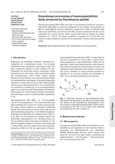
Fig. 1. Molecular structure <strong>of</strong> <strong>mannosylerythritol</strong> lipid.<br />
The length and saturation <strong>of</strong> the fatty acid residues (R2,<br />
R3) depend on the substrate and microorganism used.<br />
For example, if Pseudozyma aphidis DSM 14930 is grown<br />
on soybean oil: R2 = R3 = C7-C14 fatty acids, saturated<br />
and unsaturated. R4, R6 = acetyl or H.<br />
2 Material and methods<br />
2.1 Microorganism<br />
Pseudozyma aphidis DSM 14930 was isolated from soil,<br />
identified <strong>by</strong> and deposited at the Deutsche Stammsammlung<br />
von Mikroorganismen und Zellkulturen<br />
(DSMZ), Braunschweig, Germany. Stock cultures were<br />
grown for 3 d at 30 7C on agar medium containing 24 g<br />
potato dextrose L 21 and 20 g agar L 21 . They were stored<br />
at 4 7C and renewed every 4 wk.<br />
© 2005 WILEY-VCH Verlag GmbH & Co. KGaA, Weinheim www.ejlst.de<br />
Research Paper
374 U. Rau et al. Eur. J. Lipid Sci. Technol. 107 (2005) 373–380<br />
2.2 Media and cultivation conditions<br />
The seed culture contained 100 mL medium in 500 mL<br />
baffled shake flasks and was inoculated from agar slants.<br />
The medium for precultivation contained (all media data<br />
are related to 1 L deionised water): 30 g glucose, 1 g<br />
NH 4NO 3, 0.3 g KH 2PO 4, 1 g yeast extract, pH = 6.0 (not<br />
controlled). After 2 d on a rotary shaker at 110 rpm and<br />
30 7C, the preculture was transferred into the following<br />
bioreactor medium: 80 mL soybean oil (density<br />
0.84 g mL 21 ), 2 g NaNO 3, 0.2 g KH 2PO 4, 0.2 g<br />
MgSO 4 7H 2O, 1 g yeast extract, pH = 6.2 (not controlled),<br />
temperature 27 7C. A 72-L bioreactor (Sartorius BBI Systems<br />
GmbH, Melsungen, Germany) equipped with three<br />
Rushton turbine impellers was used. The bioreactor was<br />
filled with only 30 L medium due to foam formation. As<br />
initial impeller speed and aeration rate, 300 rpm and<br />
720 L h 21 , respectively, were applied. Soybean oil as antifoam<br />
agent and carbon source was additionally supplied<br />
throughout the cultivation. The bioreactor cultivation is<br />
described in detail elsewhere [18].<br />
2.3 Analyses<br />
In order to quantitatively detect MEL, triglycerides and<br />
fatty acids, 3 mL culture suspension was acidified with<br />
two drops <strong>of</strong> 5 N HCl to pH 2 and was subsequently<br />
extracted three times with 3 mL methyl tertiary butyl ether<br />
(MTBE). The mixture was vortexed for 1 min and centrifuged<br />
for 5 min at 6000 rpm. The organic phases were<br />
combined and analysed either <strong>by</strong> TLC or HPLC. The distinct<br />
separation <strong>of</strong> single spots for qualitative TLC analysis<br />
was carried out <strong>by</strong> the development <strong>of</strong> Silica gel 60<br />
F 254 (Merck, Germany) plates with the ternary solvent<br />
system CHCl 3/MeOH/H 20 (65:15:2). HPLC was performed<br />
on a silica gel column (Nucleosil 100-5; CS-Chromatographie<br />
Service GmbH, Langerwehe, Germany) with<br />
an evaporative light scattering detector (ACS Mass<br />
Detector model 750/14; Houston, TX, USA) using a gradient<br />
solvent program consisting <strong>of</strong> various proportions <strong>of</strong><br />
CHCl 3 and CH 3OH (from 99:1 to 0:100, vol/vol) at a flow<br />
rate <strong>of</strong> 1 mL min 21 [17]. The data <strong>of</strong> MEL, soybean oil,<br />
fatty acids and cell protein are mean values from at least<br />
two independent determinations. Differences <strong>of</strong> individual<br />
data varied between 2% and 7%. Due to non-uniform<br />
accumulation <strong>of</strong> storage material inside the cells, bio dry<br />
mass showed higher deviations from the mean value and,<br />
therefore, up to a fourfold determination was carried out.<br />
2.4 Surface tension<br />
The influence <strong>of</strong> MEL on the surface tension <strong>of</strong> water at<br />
25 7C was measured with a tensiomat (Lauda Wobser<br />
GmbH, Lauda-Königsh<strong>of</strong>en, Germany) using the ring<br />
method. The detailed description <strong>of</strong> this method is published<br />
elsewhere [17].<br />
2.5 <strong>Downstream</strong> <strong>processing</strong> <strong>of</strong> MEL<br />
2.5.1 Adsorption at XAD<br />
Different types <strong>of</strong> Amberlite XAD (Rohm and Haas, Philadelphia,<br />
PA, USA) were used as adsorbent. XAD-4 and<br />
XAD-16 are cross-linked polymers with different groups at<br />
their surface for the preferred adsorption <strong>of</strong> organic compounds<br />
with low and low-to-medium molecular weights,<br />
respectively. XAD-7HP can adsorb non-polar compounds<br />
from aqueous systems as well as polar compounds from<br />
non-polar solvents. Adsorption tests were carried out as<br />
follows: 10 mL culture suspension was added to 4 mL<br />
Amberlite beads. After 24 h <strong>of</strong> stirring, the beads were<br />
separated <strong>by</strong> filtration (glass fibre filter) and rinsed with<br />
10 mL water. The rinsing solution was extracted <strong>by</strong> the<br />
addition <strong>of</strong> 10 mL MTBE under vigorous mixing. The<br />
beads were divided and subsequently extracted with<br />
10 mL <strong>of</strong> either MTBE or methanol. The organic phases<br />
were analysed and compared <strong>by</strong> TLC.<br />
2.5.2 Extraction with organic solvents<br />
The thoroughly mixed culture suspension was first<br />
extracted three times with MTBE. Aqueous and organic<br />
phases were separated <strong>by</strong> centrifugation. The collected<br />
organic phases were evaporated and vacuum dried<br />
(60 7C, 3000 Pa). After solving the extract in methanol, it<br />
was subsequently treated also three times, but with<br />
cyclohexane. The again evaporated and dried extract still<br />
contained minor amounts <strong>of</strong> soybean oil and fatty acids.<br />
These compounds were removed <strong>by</strong> using a mixture <strong>of</strong><br />
n-hexane, methanol and water (1:6:3, pH 5.5) as well as<br />
subsequent threefold extraction <strong>of</strong> the aqueous phase <strong>by</strong><br />
n-hexane. The lyophilised aqueous phase resulted in a<br />
pure MEL fraction as transparent, brown-coloured, highly<br />
viscous fluid.<br />
2.5.3 Heating<br />
After heating <strong>of</strong> the culture suspension to 100–121 7C for<br />
1–20 min, two MEL-containing phases, a solid sticky and<br />
an aqueous one, were formed. About 90% (wt/vol) <strong>of</strong> MEL<br />
was transferred into the solid phase <strong>by</strong> this procedure,<br />
which could be easily separated <strong>by</strong> pouring <strong>of</strong>f the cell<br />
debris-containing aqueous phase.<br />
© 2005 WILEY-VCH Verlag GmbH & Co. KGaA, Weinheim www.ejlst.de
Eur. J. Lipid Sci. Technol. 107 (2005) 373–380 <strong>Downstream</strong> MEL 375<br />
3 Results<br />
3.1 Bioreactor cultivation<br />
A representative example <strong>of</strong> bioreactor cultivation for the<br />
production <strong>of</strong> MEL using Pseudozyma aphidis<br />
DSM 14930 is shown in Fig. 2. After about 1 d, nitrate<br />
and yeast extract as nitrogen sources were consumed,<br />
and the course <strong>of</strong> cell protein indicated the approach to<br />
the stationary phase. However, bio dry mass continued<br />
to increase due to the accumulation <strong>of</strong> storage material<br />
inside the cells. After growth had ceased, the formation<br />
<strong>of</strong> foam cumulatively increased; therefore, impeller<br />
speed and aeration rate had to be reduced gradually<br />
from 300 to 250 rpm and from 720 to 100 L h 21 , respectively.<br />
In spite <strong>of</strong> these modifications, the pO 2 was not<br />
influenced essentially and remained at about 60%. The<br />
reduction <strong>of</strong> impeller speed and aeration rate was not<br />
sufficient to prevent overfoaming. For this reason, soy-<br />
bean oil was additionally fed in various rates, both as<br />
carbon source and as anti-foam agent. Depending on<br />
the addition and consumption rate, different amounts <strong>of</strong><br />
soybean oil and fatty acids, released from soybean oil <strong>by</strong><br />
the lipolytic activity <strong>of</strong> P. aphidis, were detected. However,<br />
repeated cultivations showed that after soybean oil<br />
addition was stopped, a prolongation <strong>of</strong> the cultivation<br />
time <strong>by</strong> 1 d was sufficient for the total assimilation <strong>of</strong><br />
residual substrates.<br />
After 3 d, the first green to yellow MEL beads separated at<br />
the bottom <strong>of</strong> the sampling bottle. The number and width<br />
(2–10 mm) <strong>of</strong> these beads increased with time. Previous<br />
investigations [18] showed that the MEL beads were<br />
formed at a concentration greater than 40 g MEL L 21 and<br />
contained high quantities <strong>of</strong> MEL .60% (wt-%) and relatively<br />
small amounts <strong>of</strong> soybean oil ,20% (wt-%) as well<br />
as fatty acids ,10% (wt-%). After 8 d <strong>of</strong> cultivation, 90 g<br />
MEL L 21 was yielded.<br />
Fig. 2. Cultivation <strong>of</strong> P. aphidis in a 30-L<br />
bioreactor. After 1 d, the impeller speed and<br />
aeration rate were gradually decreased<br />
from 300 to 250 rpm and from 720 to<br />
100 L h 21 , respectively, depending on the<br />
foam formation. Between days 2.5 and 8,<br />
soybean oil was fed at various rates (0.2–<br />
0.67 g L 21 h 21 ). The first MEL beads were<br />
observed after 3 d.<br />
© 2005 WILEY-VCH Verlag GmbH & Co. KGaA, Weinheim www.ejlst.de
376 U. Rau et al. Eur. J. Lipid Sci. Technol. 107 (2005) 373–380<br />
3.2 Isolation and purification <strong>of</strong> MEL<br />
The MEL beads could be taken as indicators for<br />
enhanced product formation. Their consistency was<br />
similar to highly viscous oil drops, and they could not<br />
simply be separated <strong>by</strong>, e.g., filtration. Therefore, stepwise<br />
conventional extraction techniques starting with the<br />
total culture suspension and using different solvents was<br />
first applied for isolation and purification <strong>of</strong> MEL (Fig. 3).<br />
The extraction step with MTBE yielded on average 75%<br />
(wt-%) MEL, 15% (wt-%) soybean oil and 10% (wt-%)<br />
fatty acids after drying. The threefold repetition <strong>of</strong> this<br />
extraction step was necessary for exhaustive transfer <strong>of</strong><br />
MEL into the organic phase. The further enrichment to<br />
91% (wt-%) MEL and decrease to 5% (wt-%) soybean oil<br />
and 4% (wt-%) fatty acids was achieved <strong>by</strong> subsequent<br />
also threefold extraction, but using cyclohexane. The<br />
resulting purified MEL fraction was a transparent, browncoloured,<br />
highly viscous fluid at ambient temperature.<br />
During this procedure, about 20% (wt-%) MEL was lost<br />
compared to the mass contained in the primary culture<br />
suspension. The quantitative analysis <strong>of</strong> the compounds<br />
was carried out <strong>by</strong> HPLC. A complete separation <strong>of</strong> residual<br />
soybean oil and fatty acids was achieved <strong>by</strong> using<br />
n-hexane, methanol and water as solvent mixture with<br />
subsequent threefold extraction <strong>by</strong> n-hexane. The purification<br />
<strong>of</strong> MEL was documented <strong>by</strong> TLC (Fig. 3). However,<br />
the advantage to yield pure MEL was combined with an<br />
essential loss <strong>of</strong> recovery down to 8% (wt-%).<br />
Different polymeric resins (Amberlite XAD-4, XAD-16,<br />
XAD-7HP) were tested for the specific adsorption <strong>of</strong><br />
either MEL or fatty acids and soybean oil, in order to<br />
Fig. 3. Scheme <strong>of</strong> the stepwise<br />
extraction procedure <strong>by</strong> using different<br />
solvents for isolation and<br />
purification <strong>of</strong> MEL. The TLC<br />
represents the three extraction<br />
steps <strong>by</strong> n-hexane. Lanes 1–6,<br />
organic phase; lanes 7–12, aqueous<br />
phase; lanes 1, 2 17, 8, first<br />
extraction; lanes 3, 4 19, 10, second<br />
extraction; lanes 5, 6 111, 12,<br />
third extraction. MTBE, methyl tertiary<br />
butyl ether; FA, fatty acid; SO,<br />
soybean oil.<br />
© 2005 WILEY-VCH Verlag GmbH & Co. KGaA, Weinheim www.ejlst.de
Eur. J. Lipid Sci. Technol. 107 (2005) 373–380 <strong>Downstream</strong> MEL 377<br />
facilitate the downstream process. As a result, all polymers<br />
were able to accumulate MEL in different amounts<br />
that could be eluted <strong>by</strong> MTBE or methanol. However, fatty<br />
acids and soybean oil were also adsorbed. A specific<br />
adsorption was not possible <strong>by</strong> the use <strong>of</strong> these resins.<br />
Only a trend to higher adsorption capacity <strong>of</strong> MEL with<br />
simultaneous decreasing accumulation <strong>of</strong> fatty acids and<br />
soybean oil was observed in the order: XAD-16 . XAD-7<br />
. XAD-4.<br />
When the MEL-containing culture suspension was transferred<br />
into a glass bottle, the separation <strong>of</strong> aggregated<br />
MEL beads could be observed at the bottom as highly<br />
viscous fluid (Fig. 4, left picture). This viscous MEL phase<br />
and the MTBE extract <strong>of</strong> the whole culture suspension<br />
(Fig. 3) possessed a similar composition (Fig. 4). After<br />
sterilisation <strong>of</strong> the MEL-containing culture suspension at<br />
121 7C for 20 min, two MEL-containing phases, a solid<br />
sticky and an aqueous one, were formed, both fatty acid<br />
enriched as well as soybean oil depleted (Fig. 4, right<br />
picture). A small volume <strong>of</strong> a primary soybean oil-containing<br />
top phase was also observed. Related to the total<br />
mass <strong>of</strong> MEL (90 g L 21 ) yielded <strong>by</strong> MTBE extraction <strong>of</strong> the<br />
culture suspension before heating, the MEL were distributed<br />
after heating into the solid and aqueous phases <strong>by</strong><br />
89% and 11% (wt/vol), respectively. This solid phase was<br />
easy to separate <strong>by</strong> pouring <strong>of</strong>f the cell debris-containing<br />
supernatant. If necessary, dependent on the intended<br />
application <strong>of</strong> the MEL, the cell debris could be separated<br />
<strong>by</strong> solving the solid phase in ethanol and subsequent filtration<br />
using a pore width <strong>of</strong> 0.2 mm. About 11% (vol/vol)<br />
<strong>of</strong> MEL remained suspended in the aqueous cell debris<br />
phase and could additionally be recovered <strong>by</strong> extraction<br />
with ethanol, centrifugation, rotary evaporation <strong>of</strong> the<br />
solvent and vacuum drying.<br />
Variations <strong>of</strong> time (1, 5, 15, 20 min) and temperature (100,<br />
110, 115, 121 7C) <strong>of</strong> the culture suspension treatment<br />
resulted in a nonessential difference <strong>of</strong> MEL content between<br />
86.2–88.3% (wt-%) in the solid phase. Short incubation<br />
times <strong>of</strong> 5 min led to the formation <strong>of</strong> a turbid<br />
solid phase. The longer the time <strong>of</strong> treatment at each<br />
temperature, the higher was the fatty acid and the lower<br />
the soybean oil content, with a minimum <strong>of</strong> 0.3% (wt-%)<br />
soybean oil and a maximum <strong>of</strong> 13.7% (wt-%) fatty acids<br />
at 121 7C for 20 min (Fig. 4, grouped bars <strong>of</strong> solid phase).<br />
Corresponding to Fig. 4, Fig. 5 shows HPLC and TLC<br />
analyses <strong>of</strong> the MEL-containing phases before and after<br />
heat treatment, as well as the distribution <strong>of</strong> the different<br />
MEL. The maximum <strong>of</strong> 93% (wt-%) MEL transfer into the<br />
solid phase with an appropriate reduction to 7% (wt-%)<br />
MEL in the resulting aqueous phase was achieved at<br />
110 7C for 10 min and was considered as the most effective<br />
treatment for downstreaming the MEL. This solid<br />
phase contained 88.3% (wt-%) MEL, 6.6% (wt-%) fatty<br />
acids as well as 5.1% (wt-%) soybean oil and reduced the<br />
surface tension <strong>of</strong> water/air to 31 mN m 21 (critical micelle<br />
concentration 15 mg L 21 ).<br />
Fig. 4. Composition <strong>of</strong> different MEL phases from a<br />
culture suspension before (left) and after (right)<br />
treatment at 121 7C for 20 min. The analyses were<br />
carried out <strong>by</strong> HPLC.<br />
© 2005 WILEY-VCH Verlag GmbH & Co. KGaA, Weinheim www.ejlst.de
378 U. Rau et al. Eur. J. Lipid Sci. Technol. 107 (2005) 373–380<br />
4 Discussion<br />
Different microorganisms as, e.g., Kurtzmanomyces sp.<br />
I-11 [12], Candida antarctica [14] and Ustilago maydis [10]<br />
were used <strong>by</strong> other authors for shake flask production <strong>of</strong><br />
MEL. Kitamoto’s group succeeded in the formation <strong>of</strong><br />
140 g MEL L 21 <strong>by</strong> additional feeding <strong>of</strong> n-octadecane<br />
using Pseudozyma (Candida) antarctica T 34 [19]. However,<br />
only rare information about bioreactor production <strong>of</strong><br />
MEL is available. Kim et al. [15] used Candida sp. SY16 in<br />
a 5-L bioreactor for the formation <strong>of</strong> 100 g L 21 crude MEL<br />
phase containing only 4% (wt-%) pure MEL. Hitherto, 46<br />
and 165 g L 21 are the highest reported yields <strong>of</strong> MEL<br />
obtained in a bioreactor <strong>by</strong> using Pseudozyma (Candida)<br />
antarctica ATCC 20509 [16] and Pseudozyma aphidis<br />
DSM 70725 [18], respectively.<br />
The choice <strong>of</strong> method for the isolation and purification <strong>of</strong> a<br />
particular biosurfactant depends on its ionic charge, its<br />
solubility in water, and on whether the product is cell<br />
bound or extracellular. The methods used include solvent<br />
extraction, adsorption followed <strong>by</strong> solvent extraction,<br />
precipitation, crystallisation, centrifugation and foam<br />
fractionation. Desai and Desai [20] as well as Syldatk and<br />
Wagner [21] gave an overview <strong>of</strong> these procedures. Solvent<br />
extraction is the most commonly used technique for<br />
the downstream <strong>processing</strong> <strong>of</strong> biosurfactants. We also<br />
described such a multi-step extraction (Fig. 3) for MEL<br />
that is further on absolutely necessary if, e.g., spectroscopic<br />
investigations are performed or an application as<br />
pharmaceuticals is intended [3, 4]. The disadvantage <strong>of</strong><br />
this process is the production <strong>of</strong> huge amounts <strong>of</strong> waste<br />
solvents that have to be recycled. Beside the costs for<br />
bioreactor production, the recycling increases the manufacturing<br />
costs so that an acceptance <strong>of</strong> this bioprocess<br />
for industry is additionally inhibited.<br />
Fig. 5. HPLC and TLC <strong>of</strong> the solid MEL<br />
phase after heat treatment at 121 7C for<br />
20 min. The HPLC signals correspond with<br />
the grouped bars <strong>of</strong> the solid phase <strong>of</strong><br />
Fig. 4. Distribution <strong>of</strong> MEL (wt-%):<br />
A = 40.8, B = 43.4, C = 9.7, D = 6.1. The<br />
embedded TLC corresponds with the<br />
grouped bars <strong>of</strong> Fig. 4: Before heat treatment,<br />
1 = viscous MEL phase; after heat<br />
treatment, 2 = aqueous phase, 3 = solid<br />
phase.<br />
Unfortunately, the specific adsorption <strong>of</strong> MEL or soybean<br />
oil and fatty acids failed <strong>by</strong> the use <strong>of</strong> different Amberlite<br />
resins. Only a general tendency to higher accumulation <strong>of</strong><br />
MEL was observed in the order: XAD-16 . XAD-7HP .<br />
XAD-4.<br />
The MEL can also be isolated <strong>by</strong> preparative HPLC<br />
equipped with silica gel columns [15, 17, 22]. This is a<br />
superior method to produce a very pure MEL mixture or<br />
even to separate the individual MEL. However, the loss <strong>of</strong><br />
product is substantial, and so this is not a beneficial solution<br />
in order to decrease the costs for the downstream<br />
process.<br />
A very easy, solvent-free separation <strong>of</strong> a MEL-enriched<br />
solid phase could be achieved <strong>by</strong> just heating the culture<br />
suspension to a temperature <strong>of</strong> 100 7C. Related to the<br />
total MEL content <strong>of</strong> the untreated culture suspension,<br />
the heating at 110 7C for 10 min led to the maximum<br />
transfer <strong>of</strong> 93% (wt-%) MEL into this solid phase. The<br />
supernatant could be poured <strong>of</strong>f, which left a solid, highly<br />
viscous mass, on average composed <strong>of</strong> 87% (wt-%) MEL<br />
(Figs. 4, 5). The remaining 13% (wt-%) consisted <strong>of</strong> soybean<br />
oil and fatty acids in different amounts, depending<br />
on time and temperature throughout the treatment. Independent<br />
<strong>of</strong> the temperature, the longer the time <strong>of</strong> treatment,<br />
the more soybean oil was transferred to a new<br />
phase separated at the top. Furthermore, prolonged<br />
heating favoured the release <strong>of</strong> fatty acids from the<br />
remaining soybean oil.<br />
A comparison <strong>of</strong> known MEL downstream procedures is<br />
given in Tab. 1. Unfortunately, to the best knowledge <strong>of</strong><br />
the authors, only two references are available with a<br />
quantitative description <strong>of</strong> the methods. The heat treatment<br />
attained the highest yield and is the fastest method<br />
<strong>by</strong> far, but only on average 87% (wt-%) MEL is contained<br />
© 2005 WILEY-VCH Verlag GmbH & Co. KGaA, Weinheim www.ejlst.de
Eur. J. Lipid Sci. Technol. 107 (2005) 373–380 <strong>Downstream</strong> MEL 379<br />
Tab. 1. Comparison <strong>of</strong> different methods for the downstream<br />
<strong>processing</strong> <strong>of</strong> MEL { .<br />
Ref. Method § Yield<br />
[wt-%]<br />
# Purity<br />
[wt-%]<br />
[22] Ethyl acetate extraction 1<br />
preparative HPLC<br />
79 100<br />
[15] Ethyl acetate extraction 1<br />
preparative HPLC<br />
4 100<br />
Stepwise extraction with<br />
different solvents (Fig. 3)<br />
8 100<br />
Heat treatment (Fig. 4) 93 87<br />
{ Preparative HPLC was performed with silica gel columns.<br />
§ g MEL recovered<br />
Yield =<br />
g MEL before downstream 100<br />
#<br />
Purity is related to the mass fraction <strong>of</strong> MEL.<br />
in the precipitated fraction. However, this solid-enriched<br />
MEL phase should be pure enough for the most industrial<br />
applications [5–8]. For example, pure MEL A, purified<br />
100% (wt-%) MEL and a mixture <strong>of</strong> 88.3% MEL, 6.6%<br />
soybean oil and 5.1% (wt-%) fatty acids reduced the surface<br />
tension <strong>of</strong> water/air to similar data <strong>of</strong> 34.7, 26.7 and<br />
31 mN m 21 , respectively [17].<br />
5 Conclusion<br />
Bioreactor production <strong>of</strong> <strong>mannosylerythritol</strong> <strong>lipids</strong> was<br />
performed <strong>by</strong> the use <strong>of</strong> Pseudozyma aphidis<br />
DSM 14930. Up to 93% (wt-%) MEL could be transferred<br />
into a solid, highly viscous phase <strong>by</strong> heating the culture<br />
suspension to 110 7C for 10 min. This phase contained on<br />
average 87% (wt-%) MEL and could be simply isolated <strong>by</strong><br />
pouring <strong>of</strong>f the supernatant, without producing solvent<br />
waste. Together with the high MEL yield <strong>of</strong> 165 g L 21 ,<br />
obtained <strong>by</strong> foam-controlled addition <strong>of</strong> soybean oil [18],<br />
this facilitated downstream process should stimulate the<br />
industrial production <strong>of</strong> MEL.<br />
Acknowledgments<br />
We thank W. Grassl for technical assistance.<br />
References<br />
[1] G. Georgiou, S. C. Lin, M. Sharma: Surface-active compounds<br />
from microorganisms. Biotechnology 1992, 10,60–65.<br />
[2] D. Kitamoto, H. Isoda, T. Nakahara: Functions and potential<br />
applications <strong>of</strong> glycolipid biosurfactants – from energy-saving<br />
materials to gene delivery carriers. J Biosci Bioeng. 2002, 94,<br />
187–201.<br />
[3] M. Shibahara, X. Zhao, Y. Wakamatsu, N. Nomura, T. Nakahara,<br />
C. Jin, H. Nagaso, T. Murata, K. K. Yokoyama: Mannosylerythritol<br />
lipid increases levels <strong>of</strong> galactoceramide in<br />
and neurite outgrowth from PC12 pheochromocytoma cells.<br />
Cytotechnol. 2000, 33, 247–251.<br />
[4] L. Vertesy, M. Kurz, J. Wink, G. Noelken: Patent US<br />
6,472,158 (2002).<br />
[5] J. H. Im, H. Yanagishita, T. Ikegami, Y. Takeyama, Y. Idemoto,<br />
N. Koura, D. Kitamoto: Mannosylerythritol <strong>lipids</strong>,<br />
yeast glycolipid biosurfactants, are potential affinity ligand<br />
materials for human immunoglobulin G. J Biomed Mater<br />
Res. 2003, 65A, 379–385.<br />
[6] D. Kitamoto, H. Yanagishita, A. Endo, M. Nakaiwa, M.<br />
Nakane, T. Akiya: Remarkable antiagglomeration effect <strong>of</strong> a<br />
yeast biosurfactant, diacyl<strong>mannosylerythritol</strong>, on ice-water<br />
slurry for cold thermal storage. Biotechnol Progress 2001,<br />
17, 362–365.<br />
[7] Z. Hua, J. Chena, S. Luna, X. Wang: Influence <strong>of</strong> biosurfactants<br />
<strong>produced</strong> <strong>by</strong> Candida antarctica on surface properties<br />
<strong>of</strong> microorganism and biodegradation <strong>of</strong> n-alkanes. Water<br />
Research 2003, 37, 4143–4150.<br />
[8] Z. Hua, Y. Chen, G. Du, J. Chen: Effects <strong>of</strong> biosurfactants<br />
<strong>produced</strong> <strong>by</strong> Candida antarctica on the biodegradation <strong>of</strong><br />
petroleum compounds. World J Microbiol Biotechnol. 2004,<br />
20, 25–29.<br />
[9] D. Kitamoto, S. Akiba, C. Hioki, T. Tabuchi: Extracellular<br />
accumulation <strong>of</strong> <strong>mannosylerythritol</strong> <strong>lipids</strong> <strong>by</strong> a strain <strong>of</strong><br />
Candida antarctica. Agric Biol Chem. 1990, 54, 31–36.<br />
[10] S. Spoeckner, V. Wray, M. Nimtz, S. Lang: Glyco<strong>lipids</strong> <strong>of</strong> the<br />
smut fungus Ustilago maydis from cultivation on renewable<br />
resources. Appl Microbiol Biotechnol. 1999, 51, 33–39.<br />
[11] G. Deml, T. Anke, F. Oberwinkler, B. M. Gianetti, W. Steglich:<br />
Schizonellin A and B, new glyco<strong>lipids</strong> from Schizonella melanogramma.<br />
Phytochem. 1980, 19, 83–87.<br />
[12] K. Kakugawa, M. Tamai, K. Imamura, K. Miyamoto, S.<br />
Miyoshi, Y. Morinaga, O. Suzuki, T. Miyakawa: Isolation <strong>of</strong><br />
yeast Kurtzmanomyces sp. I-11, novel producer for <strong>mannosylerythritol</strong><br />
lipid. Biosci Biotech Biochem. 2002, 62, 188–<br />
191.<br />
[13] H. Kawashima, T. Nakahara, M. Oogaki, T. Tabuchi: Extracellular<br />
production <strong>of</strong> a <strong>mannosylerythritol</strong>lipid <strong>by</strong> a mutant<br />
<strong>of</strong> Candida sp. from n-alkanes and triacylglycerols. J Ferment<br />
Technol. 1983, 61, 143–149.<br />
[14] D. Kitamoto, T. Yokoshima, H. Yanagishita, K. Haraya, H. K.<br />
Kitamoto: Formation <strong>of</strong> glycolipid biosurfactant, <strong>mannosylerythritol</strong><br />
lipid, <strong>by</strong> Candida antarctica from aliphatic hydrocarbons<br />
via subterminal oxidation pathway. J Jpn Oil Chem<br />
Soc. 1999, 48, 1377–1384.<br />
[15] H.-S. Kim, B. D. Yoon, D. H. Choung, H.-M. Oh, T. Katsuragi,<br />
Y. Tani: Characterization <strong>of</strong> a biosurfactant, MEL, <strong>produced</strong><br />
from Candida sp. SY16. Appl Microbiol Biotechnol. 1999,<br />
52, 713–721.<br />
[16] M. Adamczak, W. Bednarski: Influence <strong>of</strong> medium composition<br />
and aeration on the synthesis <strong>of</strong> biosurfactant <strong>produced</strong><br />
<strong>by</strong> Candida antarctica. Biotechnol Lett. 2000, 22,<br />
313–316.<br />
[17] U. Rau, L. A. Nguyen, S. Schulz, V. Wray, M. Nimtz, H. Roeper,<br />
H. Koch, S. Lang: Formation and analysis <strong>of</strong> <strong>mannosylerythritol</strong><br />
<strong>lipids</strong> secreted <strong>by</strong> Pseudozyma aphidis. Appl<br />
Microbiol Biotechnol. 2005, 66, 551–559.<br />
[18] U. Rau, L. A. Nguyen, H. Roeper, H. Koch, S. Lang: Fedbatch<br />
bioreactor production <strong>of</strong> <strong>mannosylerythritol</strong> <strong>lipids</strong><br />
secreted <strong>by</strong> Pseudozyma aphidis. Appl Microbiol Biotechnol.<br />
2005, DOI 10.1007/s00253-005-1906-5.<br />
© 2005 WILEY-VCH Verlag GmbH & Co. KGaA, Weinheim www.ejlst.de
380 U. Rau et al. Eur. J. Lipid Sci. Technol. 107 (2005) 373–380<br />
[19] D. Kitamoto, T. Ikegami, G. Suzuki, A. Sasaki, Y. Takeyama,<br />
Y. Idemoto, N. Koura, H. Yanagishita: Microbial conversion<br />
<strong>of</strong> n-alkanes into glycolipid biosurfactants, <strong>mannosylerythritol</strong><br />
<strong>lipids</strong>, <strong>by</strong> Pseudozyma (Candida) antarctica. Biotechnol<br />
Lett. 2001, 23, 1709–1714.<br />
[20] J. D. Desai, A. J. Desai: Production <strong>of</strong> biosurfactants. In:<br />
Biosurfactants: Production, Properties, Applications.Vol48,<br />
Surfactant Science Series. Ed. N. Kosaric, Marcel Dekker,<br />
Inc., New York (USA) 1993, pp. 65–97.<br />
[21] C. Syldatk, F. Wagner: Production <strong>of</strong> biosurfactants. In: Biosurfactants<br />
and Biotechnology. Vol. 25, Surfactant Science<br />
Series. Eds. N. Kosaric, W. L. Cairns, N. C. C. Gray, Marcel<br />
Dekker, Inc., New York (USA) 1987, pp. 89–120.<br />
[22] D. Kitamoto, S. Ghosh, O. G. Y. Nakatani: Formation <strong>of</strong> giant<br />
vesicles from diacyl<strong>mannosylerythritol</strong>s and their binding to<br />
concanavalin A. Chem Comm. 2000, 10, 861–862.<br />
[Received: December 22, 2004; accepted: April 1, 2005]<br />
© 2005 WILEY-VCH Verlag GmbH & Co. KGaA, Weinheim www.ejlst.de


