Molecular characterization and 16S rRNA sequence analysis of probiotic lactobacillus acidophilus isolated from indigenous Dahi (Yoghurt)
Indigenous yoghurt is being the most popular fermented milk product in Pakistan, produced by heterogenous lactic acid bacteria (LAB). Nowadays LAB has drawn particular interest in food and nutrition science because of functional and probiotic attributes. Among various genera of LAB, Lactobacillus acidophilus is considered to be a potential candidate. Present study was designed by keeping in view the probiotic attributes of L. acidophilus. Isolation was conducted on MRS media supplemented with 0.7% bile salts to screen out bile tolerant isolates. Out of sixty lactobacilli, eighteen were identified as L. acidophilus by performing gram staining, catalase, carbohydrate fermentation test and growth at different temperatures. Only six had shown tolerance of pH 2 up to 50% and only three had shown a wide range of antimicrobial activity against tested organisms. S. epidermis found to be more sensitive with a maximum zone of inhibition (18 mm) and A. baumanii and S. aureus were least sensitive with a smallest zone of inhibition (11.5 mm and 12 mm respectively). 16S rRNA gene amplification, sequencing and phylogenetic tree construction of the obtained sequences with the most closely related lactobacillus spp. was performed. Sequences are available in GeneBank and NCBI with the accession numbers KU877440 (WFA1), KU877441 (WFA2) and KU877442 (WFA3). Presence of probiotic L. acidophilus in locally fermented product (dahi) has proved its potential along with bile salts supplementation in MRS media is helpful in initial screening of probiotic isolates.
Indigenous yoghurt is being the most popular fermented milk product in Pakistan, produced by heterogenous lactic acid bacteria (LAB). Nowadays LAB has drawn particular interest in food and nutrition science because of functional and probiotic attributes. Among various genera of LAB, Lactobacillus acidophilus is considered to be
a potential candidate. Present study was designed by keeping in view the probiotic attributes of L. acidophilus. Isolation was conducted on MRS media supplemented with 0.7% bile salts to screen out bile tolerant isolates. Out of sixty lactobacilli, eighteen were identified as L. acidophilus by performing gram staining, catalase, carbohydrate fermentation test and growth at different temperatures. Only six had shown tolerance of pH 2 up to 50% and only three had shown a wide range of antimicrobial activity against tested organisms. S. epidermis found to be more sensitive with a maximum zone of inhibition (18 mm) and A. baumanii and S. aureus were least sensitive with a smallest zone of inhibition (11.5 mm and 12 mm respectively). 16S rRNA gene
amplification, sequencing and phylogenetic tree construction of the obtained sequences with the most closely related lactobacillus spp. was performed. Sequences are available in GeneBank and NCBI with the accession numbers KU877440 (WFA1), KU877441 (WFA2) and KU877442 (WFA3). Presence of probiotic L. acidophilus in
locally fermented product (dahi) has proved its potential along with bile salts supplementation in MRS media is helpful in initial screening of probiotic isolates.
You also want an ePaper? Increase the reach of your titles
YUMPU automatically turns print PDFs into web optimized ePapers that Google loves.
Int. J. Biosci. 2016<br />
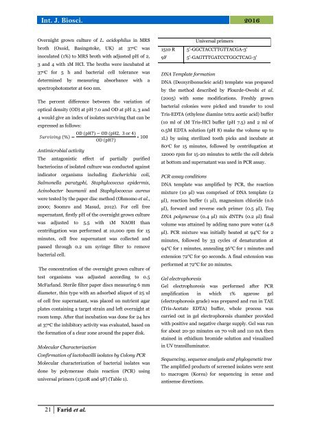
Overnight grown culture <strong>of</strong> L. <strong>acidophilus</strong> in MRS<br />
broth (Oxoid, Basingstoke, UK) at 37 o C was<br />
inoculated (1%) to MRS broth with adjusted pH <strong>of</strong> 2,<br />
3 <strong>and</strong> 4 with 1M HCl. The broths were incubated at<br />
37 o C for 5 h <strong>and</strong> bacterial cell tolerance was<br />
determined by measuring absorbance with a<br />
spectrophotometer at 600 nm.<br />
The percent difference between the variation <strong>of</strong><br />
optical density (OD) at pH 7.0 <strong>and</strong> OD at pH 2, 3 <strong>and</strong><br />
4 would give an index <strong>of</strong> isolates surviving that can be<br />
expressed as follows:<br />
Surviving (%) =<br />
Antimicrobial activity<br />
OD (pH7) − OD (pH2, 3 or 4)<br />
∗ 100<br />
OD (pH7)<br />
The antagonistic effect <strong>of</strong> partially purified<br />
bacteriocins <strong>of</strong> <strong>isolated</strong> culture was conducted against<br />
indicator organisms including Escherichia coli,<br />
Salmonella paratyphi, Staphylococcus epidermis,<br />
Acinobacter baumanii <strong>and</strong> Staphylococcus aureus<br />
were tested by the paper disc method (Ohmomo et al.,<br />
2000; Soomro <strong>and</strong> Masud, 2012). For cell free<br />
supernatant, firstly pH <strong>of</strong> the overnight grown culture<br />
was adjusted to 5.5 with 1M NAOH than<br />
centrifugation was performed at 10,000 rpm for 15<br />
minutes, cell free supernatant was collected <strong>and</strong><br />
passed through 0.2 um syringe filter to remove<br />
bacterial cell.<br />
The concentration <strong>of</strong> the overnight grown culture <strong>of</strong><br />
test organisms was adjusted according to 0.5<br />
McFarl<strong>and</strong>. Sterile filter paper discs measuring 6 mm<br />
diameter, thin type with an adsorbed aliquot <strong>of</strong> 25 ul<br />
<strong>of</strong> cell free supernatant, was placed on nutrient agar<br />
plates containing a target strain <strong>and</strong> left overnight at<br />
room temp. After that incubation was done for 24 hrs<br />
at 37 o C the inhibitory activity was evaluated, based on<br />
the formation <strong>of</strong> a clear zone around the paper disk.<br />
<strong>Molecular</strong> Characterization<br />
Confirmation <strong>of</strong> lactobacilli isolates by Colony PCR<br />
<strong>Molecular</strong> <strong>characterization</strong> <strong>of</strong> bacterial isolates was<br />
done by polymerase chain reaction (PCR) using<br />
universal primers (1510R <strong>and</strong> 9F) (Table 1).<br />
Universal primers<br />
1510 R 5’-GGCTACCTTGTTACGA-3'<br />
9F 5’-GAGTTTGATCCTGGCTCAG-3'<br />
DNA Template formation<br />
DNA (Deoxyribonucleic acid) template was prepared<br />
by the method described by Plourde-Owobi et al.<br />
(2005) with some modifications. Freshly grown<br />
bacterial colonies were picked <strong>and</strong> transfer to 10ul<br />
Tris-EDTA (ethylene diamine tetra acetic acid) buffer<br />
(10 ml <strong>of</strong> 1M Tris-HCl buffer (pH 7.5) <strong>and</strong> 2 ml <strong>of</strong><br />
0.5M EDTA solution (pH 8) make the volume up to<br />
1L) by using sterilized tooth picks <strong>and</strong> incubate at<br />
80 o C for 15 minutes, followed by centrifugation at<br />
12000 rpm for 15-20 minutes to settle the cell debris<br />
at bottom <strong>and</strong> supernatant was used in PCR assay.<br />
PCR assay conditions<br />
DNA template was amplified by PCR, the reaction<br />
mixture (10 μl) was comprised <strong>of</strong> DNA template (2<br />
μl), reaction buffer (1 µl), magnesium chloride (0.6<br />
µl), forward <strong>and</strong> reverse each primer (0.5 µl), Taq<br />
DNA polymerase (0.4 µl) mix dNTPs (0.2 µl) final<br />
volume was attained by adding nano pure water (4.8<br />
µl). PCR mixture was initially heated at 94°C for 2<br />
minutes, followed by 33 cycles <strong>of</strong> denaturation at<br />
94°C for 1 minutes, annealing 56°C for 1 minutes <strong>and</strong><br />
extension 72°C for 90 seconds. A final extension was<br />
performed at 72°C for 20 minutes.<br />
Gel electrophoresis<br />
Gel electrophoresis was performed after PCR<br />
amplification in which 1% agarose gel<br />
(electrophoresis grade) was prepared <strong>and</strong> run in TAE<br />
(Tris-Acetate EDTA) buffer, whole process was<br />
carried out in gel electrophoresis chamber provided<br />
with positive <strong>and</strong> negative charge supply. Gel was run<br />
for about 20-30 minutes on 70 volt <strong>and</strong> 110 mA then<br />
stained in ethidium bromide solution <strong>and</strong> visualized<br />
in UV transilluminator.<br />
Sequencing, <strong>sequence</strong> <strong>analysis</strong> <strong>and</strong> phylogenetic tree<br />
The amplified products <strong>of</strong> screened isolates were sent<br />
to macrogen (Korea) for sequencing in sense <strong>and</strong><br />
antisense directions.<br />
21 Farid et al.


