Carboxyhemoglobin - Masimo
Carboxyhemoglobin - Masimo
Carboxyhemoglobin - Masimo
You also want an ePaper? Increase the reach of your titles
YUMPU automatically turns print PDFs into web optimized ePapers that Google loves.
masimo corporation 40 parker irvine ca 92618 www.masimo.com<br />
<strong>Carboxyhemoglobin</strong>emia in Acute Care<br />
Carbon Monoxide Lurking Within: The Danger of<br />
<strong>Carboxyhemoglobin</strong>emia in Acute Care<br />
Summary<br />
Much is known about the causes, symptoms and detection of exogenous carbon monoxide (CO)<br />
poisoning and the treatment of the resultant carboxyhemoglobinemia seen in patients after CO<br />
exposure. However, it may be surprising to learn that CO poisoning can occur in the hospital and<br />
not just outside the hospital. In most cases, these patients present in the emergency department<br />
(ED) with equivocal, flu-like symptoms. Because the clearance of CO from the blood begins<br />
to occur immediately after the etiologic agent is removed, patients may begin to feel better in<br />
the ED and suffer a missed diagnosis as the symptoms abate. In many reported cases, these<br />
poisoned patients are sent back to the source of the odorless, colorless toxin, and poisoning<br />
continues, sometimes with lethal consequences. Others will find themselves admitted for acute<br />
care as the symptoms of CO poisoning mimic cardiac, pulmonary, and neurologic disorders that<br />
demand emergent care. Whether in the ED or other places in the hospital such as the OR, if and<br />
when carboxyhemoglobinema detection occurs, it is often after costly diagnostic procedures and<br />
protocols have been attempted with negative results.<br />
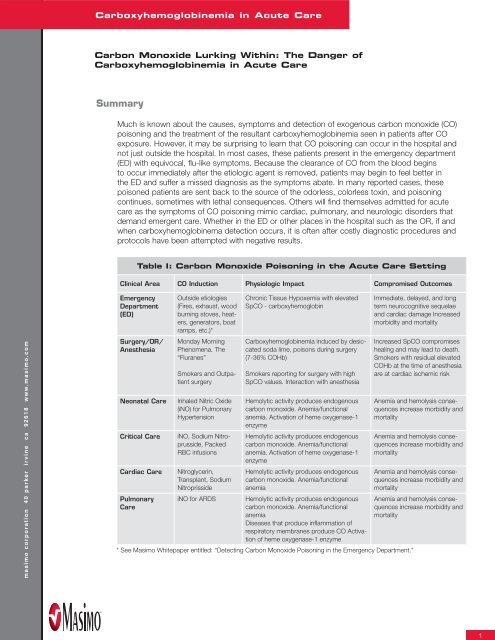
Table I: Carbon Monoxide Poisoning in the Acute Care Setting<br />
Clinical Area CO Induction Physiologic Impact Compromised Outcomes<br />
Emergency<br />
Department<br />
(ED)<br />
Surgery/OR/<br />
Anesthesia<br />
Outside etiologies<br />
(Fires, exhaust, wood<br />
burning stoves, heaters,<br />
generators, boat<br />
ramps, etc.)*<br />
Monday Morning<br />
Phenomena. The<br />
“Fluranes”<br />
Smokers and Outpatient<br />
surgery<br />
Neonatal Care Inhaled Nitric Oxide<br />
(iNO) for Pulmonary<br />
Hypertension<br />
Critical Care iNO, Sodium Nitroprusside,<br />
Packed<br />
RBC infusions<br />
Cardiac Care Nitroglycerin,<br />
Transplant, Sodium<br />
Nitroprisside<br />
Pulmonary<br />
Care<br />
Chronic Tissue Hypoxemia with elevated<br />
SpCO - carboxyhemoglobin<br />
<strong>Carboxyhemoglobin</strong>emia induced by desiccated<br />
soda lime, poisons during surgery<br />
(7-36% COHb)<br />
Smokers reporting for surgery with high<br />
SpCO values. Interaction with anesthesia<br />
Hemolytic activity produces endogenous<br />
carbon monoxide. Anemia/functional<br />
anemia. Activation of heme oxygenase-1<br />
enzyme<br />
Hemolytic activity produces endogenous<br />
carbon monoxide. Anemia/functional<br />
anemia. Activation of heme oxygenase-1<br />
enzyme<br />
Hemolytic activity produces endogenous<br />
carbon monoxide. Anemia/functional<br />
anemia<br />
iNO for ARDS Hemolytic activity produces endogenous<br />
carbon monoxide. Anemia/functional<br />
anemia<br />
Diseases that produce infl ammation of<br />
respiratory membranes produce CO Activation<br />
of heme oxygenase-1 enzyme<br />
* See <strong>Masimo</strong> Whitepaper entitled: “Detecting Carbon Monoxide Poisoning in the Emergency Department.”<br />
Immediate, delayed, and long<br />
term neurocognitive sequelae<br />
and cardiac damage Increased<br />
morbidity and mortality<br />
Increased SpCO compromises<br />
healing and may lead to death.<br />
Smokers with residual elevated<br />
COHb at the time of anesthesia<br />
are at cardiac ischemic risk<br />
Anemia and hemolysis consequences<br />
increase morbidity and<br />
mortality<br />
Anemia and hemolysis consequences<br />
increase morbidity and<br />
mortality<br />
Anemia and hemolysis consequences<br />
increase morbidity and<br />
mortality<br />
Anemia and hemolysis consequences<br />
increase morbidity and<br />
mortality<br />
1
masimo corporation 40 parker irvine ca 92618 www.masimo.com<br />
<strong>Carboxyhemoglobin</strong>emia in Acute Care<br />
However, CO poisoning may be quickly caught within the acute care setting using a state-ofthe-art<br />
<strong>Masimo</strong> Rainbow SET Pulse CO-Oximetry device. Expeditious diagnosis ensures proper<br />
treatment may ensue to minimize the known long-term cardiac and neuropsychiatric damage of CO<br />
exposure. In the case of a CO poisoned patient that presents in the ED and then transitions into<br />
the acute care environment, Pulse CO-Oximetry provides clinicians with immediate detection and<br />
subsequent continuous monitoring of carboxyhemoglobin (COHb) levels as feedback to the efficacy<br />
of treatment decisions. However, CO poisoning also occurs within the acute care setting – both<br />
endogenously and exogenously - contributing to severe tissue hypoxemia, ischemia, and death.<br />
Therefore, Pulse CO-Oximetry serves an additional role in detection and monitoring of nosocomial<br />
CO poisoning.<br />
This paper explores a sampling of several areas in which nosocomial CO exposure is possible.<br />
Several disease processes produce endogenous CO through their natural progression as in the<br />
development of systemic inflammatory response syndrome (SIRS), sepsis, pulmonary inflammation,<br />
and hemolysis. In these events, the detection and measurement of endogenously produced CO<br />
may prove to be a valuable marker of disease severity. There are also reports of CO poisoning<br />
by the Monday Morning Phenomena where carboxyhemoglobinemia is induced within the closed<br />
circuit anesthesia system during surgery, inhaled nitric oxide therapy, and anti-hypertension<br />
treatment with sodium nitroprusside. Though these acute care sources of CO may not raise blood<br />
COHb to life-threatening levels for many patients, even slight increases in CO concentrations can<br />
be life-threatening to those patients compromised by cardiac disease, anemia, loss of pulmonary<br />
reserve and a host of other diseases.<br />
In all cases, the clinical importance of continually and accurately measuring COHb within the<br />
hospital is established in the medical literature. New Pulse CO-Oximetry technology platforms,<br />
available as handheld and bedside devices, provide immediate and continuous results through noninvasive<br />
monitoring and enhance the clinical workflow by eliminating the need for a physician order,<br />
painful blood sampling, and the attendant high costs and likely delays associated with a laboratory<br />
CO-Oximeter analysis. In addition, they allow trended presentations of COHb which quickly illustrate<br />
significant changes that occur over time, changes that might be lost in visual or tabular displays of<br />
invasive COHb measurements.<br />
I. Emergency Department/ER<br />
Study shows prevalence of carbon monoxide toxicity in the ED may be higher<br />
than previously recognized<br />
In a study led by Dr. Robert Partridge and Dr. Gregory Jay of Rhode Island Hospital at Brown<br />
University Medical School, a team of researchers performed a study to assess baseline CO levels<br />
of nearly 5,000 patients presenting to the emergency room. To accomplish this, all pulse oximeters<br />
in the ED were replaced with <strong>Masimo</strong> Rainbow SET Pulse CO-Oximeters and the ED staff began<br />
assessing baseline COHb levels of all adult patients as part of the standard triage process. In<br />
addition to confirming suspected cases of CO toxicity (COT) from smoke inhalation, there were<br />
nine unsuspected cases of COT discovered, in just three months, in patients who presented with<br />
non-specific symptoms or unrelated complaints. Toxic COHb levels ranged from 16-33% and were<br />
confirmed with an invasive laboratory blood test. If this rate were indicative of all US hospitals, it<br />
would equate to as many as 50,000 cases of unsuspected CO toxicity annually.<br />
The study concluded that the use of <strong>Masimo</strong> Rainbow SET as a noninvasive test for COT can<br />
effectively and efficiently be performed at ED triage, and that “unsuspected COT may be identified<br />
using noninvasive COHb screening and the prevalence of COT may be higher than previously<br />
recognized.” 1
The team from Brown University also presented a case report of a previously healthy 52-year old non-smoking<br />
female who was brought to the ED complaining of nausea, headache, dizziness, and feeling cold. The patient<br />
had no history of carbon monoxide exposure. The <strong>Masimo</strong> Rainbow SET device recorded an SpCO level of 33%,<br />
which was later confirmed with an invasive laboratory measurement. After interviewing the woman, clinicians<br />
learned that her utilities had been shut off and she was running a gas-powered generator in her basement.<br />
In the report, researchers said that since early CO toxicity shares symptoms with other more common illnesses,<br />
“physicians must maintain a high index of suspicion to avoid incorrect diagnosis, management and disposition.<br />
Unrecognized CO poisoned patients returned to the site of exposure may develop more serious CO toxicity.”<br />
They added that the noninvasive testing provided by <strong>Masimo</strong> Rainbow SET technology “is a rapid, inexpensive<br />
method for screening large numbers of patients for CO toxicity and identifying unsuspected cases that might<br />
otherwise be missed.” 2<br />
II. Surgery/Operating Room/Anesthesia<br />
Monday Morning Phenomena and Anesthesia-Related COHb<br />
whitepaper<br />
There is increasing evidence that exposure of volatile anesthetics, i.e., desflurane, enflurane, and isoflurane<br />
(in descending order of magnitude) to desiccated carbon dioxide (CO 2) absorbents may result in reactions<br />
in anesthetic breathing circuits and production of toxic products (e.g., CO, methanol, formaldehyde). 3-6 CO 2<br />
absorbents such as soda lime are mixtures of chemicals, used in closed breathing environments, such as<br />
general anesthesia to remove CO 2 from breathing gases to prevent CO 2 retention and poisoning. There is<br />
significant evidence that potentially toxic products can be produced upon exposure of volatile anesthetics to<br />
other desiccated absorbents containing strong bases, particularly potassium and sodium hydroxide. The clinical<br />
scenario has been called the “Monday Morning Phenomenon,” as anesthesia breathing circuits may be left on<br />
and cycling through the weekend in preparation for the early morning procedures on Monday. Unfortunately,<br />
during the delivery of the anesthetic the desiccant becomes exhausted and loses its ability to properly “scrub”<br />
the anesthetic gases. CO may be produced in significant quantities to poison the patient under anesthesia (see<br />
Figure 2).<br />
The desiccant does not transition from fully functional to suddenly consumed. The transition occurs over time,<br />
and thus, some patients may be exposed to low, but clinically significant levels of CO during a time when they<br />
can ill-afford a compromise to oxygen delivery, especially to the heart and brain. Case studies demonstrate<br />
this, and in spite of the general awareness and the adjustment of guidelines to thwart the possibility of this<br />
unfortunate preventable adverse event, it occurs even today. As concern has grown, the Anesthesia Patient<br />
Safety Foundation held a conference entitled Carbon Dioxide Absorbent Desiccation: APSF Conference on<br />
Safety Considerations on April 27, 2005. From the proceedings: “There is increasing evidence that exposure of<br />
volatile anesthetics to desiccated carbon dioxide absorbents may result in exothermic reactions leading to fires in<br />
anesthetic breathing circuits and production of toxic products (e.g., carbon monoxide, compound A, methanol,<br />
formaldehyde)… In some cases this may lead to sub-clinical carbon monoxide exposure.”<br />
3
masimo corporation 40 parker irvine ca 92618 www.masimo.com<br />
<strong>Carboxyhemoglobin</strong>emia in Acute Care<br />
The exact incidence of patient exposure to CO through CO 2 absorbent desiccation is unknown.<br />
The American Society of Anesthesia (ASA) estimates that 25 million anesthetic procedures are<br />
performed each year in the US. If as little as 33 percent of these anesthetics involve isoflurane,<br />
enflurane, or desflurane, and if four cases are performed in the average operating room each day<br />
so that 25 percent of cases will be first cases, i.e., the most likely to be impacted by desiccated<br />
absorbent, then up to 2 million patients may be at risk each year for intraoperative CO exposure. 7<br />
If the published incidence of CO exposures can be extrapolated to other institutions and remains<br />
between 0.0005 and 0.005 first cases, 8 then approximately 1,000-10,000 patients may be<br />
exposed to CO annually in the US as a result of anesthetic breakdown. If these CO poisonings go<br />
undetected they can't be treated and injury and even death can occur.<br />
Reports of elevated COHb concentrations detected intraoperatively in humans have ranged from<br />
7 to 32 percent. 9-11 Berry et al. reported a patient who attained 36 percent COHb. 12 As a subject<br />
in a clinical study, the patient was a healthy female who did not appear to be adversely affected<br />
by her CO exposure. However, it may be possible that far lower CO exposure in the presence of<br />
concurrent disease may predispose patients to far greater risks. In patients with coronary artery<br />
disease, COHb levels as low as 2.9-4.5 percent can exacerbate myocardial ischemia. 13-14 Similarly,<br />
smoke inhalation with relatively mild CO exposure (COHb levels
In this case, the CO exposure of the hemolytic patient imitated CO production from anesthetic breakdown. In<br />
reality, analysis of the patient’s blood estimated CO production of 257 ml per 24 hours. Normal endogenous<br />
CO production is approximately 10 ml per day. 18 A mathematical model of CO uptake 19-21 predicts a COHb<br />
concentration between 5.6 percent and 7.3 percent using this rate of hemolysis. If this patient had received an<br />
anesthetic through a closed breathing circuit, the oxygen binding capacity of hemoglobin could have become<br />
an additional 23 percent saturated with CO during the 6-hour procedure because none of the endogenously<br />
produced CO would be removed. The model predicts that closed-circuit anesthesia during an episode of<br />
hemolysis may dangerously increase COHb concentrations. 22 As such, anesthesiologists should be aware of all<br />
sources of CO in the perioperative period and maintain constant awareness of the patient’s COHb status.<br />
2. Inhaled Nitric Oxide<br />
Inhaled nitric oxide (iNO) is occasionally used to improve arterial oxygenation in patients with the acute<br />
respiratory distress syndrome (ARDS). 23 Inhaled NO induces selective vasodilation in pulmonary vessels to relieve<br />
hypertension. The use in ARDS cases has brought to light a potential pathophysiologic mechanism linking iNO,<br />
methemoglobin (MetHb), and carboxyhemoglobin (COHb). The withdrawal of iNO in this study resulted in a parallel<br />
decline in MetHb and COHb levels. Due to the negative influence of COHb on the oxygen-carrying capacity of the<br />
blood, its iNO-induced increase (through stimulants of hemoxygenase inductions) cancelled out the slight benefit<br />
of iNO on arterial oxygenation. A case report published in 2004 demonstrated a correlation between iNO and<br />
COHb. 24 The authors do propose that not only MetHb but also COHb levels be monitored if iNO is administered<br />
during the course of ARDS, since even low levels of COHb may potentially offset any benefit of iNO.<br />
3. Sodium Nitroprusside<br />
whitepaper<br />
Sodium nitroprusside is the most widely used vasodilator drug in critically ill patients. 25-28 The drug is often<br />
administered intravenously to patients who are experiencing a hypertensive emergency and to produce controlled<br />
hypotension (low blood pressure) in anesthetized patients during surgery. Sodium nitroprusside breaks down<br />
in the blood and releases nitric oxide (NO) which enters the muscle cells in the walls of the blood vessels and<br />
causes them to relax. When the muscles relax, the vessels become wider and the blood pressure decreases.<br />
The most important toxic effects of sodium nitroprusside are cyanide poisoning, thiocyanate toxicity, and<br />
methemoglobinemia. 29 Like the reaction triggered by sepsis and pulmonary inflammation, research suggest that<br />
NO donors, such as sodium nitroprusside, can induce heme oxygenase-1, and produce CO by breakdown of<br />
heme molecules. 30-32<br />
One study examines the cases of four pediatric heart transplant cases. 33 The patients showed a moderate increase<br />
in COHb level after nitroprusside administration, and in three of these cases the withdrawal of the drug led to the<br />
normalization of COHb level. If in fact prolonged treatment with moderate or high doses of sodium nitroprusside<br />
can produce carboxyhemoglobinemia in children after heart transplant, specific medical management after<br />
pediatric heart transplant should include frequent measurement of COHb. Even low levels of carbon monoxide<br />
bound to hemoglobin in cardiac compromised patients can be lethal, starving the tissues of oxygen due to<br />
functional anemia, poor perfusion, cardiac output compromise, and suboptimal oxygen delivery mechanisms.<br />
Accepted standards of patient monitoring associated with nitroprusside administration include analysis of MetHb<br />
concentrations. 34 Research suggests that COHb levels should be evaluated as well. Current blood analysis devices<br />
that measure CO-Oximetry in each blood gas sample permit diagnosis of moderate COHb elevations that probably<br />
would not have been discovered in the past. With noninvasive and continuous Pulse CO-Oximetry, results are<br />
faster and less resource-intensive than ever before. Of significant importance is the ability to trend the changes in<br />
COHb over time (see Figure 1), and view the trend at will. Days of trend data are saved for retrospective analysis<br />
of subtle changes in COHb (and when necessary, MetHb). Without the continuous assessment, the task of<br />
associating subtle changes in the dyshemoglobins is daunting, and impossible using traditional laboratory CO-<br />
Oximetry. As well, the trend value allows the patient to serve as their own baseline. At the beginning of the trend<br />
period, carboxyhemoglobin by Pusle CO-Oximetry (SpCO) may measure 1.0 percent, but with a course of sodium<br />
nitroprusside treatment, or following the transitions into SIRS, the clinician may note trends in elevation of COHb,<br />
and interact accordingly with the patient to achieve the desired outcome.<br />
5
masimo corporation 40 parker irvine ca 92618 www.masimo.com<br />
<strong>Carboxyhemoglobin</strong>emia in Acute Care<br />
IV. COHb as a Marker<br />
Endogenous production of CO was first reported in the mid 20th century, but it has been a known<br />
poison since Claude Bernard first noted its high affinity for hemoglobin a century earlier. 35 Moderate<br />
endogenous increases in COHb levels (0.8–2 percent) have been reported in critically ill patients 36<br />
and clinical interest has grown rapidly as CO production has been proposed to induce excessive<br />
vascular relaxation, and hence a fall in blood pressure. 37 The mechanism behind this reaction is heme<br />
oxygenase (HO), the initial enzyme in heme metabolism. 38 HO produces CO during breakdown of<br />
heme molecules primarily in the liver and spleen. It is well established that metabolism of heme via<br />
heme oxidase results in production of one molecule of CO for each molecule of heme destroyed. 37<br />
Recent data suggest that CO is also produced in the lungs. A number of stress-associated agents<br />
induce the expression of heme oxygenase, including heavy metals, hyperthermia, hyperoxia, hypoxia,<br />
heat shock, endotoxin, hydrogen peroxide, cytokines, ultraviolet radiation and nitric oxide, producing<br />
CO. 39-42<br />
To investigate whether critical illness results in increased CO production researchers have measured<br />
the CO concentration in exhaled air in critically ill patients and in healthy controls. 43 Sampling exhaled<br />
CO is only an approximation of COHb levels in the blood. In patients with pulmonary compromise,<br />
high dead space to tidal volume ratios, or ventilation to perfusion mismatch, exhaled CO will correlate<br />
poorly with CO bound to hemoglobin and induce tissue hypoxemia. In a study of 95 mechanically<br />
ventilated, critically ill patients, CO production was correlated with multiple organ dysfunction<br />
score. Patients suffering from cardiac disease and critically ill patients undergoing dialysis produced<br />
significantly higher amounts of CO compared to other critically ill controls. The findings suggest that<br />
endogenous CO production might reflect the severity of acute organ dysfunction and therefore may<br />
offer clinicians an effective, non-invasive gauge of patient condition. Two examples of this correlation<br />
exist in patients with sepsis and pulmonary inflammation.<br />
1. Sepsis<br />
Sepsis is among the top causes of death in the world today. It kills 210,000 people in the U.S. each<br />
year - more than lung and breast cancer combined. Nationally, sepsis is a complication in about 3.0<br />
cases per 1,000 population, or 751,000 cases annually, 44 where related intravenous (IV) lines, surgical<br />
wounds or drains, and bedsores can be entry points for bacteria. Sepsis is caused most commonly<br />
by bacteria in the bloodstream, and is thought to be preceded by Systemic Inflammatory Response<br />
Syndrome (SIRS) with attendant hemolysis of red blood cells, producing CO. In adults, sepsis is most<br />
often a nosocomial infection seen after surgery or another invasive medical procedure in the hospital.<br />
Experts predict that sepsis will increase by 1.5 percent per year due to the high incidence of sepsis in<br />
the elderly and the overall aging of the population. They estimate that there will be 934,000 cases in<br />
the United States in the year 2010 and 1,110,000 cases in 2020. 45-46<br />
During the 1990s, CO was recognized as a new participant in the pathogenesis of sepsis syndrome.<br />
Products of the HO enzyme include COHb and bilirubin, which have protective effects in stressed<br />
states. The HO enzyme up-regulates during states of oxidative stress. The marked increase in HO<br />
activity stimulated by endotoxin suggests that overproduction of CO may contribute to the reduction<br />
in vascular tone during endotoxic shock. In support of this theory, research has demonstrated<br />
increased CO concentrations during stress, sepsis, and shock. 47-48<br />
Because early detection and intervention of patients who are sepsis/septic shock candidates has<br />
significant impact on morbidity and mortality, the clinical importance of measuring and trending CO<br />
concentrations as an ancillary marker of sepsis may prove highly valuable in treating this condition. To<br />
date, an evidence-base is being compiled to determine if monitoring subtle changes in CO production<br />
may prove to be a robust marker of sepsis or septic shock onset.
2. Pulmonary Disease<br />
Exhaled CO is increased in patients with inflammatory pulmonary diseases such as bronchial asthma,<br />
bronchiectasis, upper respiratory tract infections, and seasonal allergic rhinitis. 49-53 Treatment with inhaled and<br />
oral corticosteroids, which have been shown to reduce airway inflammation, is associated with a reduction in the<br />
exhaled levels of CO in asthma. 54 Furthermore, exhaled CO is increased in exacerbations of bronchial asthma<br />
induced by respiratory virus infections. 55 Based on these findings, it has been proposed that measurements of<br />
exhaled CO may serve as an indirect marker of airway inflammation. 56-61<br />
Exhaled CO concentration is reported to correlate closely with blood COHb in smokers and non-smokers, 62 which<br />
suggests that the COHb levels may increase in patients with inflammatory pulmonary diseases. A study that was<br />
undertaken to determine whether arterial blood COHb increases in patients with inflammatory pulmonary diseases<br />
confirmed that COHb concentrations are increased in patients with bronchial asthma, pneumonia, and idiopathic<br />
pulmonary fibrosis (IPF). 63 Increased blood levels of COHb in patients with inflammatory pulmonary diseases may<br />
reflect lung inflammation. This finding was seen as a benefit for ventilatory limited patients, especially children, who<br />
cannot perform the vital capacity maneuver to measure exhaled CO. Also, patients with lung disease demonstrate<br />
poor correlations between COHb in the blood and exhaled values. The continuous measurement of blood levels of<br />
carboxyhemoglobin allows a trending presentation that graphically depicts subtle yet clinically significant elevations<br />
in COHb, providing a simple and valuable marker to indicate pulmonary inflammation.<br />
Cystic fibrosis treatment stands to benefit in particular. Inflammation, oxidative stress, and recurrent pulmonary<br />
infections are major aggravating factors in cystic fibrosis. NO, a common marker of inflammation, is not increased<br />
in cystic fibrosis patients probably because it is metabolized to peroxynitrite, 64-65 making this measurement of little<br />
use for monitoring lung inflammation in cystic fibrosis. However, exhaled CO which is induced by inflammatory<br />
cytokines and oxidants, has been established as an effective noninvasive marker of airway inflammation and<br />
oxidative stress. 66 If CO measurement were simple and non-invasive it could be used to continuously monitor all<br />
patients with severe disease.<br />
V. Detection Systems: Expired CO, CO-Oximetry and Pulse CO-Oximetry<br />
whitepaper<br />
Two methods have been widely studied for assessing CO concentrations in clinical practice: exhaled CO and<br />
COHb levels measured via CO-Oximetry. The differences between the readings obtained from the two methods<br />
have deemed exhaled CO to be clinically acceptable for the purposes of epidemiological studies, but only in<br />
those few patients who can perform a robust, repeatable vital capacity maneuver and in those patients without<br />
cardiopulmonary compromise.<br />
While the end-expired method can be used to measure moderate and low COHb levels in individuals, patients<br />
admitted with CO poisoning or who are otherwise critically ill are not in a state to blow a sample of expired air into<br />
an analyzer sample reservoir. Therefore, taking a sample of blood is the primary method by which COHb level is<br />
measured in cases with high acuity.<br />
In hospitals, the most common means of measuring CO exposure is a CO-Oximeter. A blood sample, under a<br />
physician order, is drawn from either venous or arterial vessel and injected into a laboratory CO-Oximeter. The<br />
laboratory device measures the invasive blood sample using a method called spectrophotometric blood gas<br />
analysis. 67 Because the CO-Oximeter can only yield a single, discrete reading for each aliquot of blood sampled,<br />
the reported value is a noncontinuous snapshot of the patient’s condition at the particular moment that the sample<br />
was collected. Another issue that profoundly affects the clinical usefulness of invasive CO-Oximetry relates to the<br />
relative paucity of devices currently purchased by and installed in acute care hospitals. One recent study indicates<br />
that fewer than half of hospitals in the U.S. have the necessary equipment on site to diagnose CO poisoning. 68<br />
For those that did not have the testing equipment, the average time to receive results of a blood sample sent to<br />
another facility was over 15 hours.<br />
7
masimo corporation 40 parker irvine ca 92618 www.masimo.com<br />
<strong>Carboxyhemoglobin</strong>emia in Acute Care<br />
Conventional two-wavelength pulse oximeters are incapable of isolating the carbon monoxide<br />
contaminated hemoglobin from oxyhemoglobin. 69 Of greater potential confusion and negative<br />
consequence, two-wavelength oximeters will report carboxyhemoglobin as oxygenated hemoglobin,<br />
a false negative with potentially fatal results.<br />
The latest technology in CO poisoning detection in the acute care setting is <strong>Masimo</strong> Rainbow<br />
SET Pulse CO-Oximetry [<strong>Masimo</strong> Corporation, Irvine, CA]. This is the first technology that allows<br />
clinicians to non-invasively detect and continuously monitor CO levels in the bloodstream. Using one<br />
sensor with more than 7 wavelengths of light to distinguish the various forms of hemoglobin (oxy-,<br />
deoxy-, carboxy- and met-) the device is capable of measuring blood SpCO levels, in addition to<br />
pulse rate, arterial hemoglobin oxygen saturation during motion and low perfusion, perfusion index,<br />
plethysmograph variation index (PVI) and SpMet. The device’s accuracy has been demonstrated to<br />
40 percent SpCO, with a range of ±3 percent (at 1 Standard Deviation) around the measurement. 70<br />
The trending feature benefit of the Rainbow technology platform allows for the real-time monitoring<br />
of the critical dyshemoglobins COHb and MetHb, permitting prophylactic and/or early interventions<br />
to elevations of the critical dyshemoglobins. Since these dyshemoglobins can change their<br />
profile and effect dynamically during the course of therapy, trend monitoring through continuous<br />
evaluations is considered a significant breakthrough.<br />
Non-invasive monitoring reduces the opportunity for hospital acquired infection, sepsis and overall<br />
patient discomfort. Needle-free testing means a safer environment for patients and caregivers<br />
alike. In addition, the immediacy of results available at the point of care represents a less resource<br />
intensive, streamlined workflow. As opposed to conventional CO-Oximetry which requires a new<br />
blood sample for each time a status of dyshemoglobins is required, the continuous nature of the<br />
<strong>Masimo</strong> Rainbow SET Pulse CO-Oximeter platform enables the ability to non-invasively trend data<br />
over time.<br />
Conclusions<br />
There are several conditions that cause dangerous elevations of carbon monoxide in the blood,<br />
and thus, CO poisoning, in the acute care environment. There are also many disease states that<br />
are accompanied by a more subtle rise of COHb in the blood. These subtle elevations may be<br />
significant or insignificant. However the ability to trend analyze these subtle increases is an important<br />
breakthrough to capture the dyshemoglobins potentially as disease markers. As these are recent<br />
breakthroughs, there may be more of which we are unaware. We are likely on the rise of a steep<br />
learning curve when it comes to fully understanding heme metabolism and its affect on COHb levels<br />
in the hospital. However, the medical literature does suggest that even low levels of COHb can<br />
have serious deleterious health effects on patients with pre-existing disease states including cardiac<br />
disease, anemia, and respiratory impairment. With 71 million American adults afflicted with one or<br />
more types of cardiovascular disease, with sepsis cases growing rapidly in our aging population,<br />
and considering the other disease states that induce hemolysis and endogenously produce CO,<br />
the accurate noninvasive detection of carboxyhemoglobin concentrations as well as methemogobin<br />
concentrations will become an increasing vital clinical tool for the diagnosis and treatment of<br />
hospitalized patients.<br />
Due to the lack of onsite laboratory CO-Oximetry equipment at many hospitals, timely detection<br />
via blood draw and analysis is not practical given the severity of the conditions described in this<br />
paper. Periodic “spot-checks” do not provide enough useful clinical data to intervene. Real time<br />
measurements are important to track the COHb levels and insure that they are being adequately<br />
managed to low, innocuous levels. Access to an immediate and continuous gauge of COHb levels<br />
from the ED to the inpatient care unit and in the surgical suite is essential for optimal patient care.
Acute care is now able to realize the untapped potential of noninvasive<br />
Pulse CO-Oximetry, a technology cleared for market<br />
by the FDA, readily available, fully validated and easy to use. 71<br />
There is good reason to believe that this technology will have<br />
a positive impact on mortality and morbidity statistics in the<br />
hospital.<br />
With patient safety awareness issues elevated to<br />
unprecedented levels, the ca se for noninvasive and continuous<br />
monitoring of the critical dyshemoglobins COHb and MetHb<br />
has never been more compelling. Without monitoring COHb,<br />
patients remain vulnerable to known but preventable toxic<br />
episodes involving carbon monoxide.<br />
References<br />
1 Layne T, Snyder C, Brooks D, Enjeti. Evaluation of a New Pulse CO-Oximeter:<br />
Noninvasive Measurement of <strong>Carboxyhemoglobin</strong> in the Outpatient<br />
Pulmonary Lab and Emergency Departments. Pulmonary Physiology Department,<br />
Erlanger Health System, Chattanooga, TN.<br />
2 Partridge R, Chee KJ, Suner S, Sucov A, Jay G. Non-Invasive <strong>Carboxyhemoglobin</strong><br />
Monitoring: Screening Emergency Department Patients for Carbon<br />
Monoxide Exposure. Department of Emergency Medicine, Rhode Island<br />
Hospital, Brown Medical School, Providence, RI.<br />
3. Janshon GP, Dudziak R: Interactions of dry soda lime with enfl urane and<br />
sevofl urane. clinical report on two unusual anesthesias. Anaesthesist.<br />
1997;46:1050-3.<br />
4. Woehlck HJ, Dunning MB III, Connolly L: Reduction in the incidence of<br />
carbon monoxide exposures in humans undergoing general anesthesia.<br />
Anesthesiology. 1997;87:228-34.<br />
5. Woehlck HJ, Dunning M III, Gandhi S, Chang D, Milosavljevic D: Indirect detection<br />
of intraoperative carbon monoxide exposure by mass spectrometry<br />
during isofl urane anesthesia. Anesthesiology. 1995;83:213-7.<br />
6. Fang ZX, Eger EI II, Laster MJ, Chortkoff BS, Kandel L, Ionescu P: Carbon<br />
monoxide production from degradation of desfl urane, enfl urane, isofl urane,<br />
halothane, and sevofl urane by soda lime and Baralyme. Anesth Analg<br />
1995;80:1187-93.<br />
7. Woehlck, HJ. [Editorial Views] Severe Intraoperative CO poisoning: should<br />
apathy prevail? Anesthesiology: Volume 90(2) February 1999 pp 353-354.<br />
8. Woehlck HJ, Dunning MB III, Connolly L: Reduction in the incidence of<br />
carbon monoxide exposures in humans undergoing general anesthesia.<br />
Anesthesiology. 1997;87:228-34.<br />
“How Many People Are We Missing?”<br />
Ann left New York in 2005 to retire in<br />
the warm Florida weather. She rented<br />
a condominium in a newly renovated<br />
building in the Tampa area. Within<br />
months she became increasingly ill,<br />
collapsed, and was rushed to a local<br />
hospital for a battery of tests over several<br />
days, all negative. Her condition<br />
improved, and she was sent home<br />
where her symptoms returned. After<br />
her headaches, fatigue, and fl u-like<br />
symptoms progressed to convulsions,<br />
she was transported to a different<br />
hospital where she was properly diagnosed<br />
with carbon monoxide poisoning.<br />
Ann died from the exposure to<br />
the toxic CO gas that seeped into her<br />
condo via holes in her chimney fl ue.<br />
Because new technology is now available<br />
to detect the blood levels of carbon<br />
monoxide without a blood sample,<br />
her mourning daughter wonders why<br />
every hospital does not have access<br />
to this test that could save so many<br />
lives. Mary Russell, EdD MSN, and a<br />
Research & Organizational Preparedness<br />
Specialist at Boca Raton Community<br />
Hospital, uses and trains on<br />
the new detection technology. She<br />
asks the question: “How many people<br />
are we missing?”<br />
9. Janshon GP, Dudziak R: Interactions of dry soda lime with enfl urane and sevofl urane. clinical report on two unusual anesthesias. Anaesthesist.<br />
1997;46:1050-3.<br />
10. Woehlck HJ, Dunning MB III, Connolly L: Reduction in the incidence of carbon monoxide exposures in humans undergoing general anesthesia.<br />
Anesthesiology. 1997;87:228-34.<br />
11. Woehlck HJ, Dunning M III, Gandhi S, Chang D, Milosavljevic D: Indirect detection of intraoperative carbon monoxide exposure by mass<br />
spectrometry during isofl urane anesthesia. Anesthesiology. 1995;83:213-7.<br />
12. Berry PD, Sessler DI, Larson MD: Severe carbon monoxide poisoning during desfl urane anesthesia. Anesthesiology. 1999;90:613-6.<br />
13. Allred EN, Bleecker ER, Chaitman BR, Dahms TE, Gottlieb SO, Hackney JD, Pagano M, Selvester RH, Walden SM, Warren J: Short-term<br />
effects of carbon monoxide exposure on the exercise performance of subjects with coronary artery disease. N Engl J Med. 1989;321:1426-<br />
32.<br />
14. Anderson EW, Andelman RJ, Strauch JM, Fortuin NJ, Knelson JH: Effect of low-level carbon monoxide exposure on onset and duration of<br />
angina pectoris. a study in ten patients with ischemic heart disease. Ann Intern Med. 1973;79:46-50.<br />
15. Seger D, Welch L: Carbon monoxide controversies: neuropsychologic testing, mechanism of toxicity, and hyperbaric oxygen. Ann Emerg<br />
Med. 1994;24:242-8.<br />
16. ECRI Editorial Staff: Carbon monoxide exposure during inhalation anesthesia: the interaction between halogenated anesthetics agents and<br />
carbon dioxide absorbents (Hazard Report). Health Devices. 1998;27(11):402-4).<br />
17. Woehlck, HJ. [Editorial Views] severe intraoperative CO Poisoning: should apathy prevail? Anesthesiology: Volume 90(2) February 1999 pp<br />
353-354.<br />
18. Sethi, JM. Carbon monoxide. Crit Care Med. 2005 Vol. 33, No. 12 (Suppl.).<br />
19. Peterson JE, Stewart RD. Absorption and elimination of carbon monoxide by inactive young men. Arch Environ Health. 1970;21:165–71.
masimo corporation 40 parker irvine ca 92618 www.masimo.com<br />
<strong>Carboxyhemoglobin</strong>emia in Acute Care<br />
20. Peterson JE, Stewart RD. Predicting the carboxyhemoglobin levels resulting from carbon monoxide exposures. J Appl<br />
Physiol. 1975;39:633– 8.<br />
21. Coburn RF, Forster RE, Kane PB. Considerations of the physiological variables that determine the blood carboxyhemoglobin<br />
concentration in man. J Clin Invest. 1965;44:1899 –910.<br />
22. Wohlfeil ER, Woehlck HJ, Gottschall JL and Poole W. CRNA. Increased carboxyhemoglobin from hemolysis mistaken as<br />
intraoperative desfl urane breakdown. Anesth Analg. 2001;92:1609–10.<br />
23. Klinger JR: Inhaled nitric oxide in ARDS. Crit Care Clin. 2002;18:45– 68, vi.<br />
24. Rusca M, Oddo M, Schaller MD, Liaudet L. <strong>Carboxyhemoglobin</strong> formation as an unexpected side effect of inhaled nitric<br />
oxide therapy in severe acute respiratory distress syndrome. Crit Care Med. 2004 32;12:2537-2539.<br />
25. Friederich JA, Butterworth JF (1995) Sodium nitroprusside: twenty years and counting. Anesth Analg. 81:152–162.<br />
26. Taketomo CK, Hodding JH, Kraus DM (2003) Pediatric dosage handbook, 10th edn. Lexi-Comp, Ohio, pp 818–819.<br />
27. Benitz WE, Malachowski N, Cohen RS, Stevenson DK, Ariagno RL, Sunshine P. Use of sodium nitroprusside in neonates:<br />
effi cacy and safety. J Pediatr. (1985) 106:102–110.<br />
28. Curry SC, Arnold-Capell P. Toxic effects of drugs used in the ICU. Nitroprusside, nitroglycerin, and angiotensin-converting<br />
enzyme inhibitors. Crit Care Clin. (1991) 7:555–581.<br />
29. Curry SC, Arnold-Capell P. Toxic effects of drugs used in the ICU. Nitroprusside, nitroglycerin, and angiotensin-converting<br />
enzyme inhibitors. Crit Care Clin. (1991) 7:555–581.<br />
30. Durante W, Kroll MH, Christodoulides N, Peyton KJ, Schafer AI. Nitric oxide induces heme oxygenase-1 gene expression<br />
and carbon monoxide production in vascular smooth muscle cells. Circ Res. (1997) 80:557–564.<br />
31. Vesely MJ, Exon DJ, Clark JE, Foresti R, Green CJ, Motterlini R. Heme oxygenase-1 induction in skeletal muscle cells:<br />
hemin and sodium nitroprusside are regulators in vitro. Am J Physiol. (1998) 275:C1087–1094.<br />
32. Hara E, Takahashi K, Takeda K, Nakayama M, Yoshizawa M, Fujita H, Shirato K, Shibahara S. Induction of heme oxygenase-1<br />
as a response in sensing the signals evoked by distinct nitric oxide donors. Biochem Pharmacol (1999) 58:227–236.<br />
33. Lopez-Herce J, Borrego R, Bustinza A, Carrillo A. Elevated carboxyhemoglobin associated with sodium nitroprusside<br />
treatment. Intensive Care Med. (2005) 31:1235–1238.<br />
34. Nitropress package insert (Abbott—US), 9/90.<br />
35. Sethi, JM. Carbon monoxide.Crit Care Med. 2005 Vol. 33, No. 12 (Suppl.)<br />
36. Scharte M, Bone HG, Van Aken H, et al: Increased carbon monoxide in exhaled air of critically ill patients. Biochem Biophys<br />
Res Commun. 2000;267:423–426.<br />
37. Marks GS, Brien JF, Nakatsu K, et al. Does carbon monoxide have a physiological function? Trends Pharmacol Rev.<br />
1991;12:185–8.<br />
38. Maines MD. The heme oxygenase system: a regulator of second messenger gases. Annu Rev Pharmacol Toxicol.<br />
1997;37:517–54.<br />
39. Coburn RF. Endogenous carbon monoxide metabolism. Annu Rev Med.1973;24:241-50.<br />
40. Morse D, Sethi J, Choi AM. Carbon monoxide-dependent signaling. Crit Care Med. (2002) 30 [Suppl]:S12–S17<br />
41. Wagener FA, Volk HD, Willis D, Abraham NG, Soares MP, Adema GJ, Figdor CG. Different faces of the heme-heme oxygenase<br />
system in infl ammation. Pharmacol Rev. (2003) 55:551–571.<br />
42. Durante W, Kroll MH, Christodoulides N, Peyton KJ, Schafer AI. Nitric oxide induces heme oxygenase-1 gene expression<br />
and carbon monoxide production in vascular smooth muscle cells. Circ Res. (1997) 80:557–564.<br />
43. Scharte M, Bone H, Van Aken H and Meyer J. Increased carbon monoxide in exhaled air of critically Ill patients. Klinik<br />
und Poliklinik für Anästhesiologie und operative Intensivmedizin, Westfälische Wilhelms-Universität, Münster, D-48149,<br />
Germany.<br />
44. Angus DC, Linde-Zwirble WT, Lidicker J, et al. Epidemiology of severe sepsis in the United States: analysis of incidence,<br />
outcome, and associated costs of care. Crit Care Med. 2001;29:1303-1310.<br />
45. Moncure, M; Brathwaite, C, Samaha, E, Marburger, R, Ross, SE. <strong>Carboxyhemoglobin</strong> elevation in trauma victims. Journal<br />
of Trauma-Injury Infection & Critical Care. 46(3):424-427, March 1999.<br />
46. Y Shi, F Pan, H Li, J Pan, S Qin, D Jiang, C Shen. Carbon monoxide concentrations in paediatric sepsis syndrome. Arch<br />
Dis Child 2003;88:889–890.<br />
47. Wenzel RP, Edmond MB. Severe sepsis—national estimates. Crit Care Med. 2001;29:1472-1473.<br />
48. Sands KE, Bates DW, Lanken PN, et al. Epidemiology of sepsis syndrome in eight academic medical centers. JAMA.<br />
1997;278:234-240.<br />
49. Zayasu K, Sekizawa K, Okinaga S, et al. Increased carbon monoxide in exhaled air of asthmatic patients. Am J Respir Crit<br />
Care Med. 1997;156:1140–3.<br />
50. Horváth I, Donnelly LE, Kiss A, et al. Raised levels of exhaled carbon monoxide are associated with an increased expression<br />
of heme oxygenase-1 in airway macrophages in asthma: a new marker of oxidative stress. Thorax. 1998;53:668–72.<br />
51. Horváth I, Loukides S, Wodehouse T, et al. Increased levels of exhaled carbon monoxide in bronchiectasis: a new marker<br />
of oxidative stress.Thorax. 1998;53:867–70.<br />
52. Yamaya M, Sekizawa K, Ishizuka S, et al. Increased carbon monoxide in exhaled air of subjects with upper respiratory<br />
tract infections. Am J Respir Crit Care Med. 1998;158:311–4.<br />
53. Monma M, Yamaya M, Sekizawa K, et al. Increased carbon monoxide in exhaled air of patients with seasonal allergic<br />
rhinitis. Clin Exp Allergy. 1999;29:1537–41.<br />
54. Zayasu K, Sekizawa K, Okinaga S, et al. Increased carbon monoxide in exhaled air of asthmatic patients. Am J Respir Crit<br />
Care Med. 1997;156:1140–3.<br />
55. Yamaya M, Sekizawa K, Ishizuka S, et al. Exhaled carbon monoxide levels during treatment of acute asthma. Eur Respir J.<br />
1999;13:757–60.
56. Zayasu K, Sekizawa K, Okinaga S, et al. Increased carbon monoxide in exhaled air of asthmatic patients. Am J Respir Crit Care Med.<br />
1997;156:1140–3.<br />
57. Horváth I, Donnelly LE, Kiss A, et al. Raised levels of exhaled carbon monoxide are associated with an increased expression of heme oxygenase-1<br />
in airway macrophages in asthma: a new marker of oxidative stress. Thorax. 1998;53:668–72.<br />
58. Horváth I, Loukides S, Wodehouse T, et al. Increased levels of exhaled carbon monoxide in bronchiectasis: a new marker of oxidative stress.<br />
Thorax. 1998;53:867–70.<br />
59. Yamaya M, Sekizawa K, Ishizuka S, et al. Increased carbon monoxide in exhaled air of subjects with upper respiratory tract infections. Am J<br />
Respir Crit Care Med. 1998;158:311–4.<br />
60. Monma M, Yamaya M, Sekizawa K, et al. Increased carbon monoxide in exhaled air of patients with seasonal allergic rhinitis. Clin Exp Allergy.<br />
1999;29: 1537–41.<br />
61. Yamaya M, Sekizawa K, Ishizuka S, et al. Exhaled carbon monoxide levels during treatment of acute asthma. Eur Respir J. 1999;13:757–60.<br />
62. Jarvis MJ, Russell MAH, Saloojee Y. Expired air carbon monoxide: a simple breath test of tobacco smoke intake. BMJ. 1980;281:484–5.<br />
63. Yasuda H, Yamaya M, Yanai M, Ohrui T, Sasaki H. Increased blood carboxyhaemoglobin concentrations in infl ammatory pulmonary diseases.<br />
Thorax. 2002 Sep;57(9):779-83.<br />
64. Vreman HJ, Wong RJ, Stevenson DK. Exhaled carbon monoxide in asthma. J Pediatr. 2000;137:889–91.<br />
65. Antuni JD, Kharitonov SA, Hughes D, et al. Increases in exhaled carbon monoxide during exacerbations of cystic fi brosis. Thorax.<br />
2000;55:138–42.<br />
66. Paredi P, Shah P L, Montuschi P, Sullivan P, Hodson M E, Kharitonov S A, Barnes P J. Increased carbon monoxide in exhaled air of patients<br />
with cystic fi brosis. Thorax. 1999;54:917–920.<br />
67. Cunnington AJ, Hormbrey P. Breath analysis to detect recent exposure to carbon monoxide. Postgraduate Medical Journal. 78(918):233–<br />
237, 2002.<br />
68. Hampson NB, Scott KL, Zmaeff JL. <strong>Carboxyhemoglobin</strong> measurement by hospitals: implications for the diagnosis of carbon monoxide poisoning.<br />
J Emerg Med. 2006 Jul;31(1):13-6.<br />
69. Hampson NB. Pulse oximetry in severe carbon monoxide poisoning. Chest. 114(4):1036–1041, 1998.<br />
70. <strong>Masimo</strong> Corp. Rad-57 Pulse CO-oximeter. www.masimo. com/rad-57/index.htm. [Accessed Sept. 26, 2005].<br />
71. Annas GJ. A patients right to safety. NEJM. 2006 Volume 354(19):2063-2066.
<strong>Masimo</strong> Corporation 40 Parker Irvine, California 92618 Tel 949-297-7000 Fax 949-297-7001 www.masimo.com<br />
© 2008 <strong>Masimo</strong> Corporation. All rights reserved. <strong>Masimo</strong>, SET, and are federally registered trademarks of <strong>Masimo</strong> Corporation.<br />
Rainbow, Pulse CO-Oximeter, SpMet, and SpCO are trademarks or registered trademarks of <strong>Masimo</strong> Labs. All rights reserved.<br />
7554-4427B-0108


