Testicular Microlithiasis: Another Starry Sky Appearance - Clinical ...
Testicular Microlithiasis: Another Starry Sky Appearance - Clinical ...
Testicular Microlithiasis: Another Starry Sky Appearance - Clinical ...

Create successful ePaper yourself
Turn your PDF publications into a flip-book with our unique Google optimized e-Paper software.
<strong>Clinical</strong> Medicine & Research<br />
Volume 5, Number 3: 163-164<br />
©2007 Marshfield Clinic<br />
http://www.clinmedres.org<br />
<strong>Testicular</strong> <strong>Microlithiasis</strong>:<br />
<strong>Another</strong> <strong>Starry</strong> <strong>Sky</strong> <strong>Appearance</strong><br />
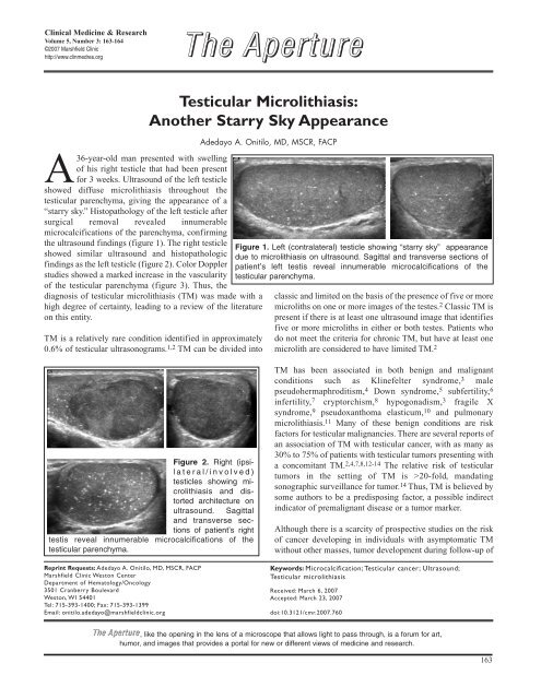
A36-year-old man presented with swelling<br />
of his right testicle that had been present<br />
for 3 weeks. Ultrasound of the left testicle<br />
showed diffuse microlithiasis throughout the<br />
testicular parenchyma, giving the appearance of a<br />
“starry sky.” Histopathology of the left testicle after<br />
surgical removal revealed innumerable<br />
microcalcifications of the parenchyma, confirming<br />
the ultrasound findings (figure 1). The right testicle<br />
showed similar ultrasound and histopathologic<br />
findings as the left testicle (figure 2). Color Doppler<br />
studies showed a marked increase in the vascularity<br />
of the testicular parenchyma (figure 3). Thus, the<br />
diagnosis of testicular microlithiasis (TM) was made with a<br />
high degree of certainty, leading to a review of the literature<br />
on this entity.<br />
TM is a relatively rare condition identified in approximately<br />
0.6% of testicular ultrasonograms. 1,2 TM can be divided into<br />
Figure 2. Right (ipsilateral/involved)<br />
testicles showing mi-<br />
DON’T MISS AN ISSUE IN 2006!<br />
crolithiasis and distorted<br />
architecture on<br />
RENEW TODAY!<br />
ultrasound. Sagittal<br />
and transverse sections<br />
of patient’s right<br />
testis reveal innumerable microcalcifications of the<br />
testicular parenchyma.<br />
Subscribe @<br />
Reprint Requests: www.clinmedres.org<br />
Adedayo A. Onitilo, MD, MSCR, FACP<br />
Marshfield Clinic Weston Center<br />
Department of Hematology/Oncology<br />
3501 Cranberry Boulevard<br />
Weston, WI 54401<br />
Xenopsylla cheopis<br />
Tel: 715-393-1400; Fax: Carrier 715-393-1399<br />
of Plague<br />
Email: onitilo.adedayo@marshfieldclinic.org<br />
The The The Aperture Aperture<br />
Adedayo A. Onitilo, MD, MSCR, FACP<br />
Figure 1. Left (contralateral) testicle showing “starry sky” appearance<br />
due to microlithiasis on ultrasound. Sagittal and transverse sections of<br />
patient’s left testis reveal innumerable microcalcifications of the<br />
testicular parenchyma.<br />
classic and limited on the basis of the presence of five or more<br />
microliths on one or more images of the testes. 2 Classic TM is<br />
present if there is at least one ultrasound image that identifies<br />
five or more microliths in either or both testes. Patients who<br />
do not meet the criteria for chronic TM, but have at least one<br />
microlith are considered to have limited TM. 2<br />
TM has been associated in both benign and malignant<br />
conditions such as Klinefelter syndrome, 3 male<br />
pseudohermaphroditism, 4 Down syndrome, 5 subfertility, 6<br />
infertility, 7 cryptorchism, 8 hypogonadism, 3 fragile X<br />
syndrome, 9 pseudoxanthoma elasticum, 10 and pulmonary<br />
microlithiasis. 11 Many of these benign conditions are risk<br />
factors for testicular malignancies. There are several reports of<br />
an association of TM with testicular cancer, with as many as<br />
30% to 75% of patients with testicular tumors presenting with<br />
a concomitant TM. 2,4,7,8,12-14 The relative risk of testicular<br />
tumors in the setting of TM is >20-fold, mandating<br />
sonographic surveillance for tumor. 14 Thus, TM is believed by<br />
some authors to be a predisposing factor, a possible indirect<br />
indicator of premalignant disease or a tumor marker.<br />
Although there is a scarcity of prospective studies on the risk<br />
of cancer developing in individuals with asymptomatic TM<br />
without other masses, tumor development during follow-up of<br />
Keywords: Microcalcification; <strong>Testicular</strong> cancer; Ultrasound;<br />
<strong>Testicular</strong> microlithiasis<br />
Received: March 6, 2007<br />
Accepted: March 23, 2007<br />
doi:10.3121/cmr.2007.760<br />
The The The Aperture Aperture Aperture,<br />
like the opening in the lens of a microscope that allows light to pass through, is a forum for art,<br />
humor, and images that provides a portal for new or different views of medicine and research.<br />
163
TM has been described to have a latency period between 15<br />
months and 11 years. 4 If no malignancy is identified on initial<br />
evaluation, close clinical follow-up with periodic (every 6 to<br />
12 months) scrotal ultrasound examination is probably<br />
indicated. In high risk patients or patients with contralateral<br />
tumors, tumor markers and biopsy may also be indicated<br />
based on clinical suspicion. Thus, microlithiases are believed<br />
to be a predisposing factor, a possible indirect indicator of<br />
premalignant disease, or a tumor marker.<br />
References<br />
1. Hobarth K, Susani M, Szabo N, Kratzik C. Incidence of<br />
testicular microlithiasis. Urology 1992;40:464-467.<br />
2. Middleton WD, Teefey SA, Santillan CS. <strong>Testicular</strong><br />
microlithiasis: prospective analysis of prevalence and<br />
associated tumor. Radiology 2002;224:425-428.<br />
3. Aizenstein RI, Hibbeln JF, Sagireddy B, Wilbur AC, O’Neil HK.<br />
Klinefelter's syndrome associated with testicular<br />
microlithiasis and mediastinal germ-cell neoplasm. J Clin<br />
Ultrasound 1997;25:508-510.<br />
4. Guzman Martinez-Valls PL, Hita Villaplana G, Fernandez<br />
Aparicio T, Minana Lopez B, Martinez Diaz F, Sanchez<br />
Gascon F. [Significance and management of testicular<br />
microlithiasis]. Arch Esp Urol 2003;56:472-477.<br />
5. Vachon L, Fareau GE, Wilson MG, Chan LS. <strong>Testicular</strong><br />
microlithiasis in patients with Down syndrome. J Pediatr<br />
2006;149:233-236.<br />
6. de Gouveia Brazao CA, Pierik FH, Oosterhuis JW, Dohle GR,<br />
Looijenga LH, Weber RF. Bilateral testicular microlithiasis<br />
predicts the presence of the precursor of testicular germ cell<br />
tumors in subfertile men. J Urol 2004;171:158-160.<br />
7. Sakamoto H, Shichizyou T, Saito K, Okumura T, Ogawa Y,<br />
Yoshida H, Kushima M. <strong>Testicular</strong> microlithiasis identified<br />
ultrasonographically in Japanese adult patients: prevalence<br />
and associated conditions. Urology 2006;68:636-641.<br />
8. Konstantinos S, Alevizos A, Anargiros M, Constantinos M,<br />
Athanase H, Konstantinos B, Michail E, Fragiskos S.<br />
Association between testicular microlithiasis, testicular<br />
cancer, cryptorchidism and history of ascending testis. Int<br />
Braz J Urol 2006;32:434-438.<br />
9. Pourbagher MA, Pourbagher A, Erol I. Fragile X syndrome<br />
associated with testicular microlithiasis in siblings. J<br />
Ultrasound Med 2005;24:1727-1729.<br />
10. Bercovitch RS, Januario JA, Terry SF, Boekelheide K, Podis<br />
AD, Dupuy DE, Bercovitch LG. <strong>Testicular</strong> microlithiasis in<br />
association with pseudoxanthoma elasticum. Radiology<br />
2005;237:550-554.<br />
11. Arslan A, Yalin T, Akan H, Belet U. Pulmonary alveolar<br />
microlithiasis associated with calcifications in the seminal<br />
vesicles. J Belge Radiol 1996;79:118-119.<br />
164<br />
Figure 3. Markedly increased vascularity of the patient’s testicular structures<br />
evident upon examination by color Doppler.<br />
<strong>Testicular</strong> <strong>Microlithiasis</strong><br />
12. Bach AM, Hann LE, Shi W, Giess CS, Yoo HH, Sheinfeld J,<br />
Thaler HT. Is there an increased incidence of contralateral<br />
testicular cancer in patients with intratesticular<br />
microlithiasis? AJR Am J Roentgenol 2003;180:497-500.<br />
13. Backus ML, Mack LA, Middleton WD, King BF, Winter TC<br />
3rd, True LD. <strong>Testicular</strong> microlithiasis: imaging appearances<br />
and pathologic correlation. Radiology 1994;192:781-785.<br />
14. Cast JE, Nelson WM, Early AS, Biyani S, Cooksey G,<br />
Warnock NG, Breen DJ. <strong>Testicular</strong> microlithiasis: prevalence<br />
and tumor risk in a population referred for scrotal<br />
sonography. AJR Am J Roentgenol 2000;175:1703-1706.<br />
Author Affiliation<br />
Adedayo A. Onitilo, MD, MSCR, FACP<br />
Department of Hematology/Oncology<br />
Marshfield Clinic Weston Center<br />
Weston, Wisconsin<br />
CM&R 2007 : 3 (October)