Embryo culture media for human IVF: which ... - JournalAgent
Embryo culture media for human IVF: which ... - JournalAgent
Embryo culture media for human IVF: which ... - JournalAgent
Create successful ePaper yourself
Turn your PDF publications into a flip-book with our unique Google optimized e-Paper software.
110<br />
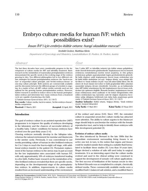
<strong>Embryo</strong> <strong>culture</strong> <strong>media</strong> <strong>for</strong> <strong>human</strong> <strong>IVF</strong>: <strong>which</strong><br />
possibilities exist?<br />
İnsan İVF‘i için embriyo kültür ortamı: hangi olasılıklar mevcut?<br />
Irmhild Gruber, Matthias Klein<br />
Department of Gynecology and Obstetrics, Landesklinikum St. Poelten, St. Poelten, Austria<br />
Abstract Özet<br />
The last three decades have seen considerable progress in the development<br />
of <strong>culture</strong> <strong>media</strong> <strong>for</strong> ART and infertility treatment. Basic<br />
research on the metabolism of mammalian preimplantation embryos<br />
demonstrated the specific needs in the evolving stage of the embryo<br />
growing in vitro. Two different philosophies led to two different <strong>culture</strong><br />
strategies <strong>for</strong> <strong>human</strong> preimplantation embryos: the ‘back-to-nature’<br />
or sequential <strong>culture</strong> principle, and ‘let-the-embryo-choose’ or<br />
one-step <strong>culture</strong> principle. Both systems are commercially available<br />
and the discussion between the different groups of scientists is ongoing.<br />
As a matter of fact, all ART <strong>culture</strong> <strong>media</strong> currently used are not<br />
optimal <strong>for</strong> the growing <strong>human</strong> preimplantation embryo. However,<br />
further research is needed to reduce stress to the <strong>human</strong> preimplantation<br />
embryo and determine how many embryos from a treatment<br />
cycle are capable of producing a live birth.<br />
(J Turkish-German Gynecol Assoc 2011; 12: 110-7)<br />
Key words: Culture <strong>media</strong>, back-to-nature, ‘let-the-embryo-choose’,<br />
<strong>media</strong> components<br />
Received: 17 March, 2011 Accepted: 22 April, 2011<br />
Introduction<br />
The goal of embryo <strong>culture</strong> in an assisted reproductive (ART)<br />
programme is to improve the quality of embryos developing<br />
in the laboratory and the chances of successful delivery of<br />
a healthy baby. Culture conditions <strong>for</strong> <strong>human</strong> embryos have<br />
evolved over the past thirty years (1-4).<br />
Cleaving embryos normally develop in the fallopian tube,<br />
whereas the natural environment <strong>for</strong> morulae and blastocysts<br />
is the uterine cavity. Previously, it was conventional to use<br />
<strong>media</strong> permitting <strong>culture</strong> of <strong>human</strong> in-vitro fertilized embryos<br />
<strong>for</strong> 2 to 3 days to reach the four-to-eight cell stage, with additional<br />
embryo transfer to the patient (5). Premature replacement<br />
of the <strong>human</strong> embryo to the uterus may in part account<br />
<strong>for</strong> the low implantation rates associated with <strong>human</strong> <strong>IVF</strong>,<br />
with only approximately 10% of embryos transferred leading<br />
to a live birth. Further basic research on the metabolism of invitro<br />
fertilized embryos revealed that there are specific needs,<br />
depending on the developmental stage of the preimplantation<br />
embryo. In addition, improvements in <strong>culture</strong> <strong>media</strong><br />
resulted from an increased understanding of the environment<br />
Son 3 yılda ART ve infertilite tedavisi için kültür ortamı geliştirilmesinde<br />
önemli ilerlemeler görülmektedir. İmplantasyon öncesi memeli<br />
embriyosu metabolizması üzerine temel araştırma, in vitro gelişen<br />
embriyonun gelişim aşamalarındaki özgün gereksinimlerini gösterdi.<br />
İki farklı düşünce şekli implantasyon öncesi insan embriyoları için<br />
iki farklı kültür stratejisine yol açtı: ‘doğaya dönüş’ veya ardışık kültür<br />
ilkesi ve ‘bırak embriyo seçsin’ veya tek-adım kültür ilkesi. Her iki<br />
sistem de ticari olarak mevcuttur ve farklı gruplardaki bilim adamları<br />
arasındaki tartışma devam etmektedir. Gerçekte, şu an kullanımda<br />
olan ART kültür ortamlarının hiç biri implantasyon öncesi insan embriyoları<br />
için optimum değildir. Bununla beraber, implantasyon öncesi<br />
insan embriyosuna stresi azaltmak ve bir tedavi döngüsünden elde<br />
edilen embriyoların kaç tanesinin canlı bir doğum oluşturma becerisine<br />
sahip olduğunu belirlemek için daha fazla araştırmaya gerek<br />
vardır. (J Turkish-German Gynecol Assoc 2011; 12: 110-7)<br />
Anahtar kelimeler: Kültür ortamı, ’doğaya dönüş’, ‘bırak embriyo<br />
seçsin’, besiyeri bileşenleri<br />
Geliş Tarihi: 17 Mart 2011 Kabul Tarihi: 22 Nisan 2011<br />
Address <strong>for</strong> Correspondence: Irmhild Gruber, M.Sc. Dr.Sc., Department of Gynecology and Obstetrics, Landesklinikum St. Poelten, <strong>IVF</strong>-Unit<br />
Propst-Fuehrer-Strasse 4, A-3100 St. Poelten, Austria Phone: +43-2742-300-71592 Fax: +43-2742-300-12429 e.mail: irmhild.gruber@stpoelten.lknoe.at<br />
doi:10.5152/jtgga.2011.25<br />
Review<br />
of the oviduct and uterus (6-8). Since 1997, the extended<br />
<strong>culture</strong> in sequential serum-free <strong>culture</strong> <strong>media</strong> has attracted<br />
more attention. The ability to <strong>culture</strong> zygotes to the blastocyst<br />
stage should help to synchronize the embryo with the female<br />
reproductive tract, and to help to identify those embryos with<br />
little development potential (3).<br />
Evolution of embryo <strong>culture</strong> <strong>media</strong><br />
The idea introduced by Bernard in the late 1800s that the<br />
im<strong>media</strong>te environment surrounding living tissues is an<br />
active one led, in turn, to the notion that organs and tissues<br />
could be studied outside their setting in a suitable fluid <strong>for</strong>mulated<br />
to facilitate these studies (9). Less than 10 years later,<br />
Ringer devised a solution of salts still used today in surgical<br />
treatments. It is an interesting aspect that the <strong>culture</strong> <strong>media</strong><br />
evolved and used in the clinical setting were construed to<br />
support the development of somatic cell <strong>culture</strong> applications.<br />
The first success of fertilization of the <strong>human</strong> oocyte in vitro<br />
by Robert Edwards was accomplished in a simple, chemically<br />
defined <strong>media</strong>. These commercially available <strong>media</strong> were a<br />
modified Earle’s balanced salt solution, and a modified Ham’s
J Turkish-German Gynecol Assoc 2011; 12: 110-7<br />
F10 or T6. They were supplemented with maternal serum thus<br />
converting them into biological <strong>media</strong> (10, 11). Menezo et al.<br />
(12) broke with the tradition of using balanced salt solution and<br />
produced a medium containing amino acids without the need<br />
of a serum supplement. Another medium specifically designed<br />
<strong>for</strong> <strong>human</strong> <strong>IVF</strong> was <strong>human</strong> tubal fluid (HTF) (13). Human tubal<br />
fluid, supplemented with either whole serum or with serum<br />
albumin, gained great popularity <strong>for</strong> the use of day 2 or day 3<br />
<strong>human</strong> embryo <strong>culture</strong>s, and has remained in use ever since.<br />
A <strong>culture</strong> medium is a <strong>for</strong>eign environment <strong>for</strong> the <strong>human</strong><br />
embryo. Hence, the design of <strong>media</strong> is complicated, because<br />
the components must be selected, and their concentrations<br />
determined in order to minimize stress <strong>for</strong> the <strong>culture</strong>d embryo<br />
(2, 14, 15). It also became clear that early embryos show an<br />
evolving need <strong>for</strong> energy substrates, moving from a pyruvatelactate<br />
preference - while the embryos are under maternal<br />
genetic control - to glucose-based metabolism after activation<br />
of the embryonic genome (7, 16). Two investigators responded<br />
to these findings by modifying the HTF <strong>media</strong>. Quinn (17)<br />
removed glucose and inorganic phosphate, QB 11, <strong>for</strong> obtaining<br />
the first glucose-free medium. Pool (18) also generated a<br />
HTF variant, called Preimplantation Stage 1 or P-1 medium, a<br />
glucose- and phosphate-free medium, but additionally containing<br />
the amino acid taurine. The improved understanding<br />
of both the physiological changes in oviduct and uterus (7)<br />
and the different metabolic needs of the cleavage-stage and<br />
blastocyst-stage embryo led to the development of stagespecific<br />
or “sequential” complex <strong>media</strong> G1/G2 (3). Barnes et<br />
al. (19) used this combination to produce pregnancy and live<br />
birth after the transfer of a single viable <strong>human</strong> blastocyst, and<br />
the <strong>media</strong> were first slightly modified and marked as the GIII/<br />
G5 series of <strong>media</strong>. Other popular sequential systems, such<br />
as Quinn’s series in the United States and the MediCult/Origio<br />
<strong>media</strong> from Europe and Cook from Australia, are also in widespread<br />
use. Lawitts and Biggers (20, 21) broke new ground to<br />
design a chemically defined <strong>media</strong>. They applied the principle<br />
of simplex optimization to determine the optimal concentration<br />
of each <strong>media</strong> component. This resulted in the <strong>for</strong>mulation of<br />
Simplex Optimizition Medium (SOM). SOM has been modified<br />
in several ways to mKSOM AA . Hence<strong>for</strong>th, it is possible to provide<br />
a one-step protocol (so-called global medium) to <strong>culture</strong><br />
<strong>human</strong> zygotes to the blastocyst stage (2, 5). Now, there exist<br />
three commercially available one-step <strong>human</strong> embryo <strong>culture</strong><br />
<strong>media</strong> (global ® , LifeGlobal, U.S.; Gynemed GM501 ® , Lensahn,<br />
Germany; SSM, Irvine Scientific, U.S.). At least nine companies<br />
now advertise <strong>media</strong> <strong>for</strong> the <strong>culture</strong> of <strong>human</strong> preimplantation<br />
embryos to the blastocyst stage (Table 1).<br />
The recovery of immature oocytes followed by in-vitro maturation<br />
(IVM) of these oocytes led to the development of specific<br />
conventional available IVM <strong>culture</strong> <strong>media</strong>.<br />
Two philosophies <strong>for</strong> <strong>human</strong> embryo <strong>culture</strong> <strong>media</strong><br />
The design of <strong>media</strong> <strong>for</strong> the <strong>culture</strong> of preimplantation embryos<br />
has been influenced by two fundamentally different philosophies<br />
(2, 22). However, growth of ART embryos is inferior to<br />
that of in vivo embryos, indicating that ART procedures invoke<br />
cellular and metabolic stress situations and the ART embryo is<br />
Gruber et al.<br />
<strong>Embryo</strong> <strong>culture</strong> <strong>media</strong> 111<br />
<strong>for</strong>ced to spend energy to adapt to this <strong>for</strong>eign environment. In<br />
particular, the <strong>culture</strong> <strong>media</strong> is an important factor in successful<br />
in vitro interactions between gametes and subsequent embryo<br />
development (3). Manufacturers of <strong>human</strong> embryo <strong>culture</strong><br />
<strong>media</strong> follow either the “back-to-nature” (sequential <strong>media</strong>)<br />
or the “let-the-embryo-choose” (global <strong>media</strong>) philosophy<br />
(23). The key components of both modern <strong>media</strong> are shown<br />
in Table 2.<br />
The sequential <strong>culture</strong>-“back-to-nature” principle<br />
The “back-to-nature” attempts to mimic the changing needs<br />
of the developing zygote and embryo in a <strong>media</strong> should<br />
approximate the concentration to <strong>which</strong> the embryo is naturally<br />
exposed (3, 23). The embryo is capable of actively controlling<br />
ionic gradients etc, and is able to regulate its internal environment.<br />
There<strong>for</strong>e, with regard to embryo physiology, it is appropriate<br />
to consider the preimplantation period in at least two<br />
phases: pre- and post-compaction (3). Such a breakdown of the<br />
preimplantation period is of importance when one considers<br />
changes to medium <strong>for</strong>mulations. Other considerations include<br />
the time at <strong>which</strong> the embryonic genome is activated (3).<br />
The mono<strong>culture</strong> “let-the-embryo-choose” principle<br />
The design of a <strong>culture</strong> medium involves the simultaneous use<br />
of all the concentrations in a mixture because the effects of<br />
each component in the medium may depend on the concentrations<br />
of the other components (23). As long as concentrations<br />
are within ‘tolerable ranges’, the embryo itself will adapt and<br />
utilize whatever it requires (2, 23, 24). This philosophy led to<br />
a family of <strong>media</strong> in <strong>which</strong> all of the substances necessary to<br />
early embryological development are provided, and there is<br />
no need <strong>for</strong> a <strong>media</strong> change. One-step <strong>for</strong>mulation is applied<br />
throughout the entire in-vitro development from fertilization to<br />
the blastocyst stage of the embryo.<br />
Four protocols can be used <strong>for</strong> the <strong>culture</strong> from fertilization to<br />
the blastocyst stage in an ART laboratory: [a] sequential <strong>media</strong><br />
protocol, with an interrupted <strong>culture</strong> where two <strong>media</strong> of different<br />
compositions are used sequentially, change of medium<br />
occurs on day 3 of embryo <strong>culture</strong>, [b] sequential <strong>media</strong> protocol<br />
with fresh medium change every day, [c] mono<strong>culture</strong>,<br />
uninterrupted <strong>culture</strong> using one medium throughout the 5 days<br />
of embryo <strong>culture</strong>, [d] interrupted <strong>culture</strong> where a mono<strong>culture</strong><br />
medium is used throughout but is renewed on day 3 of<br />
embryo <strong>culture</strong>.<br />
Key components of ART <strong>culture</strong> <strong>media</strong><br />
Studies using the development of mammalian preimplantation<br />
embryos in-vitro have played a major role in the understanding<br />
of pre-embryo physiology (<strong>for</strong> reviews, see 3, 24-30). As a result,<br />
this is also the limitation in the development of <strong>culture</strong> <strong>media</strong><br />
<strong>for</strong> the <strong>human</strong> embryo. The most widely used models <strong>for</strong> the<br />
<strong>human</strong> embryo have been the mouse and the cow.<br />
Carbohydrates<br />
In brief, the early embryo shows a rather simplistic physiology<br />
and maintains only low levels of oxidative metabolism, whereas<br />
it exhibits a somatic-cell like physiology after compaction
112<br />
Gruber et al.<br />
<strong>Embryo</strong> <strong>culture</strong> <strong>media</strong><br />
Table 1. Available commercial systems <strong>for</strong> <strong>human</strong> <strong>IVF</strong> <strong>culture</strong><br />
One <strong>media</strong> system<br />
Company Medium Culture period Website<br />
LifeGlobal global ® day-1 to day-5/6 www.lifeglobal.com<br />
Gynemed GM501 day-0 to day-5/6 www.gynemed.de<br />
IrvineScientific<br />
Sequential <strong>media</strong> system<br />
SSM day-0 to day-5/6 www.irvinesci.com<br />
Company Medium Culture period Website<br />
Cook Medical Cleavage K-SICM day-1 to day-3 www.cookmedical.com<br />
Blastocyst K-SIBM day-3 to day-5/6<br />
CooperSurgical Quinns Advantage ® Cleavage day-1 to day-3 www.coopersurgical.com<br />
Quinns Advantage ® Blastocyst day-3 to day-5/6<br />
FertiPro FERTICULT<strong>IVF</strong> Medium day-1 to day-2 www.fertipro.com<br />
FERTICULT G3 Medium day-3 to day-4<br />
InVitroCare IVC-TWO day-0 to day-3 www.invitrocare.com<br />
IVC-THREE day-3 to day-5<br />
Irvine Scientific ECM ® day-0 to day-3 www.irvinesci.com<br />
MultiBlast ® day-3 to day-5<br />
Origio <strong>Embryo</strong>Assist day-0 to day-3 www.origio.com<br />
BlastAssist day-3 to day-5<br />
ISM1 day-0 to day-3<br />
ISM2 day-3 to day-5<br />
Vitrolife G-1PLUS day-1 to day-3 www.vitrolife.com<br />
G-2PLUS day-3 to day-5<br />
<strong>IVF</strong> day-0 to day-3<br />
CCM day-3 to day-5<br />
utilizing a wider spectrum of nutrients, biosynthetic rates are<br />
increasing, along with an increased respiratory capacity and<br />
an ability to utilize glucose (8, 28). This involves a shift in the<br />
energy requirements at the time at <strong>which</strong> the embryonic genome<br />
is activated or at the post-compaction stage. Zygotes and subsequent<br />
cleavage stages prefer pyruvate as the primary source of<br />
energy, while the eight-cell-stage embryo uses glucose (31-33).<br />
Glucose is a key anabolic precursor and is required <strong>for</strong> the synthesis<br />
of triacylglycerols and phospholipids, and as a precursor <strong>for</strong><br />
complex sugars and glycoproteins. Glucose also metabolized by<br />
the pentose phosphate pathway (PPP) generates ribose moieties<br />
required <strong>for</strong> nucleic acid synthesis (34).<br />
Amino acids<br />
It has been proposed that “amino acids-(AA)”, a term <strong>which</strong><br />
includes all 20 common and naturally occurring amino acids,<br />
are important regulators of mammalian preimplantation development<br />
(<strong>for</strong> reviews, see 8, 29, 35). Prior to embryonic genome<br />
expression, the embryo utilizes carboxylic acids and AA as<br />
energy sources (29). In addition, certain AA are known to function<br />
as biosynthetic precursor molecules (36), osmolytes (37),<br />
buffers of internal pH (38), antioxidants (39) and chelators,<br />
J Turkish-German Gynecol Assoc 2011; 12: 110-7<br />
especially <strong>for</strong> heavy metals (40). It is important to note that<br />
there are also specific changes in the nitrogen requirements of<br />
the embryo (27, 28). The seven non-essential AA and glutamine<br />
stimulate the growth of the early cleavage embryo (41). In contrast,<br />
an inhibitory effect was seen on blastocyst development<br />
and viability if the thirteen essential AAs are presented at an<br />
early stage (42). At the post-compaction stage, both groups of<br />
AAs act stimulatory to the inner cell mass of blastocysts, while<br />
the non-essential AAs and glutamine lead to stimulation of the<br />
throphectoderm and hatching from the zona pellucida (43, 44).<br />
Leese at al. (45-47) have described AA turnover studies on the<br />
mammalian embryo and argued <strong>for</strong> “quiet” embryo metabolism<br />
during preimplantation embryo <strong>culture</strong> and development<br />
to produce the most viable embryos.<br />
However, AAs in <strong>culture</strong> <strong>media</strong> also spontaneously undergo<br />
breakdown to release ammonium into the <strong>culture</strong> medium<br />
with concentration being time dependent. Ammonium is toxic<br />
to the embryo and reduces viability (48). Especially L-glutamine<br />
(Gln) is highly unstable in solution, where it breaks down<br />
fairly rapidly into equimolecular amounts of ammonium and<br />
pyrrolidine-5-carboxylic acid (<strong>for</strong> review see, 49). There<strong>for</strong>e,<br />
Lane and Gardner (42) introduced a two-step (sequential) cul-
J Turkish-German Gynecol Assoc 2011; 12: 110-7<br />
Table 2. Key components of modern <strong>media</strong><br />
Components One <strong>media</strong> system Sequential <strong>media</strong> Sequential <strong>media</strong><br />
Gynemed GM501 ® G-1PLUS G-2PLUS<br />
Salts Sodium chloride Sodium chloride Sodium chloride<br />
Potassium chloride Potassium chloride Potassium chloride<br />
Calcium chloride Calcium chloride Calcium chloride<br />
Monopotassium phosphate Sodium citrate Sodium citrate<br />
Magnesium sulphate Magnesium sulphate Magnesium sulphate<br />
Sodium dihydrogen phosphate Sodium dihydrogen phosphate<br />
Buffer Sodium bicarbonat Sodium bicarbonate Sodium bicarbonate<br />
Energy Substrates Glucose Glucose Glucose<br />
Sodium lactate Sodium lactate Sodium lactate<br />
Sodium pyruvate Sodium pyruvate Sodium pyruvate<br />
Non-Essential AA’s NEAA’s 8 NEAA’s 9 NEAA’s<br />
Glutamine Dipeptide Alanyl-Glutamine<br />
Essential AA’s EAA’s 2 EAA’s 11 EAA’s<br />
Chelator EDTA EDTA none<br />
Macromolecules none Hyaluronan, HSA Hyaluronan, HSA<br />
Fatty acid none Lipoic acid none<br />
Vitamins none none 4 Vitamins<br />
Indicator Phenol Red optional none none<br />
Antibiotic Gentamicin Gentamicin Genamicin<br />
Water yes yes yes<br />
ture <strong>media</strong> protocol to remove the accumulated ammonium.<br />
Another possibility is replacing Gln with a stable dipeptide of<br />
Gln (24). It must be noted that <strong>culture</strong> <strong>media</strong> should include<br />
sufficient levels of sulphur containing amino acids to minimize<br />
apoptosis leading to monozygote twinning (50).<br />
EDTA (Ethylenediaminetetraacetic acid)<br />
Its usefulness is based on its role as a ligand and chelating<br />
agent, i.e. its ability to “sequester” metal ions. After being<br />
bound by EDTA, metal ions remain in solution but exhibit<br />
diminished reactivity. The addition of EDTA to <strong>culture</strong> <strong>media</strong><br />
alleviates the 2-cell block in mice embryos (51) and inhibits<br />
premature utilization of glycolysis by cleavage stage embryos,<br />
thereby preventing any Crabtree-like effect that is associated<br />
with arrest in <strong>culture</strong> (48). However, EDTA at a concentration<br />
of 0.1mmol/L reduces blastocyst development and cell number<br />
(52). Other investigators indicated that an EDTA concentration<br />
of 0.005-0.01 mmol/L did not have a deleterious effect on<br />
murine preimplantation or postimplantation development (53).<br />
Regulation of cell volume-osmolytes<br />
Maintenance of a constant volume in the face of extracellular<br />
and intracellular osmotic perturbations is a critical problem<br />
faced by all cells. Most cells respond to swelling or shrinkage by<br />
activating specific metabolic or membrane-transport processes<br />
Gruber et al.<br />
<strong>Embryo</strong> <strong>culture</strong> <strong>media</strong> 113<br />
that return cell volume to its normal resting state. These processes<br />
are essential <strong>for</strong> the normal function and survival of cells (54).<br />
The osmotic pressure of oviduct fluid is >360 mOsmol (55).<br />
However, the osmolarity of most commercially available ART<br />
<strong>culture</strong> <strong>media</strong> is lower-at about 250-300 mOsmol. When the<br />
NaCl concentration is <strong>for</strong>ced up to 290 mOsmol, the development<br />
of the embryo is severely impaired (56). Addition of<br />
extracellular AA, such as glycine, betaine, proline, alanine and<br />
hypotaurin <strong>which</strong> act as organic osmolytes, protects the preimplantation<br />
embryo against hypertonicity and increases embryo<br />
development (37, 56, 57).<br />
Impact of pH and buffers<br />
The pH only refers to hydrogen ion concentration and is only<br />
meaningful when applied to aqueous (water-based) solutions.<br />
When water dissociates it yields a hydrogen ion and a hydroxide<br />
ion, H 2 O H + +OH - (<strong>for</strong> review see, 58, 59). It must be noted<br />
that pH is dynamic. The balance of pH depends on the association<br />
or dissociation of compounds. The most important ions are<br />
sodium, potassium, magnesium, chloride and lactate and also the<br />
AA glycine <strong>which</strong> acts as an intracellular zwitterionic buffer (60).<br />
An acceptable pH range <strong>for</strong> embryo <strong>culture</strong> <strong>media</strong> may be set<br />
between pH 7.4 and 7.2. Culture <strong>media</strong> pH is regulated by the balance<br />
of CO 2 concentration, supplied by the <strong>media</strong> and by the concentration<br />
of bicarbonate in the <strong>media</strong>. However, the intracellular
114<br />
Gruber et al.<br />
<strong>Embryo</strong> <strong>culture</strong> <strong>media</strong><br />
pH in <strong>human</strong> cleavage embryonic cells is pH=7.2 (61) and pH<br />
is an important cellular function <strong>which</strong> is necessary to maintain<br />
intracellular homeostasis. Moreover, after the compaction stage<br />
the preimplantation embryos appear to have more control over<br />
their intracellular pH, because of the <strong>for</strong>mation of tight junctions<br />
between cells (38, 62). Hence, there is a trend to <strong>culture</strong><br />
cleavage stage embryos in a slightly lower pH and morulae and<br />
blastocysts in a slightly higher pH (low-high paradigm). Table 3<br />
provides in<strong>for</strong>mation about the recommended pH of commercial<br />
available <strong>media</strong>.<br />
In the past, handling <strong>media</strong> were used with phosphate-buffered<br />
saline solutions (PBS) or different “Good’s” buffers (63).<br />
Nowadays, especially two “Good’s” puffers are used in commercially<br />
<strong>IVF</strong> handling <strong>media</strong>. The most commonly used buffer<br />
is 4-(2-hydroxyethyl)-1-piperazineethanesulphonic acid<br />
(HEPES at 21 mmol/L), whereas some companies include<br />
3-(N-morpholino)-propanesulphonic acid (MOPS). Both buffers<br />
have a pK a value of 7.2, it is the closest of the zwitterionic buffers<br />
to the pH i of embryos of 7.12.<br />
Although both buffers have been widely used in <strong>IVF</strong> handling,<br />
studies indicated there may be species specific sensitivities<br />
to HEPES (64, 65). Results demonstrated that, in the presence<br />
of optimal <strong>culture</strong> conditions, such as pH, gas concentration,<br />
osmolarity etc., HEPES is able to support mammalian embryo<br />
development and can also act as a chelator of heavy metals<br />
such as copper (66). If using MOPS <strong>for</strong> <strong>IVF</strong> handling at 37°C the<br />
pK a <strong>for</strong> this buffer is actually 7.02 (59), <strong>which</strong> is low, because<br />
most <strong>IVF</strong> laboratories target their <strong>media</strong> pH at 7.3. Yet, MOPS<br />
can interact with DNA in cellular preparations (67). Currently,<br />
it has not yet been defined whether both buffers used have an<br />
impact on embryo osmotic regulation (59).<br />
Macromolecules<br />
Common sources <strong>for</strong> macromolecules are proteins <strong>for</strong> <strong>culture</strong><br />
<strong>media</strong> such as <strong>human</strong> serum albumin or synthetic serum. Both<br />
are added at concentrations of 5 to 20%. Today, most commercial<br />
<strong>media</strong> include synthetic serum in <strong>which</strong> the composition is<br />
well known. Protein in the <strong>for</strong>m of albumin is thought to maintain<br />
the stability of cell membranes and chelate trace amounts<br />
of toxic components presented in <strong>culture</strong> water, <strong>media</strong> components<br />
and <strong>culture</strong> dishes. Other functions include capillary<br />
membrane permeability and osmoregulation. The presence of<br />
macromolecules in embryo <strong>culture</strong> <strong>media</strong> serves to facilitate<br />
manipulation of gametes and embryos (8). However, the uses<br />
of any blood products involve the risk of potential contamination<br />
and infection of preimplantation embryos.<br />
Some investigators have used synthetic polymers such as polyvinyl<br />
alcohol (PVA) and polyvinyl pyrrolidone (PVP) in ART (29)<br />
but neither can be considered a physiological alternative to<br />
protein (68).<br />
Another physiological alternative to albumin is the glycosaminoglycan<br />
hyaluronate (also called hyaluronic acid or hyaluronan).<br />
The <strong>human</strong> embryo expresses the receptor <strong>for</strong> it throughout<br />
preimplantation development (69). While hyaluronate could<br />
not only replace serum albumin in <strong>culture</strong>, it increased the<br />
implantation rate of resultant mouse embryo blastocysts (70).<br />
There<strong>for</strong>e, hyaluronate can replace albumin as a sole macro-<br />
J Turkish-German Gynecol Assoc 2011; 12: 110-7<br />
Table 3. Recommended pH-ranges of <strong>IVF</strong> <strong>culture</strong> <strong>media</strong><br />
(adapted from Swain, 2010)<br />
Company Medium pH-range<br />
LifeGlobal global® 7.2-7.4<br />
Gynemed GM501 7.2-7.4<br />
Irvine Scientific SSM 7.28-7.32<br />
ECM® 7.2-7.25<br />
MultiBlast® 7.3-7.4<br />
Cook Medical K-SICM 7.3-7.5<br />
K-SICB 7.3-7.5<br />
Cooper Surgical Quinns Advantage®Cleavage 7.1-7.3<br />
Quinns Advantage®Blastocyst 7.2-7.4<br />
FertiPro FERTICULT<strong>IVF</strong> Medium 7.2-7.6<br />
FERTICULTG3 Medium 7.3-7.6<br />
InVitroCare IVC-TWO 7.25-7.45<br />
IVC-THREE 7.25-7.45<br />
Origio <strong>Embryo</strong>Assist 7.3-7.5<br />
BlastAssist 7.3-7.5<br />
ISM1 7.2-7.4<br />
ISM2 7.2-7.4<br />
Vitrolife G-1PLUS 7.27±0.07<br />
G-2PLUS 7.27±0.07<br />
<strong>IVF</strong> 7.35±0.10<br />
CCM 7.35±0.10<br />
molecule in an embryo transfer medium and in some infertile<br />
patients it can improve ongoing pregnancy rates (71).<br />
Vitamins<br />
The addition of vitamins as antioxidants to the <strong>culture</strong> <strong>media</strong><br />
containing glucose and phosphate helped to prevent a loss<br />
in respiration and metabolic control (72). The following possible<br />
vitamins are components of different ART <strong>culture</strong> <strong>media</strong>:<br />
ascorbic acid, cyanocobalamin, folic acid and tocopherol.<br />
Their optimum concentrations were determined using mouse<br />
zygote assays. Moderate dosages of vitamins C and E were<br />
seen to reduce oxidative damage in mouse embryo <strong>culture</strong> and<br />
improve their blastocyst development rate (73).<br />
Growth factors<br />
Mammalian embryos are naturally exposed to a complex mixture<br />
of growth factors that play a key role in growth and differentiation<br />
from the time of morula to blastocyst transition. However, defining<br />
their role and potential <strong>for</strong> improving in-vitro preimlantation<br />
development is complicated by factors such as gene expression<br />
of both the factors and their receptors. The blastocyst expresses<br />
ligands and receptors <strong>for</strong> several growth factors, many of <strong>which</strong><br />
can cross-react thus making it difficult to interpret the effect of<br />
single factors added to a <strong>culture</strong> <strong>media</strong> (74, 75).
J Turkish-German Gynecol Assoc 2011; 12: 110-7<br />
Antibiotics<br />
<strong>Embryo</strong> <strong>culture</strong> <strong>media</strong> are routinely supplemented with antibiotics<br />
to prevent bacterial contamination (76). Nowadays, commonly<br />
used antibiotics are penicillin (β-lactam; 100U/ml), streptomycin<br />
(aminoglycoside; 100 µg/ml) and gentamycin (aminoglycoside;<br />
50 µg/ml). The anti-bacterial effect of penicillin is<br />
attributed to its disturbance of cell wall integrity through the inhibition<br />
of the synthesis of peptidoglycan. Penicillin has no direct<br />
toxic effects on the preimplantation embryo. Streptomycin and<br />
gentamycin disturb bacterial protein synthesis. However, the<br />
aminoglycosides show more toxic effects (76).<br />
Literature review <strong>for</strong> comparison of <strong>media</strong> types<br />
A number of recent studies have been conducted to compare<br />
the effectiveness of commercially available ART <strong>culture</strong><br />
<strong>media</strong> types. A search was conducted on published literature.<br />
Interestingly, most studies prefer 3-day <strong>human</strong> <strong>IVF</strong> embryo<br />
<strong>culture</strong> and embryo transfer <strong>for</strong> comparison of different <strong>media</strong><br />
types (77-81). Differences in embryo quality were observed in<br />
studies that used modern <strong>for</strong>mulated <strong>media</strong> versus standard<br />
<strong>media</strong>, but no differences in pregnancy rates were reported<br />
(77, 78). Moreover, no differences between a single or one-step<br />
defined medium versus a cleavage-stage <strong>media</strong> with regard to<br />
fertilization, pregnancy implantation rates, and ongoing pregnancy<br />
were found by following studies (78, 81). Ebert et al. (79)<br />
reported similar results. Only the rate of pregnancy losses was<br />
significantly lower in patients with the one-step medium GM501<br />
as compared to the Universal <strong>IVF</strong> medium.<br />
Three studies assess pregnancy outcomes and embryo morphology<br />
after transfer of day-3, day-5 or -6 embryos (82-84). Van<br />
Langendonckt et al. (82) matched two sequential <strong>media</strong>, G1.2/<br />
G2.2 and Sydney <strong>IVF</strong> cleavage/blastocyst <strong>media</strong>. Both <strong>media</strong><br />
yielded similar outcomes in the blastocyst transfer programme,<br />
but a lower day-3 embryo quality in the G1.2 <strong>media</strong>. The other<br />
two studies compared a single-step medium versus a sequential<br />
medium. Reed et al. (83) reported no significant difference<br />
<strong>for</strong> results on day-3 transfer. However, <strong>for</strong> day-5 transfer, a<br />
greater number of blastocysts were available from the single<br />
medium. Paternot et al. (84) described similar positive results<br />
using GM501.<br />
Two other studies compared a single-step versus a sequential<br />
<strong>media</strong> system <strong>for</strong> the development of the <strong>human</strong> embryos to<br />
the blastocyst stage (85, 86). Biggers and Racowsky (85) found<br />
that significantly more <strong>IVF</strong>-embryos <strong>culture</strong>d in the single-step<br />
medium showed cytoplasmic pitting. <strong>IVF</strong>-blastocyst <strong>for</strong>mation<br />
rates were not significantly different between the two <strong>media</strong><br />
systems. Sepulveda et al. (86) referred to donor cycles, and<br />
had better development rates on days 3, 4 and 5 as well as<br />
significantly higher implantation rates <strong>for</strong> embryos <strong>culture</strong>d in<br />
the single medium.<br />
Furthermore, three other studies used the mouse model <strong>for</strong><br />
comparison of commercially available <strong>media</strong> (53, 87, 88).<br />
Biggers and colleagues (53) observed no significant differences<br />
in the proportion of the blastocysts, rates of hatching, numbers<br />
of cells in the inner cell mass and trophectoderm between<br />
KSOM AA and G1.2/G2.2. However, Perin et al. (87) reported a<br />
higher blastocyst <strong>for</strong>mation, higher cell numbers in the inner<br />
cell mass and higher hatching rates after <strong>culture</strong> in the singlestep<br />
medium KSOM AA . Hentemann and Bertheussen (88) compared<br />
two sequential <strong>media</strong>, BlastAssist M1 and M2 versus G1/<br />
G2, in a mouse model and achieved similar results.<br />
Concluding remarks<br />
In this review, two different types, ‘back-to-nature’ and ‘let-theembryo-choose’,<br />
of <strong>culture</strong> <strong>media</strong> were presented with the<br />
recent literature. Both <strong>media</strong> philosophies are part of worldwide<br />
practice in the ART laboratories. Based on recent literature,<br />
it can be concluded that global one step <strong>media</strong> are at least<br />
as useful as sequential <strong>media</strong>.<br />
Human embryos can develop in vitro in rather different types<br />
of <strong>media</strong> from basic systems to sequential complex <strong>culture</strong><br />
<strong>media</strong>. ART <strong>culture</strong> <strong>media</strong> contain only a subset of parts <strong>which</strong><br />
are found under in vivo conditions. Hence, embryos <strong>culture</strong>d<br />
in-vitro was exposed to constant stress. Suboptimal <strong>culture</strong><br />
conditions <strong>for</strong>ce the embryo to undergo adaptations, and thus<br />
lead to lower pregnancy and higher abortion rates.<br />
It is evident that all necessary steps in ART as part of the treatment<br />
of infertility can influence the epigenetic programming<br />
during early development (89). There<strong>for</strong>e, it is essential that<br />
a high level of quality control exists in the laboratory, and it is<br />
suggested that further investigations are necessary to optimize<br />
environmental conditions in <strong>which</strong> the preimplantation embryo<br />
can evolve.<br />
Conflict of interest<br />
No conflict of interest was declared by the authors.<br />
References<br />
Gruber et al.<br />
<strong>Embryo</strong> <strong>culture</strong> <strong>media</strong> 115<br />
1. Biggers JD. Pioneering mammalian embryo <strong>culture</strong>. In Bavister,<br />
B.D. (ed.). The Mammalian Preimplantation <strong>Embryo</strong>. Plenum<br />
Press, New York 1987; 1-22.<br />
2. Bigger JD. Thoughts on embryo <strong>culture</strong> conditions. Reprod Biomed<br />
Online 2001; 4: 30-8.<br />
3. Gardner DK and Lane M. Culture and selection of viable blastocysts:<br />
a feasible proposition <strong>for</strong> <strong>human</strong> <strong>IVF</strong>? Hum Repod Update 1997; 3:<br />
367-82. [CrossRef]<br />
4. Pool TB. Recent advances in the production of viable <strong>human</strong><br />
embryos in vitro: Reprod Biomed Online 2002; 4: 294-302.<br />
[CrossRef]<br />
5. Biggers JD. Fundamentals of the design of <strong>culture</strong> <strong>media</strong> that<br />
support <strong>human</strong> preimplantation development. In: Van Blerkom J,<br />
ed. Essential <strong>IVF</strong>. Norell, MA: Kluwer Academic Press 2003; 291-332.<br />
6. Leese HJ. Formation and function of oviduct fluid. J Reprod Fertil<br />
1988; 82: 843-56. [CrossRef]<br />
7. Gardner DK, Lane M, Calderon I and Leeton J. Environment of the<br />
preimplantation <strong>human</strong> embryo in vivo: metabolite analysis of<br />
oviduct and uterine fluids and metabolism of cumulus cells. Fertil<br />
Steril 1996; 65: 349-53.<br />
8. Gardner DK. Changes in requirements and utilization of nutrients<br />
during mammalian preimplantation embryo development and their<br />
significance in embryo <strong>culture</strong>. Theriogenology 1998; 49: 83-102.<br />
[CrossRef]<br />
9. Freshney RI. Culture of animal cells. 4th ed. New York: Wiley-Liss, 2000.<br />
10. Edwards RG. Test-tube babies. Nature 1981; 293: 253-6. [CrossRef]<br />
11. Edwards RG, Purdy JM, Steptoe PC et al. The growth of <strong>human</strong><br />
preimplantation embryos in vitro. Am J of Obstet and Gynecol<br />
1981; 141: 408-16.
116<br />
Gruber et al.<br />
<strong>Embryo</strong> <strong>culture</strong> <strong>media</strong><br />
12. Menezo Y, Testart J, Perone D. Serum is not necessary in <strong>human</strong> in<br />
vitro fertilization and embryo development. Fertil Steril 1984; 42: 750-5.<br />
[CrossRef]<br />
13. Quinn P, Kerin JF, Warnes GM. Improved pregnancy rate in <strong>human</strong><br />
in vitro fertilization with the use of a medium based on the<br />
composition of <strong>human</strong> tubal fluid. Fertil Steril 1985; 44: 493-8.<br />
14. Barnet DK, Bavister BD. What is the relationship between the<br />
metabolism of preimplantation embryos and their developmental<br />
competence? Molecular Reproduction and Development 1996; 43:<br />
378-83.<br />
15. Gardner DK and Lane M. <strong>Embryo</strong> <strong>culture</strong> systems. In Trouson AO,<br />
Gardner DK (eds) Handbook of in Vitro Fertilization, 2nd edn. 1999,<br />
CRC Press, Boca Raton pp. 205-64.<br />
16. Leese HJ and Barton AM. Production of pyruvate by isolated mouse<br />
cumulus cells. J Exp Zool 1985; 234: 231-6. [CrossRef]<br />
17. Quinn P. Enhanced results in mouse and <strong>human</strong> embryo <strong>culture</strong> using<br />
a modified <strong>human</strong> tubal fluid medium lacking glucose and inorganic<br />
phosphate. J Assist Reprod Genet 1995; 12: 97-105. [CrossRef]<br />
18. Pool TB, Atiee SH and Martin JE. Oocyte and embryo <strong>culture</strong>: basic<br />
concepts and recent advances. Infertil Reprod Med Clin N Am 1998;<br />
9: 181-203.<br />
19. Barnes FL, Crombie A, Gardner DK, Kausche A, Lacham-Kaplan O,<br />
Suikkari AM, et al. Blastocyst development and pregnancy after in<br />
vitro maturation of <strong>human</strong> primary oocytes, intracytoplasmic sperm<br />
injection and assisted hatching. Hum Repod 1995; 10: 3243-7.<br />
20. Lawitt JA and Biggers JD. Optimizing of mouse embryo <strong>culture</strong><br />
<strong>media</strong> using simplex methods. J Reprod Fert 1991; 91: 543-56.<br />
21. Lawitt JA and Biggers JD. Joint effects of sodium chloride,<br />
glutamine, and glucose in mouse preimplantation embryo <strong>culture</strong><br />
<strong>media</strong>. Molecular Reproduction and Development 1992; 31: 189-94.<br />
22. Biggers JD. Reflections on the <strong>culture</strong> of the preimplantation embryo.<br />
International Journal of Developmental Biology 1998; 42: 879-84.<br />
23. Summers MC, Biggers JD. Chemically defined <strong>media</strong> and the <strong>culture</strong><br />
of mammalian preimplantation embryos: historical perspective and<br />
current issues. Hum Reprod Update 2003; 9: 557-82. [CrossRef]<br />
24. Biggers JD and Summers MC. Choosing a <strong>culture</strong> medium: making<br />
in<strong>for</strong>med choices. Fertil Steril 2008; 90: 473-83. [CrossRef]<br />
25. Whitten WK. Culture of tubal ova. Nature 1956; 177: 96. [CrossRef]<br />
26. Brinster RL. IV. The effect of interaction of energy sources. J Reprod<br />
Fertil 1965; 10: 227-40.<br />
27. Leese HJ. Metabolism of the preimplantation mammalian embryo.<br />
Oxf Rev Reprod Biol 1991; 13: 35-72.<br />
28. Rieger D. Relationship between energy metabolism and<br />
development of the early embryo. Theriogenology 1992; 37: 75-93.<br />
[CrossRef]<br />
29. Bavister BD. Culture of preimplantation embryos: facts and<br />
artefacts. Hum Reprod Update 1995; 1: 91-148. [CrossRef]<br />
30. Bavister BD. Glucose and the <strong>culture</strong> of <strong>human</strong> embryos. Fertil<br />
Steril 1999; 72: 233-4. [CrossRef]<br />
31. Leese HJ and Barton AM. Pyruvate and glucose uptake by mouse<br />
ova and preimplantation embryos. J Reprod Fertil 1984; 72: 9-13.<br />
[CrossRef]<br />
32. Leese HJ, Biggers JD, Mroz EA and Lechene C. Nucleotides in<br />
a single mammalian ovum or preimplantation embryo. Anal<br />
Biochem 1984; 140: 443-8. [CrossRef]<br />
33. Leese HJ, Conaghan J, Martin KL and Hardy K. Early <strong>human</strong> embryo<br />
metabolism. BioEssays 1993; 15: 259-64. [CrossRef]<br />
34. Reitzer LJ, Wice BM, Kennell D. The pentose cycle; control and<br />
essential fuction in HeLa cell nucleic acid synthesis. J Biol Chem<br />
1980; 255: 5616-26.<br />
35. Biggers JD. McGinnis LK, Raffin M. Amino acids and preimplantation<br />
development of the mouse in protein-free potassium simplex<br />
optimized medium: Biol Reprod 2000; 63: 281-93. [CrossRef]<br />
36. Crosby IM, Gandolf F and Moor FM. Control of protein synthesis<br />
during early cleavage of sheep embryos. J Reprod Fertil 1988; 82:<br />
769-75. [CrossRef]<br />
J Turkish-German Gynecol Assoc 2011; 12: 110-7<br />
37. Van Winkel LJ, Haghighat N and Campione AL. Glycine protects<br />
preimplantation mouse conceptuses from a detrimental effect on<br />
development of the inorganic ions in oviductal fluid. J Exp Zool<br />
1990; 253: 215-9.<br />
38. Edwards LE, Williams DA and Gardner DK. Intracellular pH of the<br />
mouse preimplantation embryo: amino acids act as buffers of<br />
intracellular pH. Hum Reprod 1998; 13: 3441-8. [CrossRef]<br />
39. Liu Z and Foot RH. Development of bovine embryos in KSMO with<br />
added superoxide dismutase and taurine and with five and twenty<br />
percent O2. Biol Reprod 1995; 56: 786-90. [CrossRef]<br />
40. Lindenbaum A. A survey of natural occurring chelating ligands. Adv<br />
Exp Med Biol 1973; 40: 67-77. [CrossRef]<br />
41. Gardner DK and Lane M. Amino acids and ammonium regulate the<br />
development of mouse embryos in <strong>culture</strong>. Biol Reprod 1993; 48:<br />
377-85. [CrossRef]<br />
42. Lane M and Gardner DK. Increase in postimplantation development<br />
of <strong>culture</strong>d mouse embryos by amino acids and induction of fetal<br />
retardation and exencephaly by ammonium ions. J Reprod Fertil<br />
1994; 102: 305-12. [CrossRef]<br />
43. Lane M, Gardner DK. Non-essential amino acids and glutamine<br />
decrease the time of the first three cleavage divisions and increase<br />
compaction of mouse zygotes in vitro. J Assist Reprod Genet 1997;<br />
14: 398-403. [CrossRef]<br />
44. Lane M, Gardner DK. Differential regulation of mouse embryo<br />
developments and viability by amino acids. J Reprod Fertil 1997;<br />
109: 153-64. [CrossRef]<br />
45. Leese HJ. Quiet please, do not disturb: a hypothesis of embryo<br />
metabolism and viability. BioEssays 2002; 24: 845-9. [CrossRef]<br />
46. Leese HJ, Sturmey RG, Baumann CG, McEvoy TG. What does an<br />
embryo need? Hum Fertil 2003; 6: 180-5.<br />
47. Leese HJ, Sturmey RG, Baumann CG, McEvoy TG. <strong>Embryo</strong> viability<br />
and metabolism: obeying the quiet rules. Hum Reprod 2007; 22:<br />
3047-50. [CrossRef]<br />
48. Gardner DK and Lane M. The 2-cell block in CF1 mouse embryos<br />
is associated with an increase in glycolysis and a decrease in<br />
tricarboxylic acid (TCA) cycle activity: alleviation of the 2-cell block<br />
is associated with the restoration of in vivo metabolic pathway<br />
activities. Biol Reprod 1993; 49: suppl: 152.<br />
49. Winitz M. Chemistry of the Amino Acids, Volume 3. John Wiley and<br />
Sons, Inc. New York 1961; 1929-54.<br />
50. Cassuto G, Chavrier M, Menezo Y. Culture conditions and not<br />
prolonged <strong>culture</strong> time are responsible <strong>for</strong> monozygotic twinning<br />
in <strong>human</strong> in vitro fertilization. Fertil Steril 2003; 80: 462-3. [CrossRef]<br />
51. Abramczuk J, Solter D and Koprowski H. The beneficial effect of<br />
EDTA on development of mouse one-cell embryos in chemically<br />
defined medium. Dev Biol 1977; 61: 378-83. [CrossRef]<br />
52. Gardner DK, Lane MW and Lane M. Bovine blastocyst cell number<br />
is increased by <strong>culture</strong> with EDTA <strong>for</strong> the first 72 h of development<br />
from the zygote. Theriogenology 1997; 47: 278. [CrossRef]<br />
53. Biggers JD, McGinnis LK and Lawitts JA. One-step versus two-step<br />
<strong>culture</strong> of mouse preimplantation embryos: is there a difference?<br />
Hum Reprod 2005; 20: 3376-84. [CrossRef]<br />
54. McManus ML, Churchwell KB, and Strange K. Regulation of Cell<br />
Volume in Health and Disease. N Engl J Med 1995; 333: 1260-6.<br />
55. Borland RM, Biggers JD, Lechene CP and Tymor ML. Elemental<br />
composition of fluid in the <strong>human</strong> Fallopian tube. J Reprod Fertil<br />
1980; 58: 479-82. [CrossRef]<br />
56. Biggers JD, Lawitts JA and Lechene CP. The protective action of<br />
betaine on the deleterious effects of NaCl on preimplantation mouse<br />
embryos in vitro. Mol Reprod Dev 1993; 34: 380-90. [CrossRef]<br />
57. Ho Y, Wiggelswoth K, Eppig IJ and Schultz RM. Preimplantation<br />
development of mouse embryos in KSOM: augmentation by amino<br />
acids and anlaysis of gene expression. Mol Reprod Dev 1995; 41: 232-8.<br />
[CrossRef]
J Turkish-German Gynecol Assoc 2011; 12: 110-7<br />
58. Pool T. Optimizing pH in clinical embryology. Clin <strong>Embryo</strong>l 2004; 7: 1-17.<br />
59. Swain JE. Optimizing the <strong>culture</strong> environment in the <strong>IVF</strong> laboratory:<br />
impact of pH and buffer capacity on gamete and embryo quality.<br />
Reprod Biomed Online 2010; 21: 6-16. [CrossRef]<br />
60. Bavister BD and McKiernan SH. Regulation of hamster embryo<br />
development in vitro by amino acids. In Bavister, BD (ed.),<br />
Preimplantation <strong>Embryo</strong> Development. Springer Verlag, New York<br />
1993; 57-72.<br />
61. Phillips KP, Leveille MC, Claman P and Baltz J. Intracellular pH<br />
regulation in <strong>human</strong> preimplantation embryos. Hum Reprod 2000;<br />
15: 896-904. [CrossRef]<br />
62. Edwards LJ, Williams DA, Gardner DK. Intracellular pH of the<br />
preimplantation mouse embryo: effects of extracellular pH and<br />
weak acids. Mol Rerpdo Dev 1998b; 50: 434-42. [CrossRef]<br />
63. Good NE, Winget GD, Winter W, Connolly TN, Izawa S, and Singh<br />
RM. Hydrogen ion buffers <strong>for</strong> biological research. Biochemistry<br />
1966; 5: 467-77. [CrossRef]<br />
64. Iwasaki T, Kimura E, Totsukawa K. Studies on a chemically defined<br />
medium <strong>for</strong> in vitro <strong>culture</strong> of in vitro matured and fertilized porcine<br />
oocytes. Theriogenology 1999; 51: 709-20. [CrossRef]<br />
65. Swain JE and Pool T. Supplementation of sequential embryo <strong>culture</strong><br />
medium with synthetic organic buffers supports development of<br />
mouse embryos in an elevated CO2 environment. Reprod Biomed<br />
Online 2009; 16.<br />
66. Mash HE, Chin YP, Sigg L, Hari R, Xue H. Complexation of<br />
copper by zwitterionic aminosulfonic (good) buffers. Anal Chem<br />
2003; 75: 671-7. [CrossRef]<br />
67. Stellwagen NC, Bossi A, Gelfi C and Righetti PG. DNA and buffers:<br />
are there any noninteracting, neutral pH buffers? Anal Biochem<br />
2000; 287: 167-75. [CrossRef]<br />
68. Gardner DK. Development of serum-free <strong>media</strong> <strong>for</strong> the <strong>culture</strong> and<br />
transfer of <strong>human</strong> blastocysts. Hum Reprod 1998; 13: 218-25.<br />
69. Campbell S, Swann HR, Aplin JD, Seif MW, Kimber SJ and Elstein<br />
M. CD44 is expressed throughout pre-implantation <strong>human</strong> embryo<br />
development. Hum Reprod 1995; 10: 425-30. [CrossRef]<br />
70. Gardner DK, Lane M, and Rodriguez-Martinez H. Fetal development<br />
after transfer is increased by replacing protein with the<br />
glycoaminoglycan hyaluronate <strong>for</strong> embryo <strong>culture</strong>. HR update<br />
1997. p. 380.<br />
71. Valojerdi MR, Karimian L, Yazid PE, Gilani MAS, Madani T and<br />
Baghestani AR. Efficacy of a <strong>human</strong> embryo transfer medium: a<br />
prospective, randomized clinical trial study. J Assist Reprod Genet<br />
2006; 23: 207-12. [CrossRef]<br />
72. Lane M and Gardner DK. <strong>Embryo</strong> <strong>culture</strong> medium: <strong>which</strong> is the<br />
best? Best Practice & Reserch Clin Obstet and Gynaecology 1997;<br />
21: 83-100.<br />
73. Wang X, Falcone T, Attaran M, Goldberg JM, Agarwal A, Sharma RK.<br />
Vitamin C and vitamin E supplemaentation reduce oxidative stressinduced<br />
embryo toxicity and improve the blastocyst development<br />
rate. Fertil Steril 2002; 78: 1272-7. [CrossRef]<br />
74. Bulgurcuoglu S, Özsait B and Attar E. Büyüme Faktörlerinin Oosit ve<br />
Embriyo Gelisimi Üzerindeki Etkisi. Artemis 2003; 4: 18-26.<br />
Gruber et al.<br />
<strong>Embryo</strong> <strong>culture</strong> <strong>media</strong> 117<br />
75. Richter KS. The importance of growth factors <strong>for</strong> preimplantation<br />
embryo development and in-vitro <strong>culture</strong>. Curr Opin Obstet<br />
Gynecol 2008; 20: 292-304.<br />
76. Lemeire K, Van Merris V and Cortvrindt R. The antibiotic<br />
streptomycin assessed in a battery of in vitro tests <strong>for</strong> reproductive<br />
toxicology. Toxicol In Vitro 2007; 21: 1348-53. [CrossRef]<br />
77. Cook S, Quinn P, Kime L, Ayres C, Tyler JPP and Driscoll GL.<br />
Improvement in early <strong>human</strong> embryo development using new<br />
<strong>for</strong>mulation sequential stage-specific <strong>culture</strong> <strong>media</strong>. Fertil Steril<br />
2002; 78: 1254-60.<br />
78. Aoki VW, Wilcox AL, Peterson CM, Parker-Jones K, Hatasaka HH,<br />
Gibson M, et al. Comparison of four <strong>media</strong> types during 3-day<br />
<strong>human</strong> <strong>IVF</strong> embryo <strong>culture</strong>. RBM Online 2005; 10: 600-6. [CrossRef]<br />
79. Ebert P, Sypajlo B, Tomalak K and Völklein K. Prospective<br />
comparison of two commercially available <strong>culture</strong> <strong>media</strong> under<br />
the provisons of the German embryo protective law. J Turkish-<br />
German Gynecol Assoc 2009; 10: 10-3.<br />
80. Xella Susanna, Marsella T, Tagliasacchi D, Giulini S, La Marca A,<br />
Tirelli A, et al. Fertil Steril 2010; 93: 1859-63. [CrossRef]<br />
81. Campo R, Binda MM, VanKerkhoven G, Frederickx V, Serneels A,<br />
Roziers P, et al. Critical reappraisal of embryo quality as a predictive<br />
parameter <strong>for</strong> pregnancy outcome: a pilot study. F, V & V in ObGyn<br />
2010; 2: 289-95.<br />
82. Van Langendonckt A, Demylle D, Wyns C, Nisolle M, Donnez J.<br />
Comparison of G1.2/G2.2 and Sydney <strong>IVF</strong> cleavage/blastocyst<br />
sequential <strong>media</strong> <strong>for</strong> the <strong>culture</strong> of <strong>human</strong> embryos: a prospective,<br />
randomized, comparative study. Fertil Steril 2001; 76: 1023-31.<br />
[CrossRef]<br />
83. Reed ML, Hamic A, Thompson DJ and Caperton CL. Continous<br />
uninterrupted single medium <strong>culture</strong> without medium renewal<br />
versus sequential <strong>media</strong> <strong>culture</strong>: a sibling embryo study. Fertil Steril<br />
2009; 92: 1783-6. [CrossRef]<br />
84. Paternot G, Debrock S, D’Hooghe TM and Spiessens C. Early embryo<br />
development in a sequential versus single medium: a randomized<br />
study. Rerpod Biology and Endocrinology 2010; 8: 83. [CrossRef]<br />
85. Biggers JD and Racowsky C. The development of fertilized <strong>human</strong><br />
ova to the blastocyst stage in KSOMAA medium: is a two-step<br />
protocol necessary? RBM Online 2002; 5: 133-40. [CrossRef]<br />
86. Sepulveda S, Garcia J, Arriaga E, Diaz J, Noriega-Portella L and<br />
Noriega-Hoces L. In vitro development and pregnancy outcomes<br />
<strong>for</strong> <strong>human</strong> embryos <strong>culture</strong>d in either a single medium or in a<br />
sequential <strong>media</strong> system. Fertil Steril 2009; 91: 1765-70.<br />
87. Perin PM, Maluf M, Nicolosi Foltran Januário DA, Nascimento Saldiva<br />
PH. Comparison of the efficacy of two commercially available<br />
<strong>media</strong> <strong>for</strong> culturing one-cell embryos in the in vitro fertilization<br />
mouse model. Fertil Steril 2008; 90 Suppl 2; 1503-10. [CrossRef]<br />
88. Hentemann M and Bertheussen K. New <strong>media</strong> <strong>for</strong> <strong>culture</strong> to<br />
blastocyst. Fertil Steril 2009; 91: 878-83. [CrossRef]<br />
89. Rivera RM, Stein P, Weaver JR, Mager J, Schultz RM and Bartolomei<br />
MS. Manipulation of mouse embryos prior to implantation result in<br />
aberrant expression of imprinted genes on day 9.5 of development.<br />
Hum Mol Genet 2008; 17: 1-14.


