Skin-Lightening Agent with Different Pathways of Action ... - Dermage
Skin-Lightening Agent with Different Pathways of Action ... - Dermage
Skin-Lightening Agent with Different Pathways of Action ... - Dermage

Create successful ePaper yourself
Turn your PDF publications into a flip-book with our unique Google optimized e-Paper software.
COSMETICS<br />
SKIN-LIGHTENING<br />
To sum up, ET-1 as well as α-MSH control<br />
dendricity and have an influence on<br />
phagocytosis - the transfer <strong>of</strong> melanosomes<br />
from melanocytes to keratinocytes.<br />
In keratinocytes the melanosomes form<br />
the secondary lysosomes around the keratinocyte<br />
nucleus. Melanosomes in dark<br />
skin differ from those in skin types I and<br />
II, they are larger and packaged as single<br />
units, whereas the melanosomes in fair<br />
skin are smaller and packaged in groups.<br />
During differentiation <strong>of</strong> keratinocytes<br />
in the Stratum corneum the degradation<br />
<strong>of</strong> the melanosome unit is much stronger<br />
in skin types I and II than in dark skin,<br />
which generates a kind <strong>of</strong> »melanin dust<br />
effect« and make these skin types appear<br />
less pigmented (20).<br />
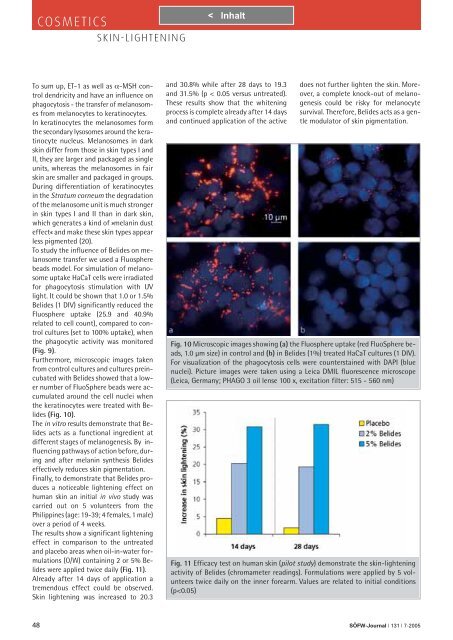
To study the influence <strong>of</strong> Belides on melanosome<br />
transfer we used a Fluosphere<br />
beads model. For simulation <strong>of</strong> melanosome<br />
uptake HaCaT cells were irradiated<br />
for phagocytosis stimulation <strong>with</strong> UV<br />
light. It could be shown that 1.0 or 1.5%<br />
Belides (1 DIV) significantly reduced the<br />
Fluosphere uptake (25.9 and 40.9%<br />
related to cell count), compared to control<br />
cultures (set to 100% uptake), when<br />
the phagocytic activity was monitored<br />
(Fig. 9).<br />
Furthermore, microscopic images taken<br />
from control cultures and cultures preincubated<br />
<strong>with</strong> Belides showed that a lower<br />
number <strong>of</strong> FluoSphere beads were accumulated<br />
around the cell nuclei when<br />
the keratinocytes were treated <strong>with</strong> Belides<br />
(Fig. 10).<br />
The in vitro results demonstrate that Belides<br />
acts as a functional ingredient at<br />
different stages <strong>of</strong> melanogenesis. By influencing<br />
pathways <strong>of</strong> action before, during<br />
and after melanin synthesis Belides<br />
effectively reduces skin pigmentation.<br />
Finally, to demonstrate that Belides produces<br />
a noticeable lightening effect on<br />
human skin an initial in vivo study was<br />
carried out on 5 volunteers from the<br />
Philippines (age: 19-39; 4 females, 1 male)<br />
over a period <strong>of</strong> 4 weeks.<br />
The results show a significant lightening<br />
effect in comparison to the untreated<br />
and placebo areas when oil-in-water formulations<br />
(O/W) containing 2 or 5% Belides<br />
were applied twice daily (Fig. 11).<br />
Already after 14 days <strong>of</strong> application a<br />
tremendous effect could be observed.<br />
<strong>Skin</strong> lightening was increased to 20.3<br />
and 30.8% while after 28 days to 19.3<br />
and 31.5% (p < 0.05 versus untreated).<br />
These results show that the whitening<br />
process is complete already after 14 days<br />
and continued application <strong>of</strong> the active<br />
does not further lighten the skin. Moreover,<br />
a complete knock-out <strong>of</strong> melanogenesis<br />
could be risky for melanocyte<br />
survival. Therefore, Belides acts as a gentle<br />
modulator <strong>of</strong> skin pigmentation.<br />
Fig. 10 Microscopic images showing (a) the Fluosphere uptake (red FluoSphere beads,<br />
1.0 µm size) in control and (b) in Belides (1%) treated HaCaT cultures (1 DIV).<br />
For visualization <strong>of</strong> the phagocytosis cells were counterstained <strong>with</strong> DAPI (blue<br />
nuclei). Picture images were taken using a Leica DMIL fluorescence microscope<br />
(Leica, Germany; PHAGO 3 oil lense 100 x, excitation filter: 515 - 560 nm)<br />
Fig. 11 Efficacy test on human skin (pilot study) demonstrate the skin-lightening<br />
activity <strong>of</strong> Belides (chromameter readings). Formulations were applied by 5 volunteers<br />
twice daily on the inner forearm. Values are related to initial conditions<br />
(p