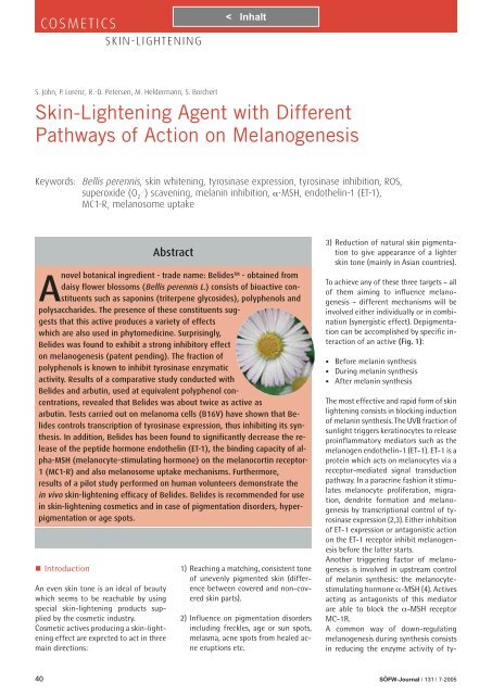
Skin-Lightening Agent with Different Pathways of Action ... - Dermage
Skin-Lightening Agent with Different Pathways of Action ... - Dermage
Skin-Lightening Agent with Different Pathways of Action ... - Dermage
You also want an ePaper? Increase the reach of your titles
YUMPU automatically turns print PDFs into web optimized ePapers that Google loves.
COSMETICS<br />
SKIN-LIGHTENING<br />
S. John, P. Lorenz, R.-D. Petersen, M. Heldermann, S. Borchert<br />
<strong>Skin</strong>-<strong>Lightening</strong> <strong>Agent</strong> <strong>with</strong> <strong>Different</strong><br />
<strong>Pathways</strong> <strong>of</strong> <strong>Action</strong> on Melanogenesis<br />
Keywords: Bellis perennis, skin whitening, tyrosinase expression, tyrosinase inhibition, ROS,<br />
superoxide (O 2 .- ) scavening, melanin inhibition, α-MSH, endothelin-1 (ET-1),<br />
MC1-R, melanosome uptake<br />
Abstract<br />
Anovel botanical ingredient - trade name: Belides TM - obtained from<br />
daisy flower blossoms (Bellis perennis L.) consists <strong>of</strong> bioactive constituents<br />
such as saponins (triterpene glycosides), polyphenols and<br />
polysaccharides. The presence <strong>of</strong> these constituents suggests<br />
that this active produces a variety <strong>of</strong> effects<br />
which are also used in phytomedicine. Surprisingly,<br />
Belides was found to exhibit a strong inhibitory effect<br />
on melanogenesis (patent pending). The fraction <strong>of</strong><br />
polyphenols is known to inhibit tyrosinase enzymatic<br />
activity. Results <strong>of</strong> a comparative study conducted <strong>with</strong><br />
Belides and arbutin, used at equivalent polyphenol concentrations,<br />
revealed that Belides was about twice as active as<br />
arbutin. Tests carried out on melanoma cells (B16V) have shown that Belides<br />
controls transcription <strong>of</strong> tyrosinase expression, thus inhibiting its synthesis.<br />
In addition, Belides has been found to significantly decrease the release<br />
<strong>of</strong> the peptide hormone endothelin (ET-1), the binding capacity <strong>of</strong> alpha-MSH<br />
(melanocyte-stimulating hormone) on the melanocortin receptor-<br />
1 (MC1-R) and also melanosome uptake mechanisms. Furthermore,<br />
results <strong>of</strong> a pilot study performed on human volunteers demonstrate the<br />
in vivo skin-lightening efficacy <strong>of</strong> Belides. Belides is recommended for use<br />
in skin-lightening cosmetics and in case <strong>of</strong> pigmentation disorders, hyperpigmentation<br />
or age spots.<br />
� Introduction<br />
An even skin tone is an ideal <strong>of</strong> beauty<br />
which seems to be reachable by using<br />
special skin-lightening products supplied<br />
by the cosmetic industry.<br />
Cosmetic actives producing a skin-lightening<br />
effect are expected to act in three<br />
main directions:<br />
1) Reaching a matching, consistent tone<br />
<strong>of</strong> unevenly pigmented skin (difference<br />
between covered and non-covered<br />
skin parts).<br />
2) Influence on pigmentation disorders<br />
including freckles, age or sun spots,<br />
melasma, acne spots from healed acne<br />
eruptions etc.<br />
3) Reduction <strong>of</strong> natural skin pigmentation<br />
to give appearance <strong>of</strong> a lighter<br />
skin tone (mainly in Asian countries).<br />
To achieve any <strong>of</strong> these three targets – all<br />
<strong>of</strong> them aiming to influence melanogenesis<br />
– different mechanisms will be<br />
involved either individually or in combination<br />
(synergistic effect). Depigmentation<br />
can be accomplished by specific interaction<br />
<strong>of</strong> an active (Fig. 1):<br />
• Before melanin synthesis<br />
• During melanin synthesis<br />
• After melanin synthesis<br />
The most effective and rapid form <strong>of</strong> skin<br />
lightening consists in blocking induction<br />
<strong>of</strong> melanin synthesis. The UVB fraction <strong>of</strong><br />
sunlight triggers keratinocytes to release<br />
proinflammatory mediators such as the<br />
melanogen endothelin-1 (ET-1). ET-1 is a<br />
protein which acts on melanocytes via a<br />
receptor-mediated signal transduction<br />
pathway. In a paracrine fashion it stimulates<br />
melanocyte proliferation, migration,<br />
dendrite formation and melanogenesis<br />
by transcriptional control <strong>of</strong> tyrosinase<br />
expression (2,3). Either inhibition<br />
<strong>of</strong> ET-1 expression or antagonistic action<br />
on the ET-1 receptor inhibit melanogenesis<br />
before the latter starts.<br />
Another triggering factor <strong>of</strong> melanogenesis<br />
is involved in upstream control<br />
<strong>of</strong> melanin synthesis: the melanocytestimulating<br />
hormone α-MSH (4). Actives<br />
acting as antagonists <strong>of</strong> this mediator<br />
are able to block the α-MSH receptor<br />
MC-1R.<br />
A common way <strong>of</strong> down-regulating<br />
melanogenesis during synthesis consists<br />
in reducing the enzyme activity <strong>of</strong> ty-<br />
40 SÖFW-Journal | 131 | 7-2005
Belides lightens<br />
and evens skin<br />
tone.<br />
Belides is produced by<br />
gentle extraction <strong>of</strong> carefully<br />
collected flowers <strong>of</strong> Bellis<br />
perennis. The extract obtained<br />
shows skin lightening activity.<br />
makes the skin look like ivory Belides<br />
CLR‘s new skin care active Belides is designed for use in depigmenting formulations. Commonly used skin lightening<br />
actives inhibit melanogenesis by reducing tyrosinase activity. Belides does more than that. Due to modulation <strong>of</strong> other<br />
biochemical pathways involved in melanin synthesis it produces synergistic effects (patent pending) by: Inhibition <strong>of</strong><br />
tyrosinase, transcriptional control <strong>of</strong> tyrosinase expression and reduction <strong>of</strong> the pro-melanogenic mediator endothelin.
COSMETICS<br />
SKIN-LIGHTENING<br />
rosinase (1). Tyrosinase catalyzes two oxidative<br />
steps in melanin synthesis. For<br />
this reason, antioxidants like polyphenols<br />
or scavengers <strong>of</strong> reactive oxygen<br />
species (ROS) are also able to interfere<br />
<strong>with</strong> melanin synthesis.<br />
Finally, depigmentation can be induced<br />
after melanin synthesis has finished<br />
by inhibiting the transfer <strong>of</strong> mature<br />
melanosomes to keratinocytes (5). Several<br />
studies have focussed on the interaction<br />
between melanocytes and keratinocytes<br />
during the transfer process<br />
(6). The most common way to achieve<br />
lightening <strong>of</strong> naturally pigmented skin is<br />
to reduce efficiency <strong>of</strong> melanin synthesis,<br />
e.g. by using tyrosinase inhibitors, antioxidants<br />
and ROS-scavengers.<br />
These different target fields <strong>of</strong> application<br />
illustrate the advantage <strong>of</strong> a whitening<br />
active which produces a cumulative<br />
effect by acting on different pigmentation<br />
pathways.<br />
It will be reported on in vitro and in vivo<br />
test results obtained <strong>with</strong> a new skinlightening<br />
product Belides, demonstrating<br />
the efficacy <strong>of</strong> skin-lightening activity<br />
and the mechanisms <strong>of</strong> action.<br />
The plant<br />
Bellis perennis L., the so-called ‘daisy<br />
flower‘ (Fig. 2), is a small perennial flowering<br />
plant (between 2 – 10 cm tall), belonging<br />
to the Asteraceae (= Compositae)<br />
family. The plant is widely distributed<br />
in Northern Europe, from the Atlantic<br />
coast <strong>of</strong> Portugal and Spain to the<br />
Western slope <strong>of</strong> the Kaukasus mountain,<br />
including the British Isles and Southern<br />
Scandinavia (7). The plant <strong>of</strong>ten grows on<br />
pastures, meadows or grassland, were it is<br />
collected.<br />
Fig. 2 Daisy flower blossoms (Bellis perennis L.)<br />
Fig. 1 Schematic Illustration <strong>of</strong> the possible approaches to interfere <strong>with</strong> melanogenesis<br />
pathways (after Briganti et al. (1))<br />
Bellis perennis has been used since ancient<br />
time in traditional folk medicine<br />
for treatment <strong>of</strong> dermatopathies like furunculosis,<br />
suppuration and slow-healing<br />
eczema. Furthermore, it has been applied<br />
against cough, fever, inflammation,<br />
dysmenorrhoe, amenorrhoe, headaches,<br />
dizziness and sleeplessness (insomnia), as<br />
well as in homeopathy (7). Phytochemical<br />
investigations <strong>of</strong> Bellis perennis have<br />
shown a wide spectrum <strong>of</strong> natural constituents<br />
such as saponins (8), flavonoids<br />
and other poly-phenolic compounds (7).<br />
Here it is reported for the first time that<br />
an active obtained from Bellis perennis<br />
inhibits the melanogenesis in human<br />
skin.<br />
� Materials and Methods<br />
Manufacturing process <strong>of</strong> Belides. Daisy<br />
flower blossoms (Bellis perennis L.) were<br />
gently extracted using an aqueous buffer<br />
system and following an internal procedure.<br />
Then, the extract was fractionated<br />
by means <strong>of</strong> ultrafiltration.<br />
Total polyphenol content <strong>of</strong> Belides was<br />
measured after a modified literature<br />
method using the Folin-Ciocalteu reaction.<br />
Gallic acid was used as an external<br />
standard for quantification.<br />
Superoxide scavenging (O 2 .- ) capacity<br />
<strong>of</strong> Belides was determined according to<br />
a literature method decribed by Lorenz<br />
et al. (10) using an enzymatic O 2 .- generation<br />
(hypoxanthin-xanthinoxidase reaction)<br />
that was visualized <strong>with</strong> NBT (nitroblue<br />
tetrazolium, Sigma) yielding a<br />
blue formazan product.<br />
Tyrosinase enzymatic activity was measured<br />
according to a modified literature<br />
method (11). Test samples <strong>of</strong> Belides (0.5,<br />
1.25, 2.5 and 10.0%) were incubated<br />
<strong>with</strong> L-tyrosine (186.9 µM) and mushroom<br />
tyrosinase (Sigma; 151.06 units/ ml)<br />
for 5 min at 40°C and L-DOPAchrome formation<br />
determined spectrophotometrically<br />
at λ max = 490 nm.<br />
Melanin inhibition assay. Mouse melanoma<br />
cells (B16V) were seeded at 2 x<br />
10 5 cells/ml in T 25 flasks (TPP) and preincubated<br />
for 3 days in vitro (DIV) in RPMI<br />
1640 medium (Biochrom; F1215) supplemented<br />
<strong>with</strong> 10% fetal calf serum<br />
(FCS), 2 mM L-glutamine and gentamycin<br />
(50 mg/l) at 37°C in a humidified 5%<br />
CO 2 atmosphere. Afterwards the medium<br />
was removed and the Belides-containing<br />
medium applied on the cell cultures<br />
as well as on controls for 7 DIV. Cells<br />
were trypsinated and lyzed <strong>with</strong> 2 ml tri-<br />
42 SÖFW-Journal | 131 | 7-2005
chloroacetic acid (5%). After centrifugation<br />
the precipitates were resuspended<br />
in 1.3 ml sodium hydroxide (1 N).<br />
The melanin absorption was measured at<br />
475 nm against blank samples. Melanin<br />
values were normalized on cell density.<br />
Optical density <strong>of</strong> control samples was<br />
set to 100%.<br />
Enzymatic quantification <strong>of</strong> tyrosinase<br />
expression. B16V mouse melanoma cells<br />
were cultivated and treated as described<br />
in the melanin inhibition assay method.<br />
After trypsination the cells were lyzed<br />
<strong>with</strong> 1.0% Triton X-100 (Sigma-Aldrich<br />
Co.) in PBS and mixed <strong>with</strong> a L-DOPA solution<br />
(0.1% L-DOPA in 0.1 M PBS, pH<br />
6.0). The transformation <strong>of</strong> L-DOPA to<br />
L-DOPAchrome was measurable <strong>with</strong>in<br />
15 - 30 min by the absorbance at 450 nm<br />
and referenced at 630 nm by a microplate<br />
reader (MRX; Dynex Technologies,<br />
Inc.).<br />
Endothelin-1 (ET-1) assay. ET-1 was quantified<br />
by using a chemiluminescent immunoassay<br />
(QuantiGlow; R&D Systems).<br />
Human epidermal keratinocytes (HaCaT,<br />
a gift <strong>of</strong> Pr<strong>of</strong>. Fusenig; DKFZ Heidelberg;<br />
Germany) were grown in Dulbecco’s Modified<br />
Eagle Medium (DMEM), supplemented<br />
<strong>with</strong> 5% FCS, 200 mM L-glutamine<br />
and 50 mg/l gentamycin (all from Biochrom<br />
KG, Berlin). Cells were seeded on<br />
a 96-well MTP <strong>with</strong> 4.5 x 10 4 cells/well<br />
and incubated for 48 h and further 3 DIV<br />
<strong>with</strong> Belides-containing medium. Then<br />
cells were stimulated for 24 h <strong>with</strong> 40<br />
ng/ml Interleukin-1 (IL-1; Sigma-Aldrich<br />
Co.). Afterwards the cells were used for<br />
the QuantiGlow ET-1 ELISA. The resulting<br />
luminescence reaction was monitored by<br />
a luminescence reader (Fluoroscan Ascent<br />
FL; Labsystems Ltd.; lag time: 1 min, reading<br />
time: 0.5 sec).<br />
The α-MSH assay. Mouse melanoma cells<br />
(B16V) were seeded at 4.5 x 10 4 cells/well<br />
in microtiter plates (MTP) and cultivated<br />
in RPMI 1640 medium <strong>with</strong> 10% FCS for<br />
2 DIV, followed by a sample application<br />
for 72 h. Then fresh medium containing<br />
1µM alpha-MSH-biotin (Phoenix Europa,<br />
Karlsruhe) was applied for 2 h. After<br />
removing unbound α-MSH, a streptavidin-horseradish<br />
peroxidase (HRP) solution<br />
(Sigma-Aldrich Co.) was added to<br />
the cells. Then o-phenylendiamine-substrate<br />
solution (Sigma-Aldrich Co.) was<br />
applied and the resulting OD values were<br />
read at 450 nm by MRX. Cells <strong>with</strong>out<br />
α-MSH-biotin served as a blank.<br />
Phagocytosis activity <strong>of</strong> keratinocytes<br />
as a function <strong>of</strong> melanosome uptake<br />
HaCaT cells were seeded at 1 x 10 3 cells/<br />
well in DMEM for 48 h in a MTP. Belidescontaining<br />
medium was applied for further<br />
3 DIV, followed by irradiation <strong>of</strong> the<br />
plates <strong>with</strong> 0.3J/cm 2 UVA and 0.03J/m 2<br />
UVB <strong>with</strong> the UV lamp SOL 500 (Dr. Hönle;<br />
Munich, Germany). Immediately after<br />
irradiation a FluoSpheres solution (beads<br />
size 1.0 µm, Molecular Probes) was added<br />
for 24 h. Then the cells were lyzed <strong>with</strong><br />
1.0% Nonident P40, 0.01% sodium dodecyl<br />
sulfate in 0.1 M Tris-HCL (pH 7.2).<br />
The fluorescence emission <strong>of</strong> the lysates<br />
was detected by a fluorescence reader<br />
(Fluoroscan Ascent FL; Labsystems Ltd.)<br />
at an excitation/emission wavelength <strong>of</strong><br />
585 nm/612 nm. Cells not treated <strong>with</strong><br />
FluoSpheres served as a blank.<br />
<strong>Skin</strong>-lightening efficacy test on human<br />
volunteers (in vivo pilot study). The in<br />
vivo pilot study was performed as a double<br />
blind study by Derma Consult GmbH<br />
(Alfter, Germany). The trials were carried<br />
out by using the inner side <strong>of</strong> the forearm<br />
as test side. After the first measurement<br />
(Chromameter), the test products<br />
were applied twice daily. Measurements<br />
were evaluated during treatment on day<br />
14, 28 and 35 about four hours after the<br />
last daily application. The L-values reflected<br />
the skin color were measured<br />
<strong>with</strong> a Minolta Chromameter CR 300<br />
(Minolta, Japan) in compliance <strong>with</strong> the<br />
Commission International de l’eclairage<br />
Scheme 1 Biosynthesis <strong>of</strong> melanin<br />
COSMETICS<br />
SKIN-LIGHTENING<br />
(CIE) system. Each L-value has an average<br />
<strong>of</strong> three recordings. Measurements<br />
are according to the guidelines <strong>of</strong> the<br />
European Society <strong>of</strong> Contact Dermatitis<br />
(12). Probability values (p) less than 0.05<br />
were considered to be significant.<br />
� Results and Discussion<br />
In the recent years there has been a growing<br />
demand for cosmetic skin-lightening<br />
products. Whereas in Western countries<br />
these products are mostly used for the<br />
prevention or treatment <strong>of</strong> irregular (hyper)pigmentation,<br />
like freckles, age- or<br />
sunspots, in Asia and Africa skin-lightening<br />
formulations are also applied to<br />
make the skin lighter and brighter.<br />
Conventional compositions developed<br />
for depigmentation generally contain<br />
ingredients that have antioxidant and<br />
tyrosinase-inhibiting activity (e.g. ascorbic<br />
acid, kojic acid, hydroquinone derivatives<br />
like arbutin etc.; for review see:<br />
(1)).<br />
In the search for a new potential skinlightening<br />
product <strong>with</strong> a broader spectrum<br />
<strong>of</strong> action on melanogenic pathways<br />
we are focusing here on Belides, a<br />
new active which is produced from daisy<br />
flowers (Bellis perennis).<br />
Melanin, responsible for pigmentation <strong>of</strong><br />
the skin, is produced in melanosomes<br />
which are located in the melanocytes.<br />
The synthesis <strong>of</strong> melanin involves oxidative<br />
processes, whereby L-tyrosine is<br />
converted into L-dihydroxy phenylalanine<br />
(L-DOPA) by the enzyme tyrosinase.<br />
L-DOPA is then finally converted<br />
to melanin by a complex chain <strong>of</strong> oxidative<br />
reactions (Scheme 1).<br />
Transcription and expression <strong>of</strong> the ty-<br />
SÖFW-Journal | 131 | 7-2005 43
COSMETICS<br />
SKIN-LIGHTENING<br />
rosinase enzymatic protein is an upstream<br />
process before melanin formation<br />
can start (Fig. 1). By inhibiting transcription<br />
<strong>of</strong> RNA on the endoplasmatic<br />
reticulum the tyrosinase expression is reduced.<br />
Investigating the influence on tyrosinase<br />
expression in melanoma cells<br />
(B16V), we show that Belides (1%) is able<br />
to reduce the biosynthesis <strong>of</strong> endogenous<br />
tyrosinase to more than 50% (Fig. 3). Belides<br />
thus avoids melanogenesis at a very<br />
early stage.<br />
Of course, these results also demonstrate<br />
that expression <strong>of</strong> tyrosinase is not completely<br />
blocked. Stronger inhibition or<br />
even complete blocking <strong>of</strong> this expression<br />
is not desirable for physiological<br />
reasons. However, Belides exerts an additional<br />
influence on the activity <strong>of</strong> remaining<br />
amount <strong>of</strong> the enzyme. To assess<br />
the inhibitory effect <strong>of</strong> Belides on<br />
tyrosinase enzymatic activity, L-tyrosin<br />
was incubated <strong>with</strong> mushroom tyrosinase<br />
and the L-DOPAchrome formation<br />
was measured. We could show that Belides,<br />
in a concentration-dependent manner,<br />
inhibits enzymatic activity which was<br />
reduced by treatment <strong>with</strong> the active<br />
(2.5%) to about 70% (Fig. 4).<br />
Furthermore, the conversion <strong>of</strong> L-tyrosine<br />
to melanin (Scheme 1) is an oxidative<br />
process that requires the presence <strong>of</strong><br />
air oxygen. Other short-lived oxidizing<br />
molecules - e.g. free radicals – namely<br />
Fig. 3 Bioassay detecting the reduction <strong>of</strong> endogenous tyrosinase<br />
expression in melanoma cells (B16V) after preincubation<br />
<strong>with</strong> Belides (7 DIV). Tyrosinase activity was measured<br />
in melanoma cells after removal <strong>of</strong> the active<br />
reactive oxygen species (ROS) can also<br />
promote melanin formation. An overproduction<br />
<strong>of</strong> ROS in the cells or tissues<br />
can alter biomolecules like L-tyrosine<br />
and represents the so-called »oxidative<br />
stress«. Antioxidants like polyphenols<br />
can scavenge ROS and inhibit oxidation<br />
<strong>of</strong> biomolecules like L-tyrosine. Belides<br />
was shown to strongly inhibit formation<br />
<strong>of</strong> one representative ROS, the superoxide<br />
(O 2 .- ). To attribute the O2 .- scavenging<br />
activity on an antioxidative principle,<br />
Belides was first standardized to the<br />
total polyphenol content. It could be<br />
demonstrated that 34.2 µg/ml polyphenol<br />
equivalents reduce the O 2 .- formation<br />
to more than 50% (IC 50 = 29.13 µg/ml<br />
polyphenol equivalents, Fig. 5).<br />
To investigate if the skin-lightening effect<br />
<strong>of</strong> Belides is only attributed to the<br />
antioxidative capacity <strong>of</strong> the polyphenols<br />
contained, it was determined the influence<br />
on melanin synthesis in the presence<br />
<strong>of</strong> a definite polyphenol amount <strong>of</strong><br />
arbutin (hydroquinone-mono-β-D-glucopyranoside)<br />
in relation to equal polyphenol<br />
amounts <strong>of</strong> Belides. Compared to<br />
Belides arbutin is a single compound and<br />
described in the literature as a tyrosinase<br />
inhibitor and a widely used skin whitening<br />
agent (13). When melanoma cells<br />
(B16V) were exposed to 13,6 µg Belides<br />
polyphenol equivalents/ml (7 DIV) the<br />
melanin accumulation was reduced to<br />
26.4% in comparison to the control cultures.<br />
When arbutin was applied at the same<br />
concentration (13.6 µg polyphenol equivalents/ml)<br />
the melanin formation was reduced<br />
only to 75.9%. The much stronger<br />
effect <strong>of</strong> Belides point to other mechanisms<br />
<strong>of</strong> action apart from tyrosinase inhibition<br />
(Fig. 6).<br />
Melanocytes as producers and distributors<br />
<strong>of</strong> the pigment melanin play the key<br />
role in pigmentation development. However,<br />
these cells are not able to express<br />
their own growth factors under nonstimulated<br />
conditions. Therefore melanocytes<br />
show a very low mitotic rate in<br />
homeostasis (14).<br />
Interaction <strong>with</strong> the surrounding cells,<br />
like keratinocytes, Langerhans cells e.c.,<br />
regulates complex biosynthesis processes<br />
<strong>of</strong> melanogenesis by controlling<br />
melanocyte growth and differentiation<br />
via release <strong>of</strong> cytokines.<br />
The peptide endothelin-1 (ET-1) is one<br />
mitogen that increases melanin synthesis<br />
as well as melanocyte proliferation.<br />
The inflammatory mediator interleukin-1<br />
(IL-1), which is induced by<br />
UV irradiation in keratinocytes, increases<br />
ET-1 expression in these cells tremendously.<br />
It is also known that in Lentigo<br />
senilis lesions a hypersecretion <strong>of</strong> ET-1<br />
occurs (15-17). This fact directed us<br />
to investigate the influence <strong>of</strong> Belides<br />
Fig. 4 Bioassay measuring tyrosinase inhibition (mushroom<br />
tyrosinase) using L-tyrosine as a substrate. Tyrosinase activity<br />
was measured over L-DOPAchrome formation. Belides<br />
concentration-dependently inhibits the tyrosinase enzymatic<br />
activity in comparison to the control (maximum inhibition<br />
= 100%)<br />
44 SÖFW-Journal | 131 | 7-2005
COSMETICS<br />
SKIN-LIGHTENING<br />
on ET-1 expression. When keratinocytes<br />
(HaCaT) where incubated <strong>with</strong> Belides<br />
(1.0, 1.5 and 2%) and stimulated <strong>with</strong> interleukin-1<br />
(IL-1) we could demonstrate<br />
that Belides reduced the expression <strong>of</strong><br />
ET-1 to 31.8, 29.9 and 27.9% in comparison<br />
to the control (100% expression).<br />
The HaCaT cells used in these experiments<br />
showed an expression <strong>of</strong> ET-1 under nonstimulated<br />
conditions too, whereby Belides<br />
was able to diminish the basal release<br />
<strong>of</strong> ET-1 as well (Fig. 7).<br />
Another factor that regulates melanogenesis<br />
is the peptide α-MSH which interacts<br />
via the melanocortin-1 receptor<br />
(MC1-R). Nowadays it is known that this<br />
peptide is not only produced in the pituitary<br />
gland but also in the cells <strong>of</strong> the skin<br />
(e.g. keratinocytes, melanocytes, Langerhans<br />
cells). α-MSH can be upregulated<br />
by UV irradiation and acts as a paracrine<br />
as well as autocrine factor <strong>of</strong> skin pigmentation.<br />
Therefore it is not a hormone<br />
in the sense <strong>of</strong> the definition (18).<br />
α-MSH binding to MC1-R enhances the<br />
second messenger cAMP which in turn<br />
activates tyrosinase and the production<br />
<strong>of</strong> eumelanin (16, 19). To prove if Belides<br />
influences the binding capacity <strong>of</strong> α-<br />
MSH to the MC1-R we carried out an experiment<br />
based on a competitive reaction<br />
between a possible bonding <strong>of</strong> Belides<br />
or the natural ligand α-MSH to<br />
the MC1-R. The results obtained <strong>with</strong><br />
melanoma cells show that 1 and 1.5%<br />
Fig. 6 Reduction <strong>of</strong> melanin formation. Belides is leading to<br />
significantly less melanin production than arbutin.<br />
Fig. 5 Analysis <strong>of</strong> the superoxide (O2 .- ) scavenging capacity <strong>of</strong> Belides. O2 .- was generated<br />
by an enzymatic reaction <strong>of</strong> hypoxanthin and xanthinoxidase (XOD). The<br />
assay is based on the spectrophotometric detection <strong>of</strong> formazan formation due to<br />
the reaction <strong>of</strong> O2 .- <strong>with</strong> NBT in competition <strong>with</strong> the added Belides polyphenol<br />
equivalents. Compared to the control (100% superoxide formation) Belides inhibits<br />
O2 .- formation concentration-dependently (IC 50 = 29.13 µg polyphenol equivalents/ml,<br />
standardized on gallic acid)<br />
Belides decreases the binding capacity<br />
<strong>of</strong> α-MSH to 38 and 12%. Therefore,<br />
Belides has antagonist features inhibiting<br />
the α-MSH binding capacity on the<br />
MC-1 R (Fig.8).<br />
On the other hand, cAMP also increases<br />
rearrangement <strong>of</strong> actin filaments which<br />
are among the major scaffolds <strong>of</strong> cell<br />
structure and cell morphology. In consequence,<br />
cAMP promotes melanocyte<br />
dendricity - a ramification connecting<br />
melanocytes <strong>with</strong> keratinocytes. This is<br />
essential for the formation <strong>of</strong> the socalled<br />
»epidermal melanin unit« which is<br />
an association <strong>of</strong> one melanocyte and<br />
surrounding 35 keratinocytes (18).<br />
Fig. 7 Influence on endothelin (ET-1) expression. Human<br />
epidermal keratinocytes (HaCaT), pretreated <strong>with</strong> Belides<br />
(3 DIV), expressed lower amounts <strong>of</strong> ET-1 <strong>with</strong> and <strong>with</strong>out<br />
IL-1 stimulation (2 DIV) in comparison to control cultures<br />
(<strong>with</strong> maximum ET-1 expression = 100%)<br />
46 SÖFW-Journal | 131 | 7-2005<br />
(µ
Fig. 8 Inhibition <strong>of</strong> α-MSH binding capacity on the MC1receptor.<br />
Belides inhibited the alpha-MSH binding capacity<br />
in melanoma cells (B16V) when the cultures were preincubated<br />
<strong>with</strong> the active (3 DIV)<br />
COSMETICS<br />
SKIN-LIGHTENING<br />
Fig. 9 Uptake <strong>of</strong> inert Fluospheres by keratinocytes (HaCaT)<br />
after UV irradiation as a model <strong>of</strong> melanosome phagocytosis.<br />
HaCaT cells incubated <strong>with</strong> Belides (3 DIV) irradiated <strong>with</strong><br />
UV light show a decreased uptake <strong>of</strong> Fluospheres in comparison<br />
to control cultures (<strong>with</strong> 100% phagocytic activity)
COSMETICS<br />
SKIN-LIGHTENING<br />
To sum up, ET-1 as well as α-MSH control<br />
dendricity and have an influence on<br />
phagocytosis - the transfer <strong>of</strong> melanosomes<br />
from melanocytes to keratinocytes.<br />
In keratinocytes the melanosomes form<br />
the secondary lysosomes around the keratinocyte<br />
nucleus. Melanosomes in dark<br />
skin differ from those in skin types I and<br />
II, they are larger and packaged as single<br />
units, whereas the melanosomes in fair<br />
skin are smaller and packaged in groups.<br />
During differentiation <strong>of</strong> keratinocytes<br />
in the Stratum corneum the degradation<br />
<strong>of</strong> the melanosome unit is much stronger<br />
in skin types I and II than in dark skin,<br />
which generates a kind <strong>of</strong> »melanin dust<br />
effect« and make these skin types appear<br />
less pigmented (20).<br />
To study the influence <strong>of</strong> Belides on melanosome<br />
transfer we used a Fluosphere<br />
beads model. For simulation <strong>of</strong> melanosome<br />
uptake HaCaT cells were irradiated<br />
for phagocytosis stimulation <strong>with</strong> UV<br />
light. It could be shown that 1.0 or 1.5%<br />
Belides (1 DIV) significantly reduced the<br />
Fluosphere uptake (25.9 and 40.9%<br />
related to cell count), compared to control<br />
cultures (set to 100% uptake), when<br />
the phagocytic activity was monitored<br />
(Fig. 9).<br />
Furthermore, microscopic images taken<br />
from control cultures and cultures preincubated<br />
<strong>with</strong> Belides showed that a lower<br />
number <strong>of</strong> FluoSphere beads were accumulated<br />
around the cell nuclei when<br />
the keratinocytes were treated <strong>with</strong> Belides<br />
(Fig. 10).<br />
The in vitro results demonstrate that Belides<br />
acts as a functional ingredient at<br />
different stages <strong>of</strong> melanogenesis. By influencing<br />
pathways <strong>of</strong> action before, during<br />
and after melanin synthesis Belides<br />
effectively reduces skin pigmentation.<br />
Finally, to demonstrate that Belides produces<br />
a noticeable lightening effect on<br />
human skin an initial in vivo study was<br />
carried out on 5 volunteers from the<br />
Philippines (age: 19-39; 4 females, 1 male)<br />
over a period <strong>of</strong> 4 weeks.<br />
The results show a significant lightening<br />
effect in comparison to the untreated<br />
and placebo areas when oil-in-water formulations<br />
(O/W) containing 2 or 5% Belides<br />
were applied twice daily (Fig. 11).<br />
Already after 14 days <strong>of</strong> application a<br />
tremendous effect could be observed.<br />
<strong>Skin</strong> lightening was increased to 20.3<br />
and 30.8% while after 28 days to 19.3<br />
and 31.5% (p < 0.05 versus untreated).<br />
These results show that the whitening<br />
process is complete already after 14 days<br />
and continued application <strong>of</strong> the active<br />
does not further lighten the skin. Moreover,<br />
a complete knock-out <strong>of</strong> melanogenesis<br />
could be risky for melanocyte<br />
survival. Therefore, Belides acts as a gentle<br />
modulator <strong>of</strong> skin pigmentation.<br />
Fig. 10 Microscopic images showing (a) the Fluosphere uptake (red FluoSphere beads,<br />
1.0 µm size) in control and (b) in Belides (1%) treated HaCaT cultures (1 DIV).<br />
For visualization <strong>of</strong> the phagocytosis cells were counterstained <strong>with</strong> DAPI (blue<br />
nuclei). Picture images were taken using a Leica DMIL fluorescence microscope<br />
(Leica, Germany; PHAGO 3 oil lense 100 x, excitation filter: 515 - 560 nm)<br />
Fig. 11 Efficacy test on human skin (pilot study) demonstrate the skin-lightening<br />
activity <strong>of</strong> Belides (chromameter readings). Formulations were applied by 5 volunteers<br />
twice daily on the inner forearm. Values are related to initial conditions<br />
(p
COSMETICS<br />
SKIN-LIGHTENING<br />
� Conclusion<br />
Belides has proven itself as a very efficient<br />
active that successfully interferes<br />
<strong>with</strong> every stage <strong>of</strong> the melanin synthesis.<br />
The extraordinary composition <strong>of</strong> this<br />
active helps to remove blemishes in a<br />
very gentle and reliable manner. Belides,<br />
a water-soluble active produced<br />
<strong>with</strong>out any organic solvent, meets the<br />
ever-growing demands <strong>of</strong> natural beauty<br />
care.<br />
References<br />
(1) Briganti S., Camera E. and Picaro, M., Chemical<br />
and Instrumental Approaches to Treat Hyperpigmentation.<br />
Pigment Cell. Res. 16 (2003)<br />
101-110<br />
(2) Imokawa G., Miyagishi M., Yada Y., Endothelin-1<br />
as new melanogen: coordinated expression<br />
<strong>of</strong> its gene and the tyrosinase gene in<br />
UVB-exposed human epidermis. J. Invest.<br />
Dermatol. 105 (1995) 32-37<br />
(3) Hara M., Yaar M., Gilchrest B.A., Endothelin-<br />
1 <strong>of</strong> keratinocyte origin is a mediator <strong>of</strong><br />
melanocyte dendricity. J. Invest. Dermatol.<br />
105 (1995) 744-748<br />
(4) Bohm M, Luger T.A., Alpha-melanocyte-stimulating<br />
hormone. Its current significance for<br />
dermatology. Hautarzt. 55 (2004) 436-445<br />
(5) Seiberg M., Paine C., Sharlow E., Costanzo M.,<br />
Andrade-Gordon P., Eisinger M., Shapiro S.S.,<br />
Inhibition <strong>of</strong> melanosome transfer results in<br />
skin lightening. J. Invest. Dermatol. 115 (2000)<br />
162-167<br />
(6) Seiberg M., Keratinocyte-melanocyte interaction<br />
during melanosome transfer. Pigment<br />
Cell Res. 14 (2001) 236-242<br />
(7) Blaschek W., Ebel S., Hackenthal E., Holzgrabe<br />
U., Keller K., Reichling J., HagerROM – Hagers<br />
Handbuch der Drogen und Arzneist<strong>of</strong>fe,<br />
Springer Verlag Berlin, Heidelberg (2003)<br />
(8) Glensk M., Wray V., Nimtz M., Schöpke T.,<br />
Triterpenoid saponins <strong>of</strong> Bellis perennis. Sci.<br />
Pharm. 69 (2001) 69-73<br />
(9) Analytical methods collection (SOP-Nr. 1999.<br />
14), University <strong>of</strong> Hamburg (Germany), Institute<br />
for Biochemistry and Food Chemistry<br />
(10) Lorenz P., Zeh M., Martens-Lobenh<strong>of</strong>fer J.,<br />
Schmidt H., Wolf G., and Horn T.F.W., Natural<br />
and newly synthesized hydroxy-1-aryl-isochromans:<br />
A class <strong>of</strong> potential antioxidants<br />
and radical scavengers. Free Rad. Res. 39<br />
(2005) 535-545<br />
(11) Jeong C.H., Shim K.H., Tyrosinase Inhibitor<br />
Isolated from the Leaves <strong>of</strong> Zanthoxylum<br />
piperitum. Biosci. Biotechnol. Biochem. 68<br />
(2004) 1984-1987<br />
(12) Fullerton A., Fischer T., Lahti A., Wilhelm K.P.,<br />
Takiwaki H., Serup J., Guidelines for measurement<br />
<strong>of</strong> skin colour and erythema. A report<br />
from the Standardization Group <strong>of</strong> the European<br />
Society <strong>of</strong> Contact Dermatitis. Contact<br />
Dermatitis 35 (1996) 1-10<br />
(13) Hori, I., Nihei, K., Kubo, I., Structural criteria<br />
for depigmentation mechanism <strong>of</strong> arbutin.<br />
Phytother. Res. 18 (2004) 475-479<br />
(14) Yada Y., Higuchi K., Imokawa G. Effect <strong>of</strong> Endothelin<br />
on Signal Transduction and Proliferation<br />
in Human Melanocytes. J. Biol. Chem.<br />
266 (1991) 18352-18357<br />
(15) Kadono S., Manaka I., Kawashima M., Kobayashi<br />
T., Imokawa G., The role <strong>of</strong> the epidermal<br />
endothelin cascade in the hyperpigmentation<br />
mechanism <strong>of</strong> lentigo senilis. J. Invest. Dermatol.<br />
116 (2001) 571-577<br />
(16) Imokawa G., Yada Y., and Kimura M., Signalling<br />
mechanisms <strong>of</strong> endothelin- induced<br />
mitogenesis and melanogenesis in human<br />
melanocytes. Biochem. J. 314 (1996) 305-312<br />
(17) Jamal S. and Schneider R.J., UV-induction <strong>of</strong><br />
keratinocyte endothelin-1 downregulates Ecadherin<br />
in melanocytes and melanoma cells.<br />
J. Clin. Invest. 110 (2002) 443-452<br />
(18) Tsatmali M., Ancans J., Thody A. J., Melanocyte<br />
Function and its Control by Melanocortin Peptides.<br />
J. Histochem. Cytochem. 50 (2002) 125-<br />
134<br />
(19) Lu D., Willard D., Patel I.D., Kadwell S., Overton<br />
L., Kost T., Luther M., Chen W., Woychik<br />
R.P., Wilkinson W.O., and Cone R.D., Agouti<br />
protein is an antagonist <strong>of</strong> the melanocytestimulating<br />
hormone receptor. Nature 371<br />
(1994) 799-802<br />
(20) Jimbow K., Sugijama S., Melanosomal translocation<br />
and transfer. In The Pigmentary System:<br />
Physiology. a. Pathophysiology. (1998),<br />
Oxford Uni. Press.<br />
Chemisches Laboratorium<br />
Dr. Kurt Richter GmbH (CLR)<br />
Bennigsenstr. 25<br />
12159 Berlin<br />
Germany<br />
Email: info@clr-berlin.com<br />
SÖFW-Journal | 131 | 7-2005 49<br />
�


