CONTENTS Applications in Biology and Medicine - Formatex ...
CONTENTS Applications in Biology and Medicine - Formatex ...
CONTENTS Applications in Biology and Medicine - Formatex ...

Create successful ePaper yourself
Turn your PDF publications into a flip-book with our unique Google optimized e-Paper software.
Modern Research <strong>and</strong> Educational Topics <strong>in</strong> Microscopy.<br />
A. Méndez-Vilas <strong>and</strong> J. Díaz (Eds.) ©FORMATEX 2007<br />
_______________________________________________________________________________________________<br />
<strong>CONTENTS</strong><br />
VOL. 1<br />
Introduction........................................................................................................................... XV<br />
<strong>Applications</strong> <strong>in</strong> <strong>Biology</strong> <strong>and</strong> Medic<strong>in</strong>e<br />
Approaches for Investigat<strong>in</strong>g Mechanobiological Dynamics <strong>in</strong> Liv<strong>in</strong>g Cells with<br />
Fluorescence <strong>and</strong> Atomic Force Microscopies<br />
A.E. Pell<strong>in</strong>g, B. M. Nicholls, Y.R. Silberberg, <strong>and</strong> M.A. Horton..........................................<br />
Challenges <strong>and</strong> Approaches - Prob<strong>in</strong>g Tumor Cell Invasion by Atomic Force<br />
Microscopy<br />
T. Ludwig, R. Kirmse <strong>and</strong> Kate Poole...................................................................................<br />
Study of Cancer Cells Used Atomic Force Microscopy<br />
K. Tomankova, H. Kolarova, M. Vujtek <strong>and</strong> H. Zapletalova................................................<br />
Atomic Force Imag<strong>in</strong>g of Ocular Tissues: morphological study of healthy <strong>and</strong><br />
cataract lenses<br />
A. Antunes, F. V. Gozzo, M. I. Borella, M. Nakamura, A. M. V. Safatle, P. S. M. Barros<br />
<strong>and</strong> H. E. Toma………………………………………………………………………………<br />
Imag<strong>in</strong>g of Bone Ultrastructure us<strong>in</strong>g Atomic Force Microscopy<br />
P. J. Thurner, E. Oroudjev, R. Jungmann, C. Kreutz, J. H. K<strong>in</strong>dt, G. Schitter, T. O.<br />
Okouneva, M. E. Lauer, G. E. Fantner, H. G. Hansma, <strong>and</strong> P. K. Hansma............................<br />
Cell Surface Membrane Remodell<strong>in</strong>g <strong>and</strong> Mitochondrial Remodell<strong>in</strong>g <strong>in</strong> Live<br />
Neurons: Practical Approaches for Study<strong>in</strong>g Dynamic Processes Us<strong>in</strong>g Confocal<br />
Microscopy<br />
N. Shulyakova, P. Kaifosh, D. Diec, <strong>and</strong> L.R. Mills...............................................................<br />
Cell-surface <strong>in</strong>teraction <strong>in</strong> biomedical implants assessed by simultaneous<br />
fluorescence <strong>and</strong> reflection confocal microscopy<br />
J. Vilches, J.I. Vilches-Perez <strong>and</strong> M. Salido..........................................................................<br />
The comb<strong>in</strong>ed application of AFM <strong>and</strong> LSCM: Chang<strong>in</strong>g the way we look at<strong>in</strong>nate<br />
immunity<br />
E. L. Adams <strong>and</strong> K. J. Czymmek............................................................................................<br />
Electron Microscopy Visualization of the Cell Surface of Trypanosomatids<br />
W. de Souza.............................................................................................................................<br />
Extend<strong>in</strong>g the Explanatory Power of Live Cell Imag<strong>in</strong>g by computationally<br />
modell<strong>in</strong>g the Execution of Apoptotic Cell Death<br />
H. Huber, G. Gomez Estrada, H. Dussmann, C. O’Connor <strong>and</strong> M. Rehm............................<br />
3-10<br />
11-22<br />
23-28<br />
29-36<br />
37-48<br />
49-59<br />
60-67<br />
68-76<br />
77-87<br />
88-99<br />
V
Modern Research <strong>and</strong> Educational Topics <strong>in</strong> Microscopy.<br />
_______________________________________________________________________________________________<br />
©FORMATEX 2007<br />
A. Méndez-Vilas <strong>and</strong> J. Díaz (Eds.)<br />
In vivo <strong>and</strong> <strong>in</strong> vitro Approaches for the Study of Adult Neurogenesis <strong>in</strong> Light,<br />
Confocal, <strong>and</strong> Electron Microscopy<br />
N. Canalia, M. Armentano, G. Ponti <strong>and</strong> L. Bonfanti............................................................<br />
Microscopy studies on uncultivated magnetotactic bacteria<br />
T. S. Silveira, J. L. Mart<strong>in</strong>s, K. T. Silva, F. Abreu <strong>and</strong> U. L<strong>in</strong>s.............................................<br />
Scann<strong>in</strong>g Electron Microscopy <strong>and</strong> Transmission Electron Microscopy of Mollicutes:<br />
Challenges <strong>and</strong> Opportunities<br />
C. T. K.-H. Stadtländer............................................................................................................<br />
Non-Invasive Fourier Transform Infrared Microspectroscopy <strong>and</strong> Imag<strong>in</strong>g<br />
Techniques: Basic Pr<strong>in</strong>ciples <strong>and</strong> <strong>Applications</strong><br />
P. Garidel <strong>and</strong> M. Boese..........................................................................................................<br />
Contribution of Electron Microscopy to the Study of the Nuclear Pore Complex<br />
Structure, Composition, <strong>and</strong> Function<br />
N. Panté...................................................................................................................................<br />
Bright Solutions to Get Sharp Images: Confocal <strong>and</strong> Two- Photon Fluorescence<br />
Microscopy <strong>and</strong> the Pros <strong>and</strong> Cons of New Multifocal Approaches<br />
R. Kurtz...................................................................................................................................<br />
Microscopy techniques <strong>and</strong> the study of synapses<br />
E. Perez-Costas, M. Melendez-Ferro <strong>and</strong> R.C. Roberts……………………………………..<br />
Microscopic Investigations <strong>in</strong> Neurodegenerative Diseases<br />
R. J. Castellani, B. A. Alexiev, D. Phillips, G. Perry <strong>and</strong> M. A. Smith..................................<br />
Study<strong>in</strong>g the temperature-dependent events of live cells under confocal <strong>and</strong> epifluorescence<br />
microscopy us<strong>in</strong>g a solid-state heat<strong>in</strong>g/cool<strong>in</strong>g system<br />
Zu-Han Huang, Huei-Hsuan Cheng, Huei-In Wu, Sh<strong>in</strong>-Hua Tseng <strong>and</strong> Yen-Chung Chang.<br />
Live imag<strong>in</strong>g genetically-encoded fluorescent prote<strong>in</strong>s <strong>in</strong> embryonic stem cells us<strong>in</strong>g<br />
confocal microscopy<br />
J. Artus <strong>and</strong> A.-K. Hadjantonakis............................................................................................<br />
Correlative Fluorescence- <strong>and</strong> Scann<strong>in</strong>g, Transmission Electron Microscopy for<br />
Biomolecular Investigation<br />
K. Jahn, D. Barton <strong>and</strong> F. Braet..............................................................................................<br />
Immunolocalization of maize transglutam<strong>in</strong>ase <strong>and</strong> its substrates <strong>in</strong> plant cells <strong>and</strong> <strong>in</strong><br />
Escherichia coli transformed cells<br />
M. Santos, E.Villalobos, P.Carvajal-Vallejos, E. Barberá, A. Campos <strong>and</strong> J.M.Torné…….<br />
Visualization of the real microarchitecture of glycoprote<strong>in</strong> matrices with scann<strong>in</strong>g<br />
electron microscopy<br />
G. Familiari, M. Relucenti, A. Familiari, E. Battaglione, G. Franchitto <strong>and</strong> R. Heyn............<br />
100-110<br />
111-121<br />
122-131<br />
132-143<br />
144-153<br />
154-163<br />
164-170<br />
171-182<br />
183-189<br />
190-202<br />
203-211<br />
212-223<br />
224-228<br />
VI
Modern Research <strong>and</strong> Educational Topics <strong>in</strong> Microscopy.<br />
A. Méndez-Vilas <strong>and</strong> J. Díaz (Eds.) ©FORMATEX 2007<br />
_______________________________________________________________________________________________<br />
Use of st<strong>and</strong>ard fluorescence microscopy to assess modifications <strong>in</strong> the plasma<br />
membrane potential <strong>and</strong> <strong>in</strong> the <strong>in</strong>tracellular concentration of <strong>in</strong>organic ions <strong>in</strong><br />
cultured cells<br />
S. Chifflet <strong>and</strong> J.A. Hernández................................................................................................<br />
Imag<strong>in</strong>g biological structures with a proton microprobe<br />
T. P<strong>in</strong>heiro, M.D.Ynsa <strong>and</strong> L.C.Alves....................................................................................<br />
Macromolecular Synthesis <strong>in</strong> Hepatocyte Mitochondria of Ag<strong>in</strong>g Mice as Revealed<br />
by Electron Microscopic Radioautography I: Nucleic Acid Synthesis<br />
T. Nagata.................................................................................................................................<br />
Macromolecular Synthesis <strong>in</strong> Hepatocyte Mitochondria of Ag<strong>in</strong>g Mice as Revealed<br />
by Electron Microscopic Radioautography II : Prote<strong>in</strong> Synthesis<br />
T. Nagata.................................................................................................................................<br />
Size Estimation of Prote<strong>in</strong> Clusters <strong>in</strong> the Nanometer Range by Us<strong>in</strong>g Spatially<br />
Modulated Illum<strong>in</strong>ation Microscopy<br />
U. J. Birk, I. Upmann, D. Toomre, C. Wagner <strong>and</strong> C. Cremer...............................................<br />
Transglutam<strong>in</strong>ase activity <strong>and</strong> localization dur<strong>in</strong>g microspore <strong>in</strong>duction <strong>in</strong> maize<br />
M. Santos, J. Alché , M.I. Rodríguez-García <strong>and</strong> J.M.Torné………………………………..<br />
A Thous<strong>and</strong> Prote<strong>in</strong>s of Light: 15 Years of Advances <strong>in</strong> Fluorescent Prote<strong>in</strong>s<br />
G. McNamara <strong>and</strong> C.A. Boswell.............................................................................................<br />
Us<strong>in</strong>g Total Internal Reflection Fluorescence Microscopy, DNA Curta<strong>in</strong>s, <strong>and</strong><br />
Quantum Dots to Investigate Prote<strong>in</strong>-DNA Interactions at the S<strong>in</strong>gle-molecule Level<br />
M-L. Visnapuu, D. Duzdevich <strong>and</strong> E. C. Greene....................................................................<br />
Fluorescence Resonance Energy Transfer us<strong>in</strong>g molecular beacon as a probe, a new<br />
approach for <strong>in</strong> vivo macromolecular <strong>in</strong>teraction study<br />
S. Ganguly...............................................................................................................................<br />
Electron-microscopic <strong>and</strong> genetic dissection of arthropod cuticle differentiation<br />
H. Schwarz <strong>and</strong> B. Moussian..................................................................................................<br />
Microscopy <strong>and</strong> egg morphology of Mayflies<br />
N. Ubero-Pascal <strong>and</strong> M.A. Puig..............................................................................................<br />
Role of Scann<strong>in</strong>g Electron Microscopy <strong>in</strong> Underst<strong>and</strong><strong>in</strong>g Insect Corneal Nipple <strong>and</strong><br />
Other Structures<br />
S. Dey......................................................................................................................................<br />
Antennal sensilla of hymenopteran parasitic wasps: variations l<strong>in</strong>ked to host<br />
exploitation behavior<br />
J. van Baaren, G. Boiv<strong>in</strong>, D. Bourdais <strong>and</strong> O. Roux...............................................................<br />
229-236<br />
237-244<br />
245-258<br />
259-271<br />
272-279<br />
280-286<br />
287-296<br />
297-308<br />
309-315<br />
316-325<br />
326-335<br />
336-344<br />
345-352<br />
VII
Modern Research <strong>and</strong> Educational Topics <strong>in</strong> Microscopy.<br />
_______________________________________________________________________________________________<br />
©FORMATEX 2007<br />
A. Méndez-Vilas <strong>and</strong> J. Díaz (Eds.)<br />
The application of confocal microscopy <strong>and</strong> 3D imag<strong>in</strong>g software <strong>in</strong> Functional,<br />
Evolutionary, <strong>and</strong> Developmental Zoology: reconstruct<strong>in</strong>g myo- <strong>and</strong> neurogenesis <strong>in</strong><br />
space <strong>and</strong> time<br />
A. Wann<strong>in</strong>ger..........................................................................................................................<br />
Comparative study of the impact of the act<strong>in</strong> cytoskeleton on cell shape <strong>and</strong><br />
membrane surface <strong>in</strong> mammalian cells <strong>in</strong> response to act<strong>in</strong> tox<strong>in</strong>s<br />
F. Lázaro-Diéguez <strong>and</strong> G. Egea……………………………………………………………..<br />
Chromosomes <strong>in</strong> Focus: Basic Cytogenetics, Light Microscopy <strong>and</strong> the Case of<br />
Neotropical Fish<br />
P.R.A.M Affonso, V S. Mir<strong>and</strong>a, A. S. Medrado, U. P Jacob<strong>in</strong>a, J. A. Bitencourt, J. S.<br />
Almeida <strong>and</strong> P. L. S. Carneiro.................................................................................................<br />
The Nuclear Area Factor (NAF): a measure for cell apoptosis us<strong>in</strong>g microscopy <strong>and</strong><br />
image analysis<br />
M. A. DeCoster.......................................................................................................................<br />
Study of the Mechanisms of Action of Medic<strong>in</strong>es <strong>in</strong> the Immune System Us<strong>in</strong>g<br />
Microscopy<br />
C. Camargo de Oliveira <strong>and</strong> D. de Freitas Buchi……………………………………………<br />
Prob<strong>in</strong>g the Structure <strong>and</strong> Function of Mammalian Sperm us<strong>in</strong>g Optical <strong>and</strong><br />
Fluorescence Microscopy<br />
J. Ramalho-Santos, A. Amaral, A.P. Sousa, A.S. Rodrigues, L. Mart<strong>in</strong>s, M. Baptista, P.C.<br />
Mota, R. Tavares, S. Amaral <strong>and</strong> S. Gamboa……………………………………………….<br />
Super-Quiet Microfluorometry: Examples of Tumor Cell Metabolic Dynamics<br />
A.J. Clark <strong>and</strong> H.R. Petty........................................................................................................<br />
Real-time visualization of develop<strong>in</strong>g viral <strong>in</strong>fection <strong>in</strong> fibroblasts<br />
K.L. Lathrop <strong>and</strong> A. Eisfeld....................................................................................................<br />
Morphology of the develop<strong>in</strong>g fetal lung – the rabbit experimental model<br />
X I. Roubliova , J. M. Biard, L. Ophalvens, D. Gallot, J.C. Jani , E.K.Verbeken, C.P. Van<br />
de Ven, D. Tibboel <strong>and</strong> J.A. Deprest.......................................................................................<br />
3D visualization <strong>and</strong> analysis of cell-matrix transformations <strong>in</strong> whole-mount <strong>and</strong> live<br />
embryos us<strong>in</strong>g confocal <strong>and</strong> multi-photon microscopy<br />
G.G. Mart<strong>in</strong>s, P. Rifes, R. Amândio, P. Camp<strong>in</strong>ho, I. Palmeirim <strong>and</strong> S. Thorste<strong>in</strong>sdóttir.....<br />
Utilization of the Laser Induced Plasma Spectroscopy for monitor<strong>in</strong>g of the metal<br />
accumulation <strong>in</strong> plant tissues with high spatial resolution<br />
J. Kaiser, M. Galiová, K. Novotný, L. Reale, K. Stejska, O. Samek, R. Mal<strong>in</strong>a, K.<br />
Páleníková, V. Adam <strong>and</strong> R. Kizek.........................................................................................<br />
Quantitative Microscopic Analysis of Histological Sections of Bra<strong>in</strong> Tissue<br />
R. A. Armstrong......................................................................................................................<br />
353-361<br />
362-369<br />
370-377<br />
378-384<br />
385-393<br />
394-402<br />
403-408<br />
409-416<br />
417-425<br />
426-433<br />
434-441<br />
442-452<br />
VIII
Modern Research <strong>and</strong> Educational Topics <strong>in</strong> Microscopy.<br />
A. Méndez-Vilas <strong>and</strong> J. Díaz (Eds.) ©FORMATEX 2007<br />
_______________________________________________________________________________________________<br />
Ultrastructural Changes In Microcoleus chthonoplastes Grow<strong>in</strong>g In the Presence of<br />
Crude Oil. <strong>Applications</strong> for Ecological Studies<br />
E. Diestra, I. Esteve, O. Castell <strong>and</strong> A. Solé........................................................................... 453-460<br />
Correlation Between flow Cytometry <strong>and</strong> Transmission Electron Microscopy<br />
A. Alvarez, F. Goñi de Cerio <strong>and</strong> E. Hilario………………………………………………... 461-468<br />
VOL. 2<br />
Introduction........................................................................................................................... XV<br />
<strong>Applications</strong> <strong>in</strong> Physical/Chemical Sciences<br />
Scann<strong>in</strong>g Force Microscopic Study of Surface Structure <strong>and</strong> Surface Properties of<br />
Organosilane Monolayers<br />
T. Koga <strong>and</strong> A. Takahara........................................................................................................<br />
Nano-Structur<strong>in</strong>g <strong>and</strong> Molecular Doma<strong>in</strong> Organizations <strong>in</strong> Lipid-Prote<strong>in</strong><br />
Membranous Interfaces<br />
K. Nag, A.K. P<strong>and</strong>a, R. Devraj <strong>and</strong> M. Fritzen-Garcia...........................................................<br />
Nanolubrication studied by Contact-Mode Atomic Force Microscopy<br />
R. Buzio <strong>and</strong> U. Valbusa.........................................................................................................<br />
Experimental contribution to the underst<strong>and</strong><strong>in</strong>g of wett<strong>in</strong>g of solid surfaces at the<br />
meso- <strong>and</strong> nano-scale us<strong>in</strong>g dynamic AFM<br />
A.B. Jódar-Reyes, A. Méndez-Vilas <strong>and</strong> M.L. González-Martín...........................................<br />
Usage of AFM, SEM <strong>and</strong> TEM for the research of carbon nanotubes<br />
K. Safarova, A. Dvorak, R. Kub<strong>in</strong>ek <strong>and</strong> M. Vujtek A. Rek..................................................<br />
Atomic Force Microscopy as a tool for unravell<strong>in</strong>g the relationship between<br />
morphology <strong>and</strong> growth dynamics of organic semiconductors<br />
M. Campione, M. Moret <strong>and</strong> A. Sassella................................................................................<br />
AFM monitor<strong>in</strong>g of size changes dur<strong>in</strong>g thermal syntheses of ferric oxide<br />
nanoparticles<br />
M. Vůjtek, R. Kubínek, R. Zbořil <strong>and</strong> L. Machala.................................................................<br />
Various approaches to control solid/solid wett<strong>in</strong>g self-assembly of organic<br />
semiconductors with STM<br />
F. Trixler <strong>and</strong> W.M. Heckl......................................................................................................<br />
Dephas<strong>in</strong>g of charge <strong>and</strong> sp<strong>in</strong> <strong>in</strong> semiconductor quantum dots<br />
W. Jacak, J. Krasnyj, <strong>and</strong> L. Jacak.........................................................................................<br />
471-482<br />
483-490<br />
491-499<br />
500-512<br />
513-519<br />
520-527<br />
528-533<br />
534-541<br />
542-549<br />
IX
Modern Research <strong>and</strong> Educational Topics <strong>in</strong> Microscopy.<br />
_______________________________________________________________________________________________<br />
©FORMATEX 2007<br />
A. Méndez-Vilas <strong>and</strong> J. Díaz (Eds.)<br />
Investigation of the nanostructured surface of s<strong>in</strong>gle-crystal silicon by the method of<br />
scann<strong>in</strong>g tunnel spectroscopy<br />
S.P. Kulyk, M.M. Melnichenko, K.V. Svezhentsova <strong>and</strong> O.M. Shmyryeva..........................<br />
Electrically characterization of Group III-Nitride nanocolumns with Scann<strong>in</strong>g Force<br />
Microscopy<br />
M. Niebelschütz, V. Cimalla, O. Ambacher, T. Machleidt, K.-H. Franke, J. Ristic, J.<br />
Gr<strong>and</strong>al, M.A. Sánchez-García <strong>and</strong> E. Calleja........................................................................<br />
Field Emission Scann<strong>in</strong>g Electron Microscopy for Structural Characterization of 3D<br />
Gold Nanoparticle Superlattices<br />
H. Yao <strong>and</strong> K. Kimura.............................................................................................................<br />
UV-VIS <strong>and</strong> TEM assessment of morphological features of silver nanoparticles from<br />
phosphate glass matrices<br />
L. Baia <strong>and</strong> S. Simon...............................................................................................................<br />
Evaluation of electronic <strong>and</strong> geometric properties of nanoparticles us<strong>in</strong>g X-PEEM<br />
O. Seifarth...............................................................................................................................<br />
Exam<strong>in</strong>ation of dent<strong>in</strong> surface us<strong>in</strong>g AFM <strong>and</strong> SEM<br />
R. Kub<strong>in</strong>ek, Z. Zapletalova, M. Vujtek, R. Novotný, H. Kolarova <strong>and</strong> H. Chmelickova......<br />
TEM analysis of the early m<strong>in</strong>eralization process of mantle dent<strong>in</strong><br />
P. Dechichi, C. Christian Gomes Moura, A. Wilsson de Almeida Filho <strong>and</strong> J. Carlos<br />
Gabrielli Biffi………………………………………………………………………………..<br />
Spatial visualization of thermally sprayed microstructure based on light microscopy<br />
P. Ctibor <strong>and</strong> R. Lechnerova...................................................................................................<br />
Analytical electron microscopy of tremolite<br />
K.N. Bozhilov <strong>and</strong> D.M. Jenk<strong>in</strong>s............................................................................................<br />
Investigat<strong>in</strong>g morphological changes on octadecyl modifiedsilicas by SEM <strong>and</strong> AFM<br />
R. Brambilla, F. Silveira <strong>and</strong> J.H.Z. dos Santos……………………………………………..<br />
Importance of Transmission Electron Microscopy for Carbon Nanomaterials<br />
Research<br />
P.R. Somani <strong>and</strong> M. Umeno....................................................................................................<br />
Discover<strong>in</strong>g the True Morphology of Amphibole M<strong>in</strong>erals: Complementary TEM<br />
<strong>and</strong> FESEM Characterization of Particles <strong>in</strong> Mixed M<strong>in</strong>eral Dust<br />
K. E. Harris, K. L. Bunker, B. R. Strohmeier, R. Hoch <strong>and</strong> R. J. Lee....................................<br />
S<strong>in</strong>gle-Molecule Fluorescence Imag<strong>in</strong>g Techniques for the Detection of Reactive<br />
Oxygen Species<br />
T. Tachikawa <strong>and</strong> T. Majima..................................................................................................<br />
550-559<br />
560-567<br />
568-575<br />
576-583<br />
584-592<br />
593-598<br />
599-605<br />
606-615<br />
616-625<br />
626-633<br />
634-642<br />
643-650<br />
651-659<br />
X
Modern Research <strong>and</strong> Educational Topics <strong>in</strong> Microscopy.<br />
A. Méndez-Vilas <strong>and</strong> J. Díaz (Eds.) ©FORMATEX 2007<br />
_______________________________________________________________________________________________<br />
Scann<strong>in</strong>g Electron Microscopy Investigation of Lead-Free High Refractive Index<br />
Glass Prepared from Local S<strong>and</strong> Used for Restoration <strong>and</strong> Conservation<br />
P. Dararutana <strong>and</strong> N. Sirikulrat...............................................................................................<br />
SEM <strong>and</strong> TEM studies <strong>in</strong> alloys developed by mechanical alloy<strong>in</strong>g<br />
J.J. Suñol <strong>and</strong> L. Escoda..........................................................................................................<br />
Scann<strong>in</strong>g Electron Microscopic Characterization of Copper (II) Phthalocyan<strong>in</strong>e<br />
Nanocrystallites Th<strong>in</strong> Films Deposited on Technologically Important Substrates<br />
B. Mallik <strong>and</strong> S. Karan............................................................................................................<br />
Surface Morphological Study of Low Temperature Plasma Treated Wool – A Time<br />
Dependence Study<br />
C.W. Kan.................................................................................................................................<br />
Fluorescence <strong>and</strong> Scann<strong>in</strong>g Electron Microscopy of Chitosan/DNA Nanoparticles for<br />
Biological <strong>Applications</strong><br />
A. Masotti, F. Mar<strong>in</strong>o, G. Ortaggi <strong>and</strong> C. Palocci...................................................................<br />
A Novel “In-situ-track<strong>in</strong>g” Approach for Evaluat<strong>in</strong>g Microstructural Variations<br />
Us<strong>in</strong>g SEM, EDS <strong>and</strong> EBSD <strong>and</strong> Its <strong>Applications</strong> <strong>in</strong> Materials Science<br />
C. Pan, Y. Huang <strong>and</strong> Q. Fu....................................................................................................<br />
Morphology Variations of Polypropylene<br />
J. Výchopňová, R. Čermák <strong>and</strong> M. Obadal.............................................................................<br />
Morphological Transition of Isotactic Polybutene-1 Tetragonal Crystals: Optical <strong>and</strong><br />
Transmission Electron Microscopy Observation<br />
M. Yamashita <strong>and</strong> T. Takahashi..............................................................................................<br />
Nanostructure of starch high-pressure treated granules discovered by low<br />
temperature scann<strong>in</strong>g electron microscopy<br />
A.D. Mol<strong>in</strong>a-García, E. Horridge, P.D. Sanz <strong>and</strong> M.N. Mart<strong>in</strong>o……………………………<br />
Nanoscale Structure of Cellulosic Materials: Challenges <strong>and</strong> Opportunities for AFM<br />
A.T. Paiva, S.M. Sequeira, D.V. Evtugu<strong>in</strong>, A.L. Kholk<strong>in</strong> <strong>and</strong> I.Portugal..............................<br />
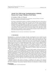
Atomic Force Microscope Nano<strong>in</strong>dentations to Reliably Measure the Young’s<br />
Modulus of Soft Matter<br />
D. Tranchida, Z. Kiflie <strong>and</strong> S. Piccarolo…………………………………………………….<br />
The Atomic Force Spectroscopy as a Tool to Investigate Surface Forces: Basic<br />
Pr<strong>in</strong>ciples <strong>and</strong> <strong>Applications</strong><br />
F.L. Leite, L.H.C. Mattoso, O.N. Oliveira Jr <strong>and</strong> P.S.P. Herrmann Jr....................................<br />
660-665<br />
666-670<br />
671-682<br />
683-689<br />
690-696<br />
697-703<br />
704-712<br />
713-718<br />
719-725<br />
726-733<br />
Techniques<br />
737-746<br />
747-757<br />
XI
Modern Research <strong>and</strong> Educational Topics <strong>in</strong> Microscopy.<br />
_______________________________________________________________________________________________<br />
©FORMATEX 2007<br />
A. Méndez-Vilas <strong>and</strong> J. Díaz (Eds.)<br />
A Guide for Atomic Force Microscopy Analysis of Soft-Condensed Matter<br />
M. Raposo, Q. Ferreira <strong>and</strong> P.A. Ribeiro................................................................................<br />
Scann<strong>in</strong>g Probe Microscopy-Based Nanofabrication for Emerg<strong>in</strong>g <strong>Applications</strong><br />
X. Liu <strong>and</strong> H. Zhang................................................................................................................<br />
Explor<strong>in</strong>g the high pressure SEM<br />
C. Mathieu, L Khouchaf <strong>and</strong> A Kadoun.................................................................................<br />
How to study biological samples by FIB/SEM?<br />
M. Milani, D. Drobne <strong>and</strong> Francesco Tatti.............................................................................<br />
Very Low Energy Scann<strong>in</strong>g Electron Microscopy<br />
I. Müllerová <strong>and</strong> L. Frank.......................................................................................................<br />
The magnetic force microscopy <strong>and</strong> its capability for nanomagnetic studies - The<br />
short compendium<br />
A. Hendrych, R. Kubínek <strong>and</strong> A.V. Zhukov...........................................................................<br />
Visualisation of Small Fluid Droplets on Biological <strong>and</strong> Artificial Surfaces Us<strong>in</strong>g the<br />
Cryo-SEM Approach<br />
S.N. Gorb, D. Voigt <strong>and</strong> E.V. Gorb........................................................................................<br />
Spherical AFM Probes for Adhesion Force Measurements on Metal S<strong>in</strong>gle Crystals<br />
B. Stegemann, H. Backhaus, H. Kloss <strong>and</strong> E. Santner............................................................<br />
Near-Field Optical Microscopy of Surface Plasmon Polaritons Nano-Optics<br />
Víctor Coello...........................................................................................................................<br />
Semiconductor nanocrystals <strong>and</strong> fluorescence microscopy <strong>in</strong> biological label<strong>in</strong>g<br />
P.M.A. Farias, B.S. Santos, A.Fontes <strong>and</strong> C.L. Cesar………………………………………<br />
Fluorescence Correlation Spectroscopy of Liv<strong>in</strong>g Cells<br />
G. Vereb, L. Ujlaky-Nagy, E. Friedländer G. Vámosi <strong>and</strong> J. Szöllősi....................................<br />
Fluorescence Correlation Spectroscopy: an Experimentalist’s View of the Basics<br />
G. Jung....................................................................................................................................<br />
L<strong>in</strong>ear fluorescence unmix<strong>in</strong>g <strong>in</strong> cell biological research<br />
B. Kraus, M. Ziegler <strong>and</strong> H. Wolff.........................................................................................<br />
High resolution Near-field Fluorescence Microscopy: from pr<strong>in</strong>ciples to applications<br />
<strong>in</strong> cell biology<br />
A. Cambi, C.G. Figdor <strong>and</strong> M.F. Garcia-Parajo…………………………………………….<br />
Two-Photon Fluorescence Microscopy: Basic Pr<strong>in</strong>ciples, Advantages <strong>and</strong> Risks<br />
S.J. Mulligan <strong>and</strong> B.A. MacVicar...........................................................................................<br />
758-769<br />
770-778<br />
779-786<br />
787-794<br />
795-804<br />
805-811<br />
812-819<br />
820-827<br />
828-839<br />
840-847<br />
848-854<br />
855-862<br />
863-872<br />
873-880<br />
881-889<br />
XII
Modern Research <strong>and</strong> Educational Topics <strong>in</strong> Microscopy.<br />
A. Méndez-Vilas <strong>and</strong> J. Díaz (Eds.) ©FORMATEX 2007<br />
_______________________________________________________________________________________________<br />
Colours Count: how the Challenge of Fluorescence was solved <strong>in</strong> Confocal<br />
Microscopy<br />
R. Borl<strong>in</strong>ghaus.........................................................................................................................<br />
Software-based three dimensional reconstructions <strong>and</strong> enhancements of focal depth<br />
<strong>in</strong> microphotographic images<br />
J. Piper.....................................................................................................................................<br />
Digital Deconvolution Microscopy: Development, Evaluation <strong>and</strong> Utilization <strong>in</strong> 3D<br />
quantitative studies of E-cadher<strong>in</strong> expression <strong>in</strong> sk<strong>in</strong> of Bufo arenarun tadpoles<br />
J.F. Adur, J.E. Diaz-Zamboni, N.B. Vicente, M.F. Izaguirre <strong>and</strong> V.H. Casco.......................<br />
Concurrent 3-D Visualization of Multiple Microscopic Structures<br />
S.J. Rehorek <strong>and</strong> T.D. Smith...................................................................................................<br />
Pr<strong>in</strong>ciples of automatic vision systems for track<strong>in</strong>g elongated microorganisms<br />
N. Babaii Rizv<strong>and</strong>i, D. Ochoa, A. Pizurica <strong>and</strong> W. Philips....................................................<br />
Registration Decisions dur<strong>in</strong>g 3D Medical Volume Reconstructions from Confocal<br />
Laser Scann<strong>in</strong>g Microscopy<br />
P. Bajcsy <strong>and</strong> Sang-Chul Lee..................................................................................................<br />
An <strong>in</strong>troduction to low dose electron tomography- from specimen preparation to<br />
data collection<br />
G. Rh. Owen <strong>and</strong> D.L.Stokes..................................................................................................<br />
Temperature controlled Microscopy<br />
A. Holz<strong>in</strong>ger............................................................................................................................<br />
An <strong>in</strong>troduction to diffractive tomographic microscopy<br />
O. Haeberlé, A. Sentenac <strong>and</strong> H. Giovann<strong>in</strong>i..........................................................................<br />
Scann<strong>in</strong>g ion conductance microscopy–a tool to <strong>in</strong>vestigate electrolyte-nonconductor<br />
<strong>in</strong>terfaces<br />
P. Happel , F. Wehner <strong>and</strong> I. D. Dietzel..................................................................................<br />
In Situ X-Ray Microscopy at High Temperature <strong>and</strong> Pressure<br />
B. Ménez, H. Bureau, J. Cauzid, V. Malavergne, A. Somogyi, A. Simionovici, M. Munoz,<br />
L. Avoscan, C. Rommevaux-Jest<strong>in</strong> <strong>and</strong> B. Gouget.................................................................<br />
Equipment for coupl<strong>in</strong>g a digital camera to a optical microscopy<br />
A.P. de Novaes, P.S. de Paula Herrmann Jr., R. Bernardes Filho <strong>and</strong> F.M. Benetti..............<br />
Virtual Microscopy <strong>in</strong> Medical Images: a Survey<br />
E. Romero, F. Gómez <strong>and</strong> M. Iregui………………………………………………………...<br />
890-899<br />
900-905<br />
906-916<br />
917-923<br />
924-930<br />
931-938<br />
939-950<br />
951-955<br />
956-967<br />
968-975<br />
976-988<br />
989-995<br />
996-1006<br />
XIII
Modern Research <strong>and</strong> Educational Topics <strong>in</strong> Microscopy.<br />
_______________________________________________________________________________________________<br />
©FORMATEX 2007<br />
A. Méndez-Vilas <strong>and</strong> J. Díaz (Eds.)<br />
Laurdan Generalized Polarization: from cuvette to microscope<br />
S. A. Sanchez, M.A.Tricerri, G. Gunther <strong>and</strong> E.Gratton........................................................<br />
Practical Aspects of Immunomicroscopy on Plant Material<br />
S. Marttila <strong>and</strong> K. Santén........................................................................................................<br />
Electron Microscopy <strong>in</strong> Develop<strong>in</strong>g Countries: What can be done to get these<br />
countries more <strong>in</strong>volved?<br />
V. B. Meyer-Rochow..............................................................................................................<br />
Inexpensive Digital Microscopy Workstations Engage Students <strong>in</strong> Integrative<br />
<strong>Biology</strong><br />
G. S. Withers <strong>and</strong> C. S. Wallace.............................................................................................<br />
1007-1014<br />
1015-1021<br />
1022-1027<br />
1028-1033<br />
XIV