Dental Asia September/October 2021
For more than two decades, Dental Asia is the premium journal in linking dental innovators and manufacturers to its rightful audience. We devote ourselves in showcasing the latest dental technology and share evidence-based clinical philosophies to serve as an educational platform to dental professionals. Our combined portfolio of print and digital media also allows us to reach a wider market and secure our position as the leading dental media in the Asia Pacific region while facilitating global interactions among our readers.
For more than two decades, Dental Asia is the premium journal in linking dental innovators and manufacturers to its rightful audience. We devote ourselves in showcasing the latest dental technology and share evidence-based clinical philosophies to serve as an educational platform to dental professionals. Our combined portfolio of print and digital media also allows us to reach a wider market and secure our position as the leading dental media in the Asia Pacific region while facilitating global interactions among our readers.

You also want an ePaper? Increase the reach of your titles
YUMPU automatically turns print PDFs into web optimized ePapers that Google loves.
User Report<br />
root; quicker healing; and fewer postoperative<br />
complications compared to<br />
conventional therapy.<br />
All steps of apicoectomy can be<br />
performed using Er:YAG laser light,<br />
including incision and granulation tissue<br />
removal. The latest protocols also involve<br />
the use of biomodulation (LLLT) with<br />
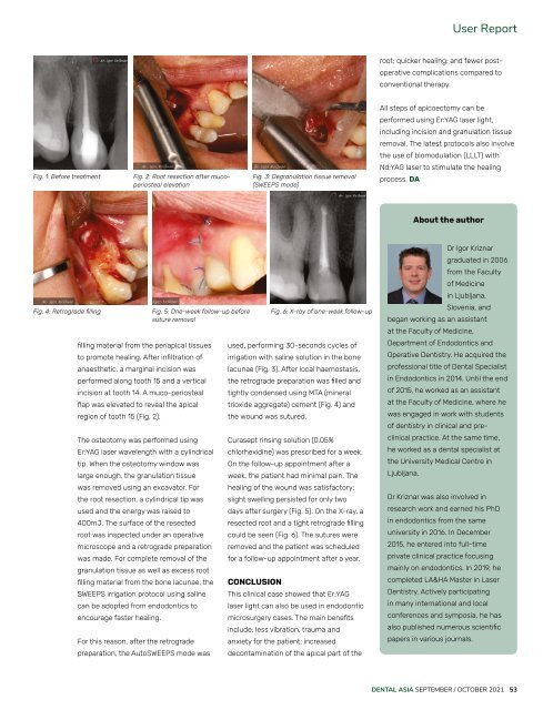
Fig. 1: Before treatment<br />
Fig. 2: Root resection after mucoperiosteal<br />
elevation<br />
Fig. 3: Degranulation tissue removal<br />
(SWEEPS mode)<br />
Nd:YAG laser to stimulate the healing<br />
process. DA<br />
About the author<br />
Fig. 4: Retrograde filling<br />
filling material from the periapical tissues<br />
to promote healing. After infiltration of<br />
anaesthetic, a marginal incision was<br />
performed along tooth 15 and a vertical<br />
incision at tooth 14. A muco-periosteal<br />
flap was elevated to reveal the apical<br />
region of tooth 15 (Fig. 2).<br />
The osteotomy was performed using<br />
Er:YAG laser wavelength with a cylindrical<br />
tip. When the osteotomy window was<br />
large enough, the granulation tissue<br />
was removed using an excavator. For<br />
the root resection, a cylindrical tip was<br />
used and the energy was raised to<br />
400mJ. The surface of the resected<br />
root was inspected under an operative<br />
microscope and a retrograde preparation<br />
was made. For complete removal of the<br />
granulation tissue as well as excess root<br />
filling material from the bone lacunae, the<br />
SWEEPS irrigation protocol using saline<br />
can be adopted from endodontics to<br />
encourage faster healing.<br />
For this reason, after the retrograde<br />
Fig. 5: One-week follow-up before<br />
suture removal<br />
preparation, the AutoSWEEPS mode was<br />
used, performing 30-seconds cycles of<br />
irrigation with saline solution in the bone<br />
lacunae (Fig. 3). After local haemostasis,<br />
the retrograde preparation was filled and<br />
tightly condensed using MTA (mineral<br />
trioxide aggregate) cement (Fig. 4) and<br />
the wound was sutured.<br />
Curasept rinsing solution (0.05%<br />
chlorhexidine) was prescribed for a week.<br />
On the follow-up appointment after a<br />
week, the patient had minimal pain. The<br />
healing of the wound was satisfactory;<br />
slight swelling persisted for only two<br />
days after surgery (Fig. 5). On the X-ray, a<br />
resected root and a tight retrograde filling<br />
could be seen (Fig. 6). The sutures were<br />
removed and the patient was scheduled<br />
for a follow-up appointment after a year.<br />
CONCLUSION<br />
Fig. 6: X-ray of one-week follow-up<br />
This clinical case showed that Er:YAG<br />
laser light can also be used in endodontic<br />
microsurgery cases. The main benefits<br />
include: less vibration, trauma and<br />
anxiety for the patient; increased<br />
decontamination of the apical part of the<br />
Dr Igor Kriznar<br />
graduated in 2006<br />
from the Faculty<br />
of Medicine<br />
in Ljubljana,<br />
Slovenia, and<br />
began working as an assistant<br />
at the Faculty of Medicine,<br />
Department of Endodontics and<br />
Operative Dentistry. He acquired the<br />
professional title of <strong>Dental</strong> Specialist<br />
in Endodontics in 2014. Until the end<br />
of 2015, he worked as an assistant<br />
at the Faculty of Medicine, where he<br />
was engaged in work with students<br />
of dentistry in clinical and preclinical<br />
practice. At the same time,<br />
he worked as a dental specialist at<br />
the University Medical Centre in<br />
Ljubljana.<br />
Dr Kriznar was also involved in<br />
research work and earned his PhD<br />
in endodontics from the same<br />
university in 2016. In December<br />
2015, he entered into full-time<br />
private clinical practice focusing<br />
mainly on endodontics. In 2019, he<br />
completed LA&HA Master in Laser<br />
Dentistry. Actively participating<br />
in many international and local<br />
conferences and symposia, he has<br />
also published numerous scientific<br />
papers in various journals.<br />
DENTAL ASIA SEPTEMBER / OCTOBER <strong>2021</strong> 53