Evidence for DNA Bending at the T7 RNA Polymerase Promoter
Evidence for DNA Bending at the T7 RNA Polymerase Promoter
Evidence for DNA Bending at the T7 RNA Polymerase Promoter

Create successful ePaper yourself
Turn your PDF publications into a flip-book with our unique Google optimized e-Paper software.
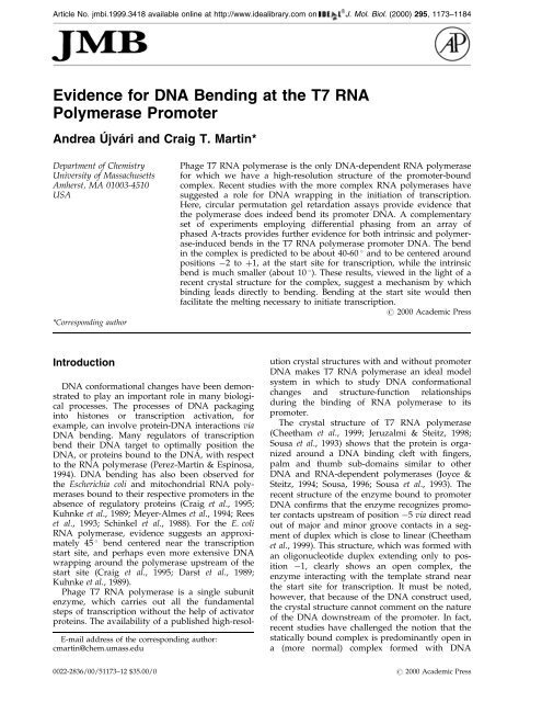
Article No. jmbi.1999.3418 available online <strong>at</strong> http://www.idealibrary.com on J. Mol. Biol. (2000) 295, 1173±1184<br />
<strong>Evidence</strong> <strong>for</strong> <strong>DNA</strong> <strong>Bending</strong> <strong>at</strong> <strong>the</strong> <strong>T7</strong> <strong>RNA</strong><br />
<strong>Polymerase</strong> <strong>Promoter</strong><br />
Andrea UÂ jvaÂri and Craig T. Martin*<br />
Department of Chemistry<br />
University of Massachusetts<br />
Amherst, MA 01003-4510<br />
USA<br />
*Corresponding author<br />
Introduction<br />
<strong>DNA</strong> con<strong>for</strong>m<strong>at</strong>ional changes have been demonstr<strong>at</strong>ed<br />
to play an important role in many biological<br />
processes. The processes of <strong>DNA</strong> packaging<br />
into histones or transcription activ<strong>at</strong>ion, <strong>for</strong><br />
example, can involve protein-<strong>DNA</strong> interactions via<br />
<strong>DNA</strong> bending. Many regul<strong>at</strong>ors of transcription<br />
bend <strong>the</strong>ir <strong>DNA</strong> target to optimally position <strong>the</strong><br />
<strong>DNA</strong>, or proteins bound to <strong>the</strong> <strong>DNA</strong>, with respect<br />
to <strong>the</strong> <strong>RNA</strong> polymerase (Perez-Martin & Espinosa,<br />
1994). <strong>DNA</strong> bending has also been observed <strong>for</strong><br />
<strong>the</strong> Escherichia coli and mitochondrial <strong>RNA</strong> polymerases<br />
bound to <strong>the</strong>ir respective promoters in <strong>the</strong><br />
absence of regul<strong>at</strong>ory proteins (Craig et al., 1995;<br />
Kuhnke et al., 1989; Meyer-Almes et al., 1994; Rees<br />
et al., 1993; Schinkel et al., 1988). For <strong>the</strong> E. coli<br />
<strong>RNA</strong> polymerase, evidence suggests an approxim<strong>at</strong>ely<br />
45 bend centered near <strong>the</strong> transcription<br />
start site, and perhaps even more extensive <strong>DNA</strong><br />
wrapping around <strong>the</strong> polymerase upstream of <strong>the</strong><br />
start site (Craig et al., 1995; Darst et al., 1989;<br />
Kuhnke et al., 1989).<br />
Phage <strong>T7</strong> <strong>RNA</strong> polymerase is a single subunit<br />
enzyme, which carries out all <strong>the</strong> fundamental<br />
steps of transcription without <strong>the</strong> help of activ<strong>at</strong>or<br />
proteins. The availability of a published high-resol-<br />
E-mail address of <strong>the</strong> corresponding author:<br />
cmartin@chem.umass.edu<br />
Phage <strong>T7</strong> <strong>RNA</strong> polymerase is <strong>the</strong> only <strong>DNA</strong>-dependent <strong>RNA</strong> polymerase<br />
<strong>for</strong> which we have a high-resolution structure of <strong>the</strong> promoter-bound<br />
complex. Recent studies with <strong>the</strong> more complex <strong>RNA</strong> polymerases have<br />
suggested a role <strong>for</strong> <strong>DNA</strong> wrapping in <strong>the</strong> initi<strong>at</strong>ion of transcription.<br />
Here, circular permut<strong>at</strong>ion gel retard<strong>at</strong>ion assays provide evidence th<strong>at</strong><br />
<strong>the</strong> polymerase does indeed bend its promoter <strong>DNA</strong>. A complementary<br />
set of experiments employing differential phasing from an array of<br />
phased A-tracts provides fur<strong>the</strong>r evidence <strong>for</strong> both intrinsic and polymerase-induced<br />
bends in <strong>the</strong> <strong>T7</strong> <strong>RNA</strong> polymerase promoter <strong>DNA</strong>. The bend<br />
in <strong>the</strong> complex is predicted to be about 40-60 and to be centered around<br />
positions 2to‡1, <strong>at</strong> <strong>the</strong> start site <strong>for</strong> transcription, while <strong>the</strong> intrinsic<br />
bend is much smaller (about 10 ). These results, viewed in <strong>the</strong> light of a<br />
recent crystal structure <strong>for</strong> <strong>the</strong> complex, suggest a mechanism by which<br />
binding leads directly to bending. <strong>Bending</strong> <strong>at</strong> <strong>the</strong> start site would <strong>the</strong>n<br />
facilit<strong>at</strong>e <strong>the</strong> melting necessary to initi<strong>at</strong>e transcription.<br />
# 2000 Academic Press<br />
ution crystal structures with and without promoter<br />
<strong>DNA</strong> makes <strong>T7</strong> <strong>RNA</strong> polymerase an ideal model<br />
system in which to study <strong>DNA</strong> con<strong>for</strong>m<strong>at</strong>ional<br />
changes and structure-function rel<strong>at</strong>ionships<br />
during <strong>the</strong> binding of <strong>RNA</strong> polymerase to its<br />
promoter.<br />
The crystal structure of <strong>T7</strong> <strong>RNA</strong> polymerase<br />
(Cheetham et al., 1999; Jeruzalmi & Steitz, 1998;<br />
Sousa et al., 1993) shows th<strong>at</strong> <strong>the</strong> protein is organized<br />
around a <strong>DNA</strong> binding cleft with ®ngers,<br />
palm and thumb sub-domains similar to o<strong>the</strong>r<br />
<strong>DNA</strong> and <strong>RNA</strong>-dependent polymerases (Joyce &<br />
Steitz, 1994; Sousa, 1996; Sousa et al., 1993). The<br />
recent structure of <strong>the</strong> enzyme bound to promoter<br />
<strong>DNA</strong> con®rms th<strong>at</strong> <strong>the</strong> enzyme recognizes promoter<br />
contacts upstream of position 5 via direct read<br />
out of major and minor groove contacts in a segment<br />
of duplex which is close to linear (Cheetham<br />
et al., 1999). This structure, which was <strong>for</strong>med with<br />
an oligonucleotide duplex extending only to position<br />
1, clearly shows an open complex, <strong>the</strong><br />
enzyme interacting with <strong>the</strong> templ<strong>at</strong>e strand near<br />
<strong>the</strong> start site <strong>for</strong> transcription. It must be noted,<br />
however, th<strong>at</strong> because of <strong>the</strong> <strong>DNA</strong> construct used,<br />
<strong>the</strong> crystal structure cannot comment on <strong>the</strong> n<strong>at</strong>ure<br />
of <strong>the</strong> <strong>DNA</strong> downstream of <strong>the</strong> promoter. In fact,<br />
recent studies have challenged <strong>the</strong> notion th<strong>at</strong> <strong>the</strong><br />
st<strong>at</strong>ically bound complex is predominantly open in<br />
a (more normal) complex <strong>for</strong>med with <strong>DNA</strong><br />
0022-2836/00/51173±12 $35.00/0 # 2000 Academic Press
1174 <strong>DNA</strong> <strong>Bending</strong> <strong>at</strong> <strong>the</strong> <strong>T7</strong> <strong>RNA</strong> <strong>Polymerase</strong> <strong>Promoter</strong><br />
extending downstream of <strong>the</strong> start site (Villemain<br />
et al., 1997).<br />
The crystal structure similarly provides no in<strong>for</strong>m<strong>at</strong>ion<br />
on <strong>the</strong> n<strong>at</strong>ure of <strong>the</strong> <strong>DNA</strong> upstream of <strong>the</strong><br />
promoter (upstream of position 17). Various<br />
models <strong>for</strong> promoter binding in more complex<br />
<strong>RNA</strong> polymerases have implic<strong>at</strong>ed a role <strong>for</strong> <strong>DNA</strong><br />
wrapping (around <strong>the</strong> polymerase) in <strong>the</strong> initi<strong>at</strong>ion<br />
of transcription (Coulombe & Burton, 1999; de<br />
Haseth & Helmann, 1995; Robert et al., 1998). The<br />
<strong>T7</strong> <strong>RNA</strong> polymerase consensus promoter is A ‡ Trich<br />
<strong>at</strong> its upstream end, and many phage promoters<br />
are A ‡ T-rich extending upstream of position<br />
17, suggesting a possible role <strong>for</strong> bending<br />
immedi<strong>at</strong>ely upstream of <strong>the</strong> promoter.<br />
Here, we have used gel electrophoretic mobility<br />
shift methods to present evidence not only <strong>for</strong> a<br />
40 -60 polymerase-induced bend in <strong>the</strong> <strong>DNA</strong>, but<br />
also <strong>for</strong> a smaller 9 intrinsic bend in <strong>the</strong> absence<br />
of polymerase. Contrary to simple expect<strong>at</strong>ions<br />
from some of <strong>the</strong> above models, <strong>the</strong> d<strong>at</strong>a suggest<br />
th<strong>at</strong> <strong>the</strong> polymerase-induced bend is not localized<br />
upstream of <strong>the</strong> promoter, but is instead loc<strong>at</strong>ed<br />
closer to <strong>the</strong> start site. To probe <strong>the</strong>se possibilities,<br />
mut<strong>at</strong>ions were introduced into <strong>the</strong> promoter to<br />
look <strong>for</strong> sequence effects on bending.<br />
Results<br />
Here, we have used two electrophoretic assays<br />
to provide evidence <strong>for</strong> bending of <strong>the</strong> <strong>T7</strong> <strong>RNA</strong><br />
polymerase promoter induced by <strong>the</strong> st<strong>at</strong>ically<br />
bound <strong>RNA</strong> polymerase. The circular permut<strong>at</strong>ion<br />
assay relies on position-dependent vari<strong>at</strong>ions in<br />
electrophoretic mobility of protein-<strong>DNA</strong> complexes.<br />
Bends loc<strong>at</strong>ed in <strong>the</strong> middle of a fragment<br />
perturb <strong>the</strong> observed mobility more than bends<br />
placed <strong>at</strong> <strong>the</strong> ends of identically sized fragments.<br />
The phasing assay places <strong>the</strong> target binding site <strong>at</strong><br />
varying distances from an intrinsic bend in <strong>the</strong><br />
<strong>DNA</strong>. A bend in <strong>the</strong> target can ei<strong>the</strong>r add to or<br />
subtract from <strong>the</strong> effects of <strong>the</strong> intrinsic bend. Position<br />
or phase-dependent vari<strong>at</strong>ions in mobility are<br />
most readily interpreted in terms of a proteininduced<br />
bend <strong>at</strong> <strong>the</strong> target site.<br />
Circular permut<strong>at</strong>ion assay<br />
The circular permut<strong>at</strong>ion assay was used initially<br />
to determine whe<strong>the</strong>r <strong>T7</strong> <strong>RNA</strong> polymerase bends<br />
its promoter, as has been demonstr<strong>at</strong>ed <strong>for</strong> <strong>the</strong><br />
E. coli and mitochondrial <strong>RNA</strong> polymerases<br />
(Kuhnke et al., 1989; Schinkel et al., 1988). An oligonucleotide<br />
representing <strong>the</strong> consensus promoter<br />
sequence (construct 1 in Figure 1) was cloned into<br />
<strong>the</strong> permut<strong>at</strong>ion vector pSL6 (Gaal et al., 1994).<br />
<strong>DNA</strong> probes of <strong>the</strong> same length, with <strong>the</strong> promoter<br />
<strong>at</strong> different positions, were gener<strong>at</strong>ed as described<br />
in M<strong>at</strong>erials and Methods. During <strong>the</strong> PCR ampli®c<strong>at</strong>ion,<br />
<strong>the</strong> <strong>DNA</strong> was uni<strong>for</strong>mly labeled via <strong>the</strong><br />
incorpor<strong>at</strong>ion of [a-32P]dATP. The PCR ampli®ed<br />
and Qiagen column-puri®ed <strong>DNA</strong> was <strong>the</strong>n<br />
digested with <strong>the</strong> indic<strong>at</strong>ed restriction enzyme,<br />
desalted on a Qiagen column, and used directly.<br />
Consequently, <strong>the</strong> circular permut<strong>at</strong>ion gels also<br />
show shorter <strong>DNA</strong> fragments arising from <strong>the</strong><br />
restriction enzyme-cleaved ends, which do not<br />
carry <strong>the</strong> promoter. Initially, digests with only four<br />
restriction endonucleases were analyzed. Two of<br />
<strong>the</strong> resulting fragments place <strong>the</strong> promoter near<br />
<strong>the</strong> center of <strong>the</strong> <strong>DNA</strong>, while two place <strong>the</strong> promoter<br />
<strong>at</strong> opposite ends of <strong>the</strong> fragment.<br />
The results shown in Figure 2 reveal th<strong>at</strong> free<br />
promoter-containing <strong>DNA</strong> does not display detectable<br />
mobility differences in this assay (but see<br />
below). In contrast, polymerase-promoter complexes<br />
carrying <strong>the</strong> promoter <strong>at</strong> <strong>the</strong> ends migr<strong>at</strong>e<br />
faster than ones with <strong>the</strong> promoter <strong>at</strong> <strong>the</strong> center.<br />
This position-sensitive variability in mobility is<br />
generally interpreted to arise from increased <strong>DNA</strong><br />
bending or ¯exibility (Wu & Cro<strong>the</strong>rs, 1984; Zinkel<br />
& Cro<strong>the</strong>rs, 1990). Kerppola & Curran (1991) have<br />
Figure 1. <strong>Promoter</strong> constructs used in <strong>the</strong> bending experiments. Construct 1 contains <strong>the</strong> conserved <strong>T7</strong> promoter<br />
sequence from position 17 to position 1 and <strong>the</strong> semi-conserved A ‡ T-rich sequence from position 21 to position<br />
18. The sequences of <strong>the</strong> second, third and fourth promoters have been modi®ed to study <strong>the</strong> effect of speci®c substitutions<br />
on bending. Modi®c<strong>at</strong>ions are indic<strong>at</strong>ed in bold letters.
<strong>DNA</strong> <strong>Bending</strong> <strong>at</strong> <strong>the</strong> <strong>T7</strong> <strong>RNA</strong> <strong>Polymerase</strong> <strong>Promoter</strong> 1175<br />
Figure 2. (a) and (b) Circular permut<strong>at</strong>ion assay with <strong>the</strong> n<strong>at</strong>ive promoter (construct 1) and (c) and (d) with <strong>the</strong><br />
promoter th<strong>at</strong> is G ‡ C-rich upstream of position 17 (construct 2). (a), (c) Comparison of <strong>the</strong> mobilities of polymerase-promoter<br />
complexes to mobilities of A-tract standards (2A through 5A, containing two to ®ve A-tract repe<strong>at</strong>s)<br />
with different bending angles. For <strong>the</strong> <strong>DNA</strong> constructs containing <strong>the</strong> <strong>T7</strong> <strong>RNA</strong> polymerase promoter, 163 bp long<br />
<strong>DNA</strong> fragments were gener<strong>at</strong>ed by restriction digestion with different endonucleases, as described in M<strong>at</strong>erials and<br />
Methods. The distances (in base-pairs) of position 6 from <strong>the</strong> end of <strong>the</strong> <strong>DNA</strong> are indic<strong>at</strong>ed on <strong>the</strong> top of <strong>the</strong> lanes.<br />
This distance is indic<strong>at</strong>ed by a broken line in <strong>the</strong> cartoon <strong>at</strong> <strong>the</strong> top of <strong>the</strong> Figure, with <strong>the</strong> promoter indic<strong>at</strong>ed by an<br />
open rectangle, and <strong>the</strong> direction of transcription from position ‡1 shown by an arrow. For <strong>the</strong> polymerase complexes,<br />
approxim<strong>at</strong>ely 5 nM <strong>DNA</strong> was incub<strong>at</strong>ed with 75 nM <strong>T7</strong> <strong>RNA</strong> polymerase, in <strong>the</strong> presence of 2 mg/ml salmon<br />
sperm <strong>DNA</strong> (®nal concentr<strong>at</strong>ions) to prevent non-speci®c binding. The letters f and c indic<strong>at</strong>e <strong>the</strong> positions of<br />
<strong>the</strong> free <strong>DNA</strong> and <strong>the</strong> <strong>T7</strong> <strong>RNA</strong> polymerase-promoter complex, respectively. (b), (d) Rel<strong>at</strong>ive mobilities of free (open<br />
circles) and bound <strong>DNA</strong> (®lled circles) were plotted versus <strong>the</strong> distance in base-pairs of position 6 from <strong>the</strong> end of<br />
<strong>the</strong> <strong>DNA</strong> fragment. D<strong>at</strong>a <strong>for</strong> <strong>the</strong> complex were ®t to equ<strong>at</strong>ion (2). The loc<strong>at</strong>ion of <strong>the</strong> bend in <strong>the</strong> bound complex was<br />
estim<strong>at</strong>ed to be position ‡1 (distance ˆ 75) <strong>for</strong> both promoter constructs, and <strong>the</strong> bend angle is expected to be<br />
approxim<strong>at</strong>ely 39 and 37 <strong>for</strong> constructs 1 and 2, respectively.<br />
pointed out th<strong>at</strong> structures o<strong>the</strong>r than a st<strong>at</strong>ic <strong>DNA</strong><br />
bend may lead to similar alter<strong>at</strong>ions in <strong>the</strong> electrophoretic<br />
mobilities, and <strong>the</strong>re<strong>for</strong>e propose <strong>the</strong> use<br />
of <strong>the</strong> term <strong>DNA</strong> ¯exure (intended to remain<br />
ambiguous) to refer to <strong>the</strong> source of <strong>the</strong> anomalous<br />
gel mobilities.
1176 <strong>DNA</strong> <strong>Bending</strong> <strong>at</strong> <strong>the</strong> <strong>T7</strong> <strong>RNA</strong> <strong>Polymerase</strong> <strong>Promoter</strong><br />
Complexes containing fragments with <strong>the</strong> promoter<br />
loc<strong>at</strong>ed symmetrically <strong>at</strong> opposite ends run<br />
differently from each o<strong>the</strong>r, suggesting th<strong>at</strong> <strong>the</strong><br />
<strong>DNA</strong> ¯exure is not loc<strong>at</strong>ed symmetrically within<br />
<strong>the</strong> promoter. To estim<strong>at</strong>e <strong>the</strong> magnitude and <strong>the</strong><br />
loc<strong>at</strong>ion of <strong>the</strong> ¯exure, <strong>the</strong> normalized mobilities of<br />
<strong>the</strong> <strong>T7</strong> <strong>RNA</strong> polymerase-complexes were ®t to<br />
equ<strong>at</strong>ion (2). Here, promoter position 6 was arbitrarily<br />
chosen as a reference point, and x is <strong>the</strong><br />
distance from position 6 to <strong>the</strong> center of <strong>the</strong><br />
bend. Fitting <strong>the</strong> d<strong>at</strong>a to equ<strong>at</strong>ions (2) and (3), <strong>the</strong><br />
center of <strong>the</strong> bend is preliminarily estim<strong>at</strong>ed to lie<br />
near position ‡1, and <strong>the</strong> magnitude of <strong>the</strong> bend is<br />
estim<strong>at</strong>ed to be 39 . For <strong>the</strong> l<strong>at</strong>ter analysis, running<br />
A-tract standards with known bending angles<br />
on <strong>the</strong> same gels as <strong>the</strong> polymerase-promoter<br />
complexes provided an internal calibr<strong>at</strong>ion of <strong>the</strong><br />
¯exure angles.<br />
The ®ts in Figure 2(b) and (d) have rel<strong>at</strong>ively<br />
few d<strong>at</strong>a points. To con®rm <strong>the</strong> interpret<strong>at</strong>ion th<strong>at</strong><br />
bending localizes near <strong>the</strong> start site and not<br />
upstream of <strong>the</strong> promotor, circular permut<strong>at</strong>ion<br />
assays of <strong>the</strong> n<strong>at</strong>ive promoter digested with ten<br />
different endonucleases are shown in Figure 3<br />
(<strong>DNA</strong> standards were interspersed on <strong>the</strong> same gel<br />
to ensure th<strong>at</strong> <strong>the</strong> obtained migr<strong>at</strong>ion differences<br />
are not due to temper<strong>at</strong>ure or voltage grad<strong>at</strong>ions<br />
in <strong>the</strong> gel). Fitting of this larger d<strong>at</strong>a set, as shown<br />
in Figure 3(b), places position 2( 3) as <strong>the</strong> predicted<br />
loc<strong>at</strong>ion of <strong>the</strong> ¯exure. This result argues<br />
strongly th<strong>at</strong> <strong>the</strong> observed <strong>DNA</strong> ¯exure is not<br />
loc<strong>at</strong>ed in <strong>the</strong> upstream part of <strong>the</strong> promoter, but<br />
is loc<strong>at</strong>ed <strong>at</strong> <strong>the</strong> downstream end of <strong>the</strong> promoter,<br />
near <strong>the</strong> start site.<br />
The apparent localiz<strong>at</strong>ion of bending to near <strong>the</strong><br />
start site does not preclude additional bending of<br />
Figure 3. Circular permut<strong>at</strong>ion<br />
assay with <strong>the</strong> n<strong>at</strong>ive promoter<br />
(construct 1) digested with ten<br />
different restriction enzymes. (a)<br />
Comparison of <strong>the</strong> mobilities of<br />
polymerase-promoter complexes to<br />
<strong>DNA</strong> sizing standards (lanes indic<strong>at</strong>ed<br />
by <strong>the</strong> letter S) ensures th<strong>at</strong><br />
<strong>the</strong> obtained migr<strong>at</strong>ion differences<br />
are not due to ``smiling'' of <strong>the</strong> gel.<br />
For <strong>the</strong> <strong>DNA</strong> fragments containing<br />
<strong>the</strong> promoter, <strong>the</strong> distances (in<br />
base-pairs) of position 6 from <strong>the</strong><br />
end of <strong>the</strong> <strong>DNA</strong> are indic<strong>at</strong>ed on<br />
<strong>the</strong> top of <strong>the</strong> lanes. Conditions<br />
were as in Figure 2. (b) Rel<strong>at</strong>ive<br />
mobilities of free and bound <strong>DNA</strong><br />
were plotted as <strong>the</strong> distance of position<br />
6 from <strong>the</strong> end of <strong>the</strong> <strong>DNA</strong><br />
fragment. D<strong>at</strong>a <strong>for</strong> <strong>the</strong> complex<br />
were ®t to equ<strong>at</strong>ion (2). The effective<br />
center of <strong>the</strong> bend was determined<br />
to be position 2<br />
(distance ˆ 78( 3).<br />
<strong>the</strong> promoter <strong>at</strong> o<strong>the</strong>r loc<strong>at</strong>ions (although <strong>the</strong> determin<strong>at</strong>ion<br />
of <strong>the</strong> bending locus would be complic<strong>at</strong>ed<br />
by <strong>the</strong> presence of more than one bend in <strong>the</strong><br />
<strong>DNA</strong>). As summarized in <strong>the</strong> introduction, <strong>the</strong>re<br />
was some reason to expect a bend in <strong>the</strong> region<br />
upstream of <strong>the</strong> start site. Both <strong>the</strong> consensus promoter<br />
sequence from position 13 to 17 and <strong>the</strong><br />
less conserved region of <strong>the</strong> bacteriophage <strong>DNA</strong><br />
upstream of position 17 are characteristically<br />
A‡T-rich. In order to test <strong>for</strong> <strong>the</strong> possibility of<br />
bending in this region which might require <strong>the</strong> presence<br />
of an A‡T-rich sequence, A T base-pairs<br />
were replaced by G C base-pairs upstream of position<br />
17 in promoter construct 2 (retaining <strong>the</strong><br />
full consensus promoter sequence), and upstream<br />
of position 13 in promoter construct 3 (altering<br />
<strong>the</strong> consensus sequence from position 14 to 17).<br />
The complex <strong>for</strong>med with <strong>the</strong> <strong>for</strong>mer construct<br />
shows an electrophoretic pro®le indistinguishable<br />
(predicting a 37 bend) from th<strong>at</strong> of <strong>the</strong> n<strong>at</strong>ive promoter<br />
(and has wild-type activity in in vitro assays<br />
of transcription; d<strong>at</strong>a not shown), indic<strong>at</strong>ing th<strong>at</strong><br />
<strong>the</strong> A ‡ T-rich region beyond <strong>the</strong> promoter is not<br />
required <strong>for</strong> <strong>the</strong> observed bending.<br />
In contrast, we were not able to obtain a gel shift<br />
using <strong>the</strong> third promoter construct (G ‡ C-rich<br />
upstream of position 13), presumably due to<br />
weaker polymerase binding to this sequence or to<br />
dissoci<strong>at</strong>ion of <strong>the</strong> complex during electrophoresis.<br />
This construct also shows a tenfold reduction in<br />
<strong>the</strong> steady-st<strong>at</strong>e syn<strong>the</strong>sis of run-off 9mer <strong>RNA</strong><br />
(d<strong>at</strong>a not shown). Consequently, although this<br />
region of <strong>the</strong> <strong>DNA</strong> is essential to binding, we can<br />
reach no conclusions regarding <strong>the</strong> requirement of<br />
this region <strong>for</strong> <strong>the</strong> observed bending.
<strong>DNA</strong> <strong>Bending</strong> <strong>at</strong> <strong>the</strong> <strong>T7</strong> <strong>RNA</strong> <strong>Polymerase</strong> <strong>Promoter</strong> 1177<br />
Phasing analysis<br />
<strong>Polymerase</strong>-promoter complex<br />
Phasing analysis is believed to be a better test of<br />
st<strong>at</strong>ic <strong>DNA</strong> bending (Ansari et al., 1995; Kerppola<br />
& Curran, 1997; Salvo & Grindley, 1987) and so<br />
was used here as a complement to <strong>the</strong> circular permut<strong>at</strong>ion<br />
assay. Phasing plasmids were constructed<br />
in which <strong>the</strong> promoter sequence is<br />
separ<strong>at</strong>ed by a variable length linker from a stretch<br />
of three phased A-tracts. An oligonucleotide carrying<br />
<strong>the</strong> consensus promoter sequence was inserted<br />
into plasmids pTK401-26 and pTK401-28 as<br />
described in <strong>the</strong> M<strong>at</strong>erials and Methods and<br />
shown in Figure 4. The complex <strong>for</strong>med shows a<br />
very clear phase-dependent vari<strong>at</strong>ion in its mobility,<br />
as shown in Figure 5(a), consistent with <strong>the</strong><br />
above permut<strong>at</strong>ion results. The rel<strong>at</strong>ive mobilities<br />
were ®t to equ<strong>at</strong>ion (4) (see Figure 5(b)) to obtain<br />
<strong>the</strong> phasing amplitude (APH). Fitting <strong>the</strong> d<strong>at</strong>a to<br />
equ<strong>at</strong>ion (5), <strong>the</strong> angle of <strong>the</strong> polymerase-induced<br />
bend is estim<strong>at</strong>ed to be 63 .<br />
The permut<strong>at</strong>ion analysis predicts a bend centered<br />
near <strong>the</strong> start site. To assess whe<strong>the</strong>r such<br />
bending requires <strong>the</strong> characteristic TATA<br />
sequence from position 4 to 1, a fur<strong>the</strong>r construct<br />
(construct 4) was prepared in which <strong>the</strong><br />
A T and T A base-pairs <strong>at</strong> positions 3 and 2<br />
have been replaced by G C and C G base-pairs,<br />
respectively. These substitutions are designed to<br />
change <strong>the</strong> TATA sequence immedi<strong>at</strong>ely<br />
upstream of <strong>the</strong> promoter start site to <strong>the</strong><br />
sequence TGCA, which is predicted to have a 3-<br />
4 kcal/mol higher energetic barrier to melting<br />
(Breslauer et al., 1986; SantaLucia et al., 1996;<br />
Sugimoto et al., 1996). In run-off transcription of<br />
a nine base transcript, while initi<strong>at</strong>ion occurs<br />
with speci®city <strong>for</strong> this construct, activity is<br />
down threefold (d<strong>at</strong>a not shown). Qualit<strong>at</strong>ive gel<br />
shift binding assays were carried out to ask<br />
whe<strong>the</strong>r <strong>the</strong> decreased transcription r<strong>at</strong>e is due<br />
to weakened binding. The amounts of polymerase-promoter<br />
complexes were quanti®ed as a<br />
function of increasing enzyme concentr<strong>at</strong>ion and<br />
<strong>the</strong> results show th<strong>at</strong> <strong>the</strong> af®nity of <strong>the</strong> promoter<br />
with a TATA to TGCA substitution (construct 4)<br />
<strong>for</strong> <strong>T7</strong> <strong>RNA</strong> polymerase appears to be on <strong>the</strong><br />
same order of magnitude as th<strong>at</strong> of <strong>the</strong> n<strong>at</strong>ive<br />
promoter (although this assay is not well-suited<br />
<strong>for</strong> accur<strong>at</strong>e measurements in <strong>the</strong> <strong>T7</strong> system, it<br />
should be able to distinguish large decreases in<br />
binding af®nity).<br />
Phasing analysis was carried out <strong>for</strong> <strong>the</strong> promoter<br />
containing <strong>the</strong> TATA to TGCA substitution, as<br />
shown in Figure 5. Large phase-dependent mobility<br />
changes are observed, and were ®t to equ<strong>at</strong>ion<br />
(4) as described above. Using equ<strong>at</strong>ion (5) <strong>the</strong><br />
angle of <strong>the</strong> bend was estim<strong>at</strong>ed to be 60 , comparable<br />
to th<strong>at</strong> <strong>for</strong> <strong>the</strong> consensus promoter.<br />
Intrinsic bending in <strong>the</strong> promoter<br />
Phasing analysis is more sensitive than <strong>the</strong> permut<strong>at</strong>ion<br />
assay, and was also able to detect intrinsic<br />
<strong>DNA</strong> bending in <strong>the</strong> absence of polymerase.<br />
Intrinsic bending angles of 9 and 10 were calcul<strong>at</strong>ed<br />
<strong>for</strong> promoters 1 and 4, respectively. To compare<br />
intrinsic <strong>DNA</strong> bending <strong>for</strong> promoters 1, 3 and<br />
4, phasing analyses were run on <strong>the</strong> free <strong>DNA</strong><br />
probes as shown in Figure 6. To ensure th<strong>at</strong> <strong>the</strong><br />
salt conditions in <strong>the</strong> gel do not effect <strong>the</strong><br />
migr<strong>at</strong>ion p<strong>at</strong>tern of <strong>the</strong> free probes, in this case<br />
TBE was used instead of <strong>the</strong> Tris-glycine gel running<br />
buffer. <strong>Bending</strong> angles of 8 and 11 were<br />
observed <strong>for</strong> <strong>the</strong> n<strong>at</strong>ive promoter 1 and <strong>for</strong> promoter<br />
4, respectively. These numbers are similar to<br />
<strong>the</strong> values determined previously with <strong>the</strong> Tris-glycine<br />
buffer, and indic<strong>at</strong>e th<strong>at</strong> <strong>the</strong> measured bending<br />
angles do not vary signi®cantly with gel<br />
conditions. Interestingly, <strong>the</strong> promoter which is<br />
G ‡ C-rich upstream of position 13 shows smaller<br />
amplitude vari<strong>at</strong>ions, and although <strong>the</strong> ®t was<br />
not as good as <strong>for</strong> <strong>the</strong> o<strong>the</strong>r constructs, <strong>the</strong> d<strong>at</strong>a are<br />
consistent with a bend of less than 4 <strong>for</strong> this<br />
sequence.<br />
Comparison of <strong>the</strong> assays<br />
Both gel mobility shift methods used in this<br />
work have advantages and disadvantages. It has<br />
been argued th<strong>at</strong> <strong>the</strong> circular permut<strong>at</strong>ion assay<br />
may be misinterpreted if <strong>the</strong> structure of <strong>the</strong><br />
protein itself has position-dependent effects on<br />
<strong>the</strong> migr<strong>at</strong>ion of <strong>the</strong> complex (Ansari et al., 1995;<br />
Kerppola & Curran, 1997). Indeed, mobility<br />
differences were observed <strong>for</strong> <strong>the</strong> Hg-MerR-<strong>DNA</strong><br />
complex in circular permut<strong>at</strong>ion assays, but not<br />
Figure 4. Design of probes <strong>for</strong> phasing analysis. The distance of position 6 (within <strong>the</strong> promoter) from <strong>the</strong> center<br />
of <strong>the</strong> A-tract was varied from 30 to 40 base-pairs, in two base-pair increments. The sequence of <strong>the</strong> non-templ<strong>at</strong>e<br />
strand <strong>for</strong> <strong>the</strong> inserts is indic<strong>at</strong>ed.
1178 <strong>DNA</strong> <strong>Bending</strong> <strong>at</strong> <strong>the</strong> <strong>T7</strong> <strong>RNA</strong> <strong>Polymerase</strong> <strong>Promoter</strong><br />
Figure 5. (a) and (b) Phasing analysis with <strong>the</strong> n<strong>at</strong>ive promoter (construct 1) and (c) and (d) with <strong>the</strong> promoter th<strong>at</strong><br />
contains a TATA to TGCA substitution (construct 4). (a), (c) Comparison of <strong>the</strong> mobilities of polymerase-promoter<br />
complexes to mobilities of A-tract standards (2A through 5A) containing different bending angles. For <strong>the</strong> <strong>DNA</strong><br />
constructs containing <strong>the</strong> <strong>T7</strong> <strong>RNA</strong> polymerase promoter, <strong>the</strong> distances (in base-pairs) of position 6 from <strong>the</strong> middle<br />
of <strong>the</strong> A-tract are indic<strong>at</strong>ed on <strong>the</strong> top of <strong>the</strong> lanes. For <strong>the</strong> polymerase complexes, approxim<strong>at</strong>ely 10 nM <strong>DNA</strong> was<br />
incub<strong>at</strong>ed with 40 nM <strong>T7</strong> <strong>RNA</strong> polymerase in <strong>the</strong> presence of 2 mg/ml salmon sperm <strong>DNA</strong> (®nal concentr<strong>at</strong>ions) to<br />
prevent non-speci®c binding. The letters f and c indic<strong>at</strong>e <strong>the</strong> positions of <strong>the</strong> free <strong>DNA</strong> and <strong>the</strong> <strong>T7</strong> <strong>RNA</strong> polymerasepromoter<br />
complex, respectively. (b), (d) Normalized mobilities of <strong>the</strong> free <strong>DNA</strong> (open circles) and <strong>the</strong> polymerase-<br />
<strong>DNA</strong> complex (®lled circles) were plotted versus <strong>the</strong> distance of position 6 from <strong>the</strong> middle of <strong>the</strong> A-tract. The<br />
normalized mobilities were <strong>the</strong>n ®t to <strong>the</strong> phasing function as described in <strong>the</strong> text. For both promoter constructs <strong>the</strong><br />
separ<strong>at</strong>ion th<strong>at</strong> results in maximum mobility was found to be 32 and 34 bp <strong>for</strong> <strong>the</strong> bound and unbound <strong>DNA</strong>,<br />
respectively. The intrinsic and polymerase induced <strong>DNA</strong> bend angles are expected to be 9 and 10 , and 63 and<br />
60 <strong>for</strong> constructs 1 and 4, respectively.<br />
in phasing analyses, and <strong>the</strong> authors concluded<br />
th<strong>at</strong> <strong>the</strong> <strong>DNA</strong> is not bent (Ansari et al., 1995;<br />
Ansari et al., 1992). The cause of mobility differences<br />
observed in phasing analysis has also been<br />
disputed <strong>for</strong> proteins with elong<strong>at</strong>ed structures.<br />
Although <strong>the</strong> mobility differences measured in<br />
phasing analyses argue <strong>for</strong> <strong>DNA</strong> bending <strong>for</strong> <strong>the</strong><br />
protein-<strong>DNA</strong> complexes of <strong>the</strong> proteins Fos and<br />
Jun, no evidence <strong>for</strong> <strong>DNA</strong> curv<strong>at</strong>ure in this system<br />
was found in a minicircle competition binding<br />
assay (Kerppola, 1997; Sitlani & Cro<strong>the</strong>rs,<br />
1998).<br />
The shape of <strong>the</strong> <strong>T7</strong> <strong>RNA</strong> polymerase molecule<br />
is rel<strong>at</strong>ively globular, so th<strong>at</strong> we do not<br />
expect <strong>the</strong> position-dependent gel mobilities of<br />
<strong>the</strong> complex to be domin<strong>at</strong>ed by <strong>the</strong> protein<br />
structure and shape, but mostly by <strong>the</strong> con<strong>for</strong>m<strong>at</strong>ion<br />
of <strong>the</strong> 163-360 base-pair <strong>DNA</strong>. By com-<br />
parison with <strong>the</strong> A-tract standards, <strong>the</strong> <strong>DNA</strong><br />
bending angle <strong>for</strong> <strong>the</strong> n<strong>at</strong>ive <strong>T7</strong> <strong>RNA</strong> polymerase<br />
promoter complex was estim<strong>at</strong>ed to be 39<br />
and 63 , from <strong>the</strong> circular permut<strong>at</strong>ion and<br />
phasing analyses, respectively. Since <strong>the</strong> circular<br />
permut<strong>at</strong>ion assay yields smaller mobility differences<br />
(15 % overall) than does <strong>the</strong> more sensitive<br />
phasing assay ( 70 %), we expect th<strong>at</strong> <strong>the</strong> actual<br />
value of <strong>the</strong> bend angle is closer to 60 .<br />
Discussion<br />
Two different approaches have been used to<br />
assess <strong>the</strong> characteristics of a potential bend in <strong>the</strong><br />
st<strong>at</strong>ic complex between <strong>T7</strong> <strong>RNA</strong> polymerase and<br />
its promoter. The circular permut<strong>at</strong>ion assay is<br />
well suited to predict <strong>the</strong> loc<strong>at</strong>ion of a bend in <strong>the</strong><br />
<strong>DNA</strong>, while <strong>the</strong> phasing analysis can provide
<strong>DNA</strong> <strong>Bending</strong> <strong>at</strong> <strong>the</strong> <strong>T7</strong> <strong>RNA</strong> <strong>Polymerase</strong> <strong>Promoter</strong> 1179<br />
Figure 6. Comparison of intrinsic bending <strong>for</strong> n<strong>at</strong>ive<br />
and modi®ed promoters, in <strong>the</strong> absence of <strong>RNA</strong> polymerase.<br />
(a) Phasing analyses with promoter constructs 1<br />
(n<strong>at</strong>ive, ®lled circles), 3 (G ‡ C-rich upstream of position<br />
13, crosses) and 4 (TATA to TGCA, open circles).<br />
(b) Normalized mobilities were plotted versus <strong>the</strong> distances<br />
(in base-pairs) of position 6 from <strong>the</strong> middle of<br />
<strong>the</strong> A-tract and were ®t to <strong>the</strong> phasing function as<br />
described in <strong>the</strong> text. The intrinsic <strong>DNA</strong> bend angles are<br />
expected to be 8 ,
1180 <strong>DNA</strong> <strong>Bending</strong> <strong>at</strong> <strong>the</strong> <strong>T7</strong> <strong>RNA</strong> <strong>Polymerase</strong> <strong>Promoter</strong><br />
drives <strong>the</strong> complex towards <strong>the</strong> open <strong>for</strong>m. These<br />
conclusions are derived from <strong>the</strong> ®tting of steadyst<strong>at</strong>e<br />
kinetic d<strong>at</strong>a as a function of <strong>the</strong> concentr<strong>at</strong>ion<br />
of GTP. However, product (pppGpG) re-associ<strong>at</strong>ion<br />
with <strong>the</strong> polymerase-promoter complex was<br />
not included in <strong>the</strong> equ<strong>at</strong>ions leading to this kinetic<br />
model (Villemain et al., 1997), and <strong>the</strong> results of Jia<br />
& P<strong>at</strong>el (1997) have demonstr<strong>at</strong>ed th<strong>at</strong> this reaction<br />
cannot be neglected. By observing <strong>the</strong> topological<br />
unwinding of <strong>the</strong> <strong>DNA</strong> in a topoisomerase assay,<br />
Villemain et al. (1997) also found th<strong>at</strong> open complex<br />
<strong>for</strong>m<strong>at</strong>ion on plasmids could be observed<br />
only in <strong>the</strong> presence of <strong>the</strong> initi<strong>at</strong>ing nucleotide<br />
(GTP). Basu & Maitra (1986) and Place et al. (1999)<br />
have reported <strong>the</strong>ir inability to footprint <strong>the</strong> polymerase-promoter<br />
complex in <strong>the</strong> absence of GTP.<br />
This most likely arises from <strong>the</strong> reasonably strong<br />
af®nity of <strong>T7</strong> <strong>RNA</strong> polymerase <strong>for</strong> non-promoter<br />
<strong>DNA</strong>, which predicts rel<strong>at</strong>ively low promoter<br />
occupancy in <strong>the</strong> presence of a large excess of nonpromoter<br />
<strong>DNA</strong>. Indeed, in our bending assays,<br />
which place <strong>the</strong> 22 bp promoter within a 160-<br />
360 bp fragment, apparent occupancy can be as<br />
low as 5 % (under conditions which would predict<br />
>90 % occupancy in <strong>the</strong> absence of competition).<br />
Thus, <strong>the</strong> lack of observed topological unwinding<br />
in <strong>the</strong> absence of GTP might simply arise from low<br />
promoter occupancy (<strong>the</strong> subsequent addition of<br />
GTP would drive more stable complex <strong>for</strong>m<strong>at</strong>ion<br />
and increase promoter occupancy).<br />
A complex between <strong>T7</strong> <strong>RNA</strong> polymerase and a<br />
short double-stranded <strong>DNA</strong> fragment corresponding<br />
to <strong>the</strong> <strong>T7</strong> promoter sequence from position 17<br />
to 1 has recently been crystallized with <strong>T7</strong> <strong>RNA</strong><br />
polymerase (Cheetham et al., 1999). The structure,<br />
shown in Figure 7, reveals only a very slight distortion<br />
in <strong>the</strong> upstream duplex promoter region<br />
centered near position 10. However, <strong>the</strong> strands<br />
are separ<strong>at</strong>ed downstream of position 5, and<br />
while <strong>the</strong> non-templ<strong>at</strong>e strand cannot be resolved<br />
downstream of this position, <strong>the</strong> templ<strong>at</strong>e strand<br />
devi<strong>at</strong>es substantially from a B-<strong>for</strong>m p<strong>at</strong>h. This<br />
suggests th<strong>at</strong> <strong>the</strong> <strong>DNA</strong> is melted in <strong>the</strong> st<strong>at</strong>ic complex<br />
and would almost certainly lead to an overall<br />
bend in <strong>the</strong> <strong>DNA</strong> centered near <strong>the</strong> transcription<br />
start site. <strong>DNA</strong> bending by <strong>the</strong> rel<strong>at</strong>ed phage SP6<br />
<strong>RNA</strong> polymerase has also recently been observed<br />
by electron microscopy (during <strong>the</strong> elong<strong>at</strong>ion<br />
phase of transcription (Bednar et al., 1999)).<br />
This observ<strong>at</strong>ion, combined with our bending<br />
results, suggests th<strong>at</strong> <strong>the</strong> polymerase bends <strong>DNA</strong><br />
not only upon initial binding, but also throughout<br />
transcription.<br />
Potential rel<strong>at</strong>ionships between <strong>DNA</strong> bending,<br />
binding and open complex <strong>for</strong>m<strong>at</strong>ion<br />
During open complex <strong>for</strong>m<strong>at</strong>ion, different con<strong>for</strong>m<strong>at</strong>ional<br />
changes of <strong>the</strong> promoter have been<br />
proposed to take place in <strong>the</strong> E. coli <strong>RNA</strong> polymerase<br />
system. According to <strong>the</strong> ``twisting-and-opening''<br />
model, binding of <strong>the</strong> polymerase to <strong>the</strong> 35<br />
and 10 elements with a slight twist could underwind<br />
<strong>the</strong> intervening <strong>DNA</strong>, which would lower<br />
<strong>the</strong> energetic barrier to base-pair opening<br />
(deHaseth & Helmann, 1995). Altern<strong>at</strong>ively (or in<br />
addition), <strong>DNA</strong> bending could also lower <strong>the</strong> barrier<br />
to melting of <strong>the</strong> <strong>DNA</strong> strands, as reviewed by<br />
Perez-Martin et al. (1994) and deHaseth et al.<br />
(1998). Gel shift experiments, <strong>at</strong>omic <strong>for</strong>ce<br />
microscopy, footprinting and chemical probing<br />
experiments all provide strong experimental evidence<br />
<strong>for</strong> polymerase-induced promoter bending<br />
in this system (Craig et al., 1995; Kuhnke et al.,<br />
1989; Rees et al., 1993). The <strong>the</strong>oretical calcul<strong>at</strong>ions<br />
of Ramstein & Lavery (1988) have shown th<strong>at</strong><br />
<strong>DNA</strong> base-pair opening in <strong>the</strong> sequence (dA5)(dT5) is strongly rel<strong>at</strong>ed to <strong>the</strong> observed intrinsic <strong>DNA</strong><br />
Figure 7. Two views of <strong>the</strong> crystal structure <strong>for</strong> <strong>T7</strong> <strong>RNA</strong> polymerase bound to its promoter (Cheetham et al., 1999),<br />
suggesting th<strong>at</strong> duplex <strong>DNA</strong> cannot continue in a linear trajectory downstream ((a) to <strong>the</strong> right, (b) out of <strong>the</strong> page)<br />
because of steric clash with amino acid residues 595-608 of <strong>the</strong> polymerase (shown in gold). The templ<strong>at</strong>e and nontempl<strong>at</strong>e<br />
strands of <strong>the</strong> <strong>DNA</strong> are shown in light and dark blue, respectively, <strong>the</strong> <strong>for</strong>mer reaching down towards <strong>the</strong><br />
active site (red). The recognition loop of <strong>the</strong> protein is shown in yellow, with <strong>the</strong> residues contacting <strong>the</strong> central<br />
major groove in green.
<strong>DNA</strong> <strong>Bending</strong> <strong>at</strong> <strong>the</strong> <strong>T7</strong> <strong>RNA</strong> <strong>Polymerase</strong> <strong>Promoter</strong> 1181<br />
bending. They suggest th<strong>at</strong> <strong>the</strong> energy invested in<br />
bending can be used to facilit<strong>at</strong>e base-pair opening,<br />
or conversely, disruption of a base-pair can lead to<br />
a n<strong>at</strong>ural tendency to bend.<br />
Studying <strong>the</strong> effects of promoter mut<strong>at</strong>ions,<br />
Schinkel et al. (1988) showed <strong>for</strong> <strong>the</strong> mitochondrial<br />
<strong>RNA</strong> polymerase th<strong>at</strong> <strong>the</strong> magnitude of enzyme<br />
induced promoter-bending correl<strong>at</strong>es with <strong>the</strong> af®nity<br />
of <strong>the</strong> polymerase <strong>for</strong> <strong>the</strong> promoter and with<br />
<strong>the</strong> ef®ciency of transcription. But no direct experimental<br />
evidence exists in any system showing th<strong>at</strong><br />
<strong>DNA</strong> bending, or wrapping of duplex <strong>DNA</strong><br />
around <strong>the</strong> polymerase, is required <strong>for</strong> open<br />
complex <strong>for</strong>m<strong>at</strong>ion.<br />
Since <strong>the</strong> <strong>DNA</strong> helix is disrupted in <strong>the</strong> melted<br />
<strong>for</strong>m of <strong>the</strong> complex, in <strong>the</strong> simplest analysis <strong>the</strong>re<br />
is no reason <strong>for</strong> <strong>the</strong> overall (net) p<strong>at</strong>h of <strong>the</strong> <strong>DNA</strong><br />
(upstream and downstream) to remain linear. If,<br />
however, <strong>the</strong> <strong>DNA</strong> starts out linear in <strong>the</strong> closed<br />
complex, <strong>the</strong>n retention of upstream and downstream<br />
protein-<strong>DNA</strong> contacts would be simplest<br />
were <strong>the</strong> net helical trajectory to remain linear. The<br />
crystal structure of <strong>the</strong> polymerase bound to a promoter<br />
fragment suggests th<strong>at</strong> <strong>the</strong> <strong>for</strong>m<strong>at</strong>ion of a<br />
closed complex cannot occur with completely linear<br />
<strong>DNA</strong>. As illustr<strong>at</strong>ed in Figure 7, positioning of<br />
fully duplex <strong>DNA</strong> so as to achieve <strong>the</strong> strong<br />
major groove contacts from positions 10 to 6<br />
(Cheetham et al., 1999; Li et al., 1996) would lead to<br />
downstream steric clash with <strong>the</strong> region of <strong>the</strong> protein<br />
shown in gold. We propose th<strong>at</strong> in order to<br />
avoid this clash, <strong>the</strong> <strong>DNA</strong> must bend. Indeed, we<br />
have recently shown th<strong>at</strong> an energetically unfavorable<br />
process is associ<strong>at</strong>ed with binding of <strong>the</strong><br />
downstream region of <strong>the</strong> promoter and th<strong>at</strong> <strong>DNA</strong><br />
constructs which completely remove this region of<br />
<strong>the</strong> <strong>DNA</strong> bind to <strong>the</strong> polymerase more tightly than<br />
does full-length <strong>DNA</strong>, as predicted by this model<br />
(UÂ jvaÂri & Martin, 1997).<br />
These consider<strong>at</strong>ions lead directly to a potential<br />
mechanistic model <strong>for</strong> promoter melting. <strong>Bending</strong><br />
of <strong>the</strong> <strong>DNA</strong> near positions 2to‡1, induced by<br />
unfavorable steric interactions downstream, would<br />
facilit<strong>at</strong>e an overall melting near <strong>the</strong> start site, as<br />
discussed above. Although <strong>the</strong> sequence TATA<br />
adjacent to <strong>the</strong> start site might facilit<strong>at</strong>e this process<br />
(UÂ jvaÂri & Martin, 1997), it does not seem to be<br />
absolutely required <strong>for</strong> binding and bending.<br />
Given <strong>the</strong> observ<strong>at</strong>ion th<strong>at</strong> E. coli <strong>RNA</strong> polymerase<br />
also bends its promoter near <strong>the</strong> start site, it is<br />
possible th<strong>at</strong> a similar steric mechanism contributes<br />
to <strong>the</strong> mechanism of melting in this and o<strong>the</strong>r<br />
more complex <strong>RNA</strong> polymerases (deHaseth et al.,<br />
1998; Perez-Martin et al., 1994).<br />
M<strong>at</strong>erials and Methods<br />
General methods<br />
Oligonucleotides were syn<strong>the</strong>sized by <strong>the</strong> phosphoramidite<br />
method on a Milligen/Biosearch Cyclone Plus<br />
<strong>DNA</strong> syn<strong>the</strong>sizer. Single strands were puri®ed trityl-on<br />
using an Amberchrom reverse phase resin as described<br />
(Schick & Martin, 1993). Plasmids were propag<strong>at</strong>ed in<br />
E. coli strains HB101 or XL1-Blue, and were puri®ed on<br />
Qiagen columns (Qiagen Inc.)<br />
Restriction enzymes, Klenow fragment of <strong>DNA</strong> polymerase<br />
I, T4 <strong>DNA</strong> ligase and T4 polynucleotide kinase<br />
were obtained primarily from New England Biolabs. Pfu<br />
and Taq polymerase were purchased from Str<strong>at</strong>agene<br />
and Gibco-BRL, respectively. <strong>T7</strong> <strong>RNA</strong> polymerase was<br />
prepared from E. coli strain BL21 carrying <strong>the</strong> overproducing<br />
plasmid pAR1219 (kindly supplied by F. W.<br />
Studier), in which <strong>RNA</strong> polymerase is expressed under<br />
inducible control of <strong>the</strong> lac UV5 promoter (Davanloo<br />
et al., 1984).<br />
PCR samples were separ<strong>at</strong>ed from <strong>the</strong> unincorpor<strong>at</strong>ed<br />
nucleotides using a Qiagen PCR puri®c<strong>at</strong>ion kit and salts<br />
and enzymes were removed from restriction digested<br />
samples by <strong>the</strong> use of a Qiagen nucleotide removal kit.<br />
Following electrophoresis, gels were dried and <strong>the</strong><br />
radioactivity was quanti®ed using a Molecular Dynamics<br />
Storm 840 Phosphorimager.<br />
Plasmid construction<br />
Plasmids pSAU1, pSAU2 and pSAU3 <strong>for</strong> <strong>the</strong> circular<br />
permut<strong>at</strong>ion assay were constructed by inserting<br />
oligonucleotides 1, 2 and 3, respectively, shown in<br />
Figure 1, between <strong>the</strong> unique EcoRI and HindIII sites<br />
of <strong>the</strong> permut<strong>at</strong>ion vector pSL6 (Gaal et al., 1994).<br />
Phasing analysis plasmids pTAU1 to 6, pTAU11 to 16<br />
and pTAU21 to 26 (where <strong>the</strong> suf®x indic<strong>at</strong>es <strong>the</strong><br />
increasing distance of <strong>the</strong> promoter from <strong>the</strong> A-tract),<br />
containing <strong>the</strong> promoter sequence 1, 3 and 4 from<br />
Figure 1, respectively, were constructed by cloning <strong>the</strong><br />
oligonucleotides between <strong>the</strong> XbaI and SalI sites of<br />
plasmids pTK401-26 and pTK401-28 (Kerppola &<br />
Currran, 1991). To gener<strong>at</strong>e plasmids with different<br />
separ<strong>at</strong>ion between <strong>the</strong> <strong>T7</strong> <strong>RNA</strong> polymerase promoter<br />
and <strong>the</strong> intrinsic <strong>DNA</strong> bend, oligonucleotides of different<br />
length, containing an additional GACC or GAC-<br />
CACAA sequence inserted between <strong>the</strong> promoter and<br />
<strong>the</strong> intrinsic bend, were used as shown in Figure 4.<br />
Sequences were initially veri®ed by digestion of <strong>the</strong><br />
plasmids with a restriction enzyme th<strong>at</strong> cuts closely after<br />
<strong>the</strong> promoter sequence, and running a transcription reaction<br />
on <strong>the</strong>se fragments with <strong>T7</strong> <strong>RNA</strong> polymerase. Only<br />
plasmids carrying <strong>the</strong> <strong>T7</strong> <strong>RNA</strong> polymerase promoter<br />
sequence produced <strong>the</strong> expected length <strong>RNA</strong> and were<br />
observed on a den<strong>at</strong>uring acrylamide gel. Mutant promoters<br />
were assayed <strong>the</strong> same way, since <strong>the</strong>y were able<br />
to direct transcription <strong>at</strong> a reduced level. Plasmid were<br />
subsequently sequenced within <strong>the</strong> region targeted <strong>for</strong><br />
PCR in order to directly verify <strong>the</strong> correct prepar<strong>at</strong>ion of<br />
<strong>the</strong> individual constructs.<br />
Prepar<strong>at</strong>ion of <strong>DNA</strong> probes <strong>for</strong> gel shift assay<br />
For <strong>the</strong> circular permut<strong>at</strong>ion assay, radioactively<br />
labeled 344 bp <strong>DNA</strong> fragments were produced by PCR<br />
ampli®c<strong>at</strong>ion of plasmids pSAU1 through 3 in <strong>the</strong> presence<br />
of [a- 32 P]dATP using <strong>the</strong> primers 5 0 -GGCGTAT-<br />
CACGAGGCCCTTTC-3 0 and 5 0 -ATGGGGTCAGGTGG<br />
GACCAAG-3 0 . The obtained <strong>DNA</strong> was digested with<br />
different restriction enzymes to gener<strong>at</strong>e a set of fragments<br />
of equal length (163 bp) containing <strong>the</strong> promoter<br />
sequence <strong>at</strong> different positions within <strong>the</strong> restriction fragment<br />
(restriction enzymes described by Gaal et al. (1994).<br />
Intrinsic <strong>DNA</strong> bend standards containing different<br />
numbers of phased A-tracts <strong>at</strong> <strong>the</strong> middle or <strong>at</strong> <strong>the</strong> end
1182 <strong>DNA</strong> <strong>Bending</strong> <strong>at</strong> <strong>the</strong> <strong>T7</strong> <strong>RNA</strong> <strong>Polymerase</strong> <strong>Promoter</strong><br />
of <strong>the</strong> probes were prepared by cutting plasmids pJT170-<br />
2 through 5 with NheI or BamH1, respectively<br />
(Thompson & Landy, 1988). Molecular mass standards<br />
were gener<strong>at</strong>ed by digestion of pUCM22 (described in<br />
Muller et al. (1989)) with <strong>the</strong> restriction enzyme HaeIII.<br />
Radioactive labeling of <strong>the</strong> standards was carried out<br />
by ®lling in <strong>the</strong> overhanging <strong>DNA</strong> ends with <strong>DNA</strong><br />
polymerase I Klenow fragment, in <strong>the</strong> presence of<br />
[a- 32 P]dATP, using standard protocols.<br />
Probes <strong>for</strong> phasing analysis were prepared by PCR<br />
ampli®c<strong>at</strong>ion of plasmids pTAU1 to 6, 11 to 16 and 21 to<br />
26 using <strong>the</strong> primers 5 0 -TAGGCGTATCACGAGGCCCT-<br />
3 0 and 5 0 -GTTAGCAATTTAACTGTGAT-3 0 . The resulting<br />
<strong>DNA</strong> fragments were 363 to 373 bp in length and<br />
contained <strong>the</strong> <strong>T7</strong> <strong>RNA</strong> polymerase promoter and <strong>the</strong><br />
intrinsic bend <strong>at</strong> <strong>the</strong> center as shown in Figure 4.<br />
Gel shift assay<br />
Buffered enzyme-<strong>DNA</strong> complex was prepared by mixing<br />
4 ml <strong>DNA</strong> (>5000 cpm) in 10 mM Tris (pH 8.5) buffer<br />
with 2 ml of5 buffer containing 250 mM Tris (pH 8.5),<br />
50 mM MgCl 2, 5 mM DTT and 10 mg/ml salmon sperm<br />
<strong>DNA</strong> and with 2 ml of 25 % (v/v) glycerol. After incub<strong>at</strong>ion<br />
on ice, 2 ml of5 <strong>T7</strong> <strong>RNA</strong> polymerase solution<br />
was added in 30 mM Tris (pH 8.5), 100 mM glycine and<br />
0.25 % (v/v) Tween-20 buffer. The 10 ml ®nal reaction<br />
conditions were: 60 mM Tris (pH 8.5), 20 mM glycine, 10<br />
mM MgCl 2, 1 mM DTT, 2 mg/ml salmon sperm <strong>DNA</strong><br />
and 5 % glycerol. Salmon sperm <strong>DNA</strong> was included in<br />
<strong>the</strong> gel shift buffer to prevent non-speci®c binding of <strong>the</strong><br />
polymerase to <strong>DNA</strong>. Gels were pre-run <strong>for</strong> three to ®ve<br />
hours and <strong>the</strong> electrophoresis buffer in both reservoirs<br />
was replaced with fresh buffer prior to sample loading.<br />
The circular permut<strong>at</strong>ion complexes were analyzed by<br />
electrophoresis in 8 % (w/v) polyacrylamide (acrylamide<br />
to bisacrylamide, 60:1, w/w) gels in 4 mM Tris, 160 mM<br />
glycine, buffer (pH 7.8) <strong>at</strong> a ®eld strength of 6 V/cm <strong>at</strong><br />
4 C <strong>for</strong> 15 hours. Phasing analysis probes were run in<br />
6 % polyacrylamide gels in 20 mM Tris, 160 mM glycine,<br />
buffer (pH 8.5) <strong>at</strong> a ®eld strength of 12 V/cm <strong>at</strong> 4 C <strong>for</strong><br />
®ve to six hours. Buffers were prepared and <strong>the</strong> pH was<br />
measured on ice. The amount of Tris or <strong>the</strong> exact pH of<br />
<strong>the</strong> buffer was found not to be important in <strong>the</strong> gel shift<br />
experiments, but pre-running <strong>the</strong> gel <strong>for</strong> more than three<br />
hours was critical to ensure homogeneous running of all<br />
samples in each lane.<br />
Free <strong>DNA</strong> probes used in Figure 6 were run on a 6 %<br />
(w/v) acrylamide gel in 0.5 TBE buffer (45 mM Tris,<br />
45 mM boric acid, 1 mM EDTA).<br />
Analysis of bending in permut<strong>at</strong>ion assays<br />
The mobilities of <strong>the</strong> free probes and <strong>the</strong> complexes<br />
were measured by <strong>the</strong> program ImageQuant (Molecular<br />
Dynamics) as <strong>the</strong> distance from <strong>the</strong> bottom of <strong>the</strong> loading<br />
well to <strong>the</strong> position of maximal intensity <strong>for</strong> <strong>the</strong> corresponding<br />
band. In order to calibr<strong>at</strong>e <strong>the</strong> migr<strong>at</strong>ion<br />
behavior of known bends under our conditions, we compared<br />
<strong>the</strong> mobilities of standard <strong>DNA</strong>s containing two<br />
to ®ve phased A-tracts. A-tracts were assumed to bend<br />
<strong>DNA</strong> toward <strong>the</strong> minor groove with an estim<strong>at</strong>ed 18<br />
per A-tract. The distances <strong>the</strong> A-tract standards traveled<br />
from <strong>the</strong> top of <strong>the</strong> gel were ®tted to <strong>the</strong> empirical<br />
equ<strong>at</strong>ion:<br />
mM ˆ cos<br />
mE ka<br />
2<br />
…1†<br />
where m M and m E are <strong>the</strong> mobilities of <strong>the</strong> fragment with<br />
<strong>the</strong> bend <strong>at</strong> <strong>the</strong> middle or <strong>at</strong> <strong>the</strong> end, respectively. a is<br />
<strong>the</strong> net bend angle <strong>for</strong> <strong>the</strong> complete A-tract<br />
(18 number of phased A-tracts). Kerppola & Curran<br />
(1991) introduced <strong>the</strong> coef®cient k into <strong>the</strong> original<br />
equ<strong>at</strong>ion of Thompson & Landy (1988) in order to better<br />
®t <strong>the</strong> observed mobilities. The ®tted value of k ˆ 1.0 to<br />
1.2 (depending on gel conditions) was used in equ<strong>at</strong>ions<br />
(3) and (5).<br />
The mobilities of <strong>the</strong> protein-<strong>DNA</strong> complexes from <strong>the</strong><br />
circular permut<strong>at</strong>ion assays were normalized to <strong>the</strong><br />
maximum mobility and ®tted to <strong>the</strong> equ<strong>at</strong>ion:<br />
x<br />
mrel ˆ ACP cos<br />
P<br />
x<br />
2p ‡ mo …2†<br />
where x is <strong>the</strong> distance in base-pairs of position 6 from<br />
<strong>the</strong> end of <strong>the</strong> fragment, x is <strong>the</strong> distance from position<br />
6 to <strong>the</strong> center of <strong>the</strong> bend, P is <strong>the</strong> length of <strong>the</strong> <strong>DNA</strong><br />
(163 bp), and ACP is <strong>the</strong> amplitude. Fitting <strong>the</strong> permut<strong>at</strong>ion<br />
d<strong>at</strong>a to equ<strong>at</strong>ion (2) gives <strong>the</strong> center of <strong>the</strong> apparent<br />
bend <strong>at</strong> x x, and from <strong>the</strong> permut<strong>at</strong>ion<br />
amplitude, <strong>the</strong> estim<strong>at</strong>ed angle of <strong>the</strong> bend (aCP) can be<br />
calcul<strong>at</strong>ed:<br />
cos kaCP<br />
2 ˆ 1 ACP …3†<br />
Analysis of bending in phasing assays<br />
Mobilities of both protein-bound and free <strong>DNA</strong>s in<br />
<strong>the</strong> phasing assay were expressed rel<strong>at</strong>ive to <strong>the</strong> average<br />
mobility and were ®tted to equ<strong>at</strong>ion (4):<br />
mrel ˆ APH S St<br />
cos<br />
2 P 2p ‡ mo …4†<br />
where S is <strong>the</strong> distance of position 6 from <strong>the</strong> center of<br />
<strong>the</strong> A-tract, St is <strong>the</strong> separ<strong>at</strong>ion th<strong>at</strong> results in maximum<br />
mobility, P is <strong>the</strong> helical periodicity (set to 10.5), and APH is <strong>the</strong> amplitude. From <strong>the</strong> ®tted phasing amplitude, <strong>the</strong><br />
apparent angle of <strong>the</strong> bend can be calcul<strong>at</strong>ed using<br />
equ<strong>at</strong>ion (5) (Kerppola & Curran, 1991):<br />
tan kaPH<br />
2<br />
APH kaC<br />
ˆ = tan<br />
2 2<br />
where a PH is <strong>the</strong> bending angle to be determined, a C is<br />
<strong>the</strong> overall bending angle of <strong>the</strong> three phased A-tracts<br />
(54 ), and k is <strong>the</strong> empirically de®ned coef®cient determined<br />
from equ<strong>at</strong>ion (1).<br />
Acknowledgments<br />
We are gr<strong>at</strong>eful to TamaÂs GaaÂl <strong>for</strong> providing us with<br />
<strong>the</strong> plasmids pSL6 and pJT170-2 through pJT170-8, and<br />
to T. Kerppola <strong>for</strong> <strong>the</strong> plasmids pTK401-26 and pTK401-<br />
28. We also thank Christy McLary <strong>for</strong> excellent assistance<br />
with several experiments.<br />
Supported by grant 1R01GM55002 from <strong>the</strong> N<strong>at</strong>ional<br />
Institutes of Health.<br />
…5†
<strong>DNA</strong> <strong>Bending</strong> <strong>at</strong> <strong>the</strong> <strong>T7</strong> <strong>RNA</strong> <strong>Polymerase</strong> <strong>Promoter</strong> 1183<br />
References<br />
Ansari, A. Z., Chael, M. L. & O'Halloran, T. V. (1992).<br />
Allosteric underwinding of <strong>DNA</strong> is a critical step in<br />
positive control of transcription by Hg-MerR.<br />
N<strong>at</strong>ure, 355, 87-88.<br />
Ansari, A. Z., Bradner, J. E. & O'Halloran, T. V. (1995).<br />
<strong>DNA</strong>-bend modul<strong>at</strong>ion in a repressor-to-activ<strong>at</strong>or<br />
switching mechanism. N<strong>at</strong>ure, 374, 371-375.<br />
Basu, S. & Maitra, U. (1986). Speci®c binding of monomeric<br />
bacteriophage T3 and <strong>T7</strong> <strong>RNA</strong> polymerases<br />
to <strong>the</strong>ir respective cogn<strong>at</strong>e promoters requires <strong>the</strong><br />
initi<strong>at</strong>ing ribonucleoside triphosph<strong>at</strong>e (GTP). J. Mol.<br />
Biol. 190, 425-437.<br />
Bednar, J., Studitsky, V., Grigoryev, S., Felsenfeld, G. &<br />
Woodcock, C. (1999). N<strong>at</strong>ure of <strong>the</strong> nucleosomal<br />
barrier to transcription: direct observ<strong>at</strong>ion of paused<br />
intermedi<strong>at</strong>es by electron cryomicroscopy. Mol. Cell,<br />
4, 377-386.<br />
Breslauer, K. J., Frank, R., Blocker, H. & Marky, L. A.<br />
(1986). Predicting <strong>DNA</strong> duplex stability from <strong>the</strong><br />
base sequence. Proc. N<strong>at</strong>l Acad. Sci. USA, 83, 3746-<br />
3750.<br />
Cheetham, G. M., Jeruzalmi, D. & Steitz, T. A. (1999).<br />
Structural basis <strong>for</strong> initi<strong>at</strong>ion of transcription from<br />
an <strong>RNA</strong> polymerase-promoter complex. N<strong>at</strong>ure, 399,<br />
80-83.<br />
Coulombe, B. & Burton, Z. F. (1999). <strong>DNA</strong> bending and<br />
wrapping around <strong>RNA</strong> polymerase: a ``revolutionary''<br />
model describing transcriptional mechanisms.<br />
Microbiol. Mol. Biol. Rev. 63, 457-478.<br />
Craig, M. L., Suli, W. C. & Record, M. T., Jr (1995). HO<br />
and <strong>DNA</strong>se I probing of Es70 <strong>RNA</strong> polymerase -<br />
lP R promoter open complexes: Mg 2‡ binding and<br />
its structural consequences <strong>at</strong> <strong>the</strong> transcription start<br />
site. Biochemistry, 34, 15624-15632.<br />
Darst, S. A., Kubalek, E. W. & Kornberg, R. D. (1989).<br />
Three-dimensional structure of Escherichia coli <strong>RNA</strong><br />
polymerase holoenzyme determined by electron<br />
crystallography. N<strong>at</strong>ure, 340, 730-732.<br />
Davanloo, P., Rosenberg, A. H., Dunn, J. J. & Studier,<br />
F. W. (1984). Cloning and expression of <strong>the</strong> gene <strong>for</strong><br />
bacteriophage <strong>T7</strong> <strong>RNA</strong> polymerase. Proc. N<strong>at</strong>l Acad.<br />
Sci. USA, 81, 2035-2039.<br />
deHaseth, P. L. & Helmann, J. D. (1995). Open complex<br />
<strong>for</strong>m<strong>at</strong>ion by Escherichia coli <strong>RNA</strong> polymerase:<br />
<strong>the</strong> mechanism of polymerase-induced strand<br />
separ<strong>at</strong>ion of double helical <strong>DNA</strong>. Mol. Microbiol.<br />
16, 817-824.<br />
deHaseth, P. L., Zupancic, M. L. & Record, M. T., Jr<br />
(1998). <strong>RNA</strong> polymerase-promoter interactions: <strong>the</strong><br />
comings and goings of <strong>RNA</strong> polymerase. J. Bacteriol.<br />
180, 3019-3025.<br />
Gaal, T., Rao, L., Estrem, S. T., Yang, J., Wartell, R. M.<br />
& Gourse, R. L. (1994). Localiz<strong>at</strong>ion of <strong>the</strong> intrinsically<br />
bent <strong>DNA</strong> region upstream of <strong>the</strong> E. coli rrnB<br />
P1 promoter. Nucl. Acids Res. 22, 2344-2350.<br />
Jeruzalmi, D. & Steitz, T. A. (1998). Structure of <strong>T7</strong> <strong>RNA</strong><br />
polymerase complexed to <strong>the</strong> transcriptional inhibitor<br />
<strong>T7</strong> lysozyme. EMBO J. 17, 4101-4113.<br />
Jia, Y. & P<strong>at</strong>el, S. S. (1997). Kinetic mechanism of transcription<br />
initi<strong>at</strong>ion by bacteriophage <strong>T7</strong> <strong>RNA</strong> polymerase.<br />
Biochemistry, 36, 4223-4232.<br />
Jia, Y., Kumar, A. & P<strong>at</strong>el, S. (1996). Equilibrium and<br />
stopped-¯ow kinetic studies of interaction between<br />
<strong>T7</strong> <strong>RNA</strong> polymerase and its promoters measured<br />
by protein and 2-aminopurine ¯uorescence changes.<br />
J. Biol. Chem. 271, 30451-30458.<br />
Joyce, C. & Steitz, T. (1994). Function and structure<br />
rel<strong>at</strong>ionships in <strong>DNA</strong> polymerases. Annu. Rev. Biochem.<br />
63, 777-822.<br />
Kerppola, T. K. (1997). Comparison of <strong>DNA</strong> bending by<br />
Fos-Jun and phased A tracts by multifactorial phasing<br />
analysis. Biochemistry, 36, 10872-10884.<br />
Kerppola, T. K. & Curran, T. (1991). <strong>DNA</strong> bending by<br />
Fos and Jun: <strong>the</strong> ¯exible hinge model. Science, 254,<br />
1210-1214.<br />
Kerppola, T. K. & Curran, T. (1997). The transcription<br />
activ<strong>at</strong>ion domains of Fos and Jun induce <strong>DNA</strong><br />
bending through electrost<strong>at</strong>ic interactions. EMBO J.<br />
16, 2907-2916.<br />
Kuhnke, G., Theres, C., Fritz, H. J. & Ehring, R. (1989).<br />
<strong>RNA</strong> polymerase and gal repressor bind simultaneously<br />
and with <strong>DNA</strong> bending to <strong>the</strong> control<br />
region of <strong>the</strong> Escherichia coli galactose operon.<br />
EMBO J. 8, 1247-1255.<br />
Li, T., Ho, H. H., Maslak, M., Schick, C. & Martin, C. T.<br />
(1996). Major groove recognition elements in <strong>the</strong><br />
middle of <strong>the</strong> <strong>T7</strong> <strong>RNA</strong> polymerase promoter. Biochemistry,<br />
35, 3722-3727.<br />
Meyer-Almes, F., Heumann, H. & Porschke, D. (1994).<br />
The structure of <strong>the</strong> <strong>RNA</strong> polymerase-promoter<br />
complex. <strong>DNA</strong>-bending-angle by quantit<strong>at</strong>ive<br />
electrooptics. J. Mol. Biol. 236, 1-6.<br />
Muller, D. K., Martin, C. T. & Coleman, J. E. (1989). <strong>T7</strong><br />
<strong>RNA</strong> polymerase interacts with its promoter from<br />
one side of <strong>the</strong> <strong>DNA</strong> helix. Biochemistry, 28, 3306-<br />
3313.<br />
Perez-Martin, J. & Espinosa, M. (1994). Correl<strong>at</strong>ion<br />
between <strong>DNA</strong> bending and transcriptional activ<strong>at</strong>ion<br />
<strong>at</strong> a plasmid promoter. J. Mol. Biol. 241,<br />
7-17.<br />
Perez-Martin, J., Rojo, F. & de Lorenzo, V. (1994). <strong>Promoter</strong>s<br />
responsive to <strong>DNA</strong> bending: a common<br />
<strong>the</strong>me in prokaryotic gene expression. Microbiol.<br />
Rev. 58, 268-290.<br />
Place, C., Oddos, J., Buc, H., McAllister, W. T. & Buckle,<br />
M. (1999). Studies of contacts between <strong>T7</strong> <strong>RNA</strong><br />
polymerase and its promoter reveal fe<strong>at</strong>ures in common<br />
with multisubunit <strong>RNA</strong> polymerases. Biochemistry,<br />
38, 4948-4957.<br />
Ramstein, J. & Lavery, R. (1988). Energetic coupling<br />
between <strong>DNA</strong> bending and base pair opening. Proc.<br />
N<strong>at</strong>l Acad. Sci. USA, 85, 7231-7235.<br />
Rees, W. A., Keller, R. W., Vesenka, J. P., Yang, G. &<br />
Bustamante, C. (1993). <strong>Evidence</strong> of <strong>DNA</strong> bending in<br />
transcription complexes imaged by scanning <strong>for</strong>ce<br />
microscopy. Science, 260, 1646-1649.<br />
Robert, F., Douziech, M., Forget, D., Egly, J. M.,<br />
Greenbl<strong>at</strong>t, J., Burton, Z. F. & Coulombe, B. (1998).<br />
Wrapping of promoter <strong>DNA</strong> around <strong>the</strong> <strong>RNA</strong> polymerase<br />
II initi<strong>at</strong>ion complex induced by TFIIF. Mol.<br />
Cell, 2, 341-351.<br />
Salvo, J. J. & Grindley, N. D. (1987). Helical phasing<br />
between <strong>DNA</strong> bends and <strong>the</strong> determin<strong>at</strong>ion of bend<br />
direction. Nucl. Acids Res. 15, 9771-9779.<br />
SantaLucia, J. J., Allawi, H. T. & Senevir<strong>at</strong>ne, P. A.<br />
(1996). Improved nearest-neighbor parameters <strong>for</strong><br />
predicting <strong>DNA</strong> duplex stability. Biochemistry, 35,<br />
3555-3562.<br />
Schick, C. & Martin, C. T. (1993). Identi®c<strong>at</strong>ion of<br />
speci®c contacts in T3 <strong>RNA</strong> polymerase-promoter<br />
interactions: kinetic analysis using small syn<strong>the</strong>tic<br />
promoters. Biochemistry, 32, 4275-4280.<br />
Schinkel, A. H., Groot, Koerkamp M. J., Teunissen, A. W.<br />
& Tabak, H. F. (1988). <strong>RNA</strong> polymerase induces
1184 <strong>DNA</strong> <strong>Bending</strong> <strong>at</strong> <strong>the</strong> <strong>T7</strong> <strong>RNA</strong> <strong>Polymerase</strong> <strong>Promoter</strong><br />
<strong>DNA</strong> bending <strong>at</strong> yeast mitochondrial promoters.<br />
Nucl. Acids Res. 16, 9147-9163.<br />
Sitlani, A. & Cro<strong>the</strong>rs, D. M. (1998). <strong>DNA</strong>-binding<br />
domains of Fos and Jun do not induce <strong>DNA</strong> curv<strong>at</strong>ure:<br />
an investig<strong>at</strong>ion with solution and gel<br />
methods. Proc. N<strong>at</strong>l Acad. Sci. USA, 95, 1404-1409.<br />
Sousa, R. (1996). Structural and mechanistic rel<strong>at</strong>ionships<br />
between nucleic acid polymerases. Trends Biochem.<br />
Sci. 21, 186-190.<br />
Sousa, R., Chung, Y. J., Rose, J. P. & Wang, B. C. (1993).<br />
Crystal structure of bacteriophage <strong>T7</strong> <strong>RNA</strong> polymerase<br />
<strong>at</strong> 3.3 AÊ resolution. N<strong>at</strong>ure, 364, 593-599.<br />
Strothkamp, R. E., Oakley, J. L. & Coleman, J. E. (1980).<br />
<strong>Promoter</strong> melting by <strong>T7</strong> ribonucleic acid polymerase<br />
as detected by single-stranded endonuclease<br />
digestion. Biochemistry, 19, 1074-1080.<br />
Sugimoto, N., Nakano, S., Yoneyama, M. & Honda, K.<br />
(1996). Improved <strong>the</strong>rmodynamic parameters and<br />
helix initi<strong>at</strong>ion factor to predict stability of <strong>DNA</strong><br />
duplexes. Nucl. Acids Res. 24, 4501-4500.<br />
Thompson, J. F. & Landy, A. (1988). Empirical estim<strong>at</strong>ion<br />
of protein-induced <strong>DNA</strong> bending angles:<br />
applic<strong>at</strong>ions to lambda site-speci®c recombin<strong>at</strong>ion<br />
complexes. Nucl. Acids Res. 16, 9687-9705.<br />
UÂ jvaÂri, A. & Martin, C. T. (1996). Thermodynamic and<br />
kinetic measurements of promoter binding by <strong>T7</strong><br />
<strong>RNA</strong> polymerase. Biochemistry, 35, 14574-14582.<br />
UÂ jvaÂri, A. & Martin, C. T. (1997). Identi®c<strong>at</strong>ion of a<br />
minimal binding element within <strong>the</strong> <strong>T7</strong> <strong>RNA</strong> polymerase<br />
promoter. J. Mol. Biol. 273, 775-781.<br />
Villemain, J., Guajardo, R. & Sousa, R. (1997). Role of<br />
open complex instability in kinetic promoter selection<br />
by bacteriophage <strong>T7</strong> <strong>RNA</strong> polymerase. J. Mol.<br />
Biol. 273, 958-977.<br />
Wu, H. M. & Cro<strong>the</strong>rs, D. M. (1984). The locus of<br />
sequence-directed and protein-induced <strong>DNA</strong> bending.<br />
N<strong>at</strong>ure, 308, 509-513.<br />
Zinkel, S. S. & Cro<strong>the</strong>rs, D. M. (1990). Compar<strong>at</strong>ive gel<br />
electrophoresis measurement of <strong>the</strong> <strong>DNA</strong> bend<br />
angle induced by <strong>the</strong> c<strong>at</strong>abolite activ<strong>at</strong>or protein.<br />
Biopolymers, 29, 29-38.<br />
Edited by R. Ebright<br />
(Received 27 January 1999; received in revised <strong>for</strong>m 15 November 1999; accepted 19 November 1999)
![Experiment 9 Research Into Alternate Syntheses of [Cr(en)3]Cl3 ...](https://img.yumpu.com/18756539/1/190x245/experiment-9-research-into-alternate-syntheses-of-cren3cl3-.jpg?quality=85)