

Steroid-sparing strategies in the management of ulcerative colitis
Steroid-sparing strategies in the management of ulcerative colitis
Steroid-sparing strategies in the management of ulcerative colitis

Create successful ePaper yourself
Turn your PDF publications into a flip-book with our unique Google optimized e-Paper software.
A<br />
Xiong WJ et al . Focal peliosis hepatis: A case report and literature review<br />
cancer treated with radical resection, was admitted to<br />
Shanghai East Hospital when a liver mass <strong>in</strong> <strong>the</strong> right liver<br />
lobe was found 11 mo after surgery dur<strong>in</strong>g <strong>the</strong> followup<br />
period. The pathological diagnosis <strong>of</strong> colon cancer<br />
was moderately differentiated adenocarc<strong>in</strong>oma. After<br />
colon cancer surgery, <strong>the</strong> patient received a number <strong>of</strong><br />
chemo<strong>the</strong>rapeutic protocols <strong>in</strong>clud<strong>in</strong>g capecitab<strong>in</strong>e (Xeloda)<br />
and oxaliplat<strong>in</strong>. She had a history <strong>of</strong> type 2 diabetes<br />
for over 20 years. She took gliclazide for <strong>the</strong> treatment <strong>of</strong><br />
diabetes, and glucose was well-controlled. She had no history<br />
<strong>of</strong> viral hepatitis or alcohol abuse.<br />
Laboratory evaluations revealed hemoglob<strong>in</strong> <strong>of</strong> 120<br />
g/L, and a normal white cell and platelet count. Prothromb<strong>in</strong><br />
time, electrolytes, BUN and creat<strong>in</strong><strong>in</strong>e were<br />
normal. Liver chemistry revealed aspartate am<strong>in</strong>otransferase<br />
24 IU/L, alan<strong>in</strong>e am<strong>in</strong>otransam<strong>in</strong>ase 16 IU/L,<br />
total bilirub<strong>in</strong> (TB) 10.6 μmol/L, direct bilirub<strong>in</strong> (DB) 4.5<br />
μmol/L, alkal<strong>in</strong>e phosphatase 97 IU/L, lactate dehydrogenase<br />
184 IU/L and album<strong>in</strong> 44 g/L. Hepatitis B virus<br />
exam<strong>in</strong>ation showed: hepatitis B surface antigen, hepatitis<br />
B e antigen, hepatitis B e antibody negative, hepatitis B<br />
surface antibody, hepatitis B core antibody positive and<br />
HBV DNA < 500 copies/mL. α-fetoprote<strong>in</strong> was 2.78<br />
ng/mL, carc<strong>in</strong>oembryonic antigen (CEA) 3.32 ng/mL,<br />
CA125 24.50 U/mL, CA153 12.29 U/mL, CA199 52.49<br />
U/mL, and CA724 1.24 U/mL. Thyroid function tests<br />
were normal. Human immunodeficiency virus was negative.<br />
CT and magnetic resonance imag<strong>in</strong>g (MRI) scan <strong>of</strong><br />
<strong>the</strong> abdomen were performed. Pla<strong>in</strong> CT scan at <strong>the</strong> level<br />
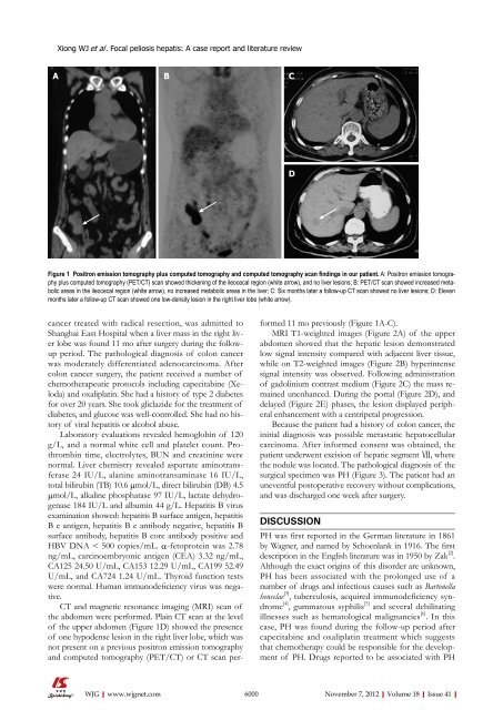
<strong>of</strong> <strong>the</strong> upper abdomen (Figure 1D) showed <strong>the</strong> presence<br />
<strong>of</strong> one hypodense lesion <strong>in</strong> <strong>the</strong> right liver lobe, which was<br />
not present on a previous positron emission tomography<br />
and computed tomography (PET/CT) or CT scan per-<br />
WJG|www.wjgnet.com<br />
B<br />
Figure 1 Positron emission tomography plus computed tomography and computed tomography scan f<strong>in</strong>d<strong>in</strong>gs <strong>in</strong> our patient. A: Positron emission tomography<br />
plus computed tomography (PET/CT) scan showed thicken<strong>in</strong>g <strong>of</strong> <strong>the</strong> ileocecal region (white arrow), and no liver lesions; B: PET/CT scan showed <strong>in</strong>creased metabolic<br />
areas <strong>in</strong> <strong>the</strong> ileocecal region (white arrow), no <strong>in</strong>creased metabolic areas <strong>in</strong> <strong>the</strong> liver; C: Six months later a follow-up CT scan showed no liver lesions; D: Eleven<br />
months later a follow-up CT scan showed one low-density lesion <strong>in</strong> <strong>the</strong> right liver lobe (white arrow).<br />
C<br />
D<br />
formed 11 mo previously (Figure 1A-C).<br />
MRI T1-weighted images (Figure 2A) <strong>of</strong> <strong>the</strong> upper<br />
abdomen showed that <strong>the</strong> hepatic lesion demonstrated<br />
low signal <strong>in</strong>tensity compared with adjacent liver tissue,<br />
while on T2-weighted images (Figure 2B) hyper<strong>in</strong>tense<br />
signal <strong>in</strong>tensity was observed. Follow<strong>in</strong>g adm<strong>in</strong>istration<br />
<strong>of</strong> gadol<strong>in</strong>ium contrast medium (Figure 2C) <strong>the</strong> mass rema<strong>in</strong>ed<br />
unenhanced. Dur<strong>in</strong>g <strong>the</strong> portal (Figure 2D), and<br />
delayed (Figure 2E) phases, <strong>the</strong> lesion displayed peripheral<br />
enhancement with a centripetal progression.<br />
Because <strong>the</strong> patient had a history <strong>of</strong> colon cancer, <strong>the</strong><br />
<strong>in</strong>itial diagnosis was possible metastatic hepatocellular<br />
carc<strong>in</strong>oma. After <strong>in</strong>formed consent was obta<strong>in</strong>ed, <strong>the</strong><br />
patient underwent excision <strong>of</strong> hepatic segment Ⅶ, where<br />
<strong>the</strong> nodule was located. The pathological diagnosis <strong>of</strong> <strong>the</strong><br />
surgical specimen was PH (Figure 3). The patient had an<br />
uneventful postoperative recovery without complications,<br />
and was discharged one week after surgery.<br />
DISCUSSION<br />
PH was first reported <strong>in</strong> <strong>the</strong> German literature <strong>in</strong> 1861<br />
by Wagner, and named by Schoenlank <strong>in</strong> 1916. The first<br />
description <strong>in</strong> <strong>the</strong> English literature was <strong>in</strong> 1950 by Zak [2] .<br />
Although <strong>the</strong> exact orig<strong>in</strong>s <strong>of</strong> this disorder are unknown,<br />
PH has been associated with <strong>the</strong> prolonged use <strong>of</strong> a<br />
number <strong>of</strong> drugs and <strong>in</strong>fectious causes such as Bartonella<br />
henselae [3] , tuberculosis, acquired immunodeficiency syndrome<br />
[4] , gummatous syphilis [5] and several debilitat<strong>in</strong>g<br />
illnesses such as hematological malignancies [6] . In this<br />
case, PH was found dur<strong>in</strong>g <strong>the</strong> follow-up period after<br />
capecitab<strong>in</strong>e and oxaliplat<strong>in</strong> treatment which suggests<br />
that chemo<strong>the</strong>rapy could be responsible for <strong>the</strong> development<br />
<strong>of</strong> PH. Drugs reported to be associated with PH<br />
6000 November 7, 2012|Volume 18|Issue 41|


